Proceeding Paper

Protein Extraction from Arthrospira platensis for Use in Food Processing †

1
Chemistry Research Centre-Vila Real (CQ-VR)—Food and Wine Chemistry Lab, University of Trás-os-Montes and Alto Douro, 5000-801 Vila Real, Portugal
2
Genetics and Biotechnology Department, University of Trás-os-Montes and Alto Douro, 5000-801 Vila Real, Portugal
3
Biology and Environment Department, University of Trás-os-Montes and Alto Douro, 5000-801 Vila Real, Portugal
4
Chemistry Department, University of Trás-os-Montes and Alto Douro, 5000-801 Vila Real, Portugal
*
Author to whom correspondence should be addressed.
Presented at the 1st International Meeting Molecules 4 Life, Vila Real, Portugal, 20–22 September 2023.
Med. Sci. Forum 2023, 23(1), 8; https://doi.org/10.3390/msf2023023008
Published: 15 January 2024
(This article belongs to the Proceedings of The 1st International Meeting Molecules 4 Life)

Abstract

:
Algae protein has emerged as a sustainable and non-allergenic alternative to animal protein as the market seeks to reduce reliance on traditional animal protein sources. To effectively utilize algae protein isolates, particularly from Arthrospira platensis, it is essential to develop an efficient method for protein extraction and isolation that can be scaled up. This work aims to optimize the extraction conditions to obtain high-purity protein extracts. HPLC-DAD was used to determine the total and free amino acid profiles, while SDS-PAGE and HPLC-MS/MS were used for the protein characterization. An optimized extraction method was selected based on achieving the highest protein content and purity.

1. Introduction

Arthrospira platensis, a cyanobacterium, is gaining recognition as a sustainable and environmentally friendly protein source [1,2,3]. With a protein content ranging from 60% to 70% of its dry weight and the presence of phycobiliproteins, it holds great promise for various applications [4]. As a non-allergenic and non-animal protein source, it offers a viable alternative to conventional proteins in several industries [5,6]. However, to realize its full potential, the development of efficient protein extraction and purification methods is essential [7,8]. Therefore, this study aimed to evaluate different protein extraction methods - such as agitation, bead milling, and ultrasound - and protein isolation via precipitation using ethanol or ammonium sulfate to optimize the protein yield and purity.

2. Materials and Methods

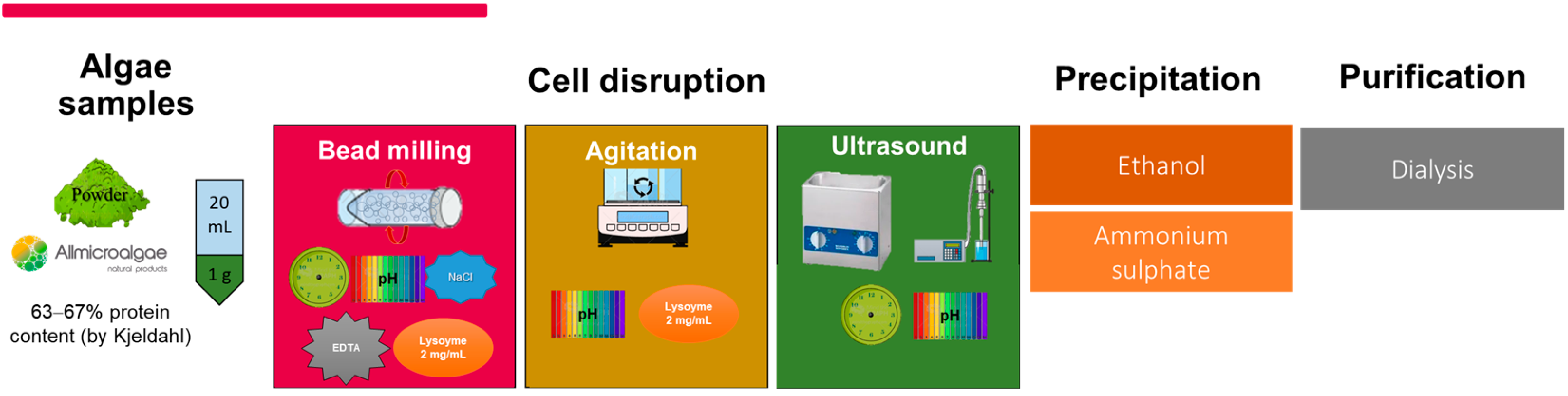
2.1. Sample and Protein Extraction

The Arthrospira platensis powder, with a protein content of 63–67% as determined using the Kjeldahl method, was obtained from a local producer (Allmicroalgae—Natural Products S.A., Portugal). However, HPLC analysis showed a total protein content of 50.79% ± 4.22. Figure 1 provides an overview of the protein extraction methods used in this study. These methods include agitation, bead milling, and ultrasound, each applied under different conditions in terms of the incubation time, pH levels, and specific chemical solutions. The extracted proteins were subsequently isolated via precipitation using ethanol or ammonium sulfate. Dialysis (MW cut-off 12–14 kDa) was the final purification step. In total, 45 different extraction conditions were tested.

2.2. Amino Acid Quantification

The amino acids were quantified via HPLC-DAD using a C18 column (AcclaimTM 120, 4.6 × 250 mm, particle size 5µm) on a Vanquish system (Thermo Fisher Scientific, Waltham, MA, USA). The procedure included derivatization with o-phthaldialdehyde in borate buffer, 2-mercaptoethanol (OPA-2MCE), and 9-fluorenylmethylchloroformate (FMOC), based on the method described by Herbert et al. [9] with some modifications. To analyze the total amino acids, 10 mg of algal biomass was hydrolyzed with 6 M HCl (1 mL) at 110 °C for 24 h. Further, 200 µL of β-alanine at 2.5 mg/mL was used as the internal standard, and an 8-point standard curve for the L-amino acids was constructed. The mobile phases consisted of eluent A, ultrapure water (100%); eluent B, methanol (MeOH) (100%); eluent C, sodium acetate buffer (0.36 M, pH 8); and eluent D, acetonitrile (100%). Proline and hydroxyproline were detected at 262 nm, while the other amino acids were detected at 337 nm.

2.3. Protein Characterization

The algae proteins were resuspended in a sample buffer comprising 2% (w/v) SDS, 40% (v/v) glycerol, 0.02% (w/v) bromophenol blue, 0.08 M Tris-HCl at a pH of 8.0, and 10% (v/v) Bolt Sample Reducing Agent (Thermo Fisher Scientific). This mixture was then heated at 65 ℃ for 30 min and separated on precast Bolt Bis-Tris Plus gels (4–12% gradient polyacrylamide concentration) using MES SDS Running Buffer (Thermo Fisher Scientific). Coomassie-stained protein bands were excised and subjected to in-gel trypsin digestion. The resulting peptides were analyzed via LC-MS/MS using an LTQ XL Linear Ion Trap Mass Spectrometer from Thermo Fisher Scientific [10].

2.4. Statistical Analysis

One-way ANOVA with a Scheffé post hoc test was employed to determine significant differences in the total amino acid profile among the extraction methods.

3. Results

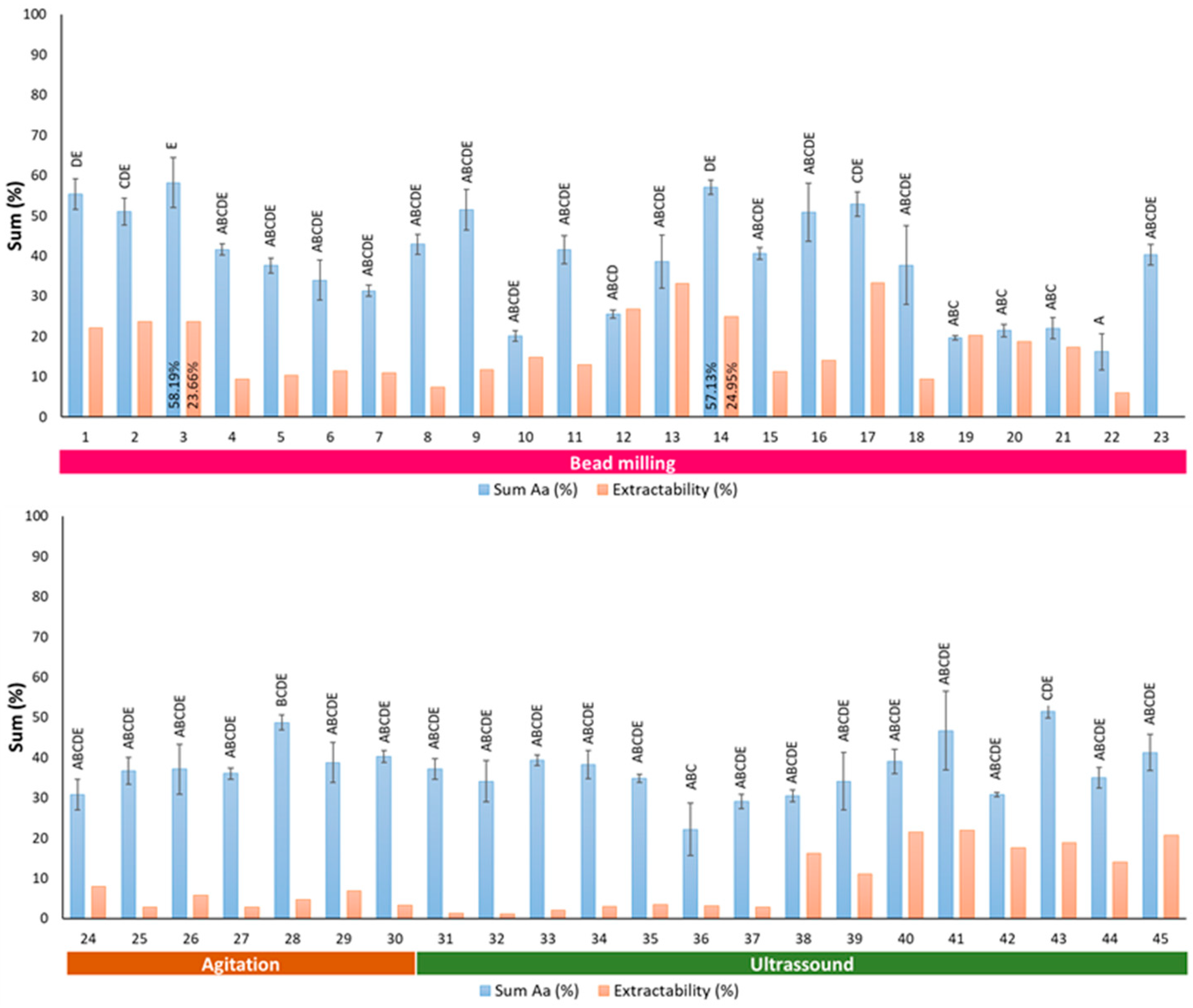
3.1. Optimization of the Protein Extraction Method

The results show that out of the 45 conditions tested, the most successful protein extraction method was method 3. This method involved bead milling for 24 h, the addition of 1 M NaCl, and pH adjustment to 7, followed by precipitation with 75% ethanol, as shown in Figure 2. This method yielded a significantly higher protein content of 58.19% ± 6.23, with an extraction yield of 23.66%. These results provide compelling evidence of the economic viability and suitability of the selected method for large-scale industrial implementation.

3.2. Quantification of Amino Acid in Extracted Proteins Using the Optimized Method

Method 3 exhibited significantly higher levels of aspartic acid (5.8 ± 0.2), glutamic acid (4.8 ± 0.3), and proline (15.0 ± 4.4). However, when the same procedure was used with a shorter extraction time (20 h, method 14), proteins with significantly higher leucine (7.9 ± 0.1), phenylalanine (7.8 ± 0.1), and isoleucine (5.1 ± 0.1) levels were obtained. This highlights the importance of carefully optimizing protein extraction protocols to achieve the desired amino acid profile. For example, proline is a critical amino acid found in conventional protein sources, such as wine-fining agents. Therefore, the selection of the optimized method is based on the highest total protein and proline content.

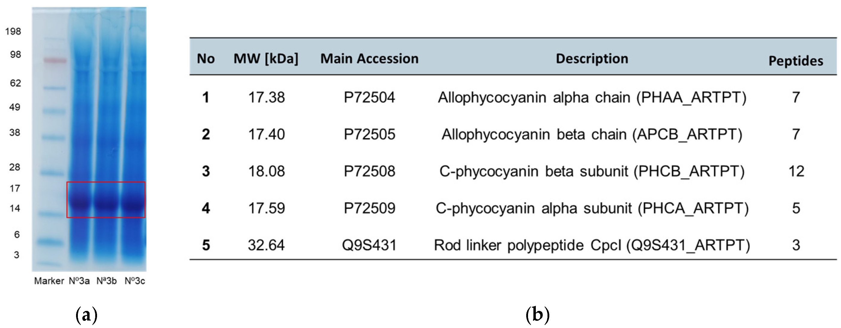
3.3. Protein Characterization

The SDS-PAGE profile showed a more concentrated and representative band in the 14–28 kDa range, consistent with the expected presence of phycobiliproteins (Figure 3a). For protein identification, a database search was performed using the OMSSA search algorithm (Figure 3b). MS/MS spectra were searched against a database containing 118,562 Arthrospira sequences retrieved from UniProt (https://www.uniprot.org/ accessed on 7 November 2023).

4. Conclusions

In this study, we have developed an efficient method for extracting and isolating proteins from Arthrospira platensis. The extraction method involved 24 h of bead milling at a pH of 7 with 1 M NaCl, followed by 75% ethanol precipitation and subsequent dialysis, resulting in a total amino acid content of 58.13%. Proline, aspartic acid, glutamic acid, and leucine were identified as the predominant amino acids in the extracted protein. Furthermore, SDS-PAGE analysis showed a prominent protein band in the 14–28 kDa range, confirming the presence of phycobiliproteins associated with Arthrospira platensis. Additionally, mass spectrometry enabled the identification and validation of five different proteins in the protein extract. These results highlight the potential of Arthrospira platensis as a protein source.

Author Contributions

Conceptualization, F.C. and F.M.N.; methodology, E.C. and M.R.; formal analysis, E.C.; investigation, E.C.; resources, E.C., F.C. and F.M.N.; data curation, E.C.; writing—original draft preparation, E.C.; writing—review and editing, E.C., M.R., L.F.-R., F.C. and F.M.N.; supervision, F.C. and F.M.N.; project administration, F.C. and F.M.N.; funding acquisition, F.C. and F.M.N. All authors have read and agreed to the published version of the manuscript.

Funding

This study received financial support from the CQ-VR Chemistry Research Centre, Vila Real (UIDB/00616/2020 and UIDP/00616/2020 https://doi.org/10.54499/UIDP/00616/2020), FCT-Portugal, and COMPETE. The authors are grateful to the Foundation for Science and Technology (FCT) for the SFRH/BD/144107/2019 grant.

Institutional Review Board Statement

Not applicable.

Informed Consent Statement

Not applicable.

Data Availability Statement

Not applicable.

Acknowledgments

This proceeding is part of the SFRH/BD/144107/2019 Ph.D. thesis.

Conflicts of Interest

The authors declare no conflicts of interest.

References

  1. Pelizer, L.H.; De Carvalho, J.C.M.; De Oliveira Moraes, I. Protein Production by Arthrospira (Spirulina) Platensis in Solid State Cultivation Using Sugarcane Bagasse as Support. Biotechnol. Rep. 2015, 5, 70–76. [Google Scholar] [CrossRef]
  2. Camacho, F.; Macedo, A.; Malcata, F. Potential Industrial Applications and Commercialization of Microalgae in the Functional Food and Feed Industries: A Short Review. Mar. Drugs 2019, 17, 312. [Google Scholar] [CrossRef] [PubMed]
  3. Kusmayadi, A.; Leong, Y.K.; Yen, H.-W.; Huang, C.-Y.; Chang, J.-S. Microalgae as Sustainable Food and Feed Sources for Animals and Humans—Biotechnological and Environmental Aspects. Chemosphere 2021, 271, 129800. [Google Scholar] [CrossRef] [PubMed]
  4. Benelhadj, S.; Gharsallaoui, A.; Degraeve, P.; Attia, H.; Ghorbel, D. Effect of PH on the Functional Properties of Arthrospira (Spirulina) Platensis Protein Isolate. Food Chem. 2016, 194, 1056–1063. [Google Scholar] [CrossRef] [PubMed]
  5. Conde, E.; Balboa, E.M.; Parada, M.; Falqué, E. Algal Proteins, Peptides and Amino Acids. In Functional Ingredients from Algae for Foods and Neutracceuticls; Elsevier: Amsterdam, The Netherlands, 2013; pp. 135–180. [Google Scholar] [CrossRef]
  6. Teuling, E.; Wierenga, P.A.; Schrama, J.W.; Gruppen, H. Comparison of Protein Extracts from Various Unicellular Green Sources. J. Agric. Food Chem. 2017, 65, 7989–8002. [Google Scholar] [CrossRef] [PubMed]
  7. Safi, C.; Ursu, A.V.; Laroche, C.; Zebib, B.; Merah, O.; Pontalier, P.-Y.; Vaca-Garcia, C. Aqueous Extraction of Proteins from Microalgae: Effect of Different Cell Disruption Methods. Algal Res. 2014, 3, 61–65. [Google Scholar] [CrossRef]
  8. Vernès, L.; Abert-Vian, M.; El Maâtaoui, M.; Tao, Y.; Bornard, I.; Chemat, F. Application of Ultrasound for Green Extraction of Proteins from Spirulina. Mechanism, Optimization, Modeling, and Industrial Prospects. Ultrason. Sonochem. 2019, 54, 48–60. [Google Scholar] [CrossRef] [PubMed]
  9. Herbert, P.; Santos, L.; Alves, A. Simultaneous Quantification of Primary, Secondary Amino Acids, and Biogenic Amines in Musts and Wines Using OPA/3-MPA/FMOC-CI Fluorescent Derivatives. J. Food Sci. 2001, 66, 1319–1325. [Google Scholar] [CrossRef]
  10. Shevchenko, A.; Tomas, H.; Havli, J.; Olsen, J.V.; Mann, M. In-Gel Digestion for Mass Spectrometric Characterization of Proteins and Proteomes. Nat. Protoc. 2006, 1, 2856–2860. [Google Scholar] [CrossRef] [PubMed]
Figure 1. Representation of the various protein extraction conditions tested.
Msf 23 00008 g001
Figure 2. Total amino acid content and extractability were obtained using the diverse methods tested. For each sum of amino acids (%), bars with the same letter are not significantly different (Scheffé test, p < 0.05).
Msf 23 00008 g002
Figure 3. (a) SDS-PAGE profile of the protein extract from method 3 (in triplicate). Sizes (in kilodaltons) of protein molecular weight markers are shown on the left (Precision Plus Protein All Blue Prestained Protein Standards, Bio-Rad, CA, USA); (b) peptide mass fingerprinting results of the most representative bands (highlighted with a red rectangle in (a)).
Msf 23 00008 g003
Disclaimer/Publisher’s Note: The statements, opinions and data contained in all publications are solely those of the individual author(s) and contributor(s) and not of MDPI and/or the editor(s). MDPI and/or the editor(s) disclaim responsibility for any injury to people or property resulting from any ideas, methods, instructions or products referred to in the content.

