Proceeding Paper

PLGA Nanoparticles Loaded with Cinnamon Extract and Coated with PVA/Poloxamer188 †

Department of Medical Nanotechnology, School of Advanced Technologies in Medicine, Tehran University of Medical Sciences, Tehran 14177-55469, Iran
*
Authors to whom correspondence should be addressed.
Presented at the 3rd International Electronic Conference on Cancers: New Targets for Cancer Therapies, 16–30 March 2023; Available online: https://iecc2023.sciforum.net/.
Med. Sci. Forum 2023, 20(1), 8; https://doi.org/10.3390/IECC2023-14262
Published: 17 March 2023

Abstract

:
Cinnamon extract has received significant attention due to its significant antibacterial, antifungal, antioxidant, and even anti-cancer properties. The purpose of this study was to create cinnamon-extract-loaded PLGA nanoparticles and evaluate their physiochemical characteristics and cytotoxicity against the C6 cell line. Physiochemical characteristics, such as the mean diameter, zeta potential, and drug loading potential, were measured. The antioxidant activity and cytotoxicity of nanoparticles were investigated by DPPH and MTT studies, respectively. The mean diameter of nanoparticles was 120 ± 24 nm. The antioxidant activity of the cinnamon extract was mostly preserved in nanoparticles and the toxicity effect on cancer cells was investigated.

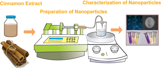
Graphical Abstract

1. Introduction

One of the challenges in treating tumors is the untargeted delivery of chemotherapeutics and their unwanted toxicity to healthy organs. Toxicity from chemotherapy drugs is hazardous and may even lead to tissue damage [1]. Today, with the use of nanotechnology, the targeted delivery of drugs has improved compared to the past [2].
Cinnamon (Cin) is a well-known spice that is also utilized in herbal medicine [3]. In addition to being an antioxidant [4], antifungal [5], and antibiotic agent [4], it is effective in treating diseases, such as obesity [6], Parkinson’s [7], cancer, and cardiovascular disease [8]. The effectiveness of this substance on various cancer cells such as leukemia [9], prostate [10], and breast cancer [11] was evaluated.
Polymeric nanoparticles (NPs) are proper nanovehicles for drug delivery, and they can be categorized as either natural or synthetic [12]. Poly(lactic-co-glycolic acid) (PLGA) is an extensively studied and widely used synthetic polymer [13], which has gained prominence due to its biodegradability, biocompatibility, low cost, and FDA approval [14]. In this study, we prepared Cin loaded in PLGA NPs (Cin/PLGA NPs) to study the physiochemical properties and cellular toxicity of NPs.

2. Materials and Methods

2.1. Preparation of Nanoparticles

Cin/PLGA NPs were prepared by following the emulsion solvent evaporation method. First of all, 30 mg of PLGA ((MW 30.000 g·mol1, 50:50) Xi’An Xinlu Biotech Company, Xi’An, China) was dissolved in 3 mL of acetonitrile and stirred for 10 min at 500 rpm. Then, 2 mg of cinnamon extract (Adonisherb Company, Tehran, Iran) was added to PLGA solution and this solution was added to 30 mL of 1% PVA/Poloxamer188 (10:1) solution simultaneously. The emulsion was sonicated for 8 min at 120 W. It was stirred for 3 h at 300 rpm. Afterward, it was centrifuged at 10,000 rpm for 25 min and washed two times.

2.2. Characterization of NPs

2.2.1. Size Distribution and Zeta Potential

DLS (ScatterScope1) and scanning electron microscope (SEM) were applied to check NPs size distribution and a Zetasizer (Malvern) was also applied to measure the zeta potential of NPs.

2.2.2. Drug Loading (DL) % and Encapsulation Efficiency (EE) %

Afterwards, 5 mg of lyophilized sample was solved in 5 mL of acetonitrile and was sonicated for 5 min. Then, the absorption was read at 286 nm. DD% and EE% were calculated.

2.2.3. In Vitro Drug Release

Next, 10 mg of lyophilized Cin/PLGA NPs was dispersed in 5 mL of PBS (pH 7.4), and then poured in a dialysis tube. Then, this was soaked in 45 mL of PBS and incubated for 7 days at 100 rpm under 37 °C. At each time point (1, 3, 6, 12, 24, 48, 72, 96, 120, 144 and 168 h), 5 mL of the medium was extracted and fresh medium was replaced.

2.2.4. Fourier Transform Infrared Spectroscopy (FTIR)

FTIR was used to confirm drug encapsulation in NPs from 400 to 4000 cm1. PVA, poloxamer188, PLGA, Cin, and Cin/PLGA NPs were used and dispersed with KBr to prepare the pellet for analysis.

2.3. Antioxidant Activity

A DPPH assay was performed to check the antioxidant activity of Cin/PLGA NPs. The serial concentrations of Cin/PLGA NPs, PLGA NPs, and Cin (1 µg/mL to 2000 µg/mL) were treated with DPPH solution in ethanol (2 mg/100 mL) for 3 h in darkness. Then, the absorption of each sample was read at 517 nm by UV-Vis spectroscopy. The level of antioxidant activity of samples was calculated as shown below:
% Inhibition = ((A0 − A1)/A1) × 100    (A0) control, (A1) sample

2.4. Blood Compatibility

To check blood compatibility, a hemolysis assay was applied. Diluted blood was encountered with PLGA NPs and Cin/PLGA NPs for 3 h and absorption was read at 540 nm.

2.5. Cellular Uptake of Nanoparticles

The cells were cultured with a DMEM-F12 medium containing 10% v/v and 1% v/v FBS and penicillin–streptomycin, respectively. Then, 1 mg of Cin/PLGA NPs, loaded with carbon quantum dots as fluorescent agents, was dispersed in a sterile PBS and poured on seeded cells. After 3 h, the cells were washed with PBS and 4% paraformaldehyde was added, followed by DAPI staining. A fluorescent microscope (Olympus BX43) was applied to capture the photos.

2.6. Cytotoxicity of Nanoparticles

C6 cells were seeded in 96-well plates. Cin and Cin/PLGA NPs were dispersed in PBS solution and added to each well. After 24 and 72 h of the treatment, the wells were washed three times with PBS (Dr. Mojallali company, Tehran, Iran) and MTT (Sigma-Aldrich, Schnelldorf, Germany) solution (0.5 mg/mL) was added. After 3 h, chemical DMSO (Dr. Mojallali company, Tehran, Iran) was used and absorptions were read via a microplate reader (Bio Tek 800 TS).

3. Results

3.1. Physiochemical Characterization of Cin/PLGA NPs

The emulsion solvent evaporation method was applied to prepare Cin/PLGA NPs. The Cin/PLGA NPs’ mean diameter was 120 ± 24 (Figure 1A–C). The zeta potential values of PLGA NPs and Cin/PLGA NPs were −10.1 ± 1.1 mV and −3.66 ± 1.8 mV, respectively (Figure 1D). The EE% and DL% of Cin in Cin/PLGA NPs were 51 ± 5% and 4.2 ± 0.7%, respectively which were calculated using UV-visible spectroscopy at 286 nm (Figure 1G). The in vitro release of cinnamon from Cin/PLGA NPs demonstrates the two phases of drug release. At first, a burst release was seen at the first 12 h, which was induced to a 16% release of Cin (Figure 1E). Moreover, 23.2%, 31.6%, and 44.6% of Cin were released during the first 24, 48, and 72 h. It was observed that the release of Cin was extended to more than 7 days and 84% of Cin was released after 168 min (7 days).
In PLGA, three sharp peaks were seen in 2940 cm−1, 1143 cm−1, and 668 cm−1. In PVA, the sharpest peak was at 1065 cm−1. In poloxamer188, the main peaks were at 2882 cm−1, 1099 cm−1, and 1144 cm−1. In the cinnamon extract, the sharpest peak was at 1047 cm−1 and two wide bands were observed at 2922 cm−1 and 3334 cm−1. In Cin/PLGA NPs, the existence of Cin, PLGA, PVA, and poloxamer188 was confirmed, according to the various peaks that were obtained (Figure 1F).

3.2. Antioxidant Activity

The DPPH scavenging activity of Cin was increased by increasing the concentration and reached 100% at the concentration of 1000 µg/mL. In the Cin/PLGA NPs, enhanced antioxidant scavenging was observed with the increase in the NPs concentration, but the slope of the graph was lower than that of the Cin (Figure 2B,D).

3.3. Blood Compatibility

The hemolysis of both Cin and Cin/PLGA NPs was concentration-dependent and was higher in Cin than Cin/PLGA NPs in all concentrations (Figure 2A).

3.4. Cellular Uptake of NPs

Figure 2F shows the fluorescent image of C6 cells which were incubated with Cin/PLGA NPs and Figure 2G shows the DAPI staining of mentioned cells. According to Figure 2H, whenever nuclei were stained, Cin/PLGA NPs were present; therefore, Cin/PLGA NPs were taken up by C6 cells.

3.5. Cytotoxicity of NPs

It was observed that the cellular toxicity of Cin and Cin/PLGA NPs in the C6 cell line was concentration- and time-dependent. C6 viability was higher in Cin-treated cells in comparison with Cin/PLGA NP-treated cells at all concentrations (1 µg/mL to 400 µg/mL) after 24 h; however, this was converted after 72 h, which means that Cin/PLGA NPs were more powerful than Cin to kill C6 cells (Figure 2C,E).

4. Discussion

Recently, there has been a great deal of focus on medicinal plants, including novel delivery systems [15]. In another study, PLGA nanoparticles containing cinnamldehyde for antifungal activity purposes were developed with a mean diameter of 130 nm and a zeta potential of −3 mV [16]. In addition with PLGA, other polymers such as PEG [4] and chitosan [17] were used for the delivery of cinnamon essential oil and cinnamon extract, respectively. The anti-tumor effect of Fe3O4 nanoparticles coated with cinnamaldeyde and FITC was seen on the breast adenocarcinoma animal model [18]. In our study, this is the first time that cinnamon extract was encapsulated in PLGA nanoparticles coated with PVA and poloxamer188.

5. Conclusions

In this study, PLGA nanoparticles containing cinnamon extract coated with PVA/poloxamer188 were prepared. The findings of this study indicate that Cin/PLGA NPs could be a promising adjuvant treatment for GBM. However, additional research is required, and we recommend using cinnamaldehyde instead of cinnamon extract for greater effectiveness and a smaller nanoparticle diameter.

Author Contributions

Conceptualization, M.K. and M.A.; methodology, F.M.; software, F.M.; validation, M.K. and M.A.; formal analysis, F.M.; investigation, F.M.; resources, F.M.; data curation, F.M.; writing—original draft preparation, F.M.; writing—review and editing M.K. and M.A.; visualization M.A., supervision, M.K. and M.A.; project administration, M.K.; funding acquisition, M.K. All authors have read and agreed to the published version of the manuscript.

Funding

This research was supported by Tehran University of Medical Sciences (grant number 51516-148-1-1400).

Institutional Review Board Statement

All experiments were compliant with research ethic committee guidelines of the Tehran University of Medical Sciences (IR.TUMS.MEDICINE.REC.1400.425).

Informed Consent Statement

Not applicable.

Data Availability Statement

Data are contained within the article.

Conflicts of Interest

The authors declare no conflict of interest.

References

  1. Haghi-Aminjan, H.; Asghari, M.H.; Farhood, B.; Rahimifard, M.; Hashemi Goradel, N.; Abdollahi, M. The role of melatonin on chemotherapy-induced reproductive toxicity. J. Pharm. Pharmacol. 2018, 70, 291–306. [Google Scholar] [CrossRef] [PubMed]
  2. Singh, S.; Sharma, A.; Robertson, G.P. Realizing the Clinical Potential of Cancer Nanotechnology by Minimizing Toxicologic and Targeted Delivery ConcernsLimitations and Approaches of Cancer Nanotechnology. Cancer Res. 2012, 72, 5663–5668. [Google Scholar] [CrossRef] [PubMed]
  3. Błaszczyk, N.; Rosiak, A.; Kałużna-Czaplińska, J. The potential role of cinnamon in human health. Forests 2021, 12, 648. [Google Scholar] [CrossRef]
  4. Hemalatha, N.; Kumar, K.R. Synthesis and characterization of PEG-cinnamon essential oil nanoparticles and their application as an insecticidal agent. J. Adv. Sci. Res. 2021, 12, 142–148. [Google Scholar] [CrossRef]
  5. Ahmed, J.; Altun, E.; Aydogdu, M.O.; Gunduz, O.; Kerai, L.; Ren, G.; Edirisinghe, M. Anti-fungal bandages containing cinnamon extract. Int. Wound J. 2019, 16, 730–736. [Google Scholar] [CrossRef] [PubMed]
  6. Sartorius, T.; Peter, A.; Schulz, N.; Drescher, A.; Bergheim, I.; Machann, J.; Schick, F.; Siegel-Axel, D.; Schürmann, A.; Weigert, C. Cinnamon extract improves insulin sensitivity in the brain and lowers liver fat in mouse models of obesity. PLoS ONE 2014, 9, e92358. [Google Scholar] [CrossRef] [PubMed]
  7. Frydman-Marom, A.; Levin, A.; Farfara, D.; Benromano, T.; Scherzer-Attali, R.; Peled, S.; Vassar, R.; Segal, D.; Gazit, E.; Frenkel, D. Orally administrated cinnamon extract reduces β-amyloid oligomerization and corrects cognitive impairment in Alzheimer’s disease animal models. PLoS ONE 2011, 6, e16564. [Google Scholar] [CrossRef] [PubMed]
  8. Shang, C.; Lin, H.; Fang, X.; Wang, Y.; Jiang, Z.; Qu, Y.; Xiang, M.; Shen, Z.; Xin, L.; Lu, Y. Beneficial effects of cinnamon and its extracts in the management of cardiovascular diseases and diabetes. Food Funct. 2021, 12, 12194–12220. [Google Scholar] [CrossRef] [PubMed]
  9. Assadollahi, V.; Parivar, K.; Roudbari, N.H.; Khalatbary, A.R.; Motamedi, M.; Ezatpour, B.; Dashti, G.R. The effect of aqueous cinnamon extract on the apoptotic process in acute myeloid leukemia HL-60 cells. Adv. Biomed. Res. 2013, 2, 25. [Google Scholar] [PubMed]
  10. Gopalakrishnan, S.; Ediga, H.H.; Reddy, S.S.; Reddy, G.B.; Ismail, A. Procyanidin-B2 enriched fraction of cinnamon acts as a proteasome inhibitor and anti-proliferative agent in human prostate cancer cells. IUBMB Life 2018, 70, 445–457. [Google Scholar] [CrossRef] [PubMed]
  11. Liu, Y.; An, T.; Wan, D.; Yu, B.; Fan, Y.; Pei, X. Targets and mechanism used by cinnamaldehyde, the main active ingredient in cinnamon, in the treatment of breast cancer. Front. Pharmacol. 2020, 11, 582719. [Google Scholar] [CrossRef] [PubMed]
  12. Madani, F.; Esnaashari, S.S.; Webster, T.J.; Khosravani, M.; Adabi, M. Polymeric nanoparticles for drug delivery in glioblastoma: State of the art and future perspectives. J. Control. Release 2022, 349, 649–661. [Google Scholar] [CrossRef] [PubMed]
  13. Tayfeh-Ebrahimi, R.; Amniattalab, A.; Mohammadi, R. Evaluation of Effect of Biologically Synthesized Ethanolic Extract of Propolis-Loaded Poly (-Lactic-co-Glycolic Acid) Nanoparticles on Wound Healing in Diabetic Rats. Int. J. Low. Extrem. Wounds, 2022; Online ahead of print. [Google Scholar] [CrossRef]
  14. Madani, F.; Esnaashari, S.S.; Bergonzi, M.C.; Webster, T.J.; Younes, H.M.; Khosravani, M.; Adabi, M. Paclitaxel/methotrexate co-loaded PLGA nanoparticles in glioblastoma treatment: Formulation development and in vitro antitumor activity evaluation. Life Sci. 2020, 256, 117943. [Google Scholar] [CrossRef] [PubMed]
  15. Husni, P.; Ramadhania, Z.M. Plant extract loaded nanoparticles. Indones. J. Pharm. 2021, 3, 38–49. [Google Scholar] [CrossRef]
  16. Gursu, B.Y.; Dag, İ.; Dikmen, G. Antifungal and antibiofilm efficacy of cinnamaldehyde-loaded poly (DL-lactide-co-glycolide)(PLGA) nanoparticles against Candida albicans. Int. Microbiol. 2022, 25, 245–258. [Google Scholar] [CrossRef] [PubMed]
  17. Alghuthaymi, M.A.; Diab, A.M.; Elzahy, A.F.; Mazrou, K.E.; Tayel, A.A.; Moussa, S.H. Green biosynthesized selenium nanoparticles by cinnamon extract and their antimicrobial activity and application as edible coatings with nano-chitosan. J. Food Qual. 2021, 2021, 6670709. [Google Scholar] [CrossRef]
  18. Shetty, V.; Jakhade, A.; Shinde, K.; Chikate, R.; Kaul-Ghanekar, R. Folate mediated targeted delivery of cinnamaldehyde loaded and FITC functionalized magnetic nanoparticles in breast cancer: In vitro, in vivo and pharmacokinetic studies. New J. Chem. 2021, 45, 1500–1515. [Google Scholar] [CrossRef]
Figure 1. (A,B) SEM image of NPs; (C) DLS result; (D) zeta potential NPs; (E) drug release profile; (F) FTIR diagram; (G) Cin absorption spectra.
Msf 20 00008 g001
Figure 2. (A) Hemolysis diagram; (B) antioxidant activity; (C) MTT result after 24 h; (D) antioxidant activity; (E) MTT results after 72 h; (FH) cellular uptake images. (* p < 0.05, ** p < 0.01, *** p < 0.001, **** p < 0.0001).
Msf 20 00008 g002
Disclaimer/Publisher’s Note: The statements, opinions and data contained in all publications are solely those of the individual author(s) and contributor(s) and not of MDPI and/or the editor(s). MDPI and/or the editor(s) disclaim responsibility for any injury to people or property resulting from any ideas, methods, instructions or products referred to in the content.