Proceeding Paper

Proteomic and Genetic Approach for Lunasin Peptide and Gene Presence Detection in Various Plants †

1
Department of Biochemistry and Biotechnology, Faculty of Biotechnology and Food Sciences, Slovak University of Agriculture, Tr. A. Hlinku 2, 949 76 Nitra, Slovakia
2
Department of Genetics and Plant Breeding, Institute of Plant and Environmental Sciences, Faculty of Agrobiology and Food Resources, Slovak University of Agriculture in Nitra, Trieda Andreja Hlinku 2, 949 76 Nitra, Slovakia
*
Author to whom correspondence should be addressed.
Presented at the 2nd International Electronic Conference on Plant Sciences—10th Anniversary of Journal Plants, 1–15 December 2021; Available online: https://iecps2021.sciforum.net/.
Biol. Life Sci. Forum 2022, 11(1), 86; https://doi.org/10.3390/IECPS2021-12004
Published: 1 December 2021

Abstract

:
Lunasin is a biologically active peptide, with a polypeptide chain consisting of 43 amino acids, originally discovered as a 2S albumin protein from the seed of the soybean coded by the GM2S-1 gene. The most significant health benefits include antioxidant activity, anti-hypertension activity, and chemoprevention activity. Lunasin peptide was initially detected in many plant species, but there is controversy about its exact origins at present. Therefore, we focused on the detection of the Lunasin gene by a polymerase chain reaction and Lunasin peptide by one and two-dimensional electrophoreses, in various plants. These results confirmed that Lunasin peptide, as well as the Lunasin gene, were observed only in soybean seeds. There was also a confirmed presence of Lunasin-like peptide in various plants (spelt wheat, bean, and oat), but gene detection was not successful.

1. Introduction

Lunasin is one of the most important bioactive peptides [1]. It occurs predominantly in soybeans [2], and other types of legumes and cereals [3]. Lunasin demonstrates positive effects on human health and is considered as a chemo-preventive agent against cancer because it shows significant bioactivity in inhibiting the formation and growth of cancer cells [4]. The most important biological property of Lunasin is its bioavailability, mainly the capability to remain unchanged and bioactive after oral ingestion. Lunasin after absorption can be distributed to metabolism. In vitro bioavailability studies have shown that isolated Lunasin is easily digested after only 2 min of incubation with simulated gastric (SGF) and simulated intestinal fluids (SIF) [5]. Lunasin is a heat-stable peptide because remains bioactivity even after boiling for 10 min [6]. The primary structure of Lunasin consists of 43 amino acids: Ser1-Lys-Trp-Gln-His-Gln-Gln-Asp-Ser-Cys10-Arg-Lys-Gln-Leu-Gln-Gly-Val-Asn-Leu-Thr20-Pro-Cys-Glu-Lys-His-Ile-Met-Glu-Lys-Ile30-Gln-Gly-Arg-Gly-Asp-Asp-Asp-Asp-Asp-Asp40-Asp-Asp-Asp [7]. N-terminal amino acids contain neutral side chains, unlike C-terminal amino acids which contain polar side chains [8]. The primary sequence of the Lunasin peptide can be cleaved into four fragments. Fragment SKWQHQQDSCRKQLQGVNLTPC has an unknown function. The second fragment is a structural preserved area of α-helix (KHIMEKIQ), which targets Lunasin to H3–H4 histones. The amino acid motif RGD internalizes Lunasin into cells, and is the third fragment which also prevents the acetylation of deacetylated histones H3 and H4. The last, fourth fragment consists of eight aspartic acid residues (DDDDDDDD) which directly bind to core histones [9]. The Lunasin peptide secondary structure consists of α-helix (29%), β-helix (28%), β-sheet (23%), and disordered regions (20%) [10]. In [11], the authors discovered that the disulfide bond creation capability of Lunasin, Cys10, and Cys22 enable two forms of Lunasin, oxidized and reduced. The high cost of producing synthetic Lunasin determined the requirement of the identification and isolation of Lunasin from natural plant sources.
The aim of the work was to investigate the proteome and genome of selected varieties of plants with an emphasis on the detection of Lunasin peptide and Lunasin peptide gene detection.

2. Materials and Methods

2.1. Biological Material

We used three genotypes of soybean (Glycine max L.), Zora, Ischigo Wase, Lokus, and kidney bean (Phaseolus vulgaris L.) Zlatý roh, Dandy, Wiscont, in our research. We also analyzed one genotype of buckwheat (Fagopyrum esculentum Moench) Mihovo, rye (Secale cereale L.) Radošínsky rekord, oat (Avena sativa L.) Veles, spelt wheat (Triticum spelta L.) Roquin, emmer wheat (Triticum dicoccum L.), peas (Pisum sativum L.) Jantar, and lentil (Lens esculentum L.) Svit. The analyzed genotypes were obtained from the Gene Bank of Seed Species of the Slovak Republic NPPC VÚRV in Piešťany.

2.2. Protein and DNA Extraction

We focused on the analysis of peptide Lunasin’s presence and detection of genetic information for this peptide in our research. Subsequently, we realized protein extraction and extraction of DNA.

2.2.1. Protein Extraction

We used systems to perform one-dimensional polyacrylamide gel electrophoresis in the presence of sodium dodecyl sulfate (1D SDS PAGE) and a system for two-dimensional electrophoretic separation in our investigation. Accordingly, we performed two different isolations of protein complexes from our samples.
The protein complex for the 1D SDS PAGE system was isolated according to the standard reference method of International Seed Testing Association (ISTA) [12]. The grain of the analyzed samples was mechanically homogenized. Protein complexes were extracted using a solution consisting of 4.25 mL storage solution (12.5 mL 1 M Tris-HCl pH 6.8; 20 mL glycerol; 4 g SDS; 20 mg Pyronin G in 100 mL of deionized water), and 0.75 mL 2-mercaptoethanol in 10 mL of deionized water. The volume of the extraction solution was calculated by a ratio 1 mg of sample = 8 μL of the extraction solution. The extraction procedure was carried out for 30 min at 100 °C, 1200 rpm in a thermal shaker followed by centrifugation (15,000× g, 10 min).
The protein complex for 2D electrophoretic separation was extracted by phenol extraction according to [13]. First, 0.1 g of the sample was mechanically homogenized and then the protein complex was extracted by adding of 200 μL extraction solution (30.88 g sucrose, 0.37 g EDTA, 6.65 mL 1M Tris-HCl pH 8.8, 0.4 mL 2-mercaptoethanol in 100 mL of deionized water) and 200 μL Tris-Phenol solution (4706× g phenol in 100 mL 1 M Tris-HCl pH 8.8). The extraction procedure included 30 min shaking on ice and centrifugation at 5000× g for 10 min. Then, 200 μL of supernatant was precipitated by 1 mL of precipitating solution (1.95 g ammonium acetate in 250 mL methanol) overnight in the fridge. The sample was then centrifugated at 15,000× g for 20 min and the pellet was resuspended in the precipitating solution and chilled at −20 °C for 20 min. The pellet was obtained by centrifugation at 5000× g for 10 min, washed with 80% acetone and 70% methanol, and centrifuged at 15,000× g for 20 min, dried, and stored. The pellet was resuspended in 200 μL of IEF rehydration buffer before use.

2.2.2. DNA Extraction

DNA was extracted using the Thermo Scientific™ GeneJET Plant Genomic DNA Purification Kit (Thermo Fisher Scientific, Waltham, MA, USA) on the basis of the standard procedure supplied by the manufacturer.

2.3. Analyses of Lunasin Peptide Presence and Detection of Genetic Information for Lunasin

The analysis of the Lunasin peptide presence was realized by 1D SDS PAGE and 2D electrophoresis. The identification of the Lunasin peptide gene was performed by polymerase chain reaction (PCR) and the PCR product was detected by agarose electrophoresis.

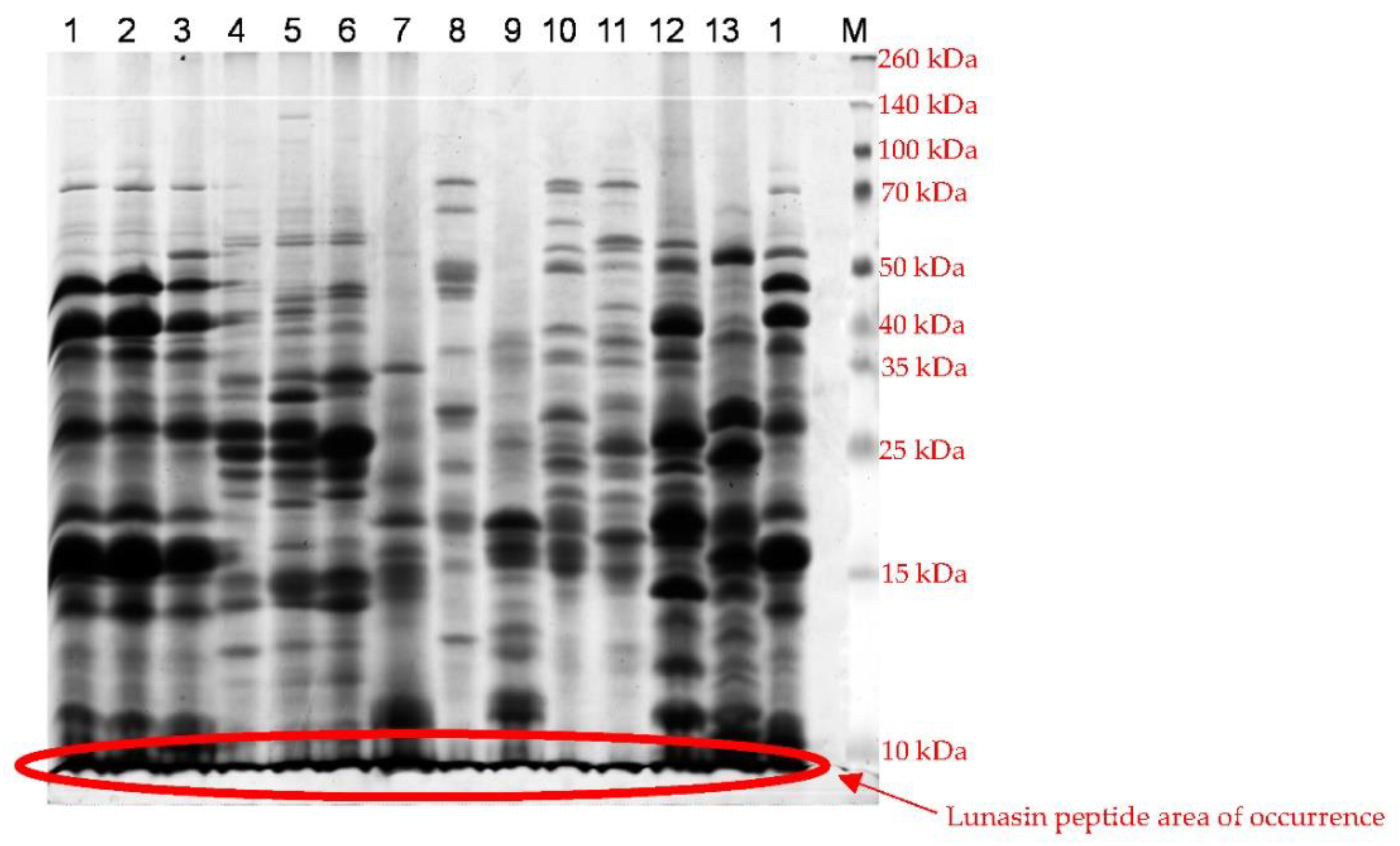
2.3.1. Electrophoretic Separation

1D electrophoresis separation was performed in a vertical discontinuous SDS-PAGE system (HeoferTM SE 600 Chroma—Fisher Scientific) according to the standard ISTA method [12]. The separation of the protein complex was performed in 22% Tris-HCl polyacrylamide stacking gel and 7.4% Tris-HCl polyacrylamide running gel. The electrophoretic buffer system was Tris-glycine buffer. The protein complex electrophoretic separation lasted 20 h (20 mA) and protein identification was performed by comparison with a broad range protein molecular weight marker (Promega). The staining system included a solution of Coomassie Brilliant Blue R250 in methanol. Gels were destained in water.
2D electrophoresis separation was performed using a combination of isoelectric focusing and 1D SDS PAGE. Isoelectric focusing was performed using Serva IPG BlueStrips 11 cm pH Gradient 3–10 (Serva Electrophoresis) according to the manual of the manufacturer on PROTEAN i12 IEF System (Bio-Rad). 1D SDS PAGE was realized by the modification of standard ISTA procedure [12] application only stacking gel. The electrophoretic separation took 15 h with a constant current of 15 mA (HeoferTM SE 600 Chroma—Fisher Scientific).

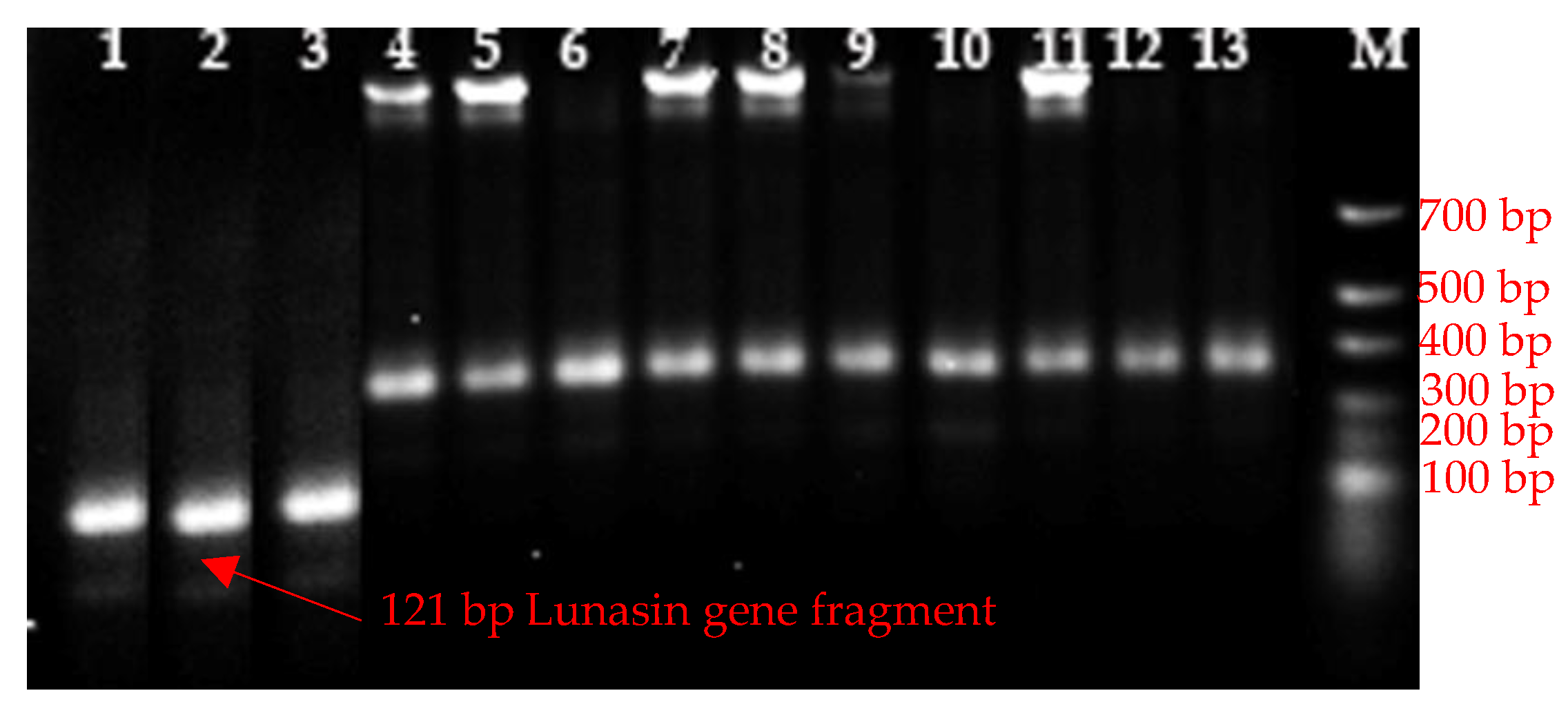
2.3.2. Polymerase Chain Reaction (PCR)

Primers and reaction conditions of PCR were performed according to the method of Dinelli et al. (2014). PCR was realized in GoTaq Green Master Mix (Promega) buffer solution. The Dinelli et al. (2014) method was modified by the optimization of reaction conditions of the PCR according to the manual of the GoTaq Green Master Mix (Promega) manufacturer. PCR products were separated and visualized in 2% agarose electrophoresis. The buffer system of agarose electrophoresis was TBE (54 g Tris, 24.5 g boric acid, 20 mL 0.5 M EDTA pH 8.0 in 1000 mL of deionized water). Electrophoresis took 90 min with a constant voltage of 100 V. The molecular weight was established by comparison with molecular standards.

2.3.3. Detection System

The analysis and evaluation of the results were performed with a ChemiDoc MP imagine system (Bio-Rad) using ImageLab software (Bio-Rad, Herculers, CA, USA).

3. Results and Discussion

Lunasin was first isolated from soybean seeds in 1987 [8] using the 60% ethanol extraction method, followed by ion exchange chromatography and reverse phase chromatography. Subsequently, Lunasin was also isolated from other plants, including wheat, triticale [3], barley [14], rye [15], oats [16], amaranth [17], and quinoa [18]. Alaswad and Krishnan used an immunological approach to detect the presence or absence of Lunasin peptide in cereals or other plant species. Their research included the use of polyclonal antibodies that responded to the 20-amino acid N-terminal sequence (SKWQHQQDSCRKQLQGVNLT) and 15-amino acid C-terminal sequence (CEKHIMEKIQGRGDD) of the peptide Lunasin. The results of this analysis showed no occurrence of the peptide Lunasin in the seeds of different plants except for soybean. The detected peptides were not identical to soy Lunasin, because, in some species, the same peptide failed to react with the N- and C-terminal peptide antibody [9]. Other researchers have focused on the detection of the Lunasin peptide gene in various plant species. Dinelli et al. [19] used the PCR method for the identification of Lunasin peptide gene, although Mitchell et al. [20] in their work realized that in silico analysis failed to identify the gene for the Lunasin peptide based on a search of the available databases.
Based on the above, we decided to monitor the presence of the Lunasin peptide and the gene for the Lunasin peptide in various plant species, in our work. We also evaluated the applicability of basic laboratory methods to identify the presence of the gene and its product, in our research. Electrophoresis and polymerase chain reaction are methods that are cheap and quite fast, but their sensitivity may not be accurate to evaluate the presence of an individual peptide or gene for that peptide.
Our results show that 1D SDS PAGE (Figure 1) is a very fast and very cheap method for the separation of the whole proteome of individual plant species, but there is no possibility to precisely identify an individual peptide. The advantage of this method is the fast screening of the presence of the possibility of the peptide of interest according to molecular weight.
The second used electrophoretic method was two-dimensional electrophoresis, which combines the utilization of peptide pI and molecular size. The first dimension of this separation method is isoelectric focusing, which separates peptides in the pH gradient according to their pI, which enables the differentiation of peptides with similar molecular weight. The second dimension of this method separates peptides according to their molecular weight. The utilization of this method is very wide, because it is widely used not only for peptide separation, but also for mass spectrometry peptide identification. We were able to identify several peptide spots with a molecular weight very close to the Lunasin peptide (5.5 kDa), but different in their pI only in soybean genotypes (Figure 2 and Table 1).
Lunasin peptide gene detection was performed by PCR according to the Dinelli method [14]. We used 2 sets of primers which were combined in 4 detection systems. Each detection system should be able to provide a product with exact molecular weight. Our results confirmed investigations obtained by 2D electrophoresis, concluding that only soybean genotypes were able to provide products with exact molecular weight (Figure 3). We decided to optimize this method according to the protocol of the used buffer manufacturer (Promega); however, the obtained results showed the same findings.

4. Conclusions

1D SDS PAGE separation is suitable for the fast separation of a protein complex and for the identification of proteins with a molecular weight close to the Lunasin peptide and, because of this, may be included mainly for basic screening of proteome. 2D electrophoresis is a method which allows for the exact detection of individual peptides and our results show that soybean genotypes consist of peptides which are very similar to the Lunasin peptide. The exact determination of the presence of theLun asin peptide in plant genotypes requires the use of a synthetic Lunasin peptide. A polymerase chain reaction is the basic method for the detection of gene presence. The application of PCR for the detection of the Lunasin peptide gene confirmed our results obtained by 2D electrophoresis.

Author Contributions

M.C., D.R., Ž.B., K.R., M.V., Z.H., L.M. and Z.G.: methodology and investigation. M.C.: writing, editing, and software. All authors have read and agreed to the published version of the manuscript.

Funding

This publication was supported by the project VEGA No. 1/0291/21, KEGA No. 026SPU-4/2021, KEGA No. 027SPU-4/2021, and by the Operational program Integrated Infrastructure within the project: Demand-driven research for the sustainable and innovative food, Drive4SIFood 313011V336, co-financed by the European Regional Development Fund.

Institutional Review Board Statement

Not applicable.

Informed Consent Statement

Not applicable.

Data Availability Statement

The data supporting the findings of this study are available from the corresponding author (Milan Chňapek) upon reasonable request.

Conflicts of Interest

The authors declare no conflict of interest. The founding sponsors had no role in the design of the study; in the collection, analyses, or interpretation of data; in the writing of the manuscript; or in the decision to publish the results.

References

  1. Mejia, E.; Vásconez, M.; De Lumen, B.; Nelson, R. Lunasin Concentration in Different Soybean Genotypes, Commercial Soy Protein, and Isoflavone Products. J. Agric. Food Chem. 2004, 52, 5882–5887. [Google Scholar] [CrossRef] [PubMed]
  2. Jeong, H.J.; Jeong, J.B.; Kim, D.S.; De Lumen, B.O. Inhibition of Core Histone Acetylation by the Cancer Preventive Peptide Lunasin. J. Agric. Food Chem. 2007, 55, 632–637. [Google Scholar] [CrossRef]
  3. Nakurte, I.; Klavins, K.; Kirhnere, I. Discovery of lunasin peptide in triticale (X Triticosecale Wittmack). J. Cereal Sci. 2012, 56, 510–514. [Google Scholar] [CrossRef]
  4. Wan, X.; Liu, H.; Sun, Y. Lunasin: A promising polypeptide for the prevention and treatment of cancer (Review). Oncol. Lett. 2017, 13, 3997–4001. [Google Scholar] [CrossRef] [PubMed] [Green Version]
  5. Hsieh, C.H.; Martínez-Villaluenga, C.; De Lumen, B.O. Updating the research on the chemopreventive and therapeutic role of the peptide lunasin: Properties of the peptide lunasin. J. Sci. Food Agric. 2018, 98, 2070–2079. [Google Scholar] [CrossRef] [PubMed] [Green Version]
  6. De Lumen, B.O. Lunasin: A Cancer-Preventive Soy Peptide. Nutr. Rev. 2005, 63, 16–21. [Google Scholar] [CrossRef] [PubMed]
  7. Odani, S.; Koide, T.; Ono, T. Amino acid sequence of a soybean (Glycine max) seed polypeptide having a poly(L-aspartic acid) structure. J. Biol. Chem. 1987, 262, 10502–10505. [Google Scholar] [CrossRef]
  8. Guha, S.; Majumder, K. Structural-features of food-derived bioactive peptides with anti-inflammatory activity: A brief review. J. Food Biochem. 2019, 43, e12531. [Google Scholar] [CrossRef]
  9. Alaswad, A.A.; Krishnan, H.B. Immunological Investigation for the Presence of Lunasin, a Chemopreventive Soybean Peptide, in the Seeds of Diverse Plants. J. Agric. Food Chem. 2016, 64, 2901–2909. [Google Scholar] [CrossRef] [PubMed]
  10. Dia, V.P.; Frankland-Searby, S.; Del Hierro, F.L. Structural property of soybean lunasin and development of a method to quantify lunasin in plasma using an optimized immunoassay protocol. Food Chem. 2013, 138, 334–341. [Google Scholar] [CrossRef]
  11. Aleksis, R.; Jaudzems, K.; Muceniece, R. Lunasin is a redox sensitive intrinsically disordered peptide with two transiently populated α-helical regions. Peptides 2016, 85, 56–62. [Google Scholar] [CrossRef] [PubMed]
  12. Wrigley, C.W. Identification of Cereal Varieties by Gel Electrophoresis of the Grain Proteins; Springer: Berlin/Heidelberg, Germany, 1992; pp. 17–41. [Google Scholar]
  13. Kuťka-Hlozáková, T.; Gregová, E.; Gálová, Z. Genetic diversity of GLU-1 in European wheat gnetic resources and varieties. J. Microbiol. Biotechnol. Food Sci. 2015, 4, 23–25. [Google Scholar] [CrossRef] [Green Version]
  14. Jeong, H.J.; Jeong, J.B.; Hsieh, C.C. Lunasin Is Prevalent in Barley and Is Bioavailable and Bioactive in In Vivo and In Vitro Studies. Nutr. Cancer 2010, 62, 1113–1119. [Google Scholar] [CrossRef] [PubMed]
  15. Jeong, H.J.; Lee, J.R.; Jeong, J.B. The Cancer Preventive Seed Peptide Lunasin From Rye Is Bioavailable and Bioactive. Nutr. Cancer 2009, 61, 680–686. [Google Scholar] [CrossRef] [PubMed]
  16. Nakurte, I.; Kirhnere, I.; Namniece, J. Detection of the lunasin peptide in oats (Avena sativa L). J. Cereal Sci. 2013, 57, 319–324. [Google Scholar] [CrossRef]
  17. Silva-Sánchez, C.; de la Rosa, A.P.; León-Galván, M.F.; de Lumen, B.O.; de León-Rodríguez, A.; de Mejía, E.G. Bioactive peptides in amaranth (Amaranthus hypochondriacus) seed. J. Agric. Food Chem. 2008, 56, 1233–1240. [Google Scholar] [CrossRef] [PubMed]
  18. Ren, G.; Zhu, Y.; Shi, Z. Detection of lunasin in quinoa (Chenopodium quinoa Willd.) and the in vitro evaluation of its antioxidant and anti-inflammatory activities. J. Sci. Food Agric. 2017, 97, 4110–4116. [Google Scholar] [CrossRef] [PubMed]
  19. Dinelli, G.; Bregola, V.; Bosi, S.; Fiori, J.; Gotti, R.; Simonetti, E.; Trozzi, C.; Leoncini, E.; Prata, C.; Massaccesi, L.; et al. Lunasin in wheat: A chemical and molecular study on its presence or absence. Food Chem. 2014, 151, 520–525. [Google Scholar] [CrossRef] [PubMed]
  20. Michell, R.; Lovergrove, A.; Shewry, P. Lunasin in cereal seeds: What is the origin? J. Cereal Sci. 2013, 57, 267–269. [Google Scholar] [CrossRef] [PubMed] [Green Version]
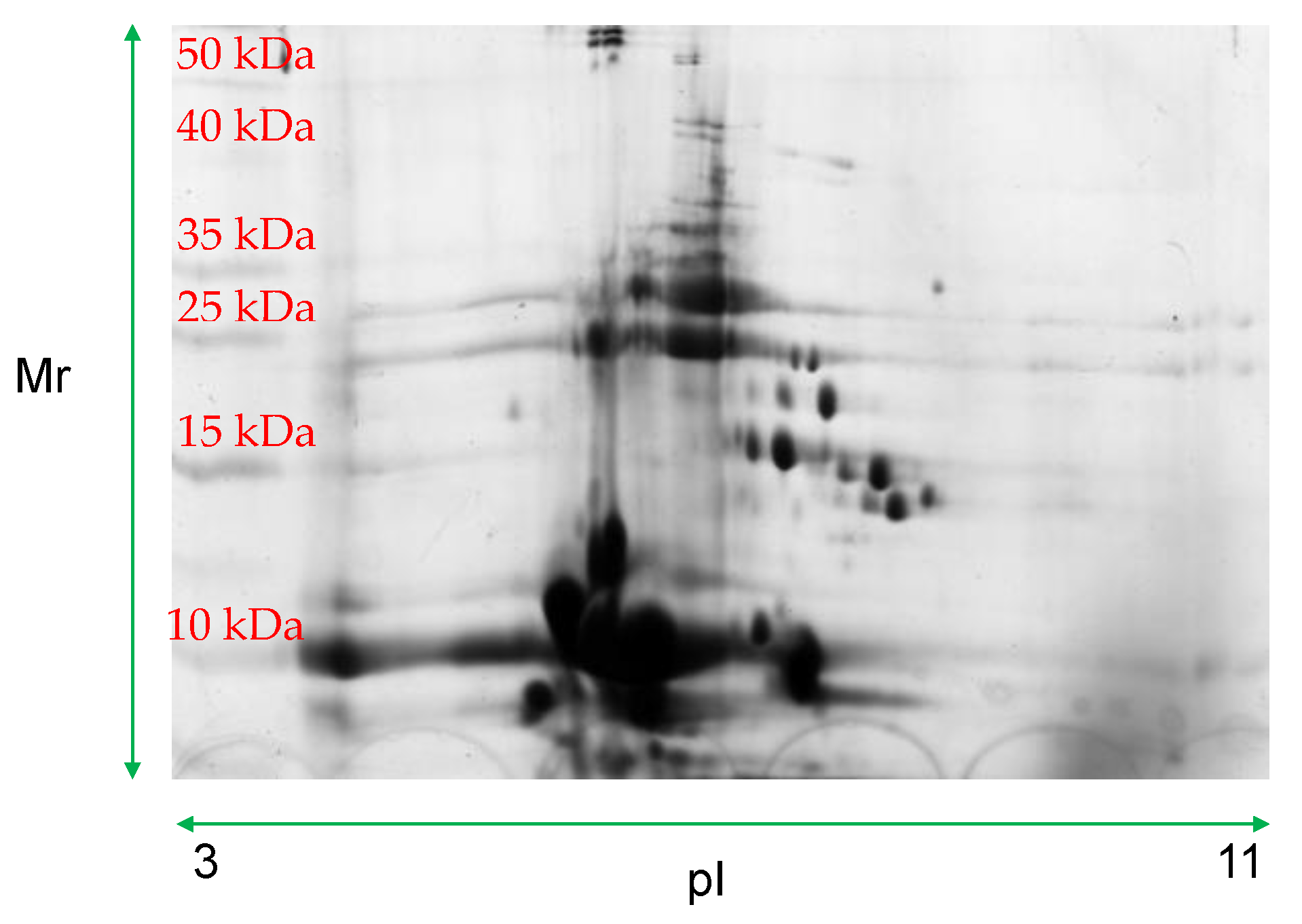
Figure 1. 1D SDS-PAGE. Explanations: 1—Soybean Zora, 2—Soybean Iscigo Wase, 3—Soybean Lokus, 4—Kidney bean Zlatý roh, 5—Kidney bean Dandy, 6—Kidney bean Wiscont, 7—Buckwheat Mihovo, 8—Rye Radošínsky rekord, 9—Oat Veles, 10—Spelt wheat, 11—Emmer wheat, 12—Bean Jantar, 13—Lentil, M—multicolor broad range protein ladder (Thermo-Fisher).
Blsf 11 00086 g001
Figure 2. 2D electrophoresis of soybean genotype Zora. Explanations: pI—isoelectric point, Mr—molecular weight—multicolor broad range protein ladder (Thermo-Fisher).
Blsf 11 00086 g002
Figure 3. PCR amplification of primer combination F1xR1 Dinelli et al. [19]. Explanations: 1—Soybean Zora, 2—Soybean Iscigo Wase, 3—Soybean Lokus, 4—Kidney bean Zlatý roh, 5—Kidney bean Dandy, 6—Kidney bean Wiscont, 7—Buckwheat Mihovo, 8—Rye Radošínsky rekord, 9—Oat Veles, 10—Spelt wheat, 11—Emmer wheat, 12—Bean Jantar, 13—Lentil, M—quick load purple 2-Log DNA ladder (New England BioLabs).
Blsf 11 00086 g003
Table 1. Lunasin-like peptide.
Spot NumberMolecular WeightIsoelectric Point
35.933.47
30015.995.82
40015.746.11
60025.977.04
70025.877.41
70035.967.67
80015.729.51
90015.8910.1
Publisher’s Note: MDPI stays neutral with regard to jurisdictional claims in published maps and institutional affiliations.

