Proceeding Paper

Overexpression of Plant-Specific Insert from Cardosin B (PSI B) in Arabidopsis Correlates with Cell Responses to Stresses †

1
Department of Biology, Faculdade de Ciências da Universidade do Porto, Rua do Campo Alegre, s/nº, 4169-007 Porto, Portugal
2
GreenUPorto—Sustainable Agrifood Production Research Center, Institute for Innovation, Training and Sustainability of Agrifood Production (Inov4Agro), Rua do Campo Alegre, s/nº, 4169-007 Porto, Portugal
*
Author to whom correspondence should be addressed.
Presented at the 2nd International Electronic Conference on Plant Sciences—10th Anniversary of Journal Plants, 1–15 December 2021; Available online: https://iecps2021.sciforum.net/.
Biol. Life Sci. Forum 2022, 11(1), 35; https://doi.org/10.3390/IECPS2021-11938
Published: 30 November 2021

Abstract

:
Under abiotic stress, several changes occur in cells regarding both their physiology and cellular mechanisms. Plants have developed modifications in the production and trafficking of proteins and the remodeling of endomembranes to overcome stress conditions. The alteration in the targeting of proteins to the vacuole by shifting their transport towards an unconventional, Golgi-independent route is a good example. Plant-specific inserts (PSIs) are known to mediate such routes, and our goal was to evaluate if transgenic Arabidopsis plants overexpressing PSI B respond differently when subjected to different abiotic stresses (osmotic, oxidative, saline and by metals). The results obtained point to a differential expression of PSI B-mCherry depending on the type of stress and a decrease of cellular and cytoplasmatic movement in all stress conditions.

1. Introduction

Most cultivated plants grow in suboptimal environments due to abiotic stress factors such as drought, salinity, oxidative stress or metal poisoning [1]. Drought and salinity are the most common abiotic stresses found, particularly in crops, and they trigger quite complex mechanisms. These types of stress are associated with a reduction in the osmotic potential of the soil solution, resulting in water stress, excess Na+ and Cl ion toxicity for the cell and nutritional imbalance. It is also related to stomatal closure, decreased photosynthetic activity, altered cell wall elasticity and ROS production [2,3,4]. Endomembrane trafficking is a fundamental cellular process in all eukaryotic cells, and its regulatory mechanisms have been extensively studied. In plants, this process is adjusted quite regularly so that it can adapt to an ever-changing environment. The main endomembrane trafficking pathways in plant cells include secretory and endocytic routes in which the molecular content is transported by vesicles between organelles and is essential for the response and adaptation to abiotic stresses [5,6]. Furthermore, some stresses, such as drought and salinity, play an important role in the process of inducing autophagy, which is quite significant in the tolerance to oxidative stress, a condition associated with most abiotic stresses [7,8]. A recent study in our lab analyzed the expression of several endomembrane-related genes involved in different pathways in Arabidopsis grown under abiotic stress conditions [9]. It was concluded that the pathway to the protein storage vacuole is favored under stress conditions, supporting the putative role of these organelles in plant stress tolerance. The plant-specific insert (PSI) corresponds to an independent domain exclusively found in some plant aspartic proteinases (APs) and consisting of about 100 amino acids [10]. Although the biological functions of plant APs are not as well characterized as those of animals, it is known that they have a role in germination, reproduction, senescence, microbial and/or insect defense, protein turnover, programmed cell death and even response to stress [11,12,13]. The PSI domain has the ability to interact with membranes and, considering that this domain undergoes structural changes depending on pH, the lipid composition of membranes govern these interactions [14]. When isolated and in vitro, this domain has a wide range of functions such as antimicrobial activity, permeabilization and membrane modulation, secretion of vesicular contents and protein processing and also functions as a vacuolar sorting signal [15,16,17,18]. It was demonstrated that the PSI domains present in cardosins A and B (two APs isolated from cardoon—Cynara cardunculus L.) and in AP1 and AP2 (APs isolated from soybean) are capable of redirecting secreted proteins to the vacuole through different pathways [19]; glycosylated PSIs mediate vacuolar transport dependent on COPII vesicles and, therefore, follow the conventional route, while non-glycosylated PSIs mediate the transport in a Golgi-independent manner. These two pathways seem to provide a certain resilience to plant cells as they can be activated depending on the stage of development and environmental conditions [17,19,20]. Taking this into account, the general objective of this work was to study the expression levels of PSI B from cardoon and its biosynthetic pathways in transgenic Arabidopsis plants overexpressing this domain coupled to m-Cherry fluorescent protein in physiological and under abiotic stress conditions. Our results show that PSI B expression is correlated with some stress conditions, although its intracellular localization remains unchanged. Interestingly, subcellular movements observed in root cells expressing PSI B-mCherry were dramatically decreased under stress conditions.

2. Material and Methods

2.1. Plant Material and Stress Assays

Homozygous PSI B-mCherry lines were obtained using the floral-dip method [21] and tested for the presence of the transgene by PCR and Western blot (data not shown). Seed germination was performed in MS (Murashige & Skoog, Duchefa Biochemie, Haarlem, The Netherlands) medium with 1.5% (w/v) sucrose and supplemented with the stress-inducing agents as described previously [9]. In short, in the abiotic stress tests, different conditions were used; S1 and S2 (salinity)—sodium chloride (50 mM and 100 mM, respectively)—mannitol (50 mM and 100 mM), hydrogen peroxide (0.5 mM) and zinc sulfate (150 μM), respectively, were added to the environment already described.

2.2. Root Biometrical Analysis

A phenotypic study was performed to evaluate biometric parameters. For this assay, the seedlings were germinated in the conditions already described, but square plates were used and incubated vertically. After a period of ten to twelve days, the plates were collected, analyzed and a photographic record was obtained. Using ImageJ/Fiji software, a metric analysis of the roots was obtained in two conditions: wild-type Arabidopsis plants and Arabidopsis overexpressing PSI B.

2.3. cDNA Preparation

To obtain total RNA preparations, 50 mg of seedlings was weighted, and the “NZY Total RNA Isolation Kit” (NZYTech, Lisboa, Portugal) was used according to the manufacturer’s instructions. Using a microdrop spectrophotometer (DeNovix DS-11, Bonsai Lab, Madrid, Spain), total RNA was quantified, and its integrity was verified in a 1% agarose gel. Total RNA was then stored at −80 °C. The cDNA preparation from RNA samples was performed using the “NZY First Strand cDNA Synthesis Kit” (NZYTech) following the protocol provided.

2.4. Quantitative RT-PCR

Three technical replicates were performed for each gene and condition in 10 µL final reaction volume, including 400 nM of each primer and 2 µL of cDNA, diluted eight times. The quantitative RT-PCR was performed using PowerUp SYBR Green Master Mix (Thermo Fisher, Waltham, MA, USA), and it was performed in a CFX96 Real-Time System (Biorad). The PCR reaction was conducted as follows: 95 °C for 3 min, followed by 40 cycles of 95 °C for 10 s, 56 °C for 10 s and 72 °C for 30 s. After 40 cycles, and to generate the melt-curve, the following conditions were used: 95 °C for 10 s, followed by a constant increase from 65 °C to 95 °C. Table 1 lists the primer pairs used for each gene. The SAND-1 and UBC9 genes were used as reference genes [22].

2.5. Confocal Microscopy

Arabidopsis seedlings expressing PSI b-mCherry, grown under the stress conditions mentioned above, were imaged using confocal laser scanning microscopy (CLSM, Leica Stellaris 8). mCherry emission was detected between 580 nm and 630 nm, using 561 nm excitation. Images were processed using ImageJ/Fiji software. For the mean intensity quantification, the same settings were used to acquire the images, and more than 20 cells were quantified.

3. Results

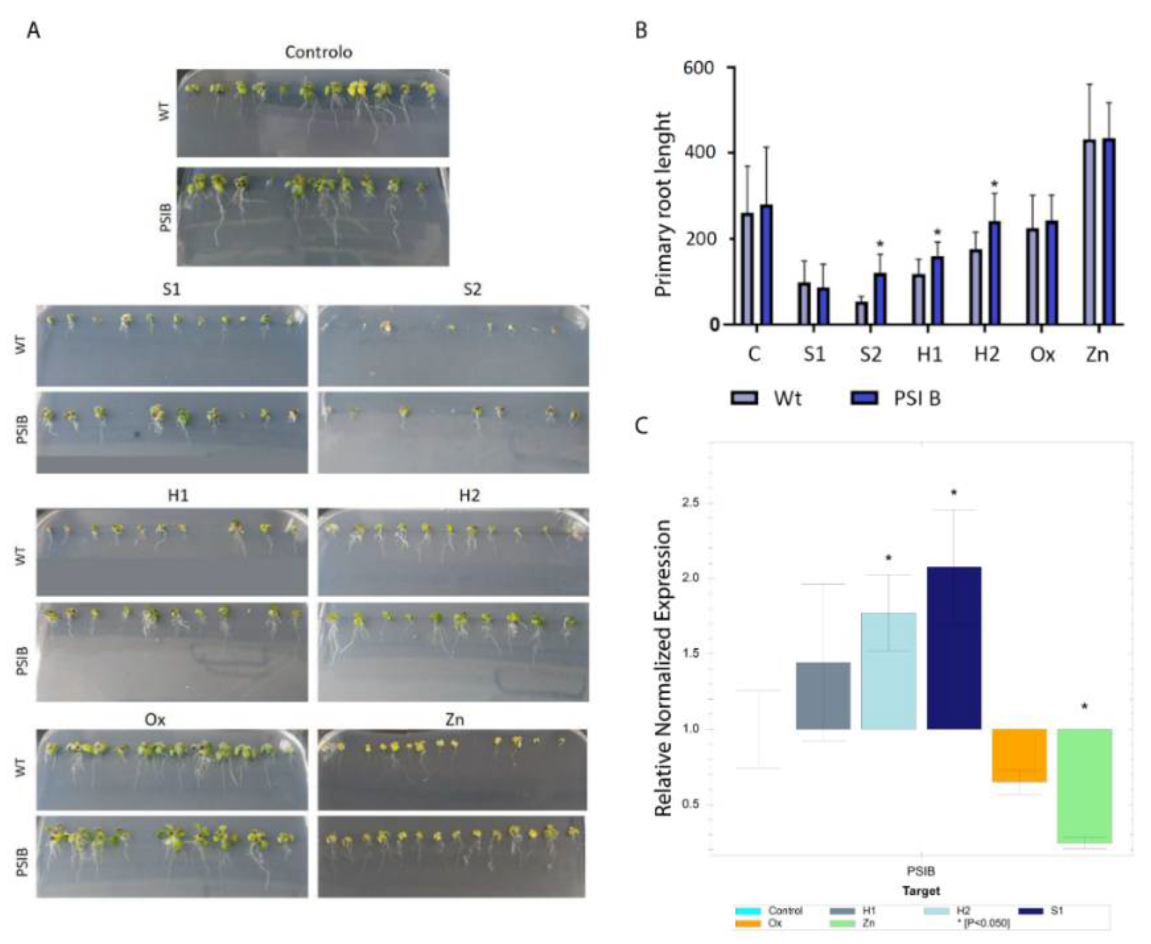
3.1. PSI B Expression Changes in Plants under Abiotic Stress

After obtaining homozygous PSI B-mCherry lines, and to study the influence of different types of stress on the expression of PSIs of Arabidopsis thaliana, abiotic stress tests were performed. Regarding the morphological aspects of the seedlings, in the lower salt concentration (S1) and high-concentration water stress (H2), the changes were quite similar, with a noticeable delay in development and some leaf chlorosis (Figure 1A).
The plants under the mild water (H1) and metal (Zn) stresses showed a slight delay in development in relation to the control, and those under Zn showed a high degree of chlorosis. Regarding oxidative stress (Ox), the plants did not show any morphological changes. Under the high salt concentration (S2), the plants did not develop, and, therefore, this condition was not used for further evaluations. Analysis of the primary root length of wild-type and Arabidopsis plants overexpressing PSI B-mCherry, in the same stress conditions, was performed (Figure 1B). In the control conditions, no changes were visible. In S1, both in wild-type and transformed plants, a marked decrease in root development was verified, with similar values in control and PSI B plants. However, in S2, and despite the plant development being quite compromised, a significant increase in the root length of plants expressing PSI B-mCherry when compared to WT plants was noticeable. The same tendency was observed in plants grown under H1 and H2 stresses. Regarding the plants under Ox and Zn conditions, no changes were observed compared to the control. The expression of PSI B-mCherry in Arabidopsis grown under abiotic stress conditions was evaluated by qPCR, using the normalized expression method with a p-value of 0.05, and the results presented are relative to control conditions (Figure 1C). From the data obtained, PSI B-mCherry was upregulated in H1-, H2- and S1-grown plants, despite only H2 and S1 having significant values. Interestingly, in Ox- and Zn-grown plants, the situation was reversed, and PSI B-mCherry was downregulated.

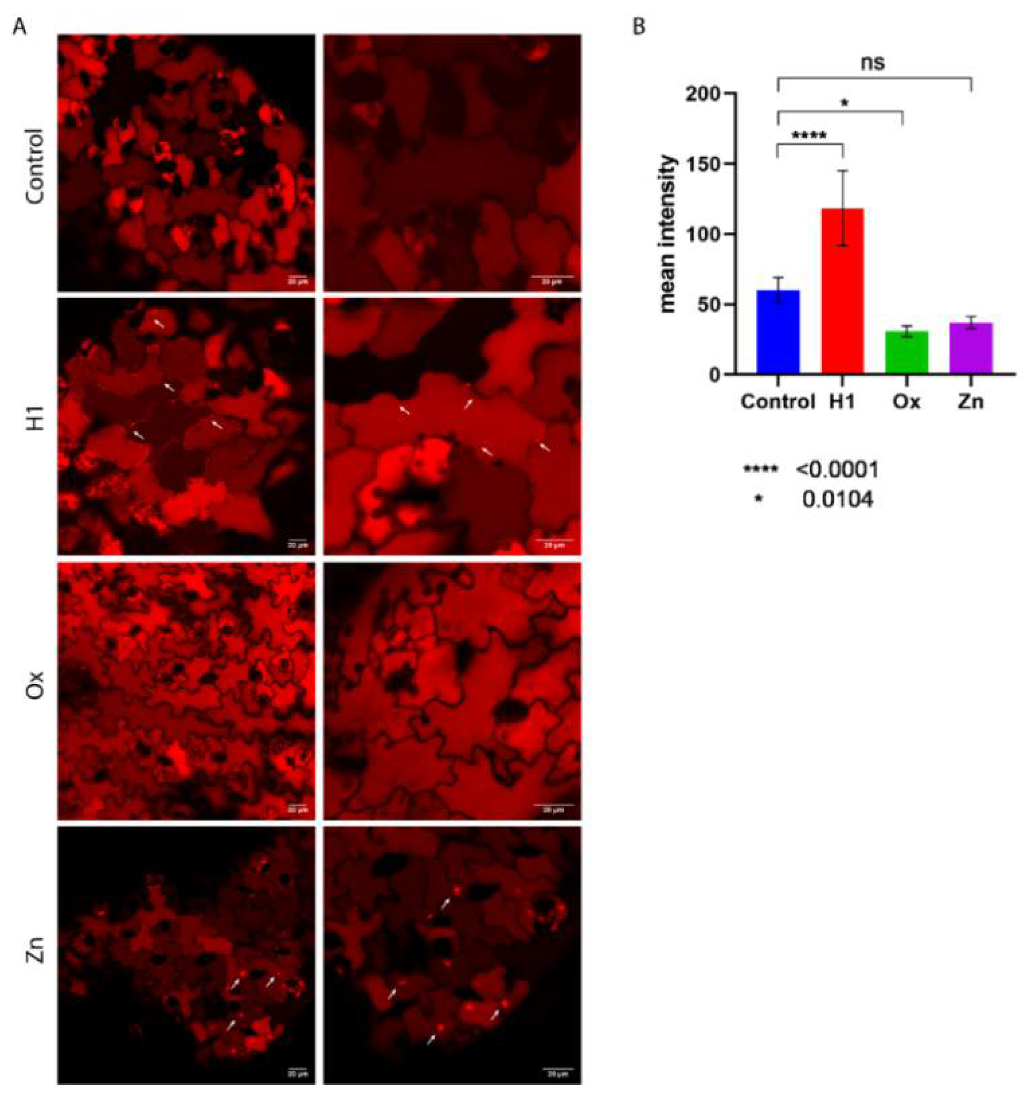
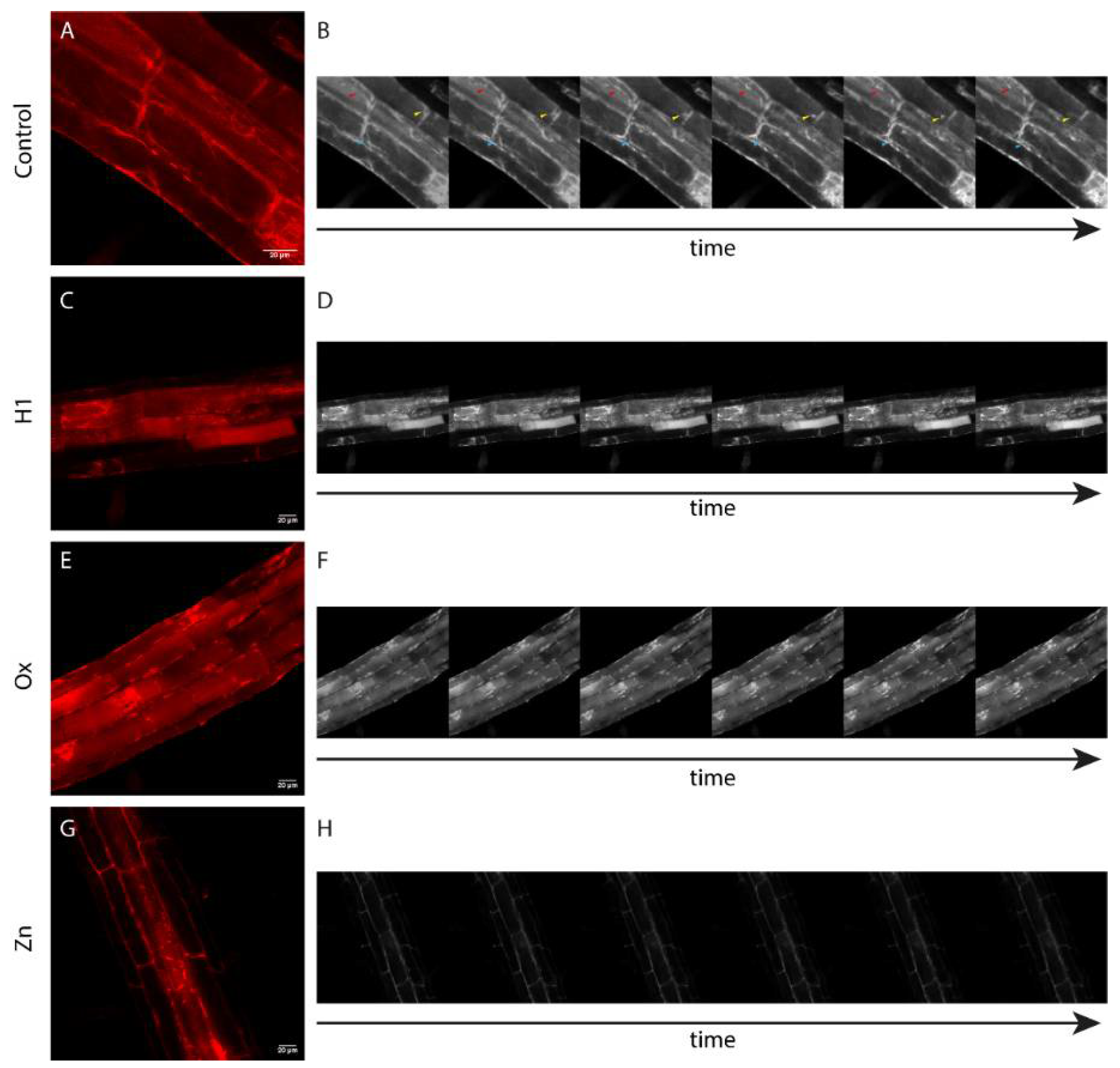
3.2. PSI B Localization in Arabidopsis Plants under Stress

Given the changes in PSI B-mCherry expression and the phenotypes observed, we checked whether the localization of PSI B was altered in the leaves and roots of plants grown in H1, Ox and Zn stress conditions when compared to the control (Figure 2 and Figure 3).
In leaves (Figure 2), PSI B-mCherry accumulated mainly in the vacuole in all the conditions studied (Figure 2A), despite some differences in the fluorescence intensity being detected, as shown by the mean fluorescence quantification (Figure 2B). A significatively higher amount of fluorescence was detected in the leaf cells of plants grown under H1 conditions, while a decrease in fluorescence was observed in those grown under Ox conditions. Interestingly, in the H1 conditions, some fluorescence was observed at the periphery of the cell (Figure 2A, H1, arrows), resembling endoplasmic reticulum patterns. In the Zn conditions, it was possible to detect some fluorescent agglomerates inside some cells (Figure 2A, Zn, arrows) that were not observed in any other condition.
In root cells, the accumulation pattern for PSI B-mCherry was quite different from that of the leaves (Figure 3). In control plants, PSI B-mCherry accumulated around the nucleus and at the cell periphery, in compartments that move within the cell (Figure 3B, colored arrowheads; Movie S1). Interestingly, in the other conditions tested, although PSI B-mCherry fluorescence was still detected in similar compartments (Figure 3C,E,G), the movements were no longer detected (Figure 3D–F; Movies S2–S4). Aditionally, in H1 conditions, several cells presented some fluorescence in the vacuole (Figure 3C), and, in Ox-grown plants, fluorescence aggregates were found inside the cells (Figure 3E).

4. Discussion

Throughout evolution, plants are exposed to adverse environmental conditions, and, to face and respond to these stresses, plants develop special mechanisms of cellular reorganization. Most abiotic stresses have similar physiological consequences, such as the induction of cell damage and, thus, induce similar signaling pathways. In this work, the conditions in which Arabidopsis plants were grown not only caused changes at the morphological level but also in the expression of PSI B. A recent study regarding APs expression suggested their involvement in defense against or tolerance to hydric and salt stress [23]. In similar conditions, our results show a higher expression of PSI B. The observed higher fluorescent accumulation of PSI B-mCherry in the H1-grown plants supports those results. The downregulation of PSI B-mCherry during metal and oxidative stresses reinforces the importance of this domain in hydric and saline stresses and may indicate a role in processes related to cellular homeostasis and water control. A study performed by our team [24] regarding Arabidopsis thaliana aspartic proteinases revealed that, depending on the abiotic stress, the three APs genes tested had different and antagonistic expression levels. In plants grown under lower salt concentration, PSI B-mCherry expression responded similarly to the overexpression of AP1 and AP3. Likewise, in the heavy metal stress assay, the downregulation of PSI B resembled the behavior of AP3. This similarity may be related to AP3′s function, being implicated in the processing and degradation of storage proteins. This is quite interesting since this overexpression in salt and mild hydric stresses suggests that degradation of storage proteins occurs to overcome and tolerate these restricted conditions. Based on these results, we can infer that PSI B may have a function related to the activation of genes responsible for the degradation of storage proteins. In addition, the study also showed the expression of several endomembrane transport-related genes involved in different vacuolar pathways in response to abiotic stress [9,24]. The results revealed that the pathway to the protein storage vacuole was enhanced, and, since PSI is a vacuolar determinant [17], it may participate in this reinforcement phenomenon. Thus, the PSI may then be associated with development and the defense system and yet operate by distinct mechanisms, as each type of stress may trigger a different response [9,14]. Regarding the biometric parameters, and, particularly, the root length, it seems that the expression of PSI B may also influence the plant’s ability to face some adverse conditions. Looking at high-salt conditions, there was a significant increase in the root length in the transformed plants, suggesting an increased stress tolerance provided by PSI B overexpression that may be triggered at certain concentrations. The plants grown under high and mild water stress conditions also showed significant changes in roots length. Given the PSI roles in membrane interaction or protein processing [15,18], these results suggest increased tolerance in order to mitigate the negative effects of environmental adversities. Interestingly, analysis of PSI B-mCherry localization in roots showed a marked decrease in cytoplasmic movements under all stress conditions, raising the question as to whether vesicle movement can be constrained by stress.

5. Conclusions

In the present study, we analyzed the expression of PSI B during stress conditions. From the results obtained, we can suggest that PSI B has an active role in adaptation and tolerance mechanisms against abiotic stress, particularly salt and hydric stress, where its expression has a positive effect on plant fitness. Despite still being very preliminary, the data presented here are encouraging, and it is worth investigating in higher detail the role of the PSIs, and aspartic proteinases in general, in plants’ responses to stress.

Supplementary Materials

The following are available online at https://www.mdpi.com/article/10.3390/IECPS2021-11938/s1, Movie S1: Time-lapse video illustrating the movement of PSI B-mCherry-labeled compartments in control conditions, Movie S2: Time-lapse video illustrating the movement of PSI B-mCherry-labeled compartments in H1 condition, Movie S3: Time-lapse video illustrating the movement of PSI B-mCherry-labeled compartments in Ox condition, Movie S4: Time-lapse video illustrating the movement of PSI B-mCherry-labeled compartments in Zn condition.

Author Contributions

I.M., A.S., S.P. and C.P. conceived and designed the experiments; I.M. performed the experiments; I.M., A.S. and C.P. analyzed the data; J.P. contributed reagents and materials; I.M. and C.P. wrote the paper; A.S., S.P. and J.P. reviewed the manuscript. All authors have read and agreed to the published version of the manuscript.

Funding

This research was performed in the frame of the scientific project PTDC/BIAFBT/32013/2017, funded by the Portuguese Foundation for Science and Technology (FCT) and also supported by national funds through FCT, within the scope of UIDB/05748/2020 and UIDP/05748/2020.

Institutional Review Board Statement

Not applicable.

Informed Consent Statement

Not applicable.

Data Availability Statement

Not applicable.

Conflicts of Interest

The authors declare no conflict of interest.

References

  1. Peleg, Z.; Apse, M.P.; Blumwald, E. Engineering Salinity and Water-Stress Tolerance in Crop Plants. Getting Closer to the Field; Academic Press: Cambridge, MA, USA, 2011; Volume 57. [Google Scholar]
  2. Evelin, H.; Kapoor, R.; Giri, B. Arbuscular mycorrhizal fungi in alleviation of salt stress: A review. Ann. Bot. 2009, 104, 1263–1280. [Google Scholar] [CrossRef] [PubMed] [Green Version]
  3. Gigon, A.; Matos, A.R.; Laffray, D.; Zuily-Fodil, Y.; Pham-Thi, A.T. Effect of drought stress on lipid metabolism in the leaves of Arabidopsis thaliana (Ecotype Columbia). Ann. Bot. 2004, 94, 345–351. [Google Scholar] [CrossRef]
  4. Sun, J.K.; Li, T.; Xia, J.B.; Tian, J.Y.; Lu, Z.H.; Wang, R.T. Influence of salt stress on ecophysiological parameters of Periploca sepium Bunge. Plant Soil Environ. 2011, 57, 139–144. [Google Scholar] [CrossRef]
  5. Scheuring, D.; Kleine-Vehn, J. On the discovery of an endomembrane compartment in plants. Proc. Natl. Acad. Sci. USA 2020, 117, 10623–10624. [Google Scholar] [CrossRef] [PubMed]
  6. Wang, X.; Xu, M.; Gao, C.; Zeng, Y.; Cui, Y.; Shen, W.; Jiang, L. The roles of endomembrane trafficking in plant abiotic stress responses. J. Integr. Plant Biol. 2020, 62, 55–69. [Google Scholar] [CrossRef] [PubMed]
  7. Liu, Y.; Xiong, Y.; Bassham, D.C. Autophagy is required for tolerance of drought and salt stress in plants. Autophagy 2009, 5, 954–963. [Google Scholar] [CrossRef] [PubMed] [Green Version]
  8. Signorelli, S.; Tarkowski, Ł.P.; Van den Ende, W.; Bassham, D.C. Linking Autophagy to Abiotic and Biotic Stress Responses. Trends Plant Sci. 2019, 24, 413–430. [Google Scholar] [CrossRef] [Green Version]
  9. Neves, J.; Sampaio, M.; Séneca, A.; Pereira, S.; Pissarra, J.; Pereira, C. Abiotic Stress Triggers the Expression of Genes Involved in Protein Storage Vacuole and Exocyst-Mediated Routes. Int. J. Mol. Sci. 2021, 22, 10644. [Google Scholar] [CrossRef] [PubMed]
  10. Faro, C.; Gal, S. Aspartic Proteinase Content of the Arabidopsis Genome. Curr. Protein Pept. Sci. 2005, 6, 493–500. [Google Scholar] [CrossRef]
  11. Kervinen, J.; Tormakangas, K.; Runeberg-Roos, P.; Guruprasad, K.; Blundell, T.; Teeri, T.H. Structure and possible function of aspartic proteinases in barley and other plants. In Proceedings of the Advances in Experimental Medicine and Biology; Springer: New York, NY, USA, 1995; Volume 362, pp. 241–254. [Google Scholar]
  12. Mutlu, A.; Susannah, G. Plant aspartic proteinases: Enzymes on the way to a function. Physiol. Plant. 1999, 105, 569–576. [Google Scholar] [CrossRef]
  13. Simões, I.; Faro, C. Structure and function of plant aspartic proteinases. Eur. J. Biochem. 2004, 271, 2067–2075. [Google Scholar] [CrossRef] [PubMed] [Green Version]
  14. Cheung, L.K.Y.; Dupuis, J.H.; Dee, D.R.; Bryksa, B.C.; Yada, R.Y. Roles of Plant-Specific Inserts in Plant Defense. Trends Plant Sci. 2020, 25, 682–694. [Google Scholar] [CrossRef]
  15. Egas, C.; Lavoura, N.; Resende, R.; Brito, R.M.M.; Pires, E.; de Lima, M.C.P.; Faro, C. The Saposin-like Domain of the Plant Aspartic Proteinase Precursor Is a Potent Inducer of Vesicle Leakage. J. Biol. Chem. 2002, 275, 38190–38196. [Google Scholar] [CrossRef] [PubMed] [Green Version]
  16. Muñoz, F.F.; Mendieta, J.R.; Pagano, M.R.; Paggi, R.A.; Daleo, G.R.; Guevara, M.G. The swaposin-like domain of potato aspartic protease (StAsp-PSI) exerts antimicrobial activity on plant and human pathogens. Peptides 2010, 31, 777–785. [Google Scholar] [CrossRef] [PubMed]
  17. Pereira, C.; Pereira, S.; Satiat-Jeunemaitre, B.; Pissarra, J. Cardosin A contains two vacuolar sorting signals using different vacuolar routes in tobacco epidermal cells. Plant J. 2013, 76, 87–100. [Google Scholar] [CrossRef] [Green Version]
  18. Terauchi, K.; Asakura, T.; Ueda, H.; Tamura, T.; Tamura, K.; Matsumoto, I.; Misaka, T.; Hara-Nishimura, I.; Abe, K. Plant-specific insertions in the soybean aspartic proteinases, soyAP1 and soyAP2, perform different functions of vacuolar targeting. J. Plant Physiol. 2006, 163, 856–862. [Google Scholar] [CrossRef] [PubMed]
  19. Vieira, V.; Peixoto, B.; Costa, M.; Pereira, S.; Pissarra, J.; Pereira, C. N-linked glycosylation modulates Golgi-independent vacuolar sorting mediated by the plant specific insert. Plants 2019, 8, 312. [Google Scholar] [CrossRef] [Green Version]
  20. Pereira, C.S.; da Costa, D.S.; Pereira, S.; de Moura Nogueira, F.; Albuquerque, P.M.; Teixeira, J.; Faro, C.; Pissarra, J. Cardosins in postembryonic development of cardoon: Towards an elucidation of the biological function of plant aspartic proteinases. Protoplasma 2008, 232, 203–213. [Google Scholar] [CrossRef] [Green Version]
  21. Clough, S.J.; Bent, A.F. Floral dip: A simplified method for Agrobacterium-mediated transformation of Arabidopsis thaliana. Plant J. 1998, 16, 735–743. [Google Scholar] [CrossRef] [Green Version]
  22. Czechowski, T.; Stitt, M.; Altmann, T.; Udvardi, M.K.; Scheible, W.-R. Genome-Wide Identification and Testing of Superior Reference Genes for Transcript Normalization in Arabidopsis. Plant Physiol. 2005, 139, 5–17. [Google Scholar] [CrossRef] [Green Version]
  23. Guo, R.; Zhao, J.; Wang, X.; Guo, C.; Li, Z.; Wang, Y.; Wang, X. Constitutive expression of a grape aspartic protease gene in transgenic Arabidopsis confers osmotic stress tolerance. Plant Cell. Tissue Organ Cult. 2015, 121, 275–287. [Google Scholar] [CrossRef]
  24. Neves, J.; Séneca, A.; Pereira, S.; Pissarra, J.; Pereira, C. Abiotic Stress Upregulates the Expression of Genes Involved in PSV and Autophagy Routes. Biol. Life Sci. Forum 2021, 4, 40. [Google Scholar]
Figure 1. Phenotypic and expression analysis of Arabidopsis plants expressing PSI B-mCherry grown under stress conditions. (A) Arabidopsis thaliana germinated for 10 to 12 days in stress conditions. (B) Primary root metric analysis. (C) PSI B-mCherry expression analysis by qPCR relative to control. * Statistically significant values. S1—Low salt concentration; S2—high salt concentration; H1—mild water stress; H2—high water stress; Ox—oxidative stress; Zn—stress induced by metals.
Blsf 11 00035 g001
Figure 2. Confocal microscopy analysis of leaves from Arabidopsis plants expression PSI B-mCherry under stress conditions. (A) Representative images of leaf cells. Arrows indicate deviation patterns relative to control. (B) Quantification of the mean fluorescent intensity in cells expressing PSI B-mCherry. * Statistically significant values. H1—mild water stress; Ox—oxidative stress; Zn—stress induced by metals.
Blsf 11 00035 g002
Figure 3. Confocal microscopy analysis of roots from Arabidopsis plants expression PSI B-mCherry under stress conditions. (A,C,E,G) Representative images of root cells expressing PSI B-mCherry. (B) Stills retrieved from time-lapse video illustrating the movement of PSI B-mCherry-labeled compartments (colored arrows). (D,F,H) Stills retrieved from time-lapse video illustrating the movement of PSI B-mCherry-labeled compartments. H1—mild water stress; Ox—oxidative stress; Zn—stress induced by metals.
Blsf 11 00035 g003
Table 1. List of genes and corresponding primer pairs used in the quantitative RT-PCR assay.
GenePrimer ForwardPrimer Reverse
UBQ_9TCACAATTTCCAAGGTGCTGCTCACAATTTCCAAGGTGCTGC
SAND-1AACTCTATGCAGCATTTGATCCACTTGATTGCATATCTTTATCGCCATC
m-CherryGACCACCTACAAGGCCAAGGTGGGAGGTGATGTCCAACT
Publisher’s Note: MDPI stays neutral with regard to jurisdictional claims in published maps and institutional affiliations.

