Proceeding Paper

Detection of Celiac Active Polypeptides in Wheat, Oat and Buckwheat Using Immunochemical Methods †

1
Faculty of Biotechnology and Food Sciences, Institute of Biotechnology, Slovak University of Agriculture in Nitra, Trieda Andreja Hlinku 2, 949 76 Nitra, Slovakia
2
Faculty of Agrobiology and Food Resources, Institute of Plant and Environmental Sciences, Slovak University of Agriculture in Nitra, Trieda Andreja Hlinku 2, 949 76 Nitra, Slovakia
3
Faculty of Economics and Management, Institute of Statistics, Operations Research and Mathematics, Slovak University of Agriculture in Nitra, Trieda Andreja Hlinku 2, 949 76 Nitra, Slovakia
*
Author to whom correspondence should be addressed.
Presented at the 2nd International Electronic Conference on Plant Sciences—10th Anniversary of Journal Plants, 1–15 December 2021; Available online: https://iecps2021.sciforum.net/.
Biol. Life Sci. Forum 2022, 11(1), 27; https://doi.org/10.3390/IECPS2021-11976
Published: 30 November 2021

Abstract

:
The aim of the study was to analyze the proteome of the wheat (Triticum aestivum L.), oat (Avena sativa) and buckwheat (Fagopyrum esculentum Moench.) genotypes using immunochemical methods and to detect the presence of celiac active polypeptides. The results of ELISA confirmed a high gluten content in wheat (43,278.51 mg·kg−1). The buckwheat varieties (average 2.03 mg·kg−1) fulfill the criteria for labeling as gluten-free food. The results confirm the oat varietal dependence on the content of allergenic proteins. The Western blot analysis revealed the strongest reactions of the polyclonal antibody with wheat proteins of 25 kDa to 50 kDa and with oat proteins of 20 kDa and 40–55 kDa. No signal was detected in buckwheat, which was confirmed by ELISA results. Buckwheat can be recommended as a safe crop for the preparation of gluten-free foods.

1. Introduction

In recent years, the number of people suffering from certain diseases resulting from civilization, such as food allergies or various intolerances, has increased. Some foods can cause an allergic reaction or problems caused by food intolerance in people who are sensitive to a particular allergen [1]. These food allergens include proteins or glycoproteins with a molecular weight of 5 kDa to 100 kDa with the ability to bind to IgE receptors [2]. Celiac disease is one of the most widespread food intolerances in the world. It is an inflammatory disease of the upper small intestine caused by the consumption of proteins in wheat; rye; barley; and in some cases, oats in people with a genetic predisposition to the disease. However, opinions on the use of oats in the preparation of foods suitable for patients with celiac disease are still discussed [3]. The disease is caused by immunological intolerance to gluten. All allergenic fragments share two celiac active tetrapeptide fragments in the N-terminal region of the protein: Pro-Ser-Gln-Gln and Gln-Gln-Gln-Pro [4,5]. Allergen-specific immunoglobulin antibodies E play a key role in the development of food allergy. Recent advances in molecular biology techniques have enabled efficient analyzes of food allergens. Allergen-specific IgE antibodies and peptide epitopes are useful indicators for identifying patients with food allergy, predicting clinical severity and detecting changes in food tolerance [6]. There are several analytical methods that allow for the quantitative and qualitative detection of allergenic food residues. The most commonly used method is ELISA, which can specifically detect proteins from allergenic sources, is sufficiently sensitive and allows for rapid determination of residue limits in industrial food processing [7]. Proteomic techniques, in combination with Western blot, make it possible to identify allergens and make a significant contribution to the acquisition of new knowledge, which serves to develop diagnostic methods by detecting the binding of IgE antibodies to specific proteins (allergens). Two-dimensional electrophoresis is also used in combination with immunoblotting and mass spectrometry, which allow for the identification and sequential processing of unknown sample extracts [8].
The aim of this work was to analyze the proteome of selected varieties of wheat, oats and buckwheat, with an emphasis on the detection of celiac active polypeptides that cause allergic reactions in people with hypersensitivity.

2. Materials and Methods

2.1. Biological Material

Grains of wheat varieties (Triticum aestivum L.) Viglanka, Elinor and PS Puqa; grains of oat varieties (Avena sativa L.) Vendelin, Valentin and Zvolen; and grains of buckwheat varieties (Fagopyrum esculentum Moench) Špačinská1, Pyra and Jana C1 were analyzed. Samples were obtained from the Gene Bank of Seed Species of the Slovak Republic NPPC VÚRV in Piešťany.

2.2. ELISA Method

The content of celiac active proteins in the grains of the analyzed crops was determined by ELISA method using the AgraQuant Gluten G12 test (Romer Labs). Protein extraction was performed according to the manufacturer’s instructions; 100 µL of standards and samples were added to the wells of the microtiter plate. After a 20 min incubation, 100 µL of conjugate was applied, then 100 µL of substrate was added and the reaction was stopped by adding 100 µL of stop solution. The absorbance of the standards and samples at a wavelength of 450 nm was measured on an ELISA reader (BioTek Instruments, Winooski, VT, USA). The amount of protein in the analyzed sample was determined by subtracting the values from the calibration curve.

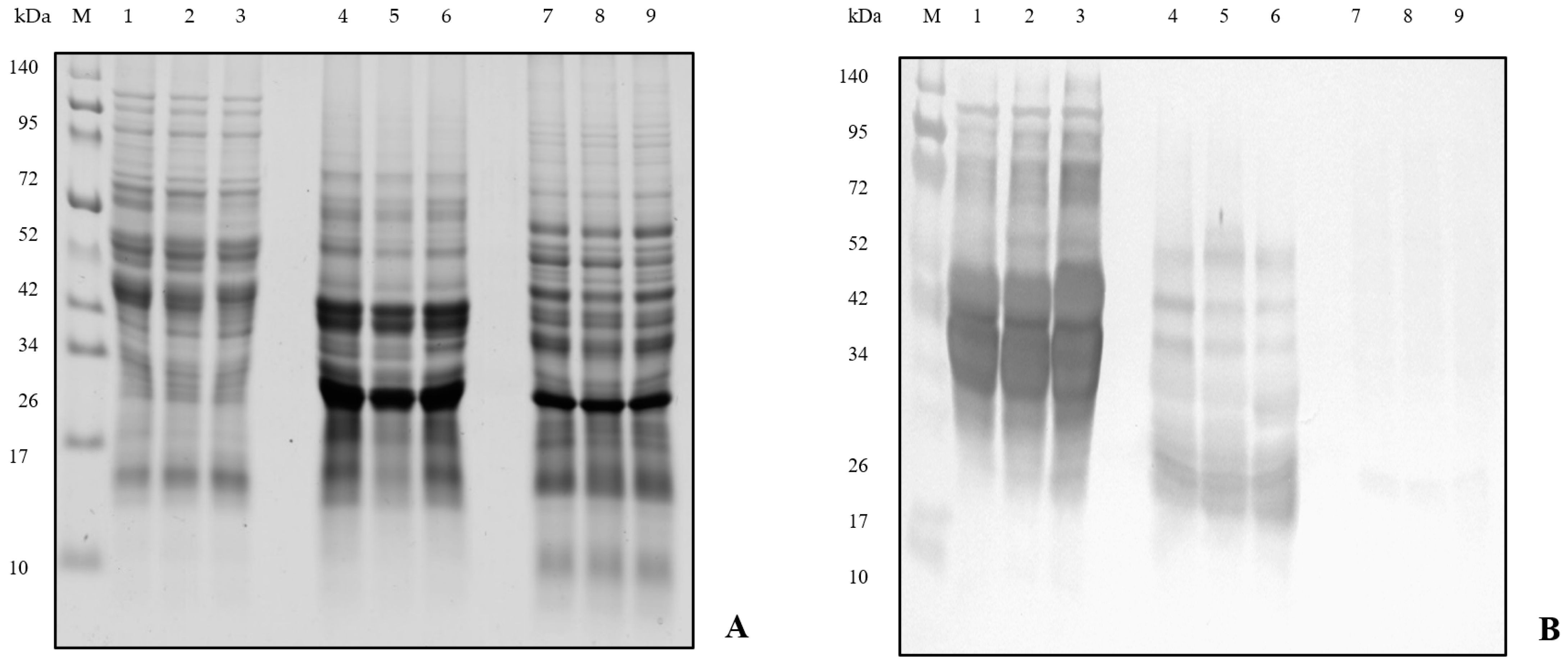
2.3. Western Blot Analysis

Protein extraction was performed according to the Schägger methodology [9]. The extracted proteins were denatured at 100 °C for 5 min before loading onto the gel. Electrophoretic analysis of proteins was performed in Tris-tricine SDS-PAGE according to the methodology of Schägger [9] in the Bio-Rad Mini-Protean Tetra System; 10 µL of the broad-spectrum protein ladder Spectra Multicolor and 3 μL of the protein samples were loaded onto the gel. Protein separation was performed for 40 min at 30 volts and 120 min at 90 volts. The gel was stained overnight in Coomassie Brilliant Blue R-250 solution, and then, the background was decolorized in 10% acetic acid.
Electrotransfer of proteins from the gel to the immobilon-P polyvinylidene fluoride membrane (Millipore) was performed in an OmniBLOT Mini Blotting system in a buffer medium for 90 min at 170 mA. The membrane was placed in a TBS/BSA solution overnight, then incubated with the primary antibody and then incubated with the secondary antibody. The resulting immunocomplexes were detected with the chromogenic substrate SIGMAFAST 3,3′-diaminobenzidine. Membranes were read on a GS-800 Calibrated Densitometer, modified in Quantity One and evaluated by Image Lab.

3. Results and Discussion

Individuals with a genetic predisposition to celiac disease are dependent on gluten-free foods [10,11,12]. The AgraQuant® Gluten G12 ELISA with the G12 monoclonal antibody, which forms a complex with prolamins and allows for the quantification of proteins in the range of 4 to 200 mg·kg−1, was applied to determine the celiac active protein. The G12 antibody recognizes amino acid sequences QPQLPY and QPQLPF, which are less common; occur only in α-gliadins and in some ω-1,2-gliadins and γ-gliadins; and bind to amino acid motifs QPQLPL, QPQQPY, QPQQPF and QPELP [13].
The gluten content in the wheat varieties analyzed (Table 1) ranged from 23,899.63 mg·kg−1 (Elinor) to 67,385.83 mg·kg−1 (PS Puqa), on the basis of which it can be stated that the gluten content far exceeds the set limit, and therefore, it is not a suitable crop for a gluten-free diet.
The content of celiac active polypeptides in the oat samples analyzed varied considerably in individual varieties. The variety Zvolen with a gluten content of 10.57 mg·kg−1 meets the criteria of gluten-free food, while the varieties Vendelin (44.71 mg·kg−1) and Valentin (57.37 mg·kg−1) belong among the low-gluten foods. Discussions about the suitability of using oats in a gluten-free diet vary widely [3,4].
A very low gluten content was determined in buckwheat samples (Table 1) with an average value of 2.03 mg·kg−1. The content of gluten proteins in buckwheat ranged from 2.13 mg·kg−1 (Pyra) to 3.97 mg·kg−1 (Jana C1), while in the genotype Špačinská 1, it was below the detection limit. These results show that all buckwheat samples meet the limit for the label “gluten-free” and can be used to produce foods suitable for patients with celiac disease.
The results obtained correspond to the conclusions of work by other authors. The content of celiac active proteins in 23 summer wheat genotypes using two different ELISA tests with antibody R5 and G12 was determined by [14]. The content of celiac active prolamins determined by the G12 antibody in the analyzed samples ranged from 58,500 mg·kg−1 to 106,500 mg·kg−1. The presence of gluten in a set of different crops using the RIDASCREEN® ELISA test was detected by [15], and the gluten content in wheat samples was determined to be from 5240 mg·kg−1 to 115,070 mg·kg−1, while in oat samples, it ranged from 60 mg·kg−1 to 21,300 mg·kg−1 depending on the variety. Ref. [16] found the gluten content of oats, flour and oat products using the RIDASCREEN® Fast Gliadin test, showing that the average gluten content in the oat sample was 316.6 mg·kg−1 and in the flour sample was 522.2 mg·kg−1. According to the results of [17], the gluten content in all analyzed buckwheat samples using the R5 antibody was below the detection limit.
Western blot is a technique by which isolated proteins are separated by SDS-PAGE and subsequently transferred to a PVDF membrane. Next, the target protein is labeled with an appropriate primary and secondary antibody and detected with a specific substrate. The intensity of the protein band corresponds to the amount of protein present [18]. The presence of potentially allergenic proteins (Figure 1), which are the most common triggers of celiac disease and other protein-induced diseases of various crops, was detected in the wheat, oat and buckwheat samples analyzed using the Western blot method. The resulting membranes were evaluated based on the molecular weights and intensity of the blotted prolamins after immunization with the antibody and were compared with the electrophoretic spectrum of the proteins from SDS-PAGE.
The protein profiles of all three analyzed wheat genotypes were very similar. The molecular weight of wheat proteins was detected in the range from 120 kDa to 13 kDa. Evaluation of the membrane (Figure 1) revealed a high concentration of celiac active proteins in all wheat samples. The polyclonal antibody reacted with almost all wheat proteins with a molecular weight from 120 kDa up to 20 kDa. The strongest signal was given by proteins with a molecular weight of 50 kDa to 25 kDa, which are the most risky protein fractions due to the presence of celiac active proteins. Only proteins with a molecular weight of less than 20 kDa did not react with the antibody.
The molecular weight of the proteins in the individual oat genotypes on SDS-PAGE ranged from 60 kDa to 13 kDa, with the oat proteins showing a weaker reaction with the polyclonal antibody compared with the wheat samples. An evaluation of the membrane revealed that the allergenic oat proteins had a molecular weight of 55 kDa to 17 kDa. The strongest signal of oat proteins was shown in proteins with a molecular weight of about 20 kDa and from 55 kDa to 40 kDa. Proteins with a molecular weight of approximately 34 kDa, which were most intense on SDS-PAGE, did not react with the antibody.
Buckwheat proteins separated in an SDS-PAGE gel had a molecular weight of 95 kDa to 5 kDa, with the most intense buckwheat proteins detected in the region of 50 kDa to 30 kDa and about 20 kDa. These proteins showed almost no reaction with the polyclonal antibody. A very weak reaction was observed in proteins with a molecular weight of about 20 kDa, which could probably be due to a cross-reaction. The results of the Western blot confirmed the conclusions of the ELISA method, and it can be stated that buckwheat is a suitable raw material for the preparation of gluten-free foods.
The results achieved are in accordance with the work of other authors. Ref. [19] compared the prolamin proteins of wheat, barley, rye and triticale using electrophoretic and immunochemical methods. All proteins with a molecular weight higher than 25 kDa were labeled by immunoblotting in wheat samples using a polyclonal antibody. The antibody did not react with proteins with a molecular weight of 15 kDa to 25 kDa. Proteins with a molecular weight of 35 kDa to 45 kDa gave the strongest signal. Ref. [20] analyzed the allergenicity of buckwheat proteins before and after hydrolysis by proteases. By monitoring the protein profile of the buckwheat in an SDS-PAGE gel, they detected the presence of proteins with a molecular weight of 50 kDa to 30 kDa, with the most intense band having a molecular weight of about 20 kDa. These proteins did not react with the antibody and did not detect the presence of any allergenic proteins in buckwheat samples.
Ref. [4] performed a proteomic analysis of seed proteins of six oat genotypes, with the molecular weight of the proteins ranging from 80 kDa to 10 kDa. The proteins were transferred to a membrane where the presence of allergenic oat proteins in the region with a molecular weight of 70 kDa to 25 kDa was detected. These proteins reacted with the antibody, and proteins with a molecular weight of about 37 kDa showed the strongest signal.
Many other proteomic techniques are used to further identify proteins, such as liquid chromatography coupled to mass spectrometry (LC-MS/MS), which is one of the most promising non-immunological methods for the identification, quantification and resolution of gluten-forming proteins and is based on accurate molecular weight peptide biomarkers [21].

4. Conclusions

The ELISA method and the Western blot have been shown to be accurate and sufficiently sensitive analyses by which it is possible not only to accurately detect but also to quantify the content of celiac agents in plant samples. The gluten content of wheat exceeds the set limit, while buckwheat met the standard for a gluten-free crop. It has been shown that the allergenicity of oats is variety-dependent, and the applied methods can be used to detect this varietal specificity and, thus, to select varieties suitable for food production and for consumption by individuals with predisposed conditions. Using immunochemical methods, it is possible to quickly and reproducibly detect allergenic proteins in raw materials prepared for the production of foods suitable for risk groups in the population of people who suffer from food allergies or intolerance to certain protein components of grains.

Author Contributions

M.C., D.R., M.V., K.R., Z.H. and L.M., methodology and investigation; Z.G., writing and editing; Ž.B. and J.D., software. All authors have read and agreed to the published version of the manuscript.

Funding

This research received no external funding.

Institutional Review Board Statement

Not applicable.

Informed Consent Statement

Not applicable.

Data Availability Statement

The data supporting the findings of this study are available from the corresponding author (Zdenka Gálová) upon reasonable request.

Acknowledgments

This publication was supported by the projects VEGA No. 1/0291/21, KEGA No. 026SPU-4/2021 and KEGA No. 027SPU-4/2021 and by the Operational program Integrated Infrastructure within the project: Demand-driven research for sustainable and innovative food, Drive4SIFood 313011V336, co-financed by the European Regional Development Fund.

Conflicts of Interest

The authors declare no conflict of interest. The sponsors had no role in the design of the study; in the collection, analyses or interpretation of data; in the writing of the manuscript; or in the decision to publish the results.

References

  1. Mills, E.N.C.; Shewry, P.R. Plant Food Allergens, 1st ed.; Blackwell Science: Oxford, UK, 2004; 248p, ISBN 0-632-05982-6. [Google Scholar]
  2. Breiteneder, H.; Radauer, C. A classification of plant food allergens. J. Allergy Clin. Immunol. 2004, 113, 821–830. [Google Scholar] [CrossRef] [PubMed]
  3. Duta, D.; Culetu, A. Evaluation of rheological, physicochemical, thermal, mechanical and sensory properties of oat-based gluten free cookies. J. Food Eng. 2015, 162, 1–8. [Google Scholar] [CrossRef]
  4. Comino, I.; Bernardo, D.; Bancel, E.; Moreno, M.D.L.; Sánchez, B.; Barro, F.; Šuligoj, T.; Ciclitira, P.J.; Cebolla, Á.; Knight, S.C.; et al. Identification and molecular characterization of oat peptides implicated on coeliac immune response. Food Nutr. Res. 2016, 60, 30324. [Google Scholar] [CrossRef] [PubMed] [Green Version]
  5. Roby, K.F. 17 Beta Estradiol. In Reference Module in Biomedical Sciences; Elsevier: Amsterdam, The Netherlands, 2019; ISBN 978-0-12-801238-3. [Google Scholar]
  6. Matsuo, H.; Yokooji, T.; Taogoshi, T. Common food allergens and their IgE-binding epitopes. Allergol. Int. 2015, 64, 332–343. [Google Scholar] [CrossRef] [PubMed] [Green Version]
  7. Baumert, J.L. Chapter thirteen–Detecting and Measuring Allergens in Food. In Risk Management for Food Allergy; Academic Press: Oxford, UK, 2014; pp. 215–226. ISBN 978-0-12-381988-8. [Google Scholar]
  8. Sancho, A.; Mills, E. Proteomic approaches for qualitative and quantitative characterisation of food allergens. Regul. Toxicol. Pharmacol. 2010, 58, S42–S46. [Google Scholar] [CrossRef] [PubMed]
  9. Schägger, H. Tricine-SDS-PAGE. Nat. Protoc. 2006, 1, 16–22. [Google Scholar] [CrossRef] [PubMed]
  10. Aydin, S. A short history, principles, and types of ELISA, and our laboratory experience with peptide/protein analyses using ELISA. Peptides 2015, 72, 4–15. [Google Scholar] [CrossRef] [PubMed]
  11. Leonard, M.M.; Sapone, A.; Catassi, C.; Fasano, A. Celiac disease and nonceliac gluten sensitivity: A review. J. Am. Med. Assoc. 2017, 318, 647–656. [Google Scholar] [CrossRef] [PubMed]
  12. Schopf, M.; Scherf, K.A. Wheat cultivar and species influence variability of gluten ELISA analyses based on polyclonal and monoclonal antibodies R5 and G12. J. Cereal Sci. 2018, 83, 32–41. [Google Scholar] [CrossRef]
  13. Lexhaller, B.; Tompos, C.; Scherf, K.A. Fundamental study on reactivities of gluten protein types from wheat, rye and barley with five sandwich ELISA test kits. Food Chem. 2017, 237, 320–330. [Google Scholar] [CrossRef] [PubMed]
  14. Hajas, L.; Scherf, K.; Török, K.; Bugyi, Z.; Schall, E.; Poms, R.E.; Koehler, P.; Tömösközi, S. Variation in protein composition among wheat (Triticum aestivum L.) cultivars to identify cultivars suitable as reference material for wheat gluten analysis. Food Chem. 2018, 267, 387–394. [Google Scholar] [CrossRef] [PubMed]
  15. Gálová, Z.; Palenčárová, E.; Chňapek, M.; Balážová, Ž. Využitie Obilnín, Pseudoobilnín a Strukovín v Bezlepkovej Diéte, 1st ed.; Slovenská Poľnohospodárska Univerzita: Nitra, Slovakia, 2012; ISBN 978-80-552-0826-8. [Google Scholar]
  16. Mickowska, B.; Litwinek, D.; Gambuś, H. Oat raw materials and bakery products—Amino acid composition and celiac immunoreactivity. Acta Sci. Pol. Technol. Aliment. 2015, 15, 89–97. [Google Scholar] [CrossRef] [PubMed] [Green Version]
  17. Socha, P.; Mickowska, B.; Mazur, E.; Urminská, D.; Cieślik, E. Application of Western Blot Analysis for Detection of Prolamin Proteins in Cereal Grains and Bread. Potravinárstvo 2011, 1, 51–55. [Google Scholar] [CrossRef]
  18. Su, W.H.; Arvanitoyannis, I.S.; Sun, D.W. Chapetr 18-Trends in Food Authentication. In Modern Techniques for Food Authentication, 2nd ed.; Academic Press: London, UK, 2018; pp. 731–757. ISBN 978-0-12-814264-6. [Google Scholar]
  19. Mickowska, B.; Socha, P.; Urminská, D.; Cieślik, E. The comparison of prolamins extracted from different varieties of wheat, barley, rye and triticale species: Amino acid composition, electrophoresis and immunodetection. J. Microbiol. Biotechnol. Food Sci. 2012, 1, 742–752. [Google Scholar]
  20. Sung, D.-E.; Lee, J.; Han, Y.; Shon, D.-H.; Ahn, K.; Oh, S.; Do, J.-R. Effects of enzymatic hydrolysis of buckwheat protein on antigenicity and allergenicity. Nutr. Res. Pract. 2014, 8, 278–283. [Google Scholar] [CrossRef] [PubMed] [Green Version]
  21. Alves, T.D.O.; D’Almeida, C.T.D.S.; Ferreira, M.S.L. Determination of Gluten Peptides Associated with Celiac Disease by Mass Spectrometry; IntechOpen: London, UK, 2017; pp. 43–58. [Google Scholar]
Figure 1. SDS-PAGE and Western blot analyses of wheat, oat and buckwheat samples. Explanations: (A) SDS-PAGE; (B) Western blot; M—molecular marker Spectra Multicolor Broad Range Protein Ladder (Thermo Scientific); 1–3, wheat (PS Puqa, Viglanka, Elinor); 4–6, oats (Vendelin, Zvolen, Valentin); and 7–9, buckwheat (Špačinská 1, Jana C1, Pyra).
Blsf 11 00027 g001
Table 1. Gluten content of analyzed crops determined by the ELISA method.
WheatGluten
mg·kg1
OatsGluten
mg·kg−1
BuckwheatGluten
mg·kg−1
PS Puqa67,385.83Vendelin44.71Špačinská 1<LOD
Viglanka38,550.06Zvolen10.57Jana C13.97
Elinor23,899.63Valentin57.37Pyra2.13
X43,278.51X37.55X2.03
σ18,065.27σ19.77σ1.62
VK41.74VK52.64VK79.78
Explanations: X—mean, σ—standard deviation, VK—coefficient of variation.
Publisher’s Note: MDPI stays neutral with regard to jurisdictional claims in published maps and institutional affiliations.

