Article

Analyses of Stalked Jellyfish in Kitsunezaki, Japan: Calvadosia nagatensis, and Two Lineages of Haliclystus inabai with Early Life Stages Observed in an Aquarium in Canada

1
Faculty of Agriculture, Tohoku University, Sendai 980-8572, Japan
2
Coastal Branch of Natural History Museum and Institute, Katsuura 299-5242, Japan
3
Smithsonian National Museum of Natural History, 10th & Constitution Ave NW, MRC 0153, Washington, DC 20560, USA
4
National Systematics Lab, National Oceanographic and Atmospheric Administration, 10th & Constituiton Ave NW, MRC 0153, Washington, DC 20560, USA
5
Aquarium du Québec, Québec City, QC G1W 4S3, Canada
6
Graduate School of Agricultural Science, Tohoku University, Sendai 980-8572, Japan
7
Kamo Aquarium, Tsuruoka 997-1206, Japan
8
Kagoshima City Aquarium, Kagoshima City 892-0814, Japan
*
Author to whom correspondence should be addressed.
Hydrobiology 2022, 1(3), 252-277; https://doi.org/10.3390/hydrobiology1030019
Submission received: 6 April 2022 / Revised: 15 May 2022 / Accepted: 18 May 2022 / Published: 21 June 2022
(This article belongs to the Special Issue Marine and Freshwater Biodiversity Conservation)

Abstract

:
In this work, staurozoans of two distinct morphotypes are reported in Kitsunezaki (Ishinomaki City, Miyagi, Japan) in the years following the Great East Japan Earthquake and Tsunami. Staurozoa specimens were collected from Eisenia and Gelidium macroalgal beds at the Kitsunezaki survey site (October 2019–July 2021). Morphological observations indicated that the Kitsunezaki staurozoans represented two species, Haliclystus inabai and Calvadosia nagatensis, but molecular analyses of the genetic markers 16S rRNA and COI suggested that the former actually encompasses two distinct lineages, H. inabai and a cryptic as yet unnamed species. Phylogenetic analysis reveals the two H. inabai lineages are separated by significant divergences for both gene markers. H. inabai lineage 1 includes specimens sampled with molecular sequences from Hokkaido (Japan) and Kitsunezaki (Japan), whereas H. inabai lineage 2 includes sequences from Victoria (Australia), Kitsunezaki, as well as populations that appeared in a lab in Germany and aquariums in Tsuruoka and Kagoshima (Japan) and Québec (Canada). Conversely, C. nagatensis from Kitsunezaki appears to be a species distributed only in the temperate NW Pacific. Observations on early life stages of H. inabai lineage 2 within aquarium tanks permitted confirmation of the presence of “microhydrula” settled larva, frustules, and elongated settled larvae. C. nagatensis was collected from the Kitsunezaki survey site in warm months only, and always exhibited gonads, while H. inabai stauromedusae were collected in most months throughout the year, with gonads usually present irrespective of season. An extensive literature review covering more than 100 years and observations in this study revealed seaweed and seagrass as the primary substrates for these two Staurozoa species. Our findings show C. nagatensis is associated with just two types of algal substrates and seagrass, while H. inabai has a much broader substrate preference, consistent with its wider geographic distribution. These findings have contributed to our understanding of Staurozoa epibiotic associations in exposed bays during the recovery period following a major natural disaster.

1. Introduction

The phylum Cnidaria includes sessile animals such as sea anemones and corals in the subphylum Anthozoa, unusual parasitic forms known as myxozoans [1], and taxa including free-swimming jellyfish in the subphylum Medusozoa [2]. Medusozoans are strikingly diverse, ranging from colonial siphonophores and hydroids to solitary polyps and medusae, and often have complex life cycles linking divergent forms, and thus have undergone a long history of taxonomic revision [3,4,5,6]. Within Medusozoa is the lesser-known class Staurozoa, or stalked jellyfish, a group of benthic cnidarians that includes about 50 known species [7]. Oddly, it was not until the discovery of the gastric cirri in stauromedusae (the sexually reproductive life stage of staurozoans) that scientists first associated these organisms with jellyfishes (reviewed in [6]. Early phylogenetic studies revealed the monophyly of the class Staurozoa within Medusozoa [2,8], but an insufficient grasp of suitable morphological characters for delineating these taxa rendered the systematic relationships among the species poorly understood. More recently, a comprehensive morphological and molecular analysis of a large number of staurozoans provided a phylogenetic revision with two suborders comprising the class: Myostaurida, taxa possessing interradial longitudinal muscles in the peduncle, and Amyostaurida, those lacking such muscles [6,9].
Like all cnidarians, staurozoans have nematocysts (stinging capsules used for prey-capture, adhering to substrates, and protection), are radially symmetrical, can reproduce sexually and asexually, and have two body layers (endoderm and ectoderm) with a central gelatinous layer (mesoglea). Staurozoans typically inhabit temperate and boreal waters; however, some reports exist from the tropics (reviewed in Miranda et al. 2018). Stauromedusae are usually found attached to substrates such as macroalgae, seagrass, and rocks in shallow waters, but occur even in deep-sea hydrothermal vents where they are found in dense monospecific populations (reviewed in Mills and Hirano 2007; Miranda et al. 2018). However, little is known about staurozoan substrate preference during early development. Furthermore, their small size (several millimeters to centimeters in length), color that often matches the substrate, and frilly calyx, make them difficult to detect on sea algae and seagrass [7], hence the challenging nature of field studies on stalked jellyfish (reviewed in [7,10]). Major knowledge gaps exist surrounding staurozoan sexual reproduction and early life history. The absence of a pelagic medusa in the life cycle (i.e., maturation in the absence of fission or budding) makes staurozoans unique among non-hydrozoan medusozoans.
A review [11] showed that, of nine species comprising five genera (including three Haliclystus species), peak abundance varies in the wild, but an annual life cycle is thought to be a common trait, with some exceptions. Curiously, frequent reports of disappearance of stauromedusae for several months prior to discovery of juvenile stages in the field suggests that larvae enter a dormant stage (cysts) until conditions are right for completing the life cycle (reviewed in [11]). For example, it was previously perceived that the gradual disappearance of stauromedusae coincided with seasonal death of Sargassum (brown algae) at different regions across Japan [12], and that the life cycle of Haliclystus inabai (as Stenoscyphus inabai) was concurrent with the life cycle of the brown alga Cystophora expansa at Black Rock, Australia [13]. Currently, environmental factors contributing to synchronized spermcasting events or triggering early development are not understood, although multiple studies have indicated light may be an important cue, e.g., [14,15]. Individual abiotic requirements (e.g., temperature) of each species, or relationship to the life cycle of their preferred substrate (e.g., algae or bivalves), are not understood either.
An early work provides an extensive account of the life cycle of the staurozoan Haliclystus octoradiatus based on live rearing in the lab [15]. Ripe H. octoradiatus individuals were collected in Roscoff (France) during all seasons, with the fewest present in the spring and the most in the summer. These life cycle studies on H. octoradiatus mention a “settled larva” form with a hemispherical body covered in nematocysts that gives rise laterally to motile frustules (frustulation), which then settle and, like the settled larva from which they are derived, go through several development stages towards becoming stauropolyps. In recent years, the “settled larva” stage has been dubbed the “microhydrula” stage after the hemispherically shaped Microhydrula limopsicola, originally described as a species of Hydrozoa, was determined genetically to be the settled larvae of Haliclystus antarcticus [16]. Through asexual reproduction, the microhydrula stage would permit proliferation in favorable environments; however, thus far, the microhydrula stage has been found in only two species of Haliclystus (the most species-rich staurozoan genus). While speculation exists that this early development stage may be a synapomorphy of the genus Haliclystus [7], for most of the 11 Staurozoa genera currently recognized, little is known of their life cycles.
In this study, we focus on two staurozoan morphotypes discovered in Kitsunezaki (Ishinomaki City, Miyagi, Japan)—an area that was devastated by the 2011 Great East Japan Earthquake and Tsunami [17]. We analyzed the respective morphologies, cnidomes (repertoire of nematocyst stinging capsules), and degree of gonad conspicuousness of individuals collected over a non-continuous 18-month period. Phylogenetic analyses using two mitochondrial molecular markers (16S rRNA gene and COI) clarified the Kitsunezaki specimens as representing three co-occurring species, two conforming to the description of Haliclystus inabai (Myostaurida) and one to Calvadosia nagatensis (Amyostaurida). Analysis of our additional substantial sequence data revealed that H. inabai comprises two distinct species—one including only samples from Japanese localities (H. inabai lineage 1), and the other including samples from Japan and several continents (H. inabai lineage 2), either from nearshore waters or found in aquariums. This study underlines the importance of collaboration between academia and aquariums. Scientists in both settings can produce research findings of reciprocal relevance, and collaborations can take advantage of complementary expertise, e.g., systematics and live jellyfish husbandry, to make more rapid progress in advancing our understanding of marine biodiversity.

2. Materials and Methods

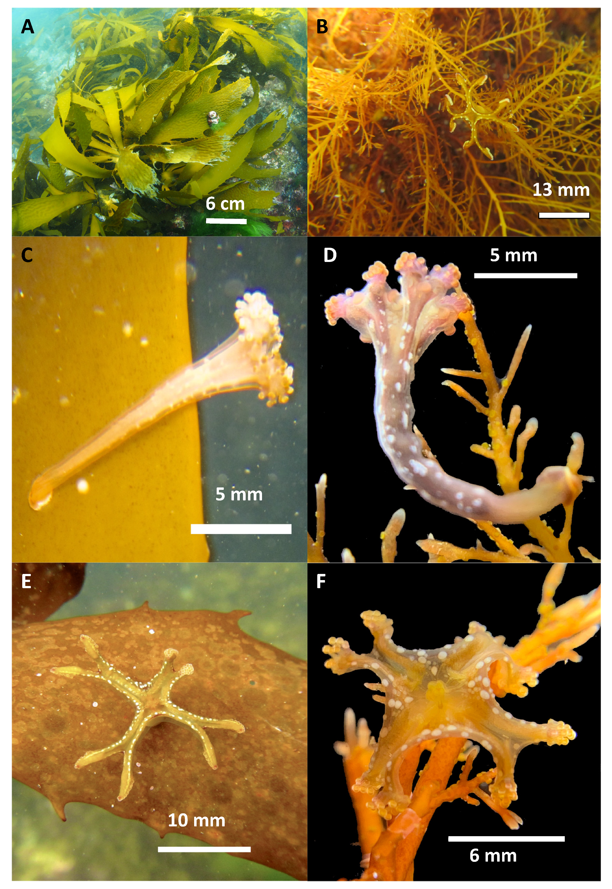
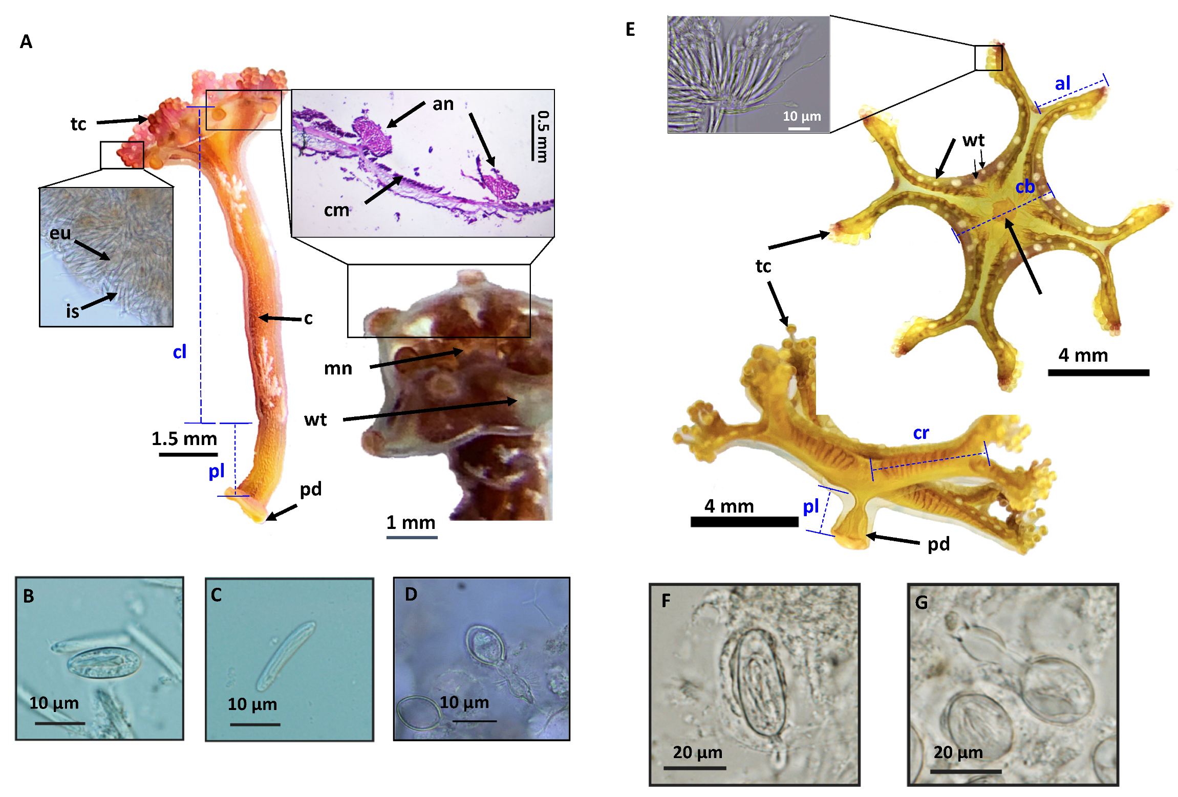
Stauromedusae of two distinct morphotypes were collected (
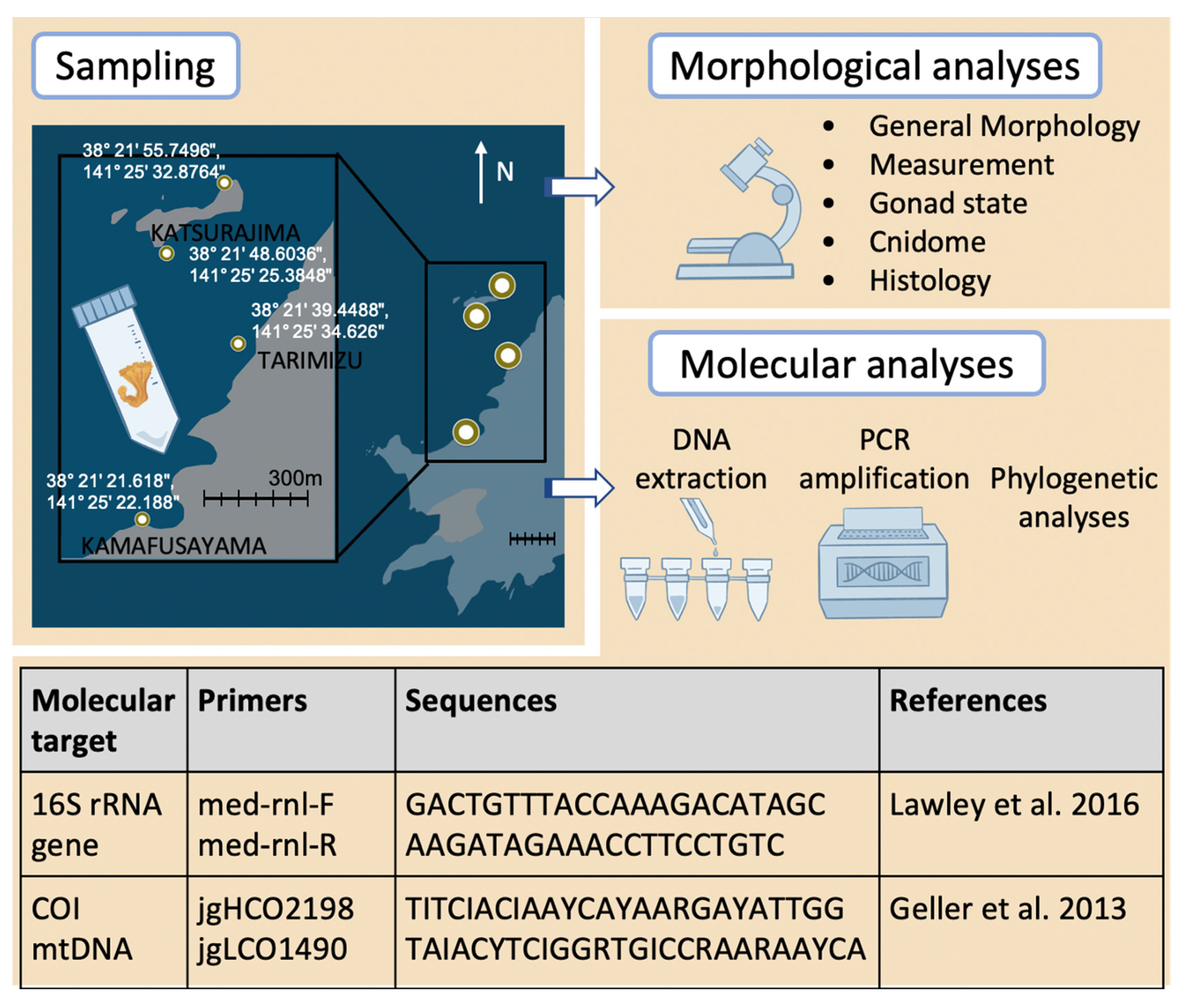
Figure 1) at the study site in Kitsunezaki, Ishinomaki, Oshika Peninsula, Miyagi Prefecture (38°21′ N, 141°25′ E, and vicinity—details in Figure 2). This location on Honshu was hit directly by the Great East Japan Earthquake on 11 March 2011. Following recovery efforts, an active survey site for research on seaweeds and seagrasses was established in July of the same year (for sampling site details see [18,19]). Eisenia bicyclis kelp (Figure 1A) and Gelidium (Figure 1B) grow abundantly on inshore (0–30 m) rocky bottoms (1–4 m depth), while the seagrass Zostera caespitosa dominates the offshore (30–70 m) sandy bottoms (4–6 m depth).
Prior to this study, the two distinct stauromedusae (Figure 1B–F) were first collected from the location in 2018, by one of the co-authors (M.A.) and, subsequently, this study began one year later in October 2019. Seawater temperature was measured using HOBO data logger (Supplementary File S1) and dive computer (ScubaPro M2). The stauromedusae were collected by hand while SCUBA diving, then placed into 50 mL Falcon tubes filled with seawater (Figure 2) along with a small piece of the macroalgal substrate, before being transported live in an insulated cooler to the laboratory of Marine Plant Ecology, Graduate School of Agricultural Science, Faculty of Agriculture, Tohoku University.
Out of the total specimens collected at Kitsunezaki (ca. 100), a portion was preserved immediately after collection in pure ethanol (99.5%), and then stored at −30 °C for future molecular analysis; some were fixed in 70% ethanol or 5–8% formalin solution for morphological study. Several stauromedusae (ca. 25) were kept alive for observations and fed three times a week on Artemia nauplii. However, rearing stauromedusae collected in the wild using artificial seawater proved to be challenging, and most died within a few days, with only a handful living for up to 79 days in simple aquariums.
In addition to the Tohoku specimens obtained at the survey site, additional H. inabai specimens that had unexpectedly appeared in aquariums in Japan (Kamo Aquarium and Kagoshima Aquarium) and Canada (Aquarium du Québec) were obtained for morphological and molecular analyses. Life history observations were made in the Aquarium du Québec.

2.1. H. inabai Stauromedusae Appearing in Aquariums

In the Aquarium du Québec, several 
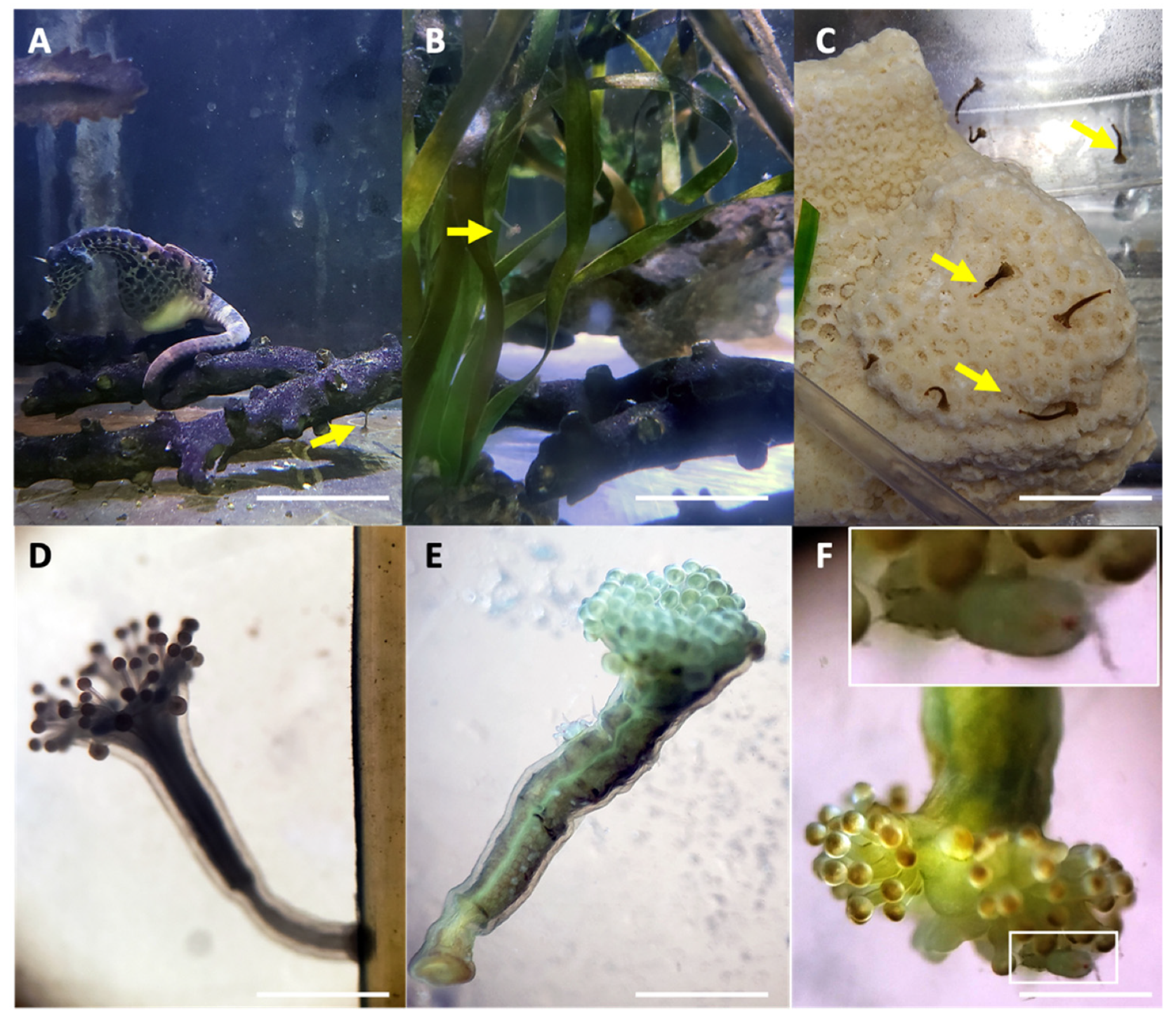
Haliclystus inabai stauromedusae were first discovered on 16 April 2020 in an exhibit tank of 1016 L in volume (totaling 2600 L including life support and sump). Within the exhibition tank, with a sand substrate, were potbelly seahorses and other decor including dead coral rocks or plastic ones. Both adult and juvenile H. inabai stauromedusae as well as early life stages (discussed below) were found on artificial plants that were frequently cleaned, including Eelgrass Zostera (bio models, Aguanga, CA, USA) as well as some uncategorized types, and Green Cardamine (resin ornament, PetSave Direct, Woodbridge, ON, Canada), the latter of which hosted mainly early life stages on one particularly clean leaf. Following the discovery, additional stauromedusae were noted in a smaller quarantine tank (182 L) also containing potbelly seahorses and plastic plant decor (Figure 3A). The temperature in both artificial seawater tanks (Instant Ocean, 32 g/L, potassium iodide 0.06 to 0.08 ppm), was kept at about 16 °C. Over a two-month period (28 November 2020–1 February 2021) a total of 31 stauromedusae (approximately 12.5 mm; range 11–15 mm in length) were counted in the smaller tank attached to coral rocks, shells, air stone weights, and plastic plants (Figure 3B,C), and one individual was found attached to a seahorse. Though easily removed from the latter with seawater forced using a pipette, it was difficult to detach the stauromedusae from harder surfaces, such as aquarium walls and coral rocks. Plastic leaves bearing staurozoans were transferred to a new tank in a sea-table (6 L plastic tank HerpHeaven, Brunswick, ME, USA), with a small pump for water circulation and air tubing for aeration (gentle water flow; fluctuating 16–19 °C), in order to conduct observations on feeding and life history. Staurozoans were fed on nauplii (48 h after hatching, 550 μm in length) of Artemia brine shrimp (SELCO enriched, Artemia International, Fairview, TX, USA). Juvenile stauromedusae were typically beige-brown with a hint of pink (possibly due to ingested brine shrimp), while adults were brown with a white “snowflake” pattern along the length, or took on a green hue (Figure 3D–F). Photo and video documentation was made via Samsung Galaxy Smartphone. Six of these individuals were preserved in 95% ethanol and shipped to Tohoku University on 20 January 2021 for molecular analysis.
Similarly, Haliclystus stauromedusae were also discovered at Kamo Aquarium (Tsuruoka, Japan), appearing in reserve aquarium tanks (144 L, with a 100 L filtration tank—mesh sizes of 10 μm, then 5 μm, 15 °C) and Kagoshima Aquarium (Kagoshima, Japan). Individuals were sent to Tohoku University, both living and preserved in 99.5% ethanol, for further analyses.

2.2. Morphology, Microscopy

Specimens collected from Kitsunezaki and preserved in 70% ethanol or 5–8% formalin were observed under a dissecting microscope (Olympus SZX12), imaged with a digital camera (Olympus DP72), and measured (CellsSens software version 1.8, Olympus, Tokyo, Japan). The presence or absence of gonads was confirmed, and the cnidome (the repertoire of stinging capsules, called nematocysts) was characterized using compound microscopy (Olympus BX61 or Nikon Eclipse Ni-U and camera) (Figure 2).
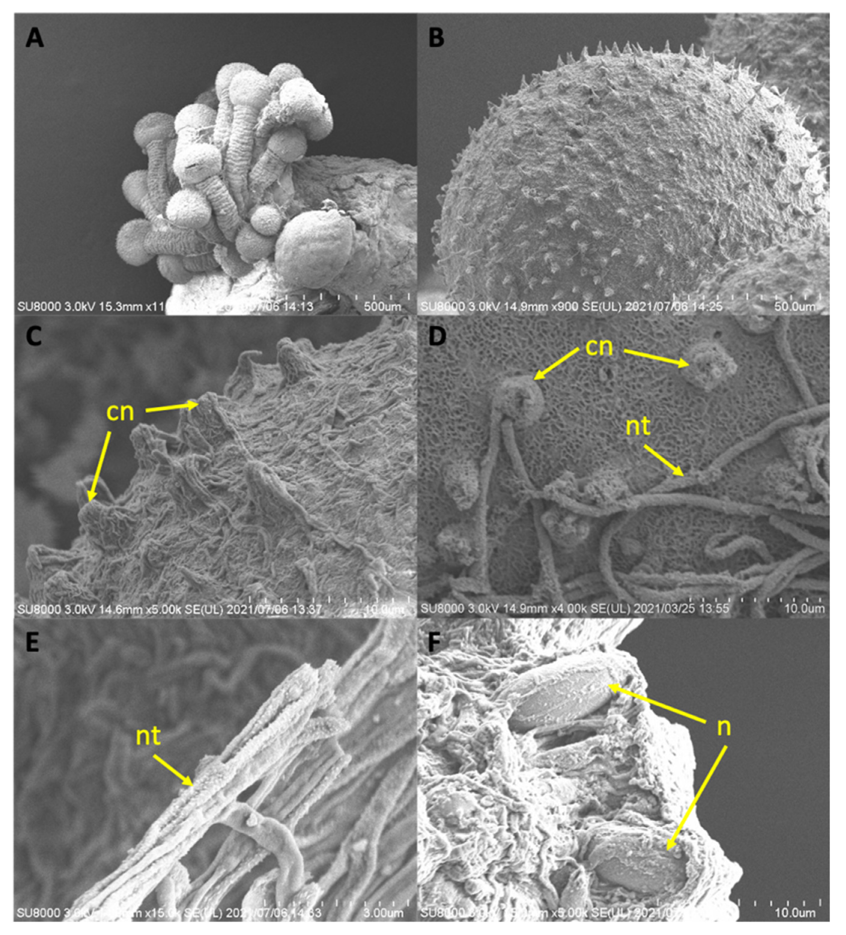
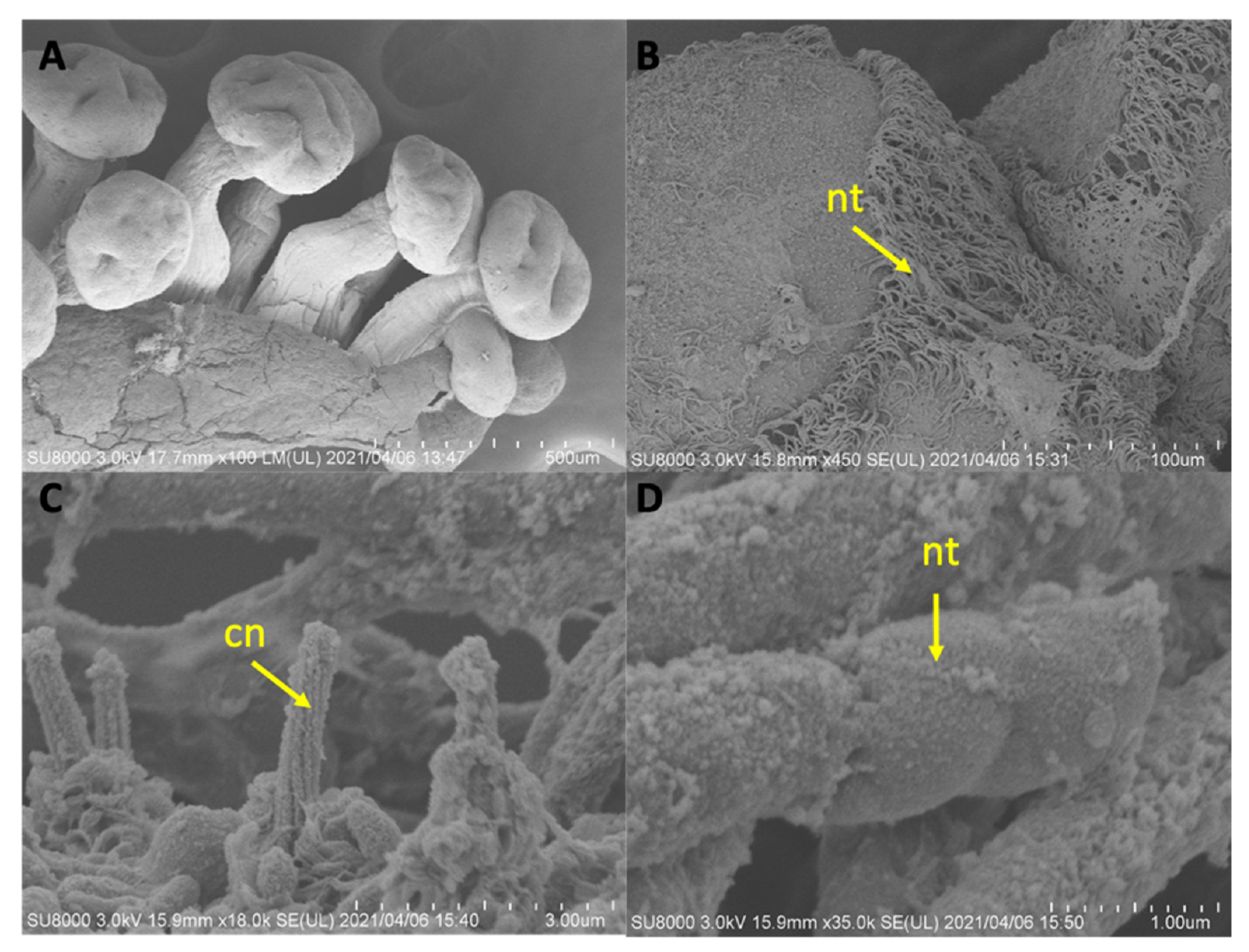
The ultrastructure of finer features was documented with Scanning Electron Microscopy (SEM) (Hitachi SU8000 (Type II)) at the Electron Microscopy Section in Tohoku University Graduate School of Agricultural Science. For SEM preparation, samples previously preserved in 70% ethanol were subjected to a graded rehydrated series (70–25%, 5–10 minutes each) of 1X PBS (Phosphate Buffered Saline, pH 7.4); fixed in isosmotically adjusted PBS buffer glutaraldehyde solution (2.5%) overnight at 4 °C; washed with isosmotic 1X PBS buffer and milli-Q water twice to remove salts; and subjected to a graded dehydration series of ethanol (25–100%, 5–10 minutes each). Dried samples were mounted onto an SEM stub (13 mm) using an adhesive carbon strip, and subjected to critical point dry, and coated in gold/palladium (MSP-1S sputtering device) before observation with SEM.
General histological procedures [22] were carried out on staurozoans preserved in 8% formalin. Specimens were dehydrated in a series of graded ethanol (70–100%); cleared in Lemosol (2 steps), infiltrated and embedded in paraffin; serially sectioned transversely (8.0–10.0 µm thick); washed with distilled water, and stained using hematoxylin-eosin. Prepared slides were photographed with light microscopy.

2.3. Molecular Barcoding

DNA was extracted from Staurozoa samples using the DNeasy Blood and Tissue Kit (Qiagen, Hilden, NW, Germany), and total DNA yield was quantified (ng/μL) using a fluorometer (QuantiFluor, Promega, Tokyo, Japan). The targets for DNA amplification were two mitochondrial gene fragments: 16S rRNA gene (565 bp) and cytochrome oxidase subunit 1 (COI) gene (720 bp). Each reaction (50 μL) contained 25 μL Taq PCR Master Mix (Qiagen), template DNA (~200 ng), nuclease free water, and 2 μL each of forward and reverse primers (10 μM; Figure 2).
PCR (polymerase chain reaction) was used to amplify the two molecular targets in a thermal cycler (BentoLab, Bioworks, London, UK or Takara Bio, Kusatsu, Shiga, Japan). Cycling profile for 16S primers included initial denaturation at 94 °C for 3 minutes, followed by 38 cycles of denaturation, annealing and extension (94 °C for 30 s, 54 °C for 30 s, and 72 °C for 45 s), and a final extension at 72 °C for 10 minutes [23]. Cycling profile for COI primer included denaturation at 94 °C for 5 minutes, followed by 38 cycles of denaturation, annealing, and extension (94 °C for 30 s, 46 °C for 30 s, and 72 °C for 1 min), and a final extension at 72 °C for 10 minutes. PCR products were visualized on a 1% agarose gel using gel electrophoresis and visualized with a gel illuminator and nucleic acid gel stains (GelGreen or GelRed). Amplicons were purified with ExoSap-IT (Applied Biosystems, Tokyo, Japan) PCR Product Cleanup toolkit prior to Sanger sequencing (Eurofins or Genewiz, Tokyo, Japan).
Generated 16S rRNA and COI gene sequences were edited in Geneious Prime (v.2022.0.1, www.geneious.com: accessed on 11 November 2021). Additional Staurozoa sequences were acquired from GenBank (https://www.ncbi.nlm.nih.gov/genbank/: accessed on 11 November 2021). 
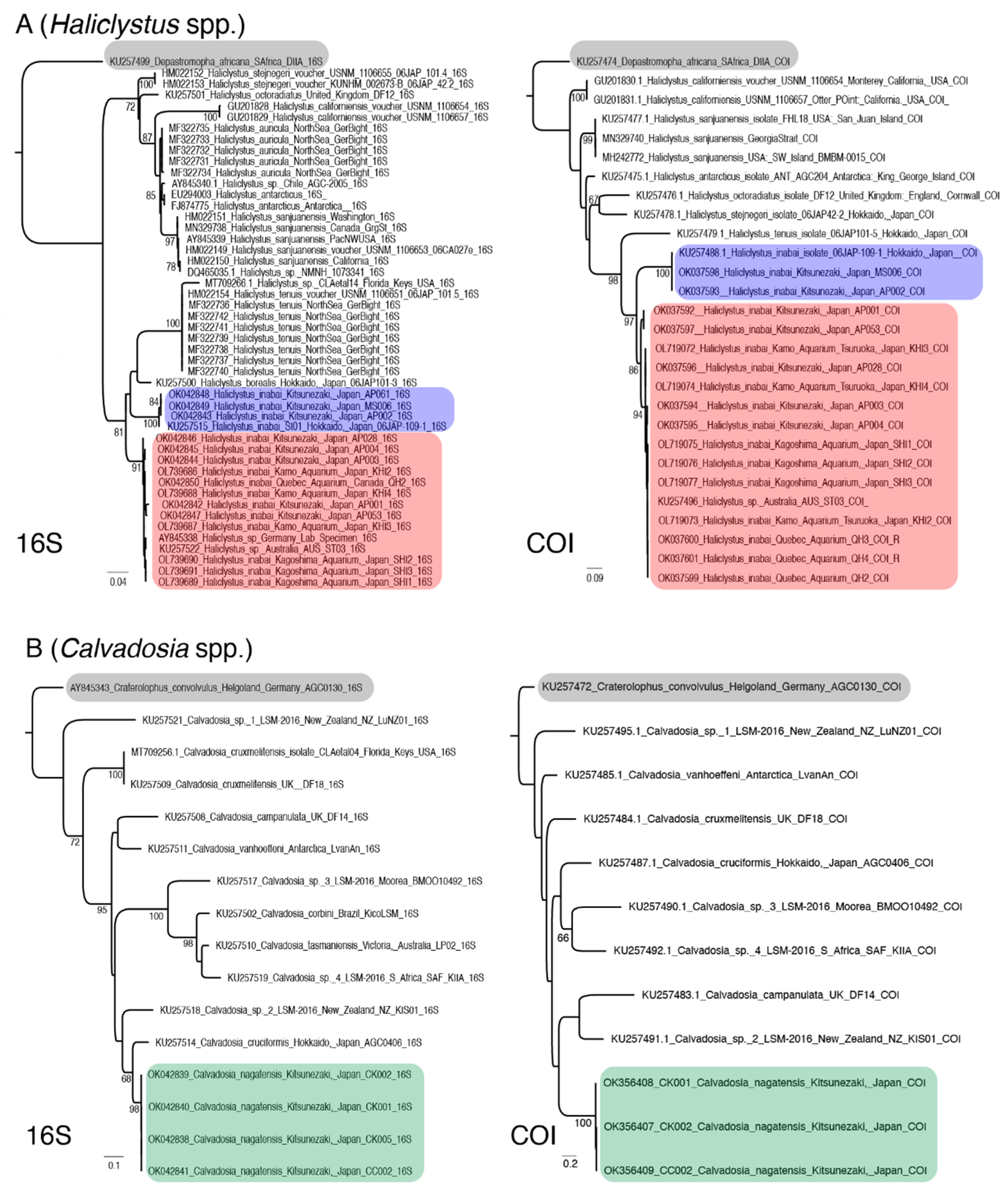
Depastromorpha africana and Craterolophus convolvulus were used as outgroups in the analyses of Haliclystus and Calvadosia species, respectively. Sequences were aligned in MAFFT using the E-INS-i and G-INS-i algorithms, respectively, for 16S and COI. Maximum likelihood trees, with nodes assessed by 400 bootstrap replicates, were constructed using PHYML as implemented in Geneious Prime (v.2022.0.1) and assuming the GTR model of nucleotide substitution. Sequences generated in this study were accessioned into GenBank, and morphological vouchers were deposited as museum specimens (Table 1). Alignments and resulting topologies are provided as Supplementary Files S2–S9.

3. Results

3.1. Haliclystus inabai

Haliclystus staurozoans collected in this study conformed with the original description of Haliclystus inabai [24]. The type locality of H. inabai is Kata-ura, Kii Province (now Wakayama), and it was described as having a dark brown color with white accents [25]. Subsequent records show 16 localities distributed in temperate regions and the species has been recorded in the Southern and Northern Hemispheres (reviewed in [7]). Haliclystus is the most diverse genus of Class Staurozoa; its 13 valid species have a widespread, antitropical distribution and have been reported on algae, rocks, and shells in tide pools to 40–60 m deep in every continent except Africa [7].

3.1.1. Taxonomy

Haliclystus inabai (Kishinouye, 1893)
Synonymized names:
Stenoscyphus inabai (Kishinouye, 1893)
Depastrum inabai Kishinouye, 1893

3.1.2. General Morphology of H. inabai

Haliclystus inabai (Figure 1C,D, Figure 3D–F, Figure 4A and Figure 5A) has a long conical body that becomes slender and tubular towards the pedal disk. The average total length of specimens was 13.3 mm (range: 4.5–29.5 mm, n = 64), which was also consistent with measurements for H. inabai specimens received from aquariums, although aquarium specimens had a much narrower size range (see details above). Their color varies from light green to brown to almost black; the body of some specimens bore white stripes or snowflake-like flecks along the length.
H. inabai, like all stalked jellyfish of the genus Haliclystus, has tetramerous symmetry—bearing eight arms terminating in secondary tentacle clusters [26]. These tentacle clusters emerge from the subumbrella, each consisting of about 25 short, capped, and highly adhesive secondary tentacles (Figure 5A). The calyx is narrow and elongated with a widened oral end, taking the shape of a funnel with a long and wide tube, bearing eight large, oval anchors, perradially and interradially, without remnant of primary tentacles [25]. Variation in anchor number (sometimes nine or ten) has been reported, as well as pentamerous, hexamerous, and octamerous symmetry [27]. The coronal or marginal muscle that lines the calyx is ring-shaped [25], although it is sometimes difficult to visualize in smaller specimens (Figure 5A). The size of specimens observed in this study reaches 25 mm in length without the peduncle, about 2–3 times the width of the calyx margin. The peduncle is short, with four interradial grooves, ending with an adhesive surface used for adhering to various substrates (discussed below). The exumbrella is generally smooth with small groups of nematocysts in some places, while the subumbrella surface is covered with clusters of variably sized spherical white spots occurring primarily in the perradii. The manubrium (buccal stomach) is short and is shaped like a cross transversely. Numerous long gastric filaments are found in the central stomach. Eight gonad rows run in parallel along the interradial muscles, from the base of the calyx to the proximal edge of each arm. In old specimens, each gonad row may consist of about forty sacs arranged approximately in two rows; younger specimens have fewer and much smaller gonad sacs arranged in a single row [25].

3.1.3. Cnidome of H. inabai

In H. inabai, four types of nematocyst capsules were observed in all specimens examined (Figure 5A–D), with three types previously reported [28]—type I (sac-like) isorhizas, type II (rod-like) isorhizas, and lemon-shaped euryteles—as well as birhopaloids, reported here for the first time. Secondary tentacle tips (Figure 5A, magnified area of tentacle) have almost exclusively type II isorhizas (average length 16.3 µm, range: 13.1–18.8 µm, n = 54) and a small proportion of euryteles (average length 12.9 µm, range: 9.5–16.3 µm, n = 11); raw measurements provided in Supplementary File S10. Anchors and white spots have primarily oval birhopaloids (Figure 5D) and type I isorhizas, though the latter are scarce. Intact nematocyst capsules were also documented by SEM in the ectoderm of the calyx of H. inabai (Figure 6F).
The surface of the tentacles bore a fuzzy texture (Figure 6A,B) upon examination using SEM. Further ultrastructural observations of the surface of the secondary tentacles revealed numerous nematocyst tubules and “bumps” resembling the bilaterally symmetrical, mechanosensory cnidocil complex (apparatus) at the apex of each nematocyst capsule (Figure 6C,D). Though not as clearly visible in H. inabai (Figure 6B–D) as in C. nagatensis (see Figure 7C below), this important sensory component of the nematocyst has been described as a central cilium surrounded by a crescent of stereocilia (for a detailed description see Hausmann and Holstein 1985 [29]). Stimulation (light, chemical, touch) of the cnidocil complex triggers the firing of the nematocysts, resulting in the venom contained therein being expelled through the everted tubule (Figure 6E).

3.2. Calvadosia nagatensis

Calvadosia staurozoans collected in this study fit the description of Calvadosia nagatensis (Oka, 1897). The type locality of C. nagatensis is Kogushi (Nagato, Western Honshu, Japan), where it was found at a 6–8 m depth [30] (Figure 4B). Calvadosia comprises 11 valid species and a unique distribution within the staurozoans—occurring in temperate to polar water environments, but also discovered in the tropics, attached to substrates such as Zostera marina (common eelgrass), rocks, shells, and corals, over a broad bathymetric range (from tide pools to 137 m deep), and on a seasonal cycle of one generation per year, with the peak month varying by species [7].

3.2.1. Taxonomy

Calvadosia nagatensis (Oka, 1897)
Synonymized names:
Kishinouyea nagatensis (Oka, 1897)
Schizodiscus nagatensis (Oka, 1897)—Schizodiscus Kishinouye 1902 was preoccupied
Lucernaria nagatensis Oka, 1897

3.2.2. General Morphology of C. nagatensis

The body of Calvadosia nagatensis (Figure 1E,F, Figure 4B and Figure 5E) is divided into a wide, open calyx and a short peduncle; the calyx is wider than it is long, and cruciform in shape, with arms united in pairs in the interradii. Of specimens observed in this study, the average arm length was 3.25 mm (range: 1.5–7 mm, n = 16), the average calyx diameter was 11.6 mm (range: 5–20 mm, n = 16), and the average calyx base was 4.6 mm (range: 2–6 mm, n = 16). Their color includes variations of ochre and green, some with accents of red in the perradial U-notches and interradial V-notches of the calyx and arm tips.
The peduncle is short and continues directly to a broad, adhesive, pedal disk at the base. There is a central cavity on the inferior portion of the pedal disk, which is possibly used for adhesion, though the function of the cavity is unknown. The peduncle is without interradial longitudinal muscle bands. The manubrium bears four perradial pleated lips. Gastric filaments are abundant in the gastrovascular cavity. The calyx is without anchors (rhopalioids) or primary tentacles. Each arm bears secondary tentacles (hollow, knobbed) with numbers varying among the arms of each individual and between individuals (average: 78; range: 51–124, n = 16). No pad-like adhesive structure (hypothesized synapomorphy of Calvadosia) was noted, nor mentioned in the original description (see also Grohmann et al. 1999 [31]). Four U-shaped perradial notches between arm pairs are about two times as deep as the V-shaped interradial notches between a pair of arms. The subumbrella of each U-notch is lined peripherally with a row of large white nematocyst spots extending to the arm tip; these flank the eight rows of gonads embedded in the gastrovascular cavity, radiating subdermally from the manubrium base to the proximal edge of each arm. Additional white nematocyst spots line the inner region of the perradial sectors, and a few nematocyst spots are also distributed along the V-notches.

3.2.3. Cnidome of C. nagatensis

In C. nagatensis, four types of nematocysts were observed (Figure 5E–G) for all specimens examined—type I (sac-like) isorhizas, type II (rod-like) isorhizas, and lemon-shaped euryteles—as well as birhopaloids, rarely reported in stauromedusae.
Secondary tentacle tips (Figure 5E, magnified area of tentacle) have almost exclusively type II isorhizas. The surface of the tentacles appeared fuzzy under SEM (Figure 7A), and upon closer examination was shown to bear clusters of discharged tubules of isorhiza nematocysts (Figure 7B) and the corresponding cnidocil complex (Figure 7C) similar to that described above for H. inabai. No spines were seen on discharged tubules, suggesting the type II isorhizas are of the atrichous type (Figure 7D). Its white spots have very few type I isorhizas (average total length: 41.1 µm, range: 35–46.2 µm, n = 3) (Figure 5F), and abundant birhopaloids (average total length: 23.0 µm, range: 19.3–27.2 µm, n = 81) (Figure 5G); raw measurements provided in Supplementary File S10.

3.3. Molecular Analysis

Phylogenetic analyses comparing new sequences from Kitsunezaki, both 16S rRNA and COI, with those in Genbank showed the specimens belong to the staurozoan genera Haliclystus and Calvadosia. This was consistent with our morphological identification of the two as H. inabai and C. nagatensis. H. inabai samples from this study fall into two distinct clades (Figure 8A) both of which include specimens from Kitsunezaki. Because we were unable to differentiate specimens of H. inabai using morphological characters, the two lineages discerned by the molecular phylogenetic tree are referred to herein as H. inabai 1—the “Japan clade”, which includes sequences corresponding only to samples collected in the field in Japan, from Kitsunezaki (Ishinomaki City, Japan) and Hokkaido (Oshoro, Japan); and H. inabai 2—the “Australia clade”, a more cosmopolitan clade, corresponding to specimens collected in Kitsunezaki (Japan), Australia, as well as specimens discovered in aquariums in Québec (Québec City, Canada), Tsuruoka (Japan) and Kagoshima City (Japan) (all analyzed in this study), and in a lab in Germany (acquired from GenBank). Our analyses of sequence data for C. nagatensis from Kitsunezaki shows it as distinct from all other Calvadosia specimens thus far sampled (Figure 8B). However, these data do not provide a strong signal about the precise phylogenetic position of C. nagatensis within the genus.
For both genetic markers, mean evolutionary divergence (based on between-group mean distances) for the two H. inabai subclades (Japan clade and the international clade Figure 8A,B) was estimated at 0.0859 (SE 0.0131) for the 16S rRNA gene and 0.1710 (SE 0.0200) for the COI gene (18 sequences per analysis), based on the number of base substitutions per site (and standard error), estimated by averaging over all 18 sequence pairs between clades in MEGA 11 [32]. This analysis was performed using the Jukes-Cantor model and rate variation among sites modeled with a gamma distribution (shape parameter = 1). All ambiguous positions were removed for each sequence pair (pairwise deletion option).

3.4. Substrate Preference

Observations in the field and aquariums, as well as literature surveys (1893–present), revealed a broad distinction in substrate specificity between the two species (Figure 9). Both species have been found on macroalgae and seagrass, although there is a much broader type of substrate attachment for H. inabai (irrespective of lineage)—including surfaces of plastic, glass, coral (dead), rock, and a seahorse recorded in the Aquarium du Québec, Canada.
H. inabai has been recorded attaching to primarily brown algae: Sargassum spp., Sargassum confusum, Cystophora expansa, Macrocystis pyrifera, and Eisenia bicyclis (Supplementary File S11). Conversely, there were not as many records of C. nagatensis substrate attachment, with algal substrate being limited to brown and red algae. This study includes the first record of the two stauromedusae attaching to the red alga Gelidium.

3.5. Gonad State and Occurrence of Staurozoa in Kitsunezaki

Temperature data were recorded along with observation of jellyfish occurrence on site (Figure 10A). The gonad states of the stauromedusae were observed with light microscope, with greater detail shown using SEM (Figure 10B,C).
In situ observations were logged non-continuously over an 18-month period in an attempt to establish the seasonal presence of each species. However, unexpected cancellations due to poor weather conditions, as well as COVID-19 restrictions, prevented regular monthly sampling, which led to patchy data (Figure 10A). Water temperature data were not obtained for every month during the study period. Consequently, these were supplemented with monthly mean air temperature data obtained from the Japan Meteorological Agency (https://www.data.jma.go.jp/obd/stats/data/en/smp/index.html: accessed on 31 July 2021). Additionally, collected staurozoans were examined to determine the presence of conspicuous or immature gonads, or whether they were entirely absent. Of the total stauromedusae collected, 25.8% of all observed specimens had conspicuous gonads, 20.2% inconspicuous, and the remaining portion was unconfirmed (examinations were not conducted). These findings were plotted against temperature in an attempt to infer a putative spawning season, but none was obvious; the presence or absence of H. inabai (irrespective of lineage) did not seem to be related to changes in temperature. Conversely, although C. nagatensis was only collected in the warmer months (July and August), in all cases gonads were conspicuous. However, with so few specimens, it is premature to assume spawning is restricted to the summer months.

3.6. Haliclystus inabai Early Life Stages Observed in Aquarium

Early life cycle observations were made on H. inabai that appeared in the Aquarium du Québec (Figure 11; Table 2), revealing that H. inabai exhibits similar early life stages to both H. octoradiatus and H. antarcticus.
On 6 January 2021, the motile larval stage (ca. 150 µm in length) of H. inabai was first detected in the Aquarium du Québec during microscopic examination of water removed from a tank during a regular water change. Motile larvae appeared as microscopic peduncles, each with what appeared to be a pedal disk attached to Green Cardamine (resin ornament) and/or eel grass leaf (see Methods above) floating on the water surface in the reserve aquarium tank (one to six individuals per day were noted during a one-month observation period). Higher magnification (400X) of the plastic leaves (Supplementary Video S12) revealed that the life forms were most similar to the frustules described for H. octoradiatus in an earlier work [15] and H. antarcticus [33] (as the hydrozoan Microhydrula limopsicola). Bearing nematocysts posteriorly, and comprising approximately 25 to 35 endodermal cells along the body for H. octoradiatus, our findings are consistent with early observations on the number of endodermal cells in H. octoradiatus frustules [15]. Nematocysts of H. haliclystus frustules in this study appear to be rhopaloids similar in capsular shape and shaft morphology to birhopaloids found in the adult stauromedusae; however, whether the shaft bore a single dilation (eurytele) or two dilations (birhopaloid) could not be determined. Also present on the leaves of Green Cardamine were transparent hemispherical larval forms bearing nematocysts in the periphery (Figure 11A,C–E) that resembled settled larva known as the “microhydrula” stage (ca. 100 µm in length) (Supplementary Video S12), previously described for H. octoradiatus and H. antarcticus [15,16]. During this study, examination on 7 May 2021 of the surface of both sides of the floating Green Cardamine leaf in tanks in the Aquarium du Québec revealed numerous similar microhydrula individuals (n = 58). Both the microhydrula and frustules stung and paralyzed Artemia nauplii, whose length was approximately four times that of the frustules; ingestion of the shrimp was not confirmed.
In this study, a superficial comparison of three main characteristics was made between the elongated settled larval form of H. inabai and that previously described in detail using histology for H. octoradiatus [15]. First, elongated settled larvae of H. inabai possessed at the base a hard layer in contact with the substrate (Figure 12A), similar to the chitinous casing secreted by the tightly packed basal cells reported for H. octoradiatus (Wietrzykowski 1912). Secondly, for H. octoradiatus, a novel structure called the pedal gland forms just before the two-tentacle young stauropolyp stage, which arises as a result of an invagination of the basal lamina when cells there become elongated and glandular (Wietrzykowski 1912). We noted a similar invagination at the base of the elongated larval form of H. inabai (Figure 12A,B), and surmise that it is homologous with the pedal invagination of H. octoradiatus. Thirdly, in what is referred to as “stage 2” for the elongated settled larvae of H. octoradiatus, on the two opposite points situated in the horizontal plan (separated by the hypostome of the calyx), ectoderm cells emerge that are notably raised compared to those around them, and these thickened regions also bear nematocysts (Wietrzykowski 1912). According to Wietrzykowski (1912), these are the start of the formation of the two primary tentacles, which appear quite fast once the gastric cavity forms two diverticula that underline the ectoderm in two thick regions. We also report similar thickening of the ectoderm in H. inabai, which resulted in two swollen lateral regions, interspersed with nematocysts (Figure 12A,B) similar to the regions from which the two primary tentacles reportedly emerge in H. octoradiatus [15]. However, further development to the two-tentacle-stage stauropolyp was not observed in this study.

4. Discussion

4.1. Staurozoan Identification and Cnidome

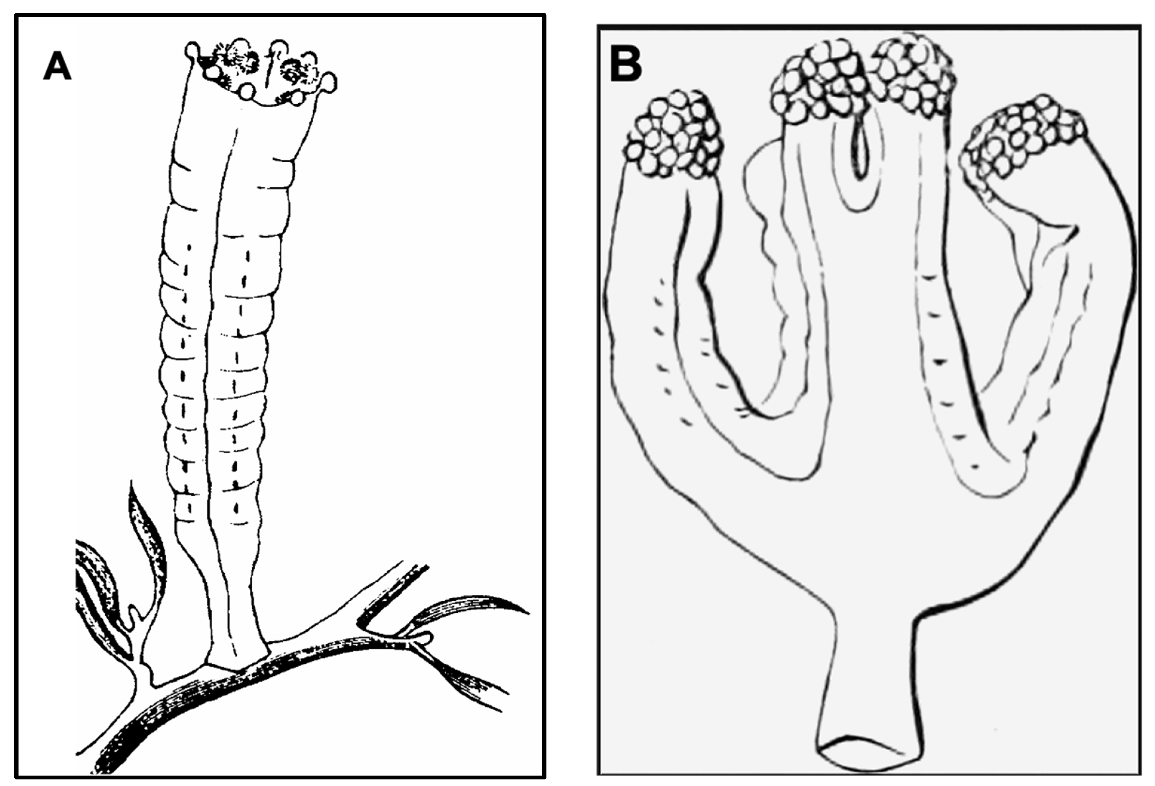
This study employed molecular barcoding, morphological analyses, light microscopy and SEM, and collaborations with aquariums in Japan and Canada, to identify three species of Staurozoa reported herein for the first time in Kitsunezaki—two as Haliclystus inabai (Kishinouye, 1893) and one as Calvadosia nagatensis (Oka, 1897), of the respective suborders Myostaurida and Amyostaurida.
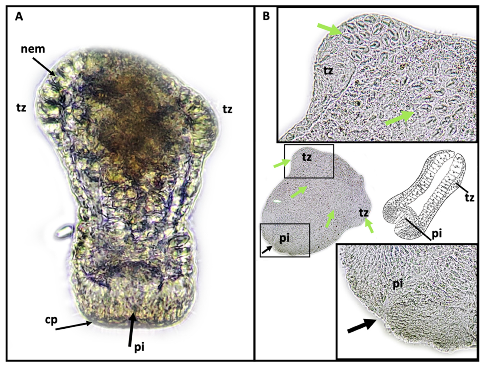
The use of SEM in this study to conduct ultrastructural observations of secondary tentacles of both staurozoan species provided a clear picture of the structure of the bilaterally symmetrical mechanosensory cnidocil complex (apparatus) at the apex of each nematocyst capsule, which consists of central cilium surrounded by a crescent of stereocilia. This sensory component of the nematocyst controls the firing of the nematocysts, and is vital for nematocyst discharge. Though well-studied in several hydrozoan species, e.g., Hydra [36] and Craspedacusta sowerbii [29], only a single mention exists of the cnidocil complex in a Staurozoa (James-Clark 1878); the structure is documented herein for the first time in the class with SEM. More detailed observations using TEM techniques are required to verify whether the ciliary pattern of the cnidocil apparatus follows the same as that seen in hydrozoans.

4.2. Molecular Phylogenetics

Our molecular analyses revealed that H. inabai consists of two distinct lineages: Haliclystus inabai lineage 1 is so far known only from Japan, comprised exclusively of specimens collected in the wild (Kitsunezaki and Hokkaido); H. inabai lineage 2 is more broadly distributed, with specimens collected from Kitsunezaki (Japan), Victoria (Australia), aquariums in Québec (Canada), Tsuruoka (Japan), Kagoshima (Japan), and a university lab (Germany).
Despite their broad average genetic differences, no apparent morphological distinction was detected, suggesting that the two clades represent two distinct but cryptic species. More extensive sampling and study of both lineages could permit morphological distinction of the two species, determination of which lineage represents the true H. inabai, and ultimately a formal naming of the new species.
Our molecular analysis identified a sequence labeled as Haliclystus sp. from a lab in Germany (AY845338.1) clustering with the H. inabai sequences, revealing a case of clarified identity, as this sequence was originally deposited as Haliclystus sp. (Figure 8A). Furthermore, Haliclystus sp. (KU257522, KU257496) from Victoria (Australia, in the Southern Hemisphere) was previously hypothesized to be a separate species from the Hokkaido one (Northern Hemisphere) based on molecular data for an individual from each population (Miranda et al. 2016b). However, increased sample analyses in this study, for both gene markers, revealed that the Australian Haliclystus resembling H. inabai is also present in Japan, and appearing spontaneously in aquariums in other parts of the world where the samples’ origins are unknown (Figure 8A,B).
The exchange of live aquatic samples, which is part-and-parcel to scientific collaboration between academic institutions and aquariums, may have played a role in the distribution of 
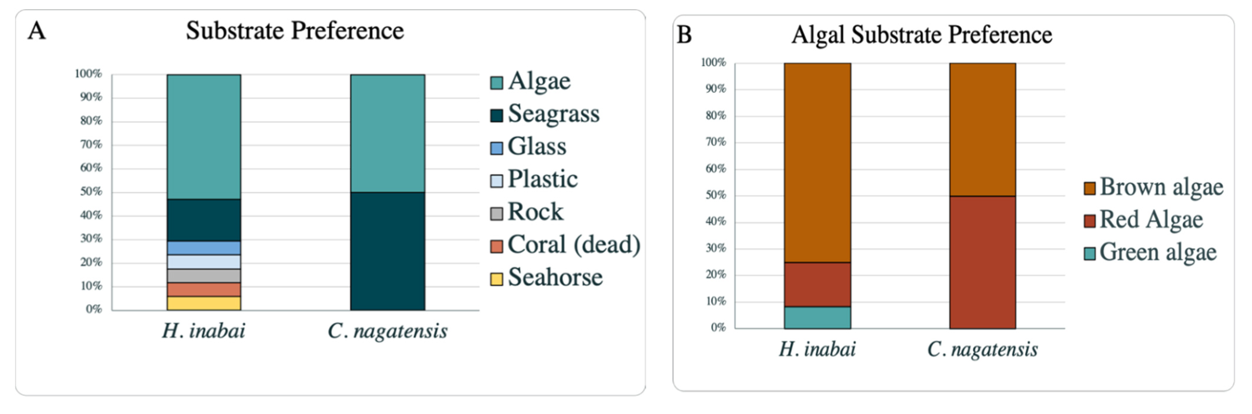
H. inabai beyond the walls of aquariums on different continents. In addition to observations on substrate preference in this study, the survey of literature revealed H. inabai to have a broad substrate preference, consistent with the records of this species from multiple countries. Furthermore, in all cases in this study where H. inabai appeared in aquariums, no stauromedusae had been placed in those aquarium tanks, suggesting H. inabai was likely introduced in the form of one of the microscopic early life cycle stages attached to a substrate (i.e., synthetic plants, coral heads, or even a live seahorse). How staurozoans are distributed across oceans is still a mystery, as no studies on staurozoan mobility or recruitment exist. Although speculation exists about the potential for rafting and ocean currents to aid in distribution of staurozoans across oceans, an inferred dispersal mode for over 100 non-staurozoan medusozoans [37] to date, no reports exist that test that hypothesis. Recently, eDNA (environmental DNA) signatures of two staurozoan species, Calvadosia cruxmelitensis and Haliclystus cf. tenuis, were detected in the Florida Keys (Key Largo and Fleming Key, FL, USA) [23], where they had not been reported previously, suggesting that these cryptic animals may have more widespread distributions than previously appreciated. Novel approaches to eDNA detection (e.g., [38]) should aid our understanding of global distribution paths of staurozoans and their underlying mechanisms of dispersal.
This study is the first to accession the molecular markers 16S rRNA and COI of C. nagatensis to GenBank, showing the limited resources available for this species, whose more narrow distribution range is consistent with the dearth of records on substrate preference of this species (as discussed in the previous section). While C. nagatensis appears to have less adaptive potential to different environments and substrates compared to H. inabai, population declines or extirpations are purported to be connected to pollution or loss of algal/seagrass substrates [7], but additional studies are needed to better understand potential threats to staurozoan species stemming from human activities.

4.3. Distribution and Seasonal Variation

During our 18-month field and lab study, we gained a better understanding of the two Staurozoa species documented for the first time at the Kitsunezaki survey site. However, due to unforeseen circumstances related to weather and the impact of the COVID-19 pandemic countermeasures (reviewed in Abramo et al. 2022 [39]), sporadic sampling impeded diligent sampling efforts and our ability to document sexual reproduction or early life stages of either species in the field.
In this study, we collected stauromedusae of both H. inabai (both lineages) and C. nagatensis on Eisenia bicyclis kelp and Gelidium seaweed, but no early life stages were witnessed in the field. Differential use of substrates during ontogeny has been documented in staturozans. Wietrzykowski (1912) reported that H. octoradiatus adult stauromedusae were mainly found on Zostera eelgrass, while early life stages were exclusively found on filamentous Ceramium red algae; the interstitial staurozoan Stylocoronella riedli, on the other hand, settles on coarse sand mixed with shell during early development [35], while its stauromedusa stage has been found on a sea cucumber [40]. Additionally, the microhydrula stage of Haliclystus antarcticus was discovered upon the shell of lamellibranch bivalve (Limopsis hirtella), which burrows in deep sediment (30 m) in the Southern Ocean, by Jarms and Tiemann (1996) (as the hydrozoan Microhydrula limopsicola), while the stauromedusa stage is witnessed mostly on rocks and macroalgae (0–8 m deep) [7,16]. In light of these reports, a more thorough survey of the benthos at the Kitsunezaki survey site could lead to the discovery of early life stages of these staurozoans, on yet unexamined substrates, such as other algae species or even bivalve shells.
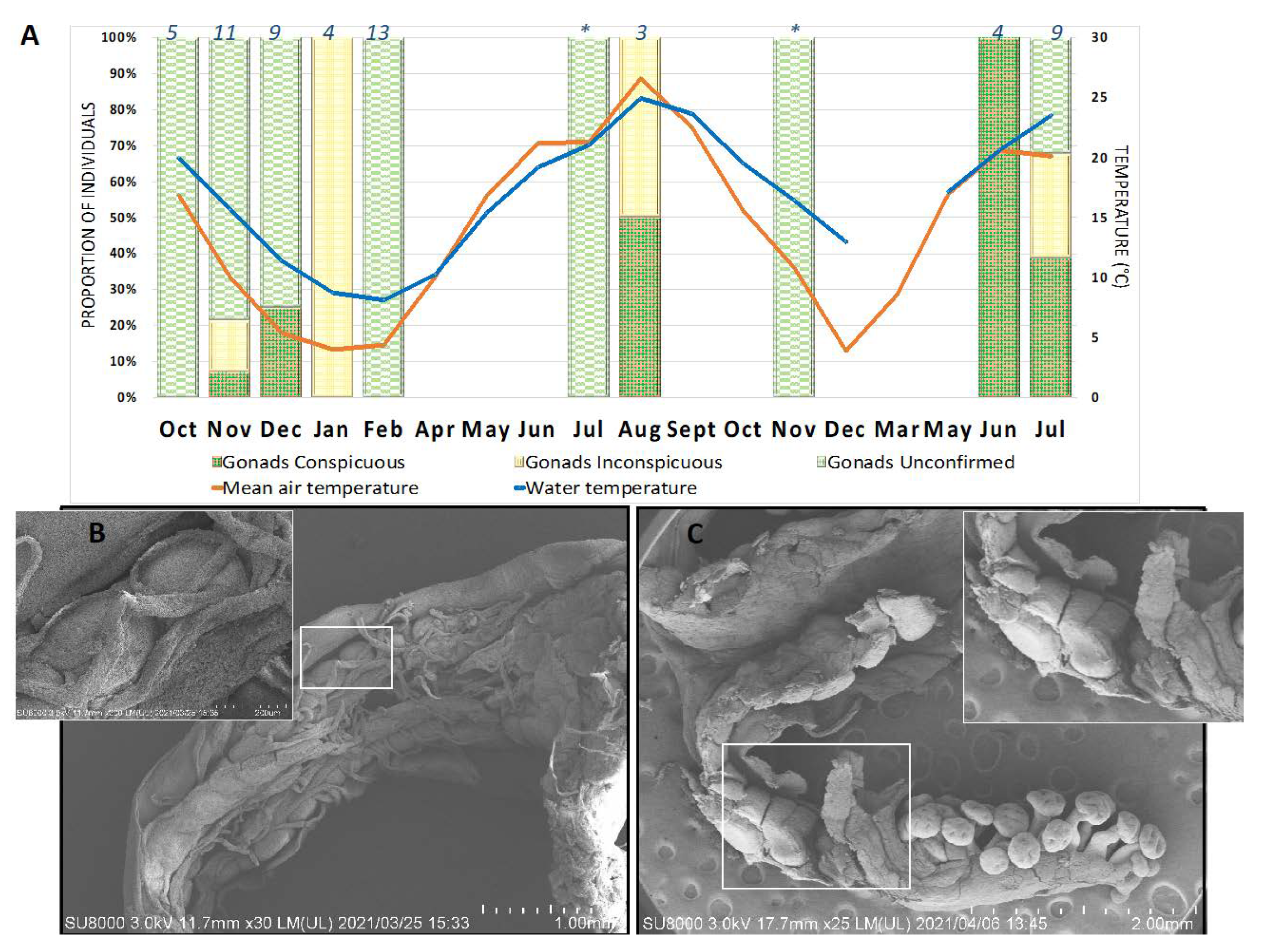
Our stauromedusa distribution data are inconclusive regarding the putative seasonality of reproduction in H. inabai, as individuals with gonads were collected both in cold (October–May) and warm months (June–September). This is in part due to insufficient sampling at the Kitsunezaki collection site during all months of the year, but also due to our inability to differentiate the two lineages of H. inabai, prior to molecular analysis. However, as spawning was not observed in this study, systematic long-term observations are needed to test the hypothesis of whether, as with H. octoradiatus (see Wietrzykowski 1912), one or both lineages of H. inabai are capable of spawning throughout the year.
The absence of Calvadosia nagatensis samples during colder months during this study—September through June—suggests this species has a seasonal appearance during the summer months in the Tohoku region. However, Oka (1897) described C. nagatensis (as Lucernaria nagatensis) based on a specimen collected in April, while Kishinouye (1902) subsequently reported this species in Japan during winter months as well (e.g., in Misaki) and Uchida (1929) in both summer and winter months. Therefore, long-term quantitative studies involving surveys of the algal beds at the Kitsunezaki site are needed to understand the seasonality and early life history of C. nagatensis in the Tohoku region.

4.4. Haliclystus inabai Life Cycle

The life cycles of most Staurozoa species remain enigmas, and despite having reared the stauromedusae specimens collected from Kitsunezaki for more than two months in the lab, it was impossible to further investigate their life history. However, thanks to our collaboration with aquariums specializing in rearing jellyfish for exhibition, we were able to gain an initial understanding of the early life history of H. inabai lineage 2, based on photo and video documentation from the Aquarium du Québec and comparisons with earlier works on other Haliclystus species (Miranda et al. 2010; Wietrzykowski 1912).
In life cycle studies on H. octoradiatus [
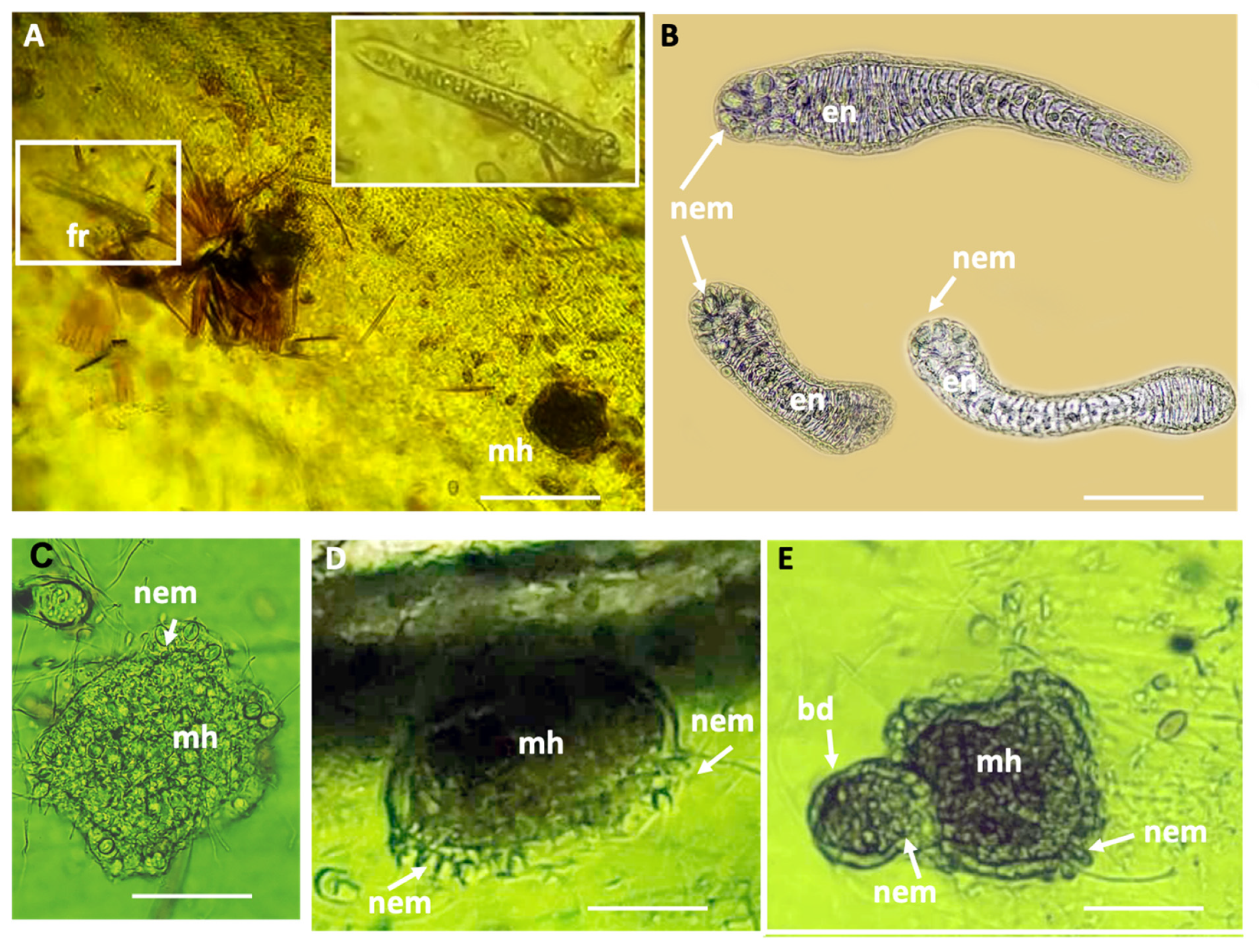
15], the hemispherical settled larval stage, whose ectoderm is replete with nematocysts, gives rise laterally to motile frustules (undergoing frustulation), which then settle and go through several development stages before becoming stauropolyps. In this study, we documented the settled larval stages for H. inabai, which were previously only known in two other species (H. octoradiatus and H. antarcticus) for which it was dubbed the “microhydrula” stage [16]. Similar to those reports, the microhydrula stage in H. inabai has numerous nematocysts interspersed in the ectoderm. We also witnessed what appeared to be a lateral bud producing a motile frustule, the latter of which we documented in abundance. While frutules are on the substrate, the large size and number of penetrant rhopaloids nematocysts around the distal mouth permit them to capture relatively large prey (such as Artemia, seen captured in this study). Though only a single putative asexual bud was witnessed in this study, reportedly, asexual buds can be rapidly produced and number up to four in H. octoradiatus [15].
In this study, the cnidome of H. inabai at both frustule and microhydrula stages appear to contain only one type of rhopaloid, in contrast to stauromedusae, which have at least four types of nematocysts—type I and type II isorhizas, euryteles, and birhopaloids. It is not uncommon for cnidomes to vary at different development stages in a single species, and the putative ontogenetic changes in cnidome witnessed here are similar to those for H. antarcticus [16]. Further examination of the cnidome of H. inabai is needed to delineate nematocyst types present at different life stages.

4.5. Ecological Response and Conservation

The rocky coastal environment of Kitsunezaki has historically been dominated by several species of algae including Sargassum spp, Gelidium elegans, and Eisenia bicyclis (Suzuki et al. 2017), the latter two being macroalgae species associated with the two staurozoan species described in the present study. However, on 11 March 2011, a megathrust earthquake (of magnitude 9.0), known as the Great East Japan Earthquake, occurred off the Pacific coast of northeastern Japan, which caused a tsunami with an estimated height of 15 m in the Oshika Peninsula and devastation to the Tohoku region (Geospatial Information Authority of Japan 2011). Since then, studies have examined correlations between the earthquake and resulting subsidence and the decline of macroalgae epibionts such as abalone, sea urchin, and other fishery resources (reviewed in [41]). In the Kitsunezaki site, where subsidence of 0.9 m occurred, a shift of Eisenia bicyclis distribution towards the shallower area and a decline in juvenile recruitment of the kelp was observed at the offshore margin of the study site [19].
While our study is the first report of staurozoans from Kitsunezaki, staurozoan specimens corresponding to Haliclystus tenuis and Calvadosia cruciformis from Ayukawa, Miyagi Prefecture (Tohoku Region, Japan), a little south on the Oshika Peninsula from our study site, were previously accessioned into the Saito Ho-on Kai Museum of Natural History [42]. In addition to the above two species, a single specimen of Calvadosia tsingtaoensis was reported (as Sasakiella tsingtaoensis) from Onagawa Bay, north of Kitsunezaki on the opposite side of the peninsula [43], but it lacked both interradial and perradial primary tentacles, suggesting it may instead corresponded to C. nagatensis. No further report exists for C. tsingtaoensis in Japan. The absence of biodiversity census data for the Kitsunezaki study site just prior to the Great East Japan Earthquake and Tsunami in 2011 precludes pinpointing of the origin or introduction timeframe of C. nagatensis and H. inabai (either lineage) in this geographic locality. Some studies have reported a decline or complete loss of staurozoan biodiversity in areas with strong anthropogenic effects (e.g., pollution), while others suggest that substrate damage, even from natural disasters, could also result in the disappearance of staurozoans (reviewed in Miranda et al. 2018).
Breakwater restoration work commenced at Kitsunezaki in July 2014, and a subsequent study suggested that the breakwater restoration work had a negative effect on the density and age structure of Eisenia bicyclis population in the Kisunezaki survey site [44]. The ratio of young plants decreased while the whole population became dominated by older plants; inevitable senescence of the adult population could lead to a decline in the E. bicyclis population in the future [44], and consequently reduce staurozoan recruits on E. bicyclis. Studies are needed to understand linkage between the population dynamics of the macroalgae and stauromedusa presence, reproduction and early life cycle, or even seasonality, as this study did not find any obvious pattern with respect to gonad state.

5. Conclusions

We present the first genetic barcode data for C. nagatensis in Japan, and compare these data with that of global species of Calvadosia for which sequence data are generally lacking. Likewise, prior to this study, publicly available molecular sequence data existed only for one H. inabai lineage 1 individual from Japan (Hokkaido), as well as one individual of H. inabai lineage 2 from Australia (Victoria). Analysis of our additional substantial sequence data reveals that H. inabai comprises two distinct species—one including only samples from Japanese localities (H. inabai lineage 1), and the other including samples from locations in several continents (H. inabai lineage 2), either from nearshore waters or found in aquariums. Increased sampling efforts and further comparative analyses are needed before a formal new species description can be made for the unnamed lineage. A consolidation of 100 years of literature documenting substrate variation for these two species revealed a clear propensity by the two named species of stauromedusae for settlement on macroalgae and seagrass. While C. nagatensis is limited to several species of seaweed (algae or kelp) and seagrass, H. inabai (lineage not considered) inhabits a much more diverse suite of substrates.
In addition to analyzing H. inabai specimens collected from the wild from both lineages 1 and 2, we also documented individuals of H. inabai lineage 2 thriving in captivity, in two major aquariums in Japan (Kamo Aquarium in Tsuruoka and Kagoshima Aquarium in Kagoshima) and in Canada (Aquarium du Québec). Photo and video documentation of early life stages in the latter permitted confirmation that the life cycle of H. inabai lineage 2 includes “microhydrula” and frustule stages, similar to those described in two other species of Haliclystus: H. octoradiatus and H. antarcticus [15,16]. In light of these new findings, we discuss whether the broad, global distribution of H. inabai lineage 2 is linked to its multiphasic life cycle, which includes the microscopic sessile microhydrula stage that buds motile frustules during its life cycle, and surmise that its non-specific substrate preference facilitates global distribution of H. inabai lineage 2, potentially through the aquarium trade. This study underlines the importance of collaboration between academia and aquariums. Scientists in both settings can produce research findings of reciprocal relevance, and collaborations can take advantage of complementary expertise, e.g., systematics and live jellyfish husbandry, to make more rapid progress in advancing our understanding of marine biodiversity.
The discovery of these staurozoan species in the Tohoku Region of Japan—an area that was devastated by the 2011 Great East Japan Earthquake—underlines the importance of documenting marine species in both healthy and recovering ecosystems. The crucial relationship between the condition of the macroalgae and its epibionts and their respective responses to environmental disaster—both natural and anthropogenic—are being increasingly highlighted. Although this study focused on species of stalked jellyfish, many other species—including important commercial fisheries species—require macroalgae substrates to complete their life cycles. The discoveries herein were made possible by scientific collaboration with aquarists, and would be very difficult to observe in nature. This work is proof that multi-disciplinary research facilitates a synergistic approach to addressing issues related to ocean ecosystems, biodiversity, and conservation.

Supplementary Materials

The following are available online at https://www.mdpi.com/article/10.3390/hydrobiology1030019/s1, Supplementary File S1: S1_Compiled_Temperature_DataLogger, Supplementary File S2: S2_202112_Haliclystus_alignment_COI.fasta, Supplementary File S3: S3_202112_Haliclystus_alignment_COI consensus tree.newick, Supplementary File S4: S4_202112_Haliclystus_alignment_16S.fasta, Supplementary File S5: S5_202112_Haliclystus_alignment_16S consensus tree.newick, Supplementary File S6: S6_202112_Calvadosia_alignment_coi.fasta, Supplementary File S7: S7_202112_Calvadosia alignment FastTree Tree COI.newick, Supplementary File S8: S8_202112_Calvadosia_alignment_16S.fasta, Supplementary File S9: S9_202112_Calvadosia 16S alignment FastTree Tree.newick, Supplementary File S10: S10_nematocyst_raw_measurement.xlsx, Supplementary File S11: S11_Substrate_preference_reference.xlsx, Supplementary Video S12: EarlyLifeStage_HI.

Author Contributions

Conceptualization, A.S.A., A.A., M.A. and C.L.A.; methodology, M.A. and C.L.A.; software, C.L.A.; validation, A.S.A. and C.L.A.; formal analysis, A.S.A., A.G.C. and C.L.A.; investigation, A.S.A., A.A., A.G.C., Y.H., M.-L.D., D.P., S.S., S.I., K.O., M.C., M.A. and C.L.A.; resources, A.S.A., A.A., A.G.C., Y.H., M.-L.D., D.P., S.S., S.I., K.O., M.C., M.A. and C.L.A.; data curation, A.S.A., A.A., S.I., K.O., M.-L.D. and C.L.A.; writing—original draft preparation, A.S.A., A.A., S.I., K.O., M.-L.D. and C.L.A.; writing—review and editing, A.S.A., A.A., A.G.C., Y.H., M.-L.D., S.I., K.O.,M.C., M.A. and C.LA.; visualization, A.S.A. and C.L.A.; supervision, A.G.C., Y.H., M.A. and C.L.A.; project administration, C.L.A.; funding acquisition, A.S.A., C.L.A. All authors have read and agreed to the published version of the manuscript.

Funding

A.S.A acknowledges the Tohoku University (Sendai, Japan) President Fellowship and Japan MEXT Honors Scholarship for Privately Financed International Students through Tohoku University (Sendai, Japan). C.L.A. acknowledges partial funding for equipment through a Tohoku University (Sendai, Japan) TUMUG Gender Equality Promotion Start-Up Grant 2019–2020. Funding for investigation and sampling was provided by Tohoku Ecosystem-Associated Marine Sciences (TEAMS) Fund of the Ministry of Education, Culture, Sports, Science and Technology (MEXT), Japan.

Institutional Review Board Statement

Not applicable.

Informed Consent Statement

Not applicable.

Data Availability Statement

Data supporting reported results can be found at https://github.com/amandasrwti/staurozoa_kitsunezaki; accessed on 5 April 2022.

Acknowledgments

Ryo Watanabe for spotting the stauromedusae at Kagoshima Aquarium; Isabel Séguin, Mélanie Bélanger, and Sandra Le Tourneux (Aquarium du Québec) for assistance with culturing; Goh Nishitani for equipment support; Kozue Shouji (SEM lab) and Kellee Collier for assistance with molecular analysis; Shenelle Lim and Maria Mokrina for technical support (Tohoku University, Faculty of Agriculture); Scott Whittaker (Smithsonian National Museum of Natural History Imaging Lab), Hidemasa Hiratsuka (Ishinomaki East Branch Office of the Miyagi Federation of Fisheries Cooperative Associations), and Shihoko Kodama (Diving Stage Ariel) for assistance with field work; Katie Ahlfeld (Smithsonian U.S. National Museum Invertebrates Collections) and Kensuke Yanagi (Coastal Branch of National History Museum and Institute, Chiba) for curation and accession of museum specimens. Finally, we are grateful for reviews by three anonymous reviewers.

Conflicts of Interest

The authors declare no conflict of interest.

References

  1. Okamura, B.; Gruhl, A.; Reft, A.J. Cnidarian Origins of the Myxozoa. In Myxozoan Evolution, Ecology and Development; Springer: Cham, Switzerland, 2015; pp. 45–68. [Google Scholar]
  2. Marques, A.C.; Collins, A.G. Cladistic analysis of Medusozoa and cnidarian evolution. Invertebr. Biol. 2004, 123, 23–42. [Google Scholar] [CrossRef]
  3. Collins, A.G. Phylogeny of Medusozoa and the evolution of cnidarian life cycles. J. Evol. Biol. 2002, 15, 418–432. [Google Scholar] [CrossRef] [Green Version]
  4. Zapata, F.; Goetz, F.E.; Smith, S.A.; Howison, M.; Siebert, S.; Church, S.H.; Sanders, S.M.; Ames, C.L.; McFadden, C.S.; France, S.C.; et al. Phylogenomic analyses support traditional relationships within Cnidaria. PLoS ONE 2015, 10, e0139068. [Google Scholar] [CrossRef] [Green Version]
  5. Dumont, H.J. The distribution and ecology of the fresh- and brackish-water medusae of the world. Hydrobiologia 1994, 272, 1–12. [Google Scholar] [CrossRef]
  6. Miranda, L.S.; Hirano, Y.M.; Mills, C.E.; Falconer, A.; Fenwick, D.; Marques, A.C.; Collins, A.G. Systematics of stalked jellyfishes (Cnidaria: Staurozoa). PeerJ 2016, 4, e1951. Available online: https://peerj.com/articles/1951 (accessed on 5 April 2022). [CrossRef] [Green Version]
  7. Miranda, L.S.; Mills, C.E.; Hirano, Y.M.; Collins, A.G.; Marques, A.C. A review of the global diversity and natural history of stalked jellyfishes (Cnidaria, Staurozoa). Mar. Biodivers. 2018, 48, 1695–1714. [Google Scholar] [CrossRef]
  8. Collins, A.; Schuchert, P.; Marques, A.; Jankowski, T.; Medina, M.; Schierwater, B. Medusozoan phylogeny and character evolution clarified by new large and small subunit rDNA data and an assessment of the utility of phylogenetic mixture models. Syst. Biol. 2006, 55, 97–115. [Google Scholar] [CrossRef] [Green Version]
  9. Miranda, L.S.; Collins, A.G.; Hirano, Y.M.; Mills, C.E.; Marques, A.C. Comparative internal anatomy of Staurozoa (Cnidaria), with functional and evolutionary inferences. PeerJ 2016, 4, e2594. [Google Scholar] [CrossRef] [Green Version]
  10. Mills, C.E.; Hirano, Y.M. Stauromedusae. Encycl. Tidepools Rocky Shores 2007, 541–543. [Google Scholar]
  11. Miranda, L.S.; Morandini, A.C.; Marques, A.C. Do Staurozoa bloom? A review of stauromedusan population biology. Hydrobiologia 2012, 690, 57–67. Available online: http://link.springer.com/10.1007/s10750-012-1048-6 (accessed on 5 April 2022). [CrossRef]
  12. Uchida, T. Studies on the Stauromedusae and Cubomedusae, with Special Reference to their Metamorphosis. Jpn. J. Morphol. 1929, 2, 103–193. [Google Scholar]
  13. McInnes, D.E. A stalked jellyfish (Stauromedusae), found at Black Rock, Port Phillip Bay. A first recording in Australia. Vic. Nat. 1989, 106, 86–92. [Google Scholar]
  14. Otto, J.J. Early Development and Planula Movement in Haliclystus (Scyphozoa: Stauromedusae). In Coelenterate Ecology and Behavior; Mackie, G.O., Ed.; Springer: Boston, MA, USA, 1976; pp. 319–329. [Google Scholar]
  15. Wietrzykowski, W. Recherches sur le de veloppement des Lucernaires. Arch. Zool. Exp. Gen. 5th Ser. 1912, 10, 1–95. [Google Scholar]
  16. Miranda, L.S.; Collins, A.G.; Marques, A.C. Molecules clarify a cnidarian life cycle—The “Hydrozoan” Microhydrula limopsicola is an early life stage of the staurozoan Haliclystus antarcticus. PLoS ONE 2010, 5, e10182. [Google Scholar] [CrossRef] [Green Version]
  17. Geospatial Information Authority of Japan. Crustal Movement Electronic Reference Point Obtained from Continious GPS Observation. (In Japanese). 2011. Available online: https://www.gsi.go.jp/chibankansi/chikakukansi40005.html (accessed on 31 March 2022).
  18. Ito, K.; Hamaguchi, M.; Inomata, E.; Agatsuma, Y.; Aoki, M.N. Vertical distribution of epifauna on Sargassum horneri, with special reference to the occurrence of bivalve spat. Plankt. Benthos Res. 2019, 14, 114–123. [Google Scholar] [CrossRef] [Green Version]
  19. Suzuki, H.; Aoki, T.; Kubo, Y.; Endo, H.; Agatsuma, Y.; Aoki, M.N. Distributional changes of the kelp community at a subtidal reef after the subsidence caused by the 2011 Tohoku Earthquake. Reg. Stud. Mar. Sci. 2017, 14, 73–83. [Google Scholar] [CrossRef]
  20. Lawley, J.W.; Ames, C.L.; Bentlage, B.; Yanagihara, A.; Goodwill, R.; Kayal, E.; Hurwitz, K.; Collins, A.G. Box Jellyfish Alatina alataHas a Circumtropical Distribution. Biol. Bull. 2016, 231, 152–169. [Google Scholar] [CrossRef] [Green Version]
  21. Geller, J.; Meyer, C.; Parker, M.; Hawk, H. Redesign of PCR primers for mitochondrial cytochrome c oxidase subunit I for marine invertebrates and application in all-taxa biotic surveys. Mol. Ecol. Resour. 2013, 13, 851–861. [Google Scholar] [CrossRef]
  22. Osada, M.; Nakamura, S.; Kijima, A. Quantitative analysis of pattern of gonial proliferation during sexual maturation in Japanese scallop Patinopecten yessoensis. Fish. Sci. 2007, 73, 1318–1324. [Google Scholar] [CrossRef]
  23. Ames, C.L.; Ohdera, A.H.; Colston, S.M.; Collins, A.G.; Fitt, W.K.; Morandini, A.C.; Erickson, J.S.; Vora, G.J. Fieldable environmental DNA sequencing to assess jellyfish biodiversity in nearshore waters of the Florida Keys, United States. Front. Mar. Sci. 2021, 8, 369. [Google Scholar] [CrossRef]
  24. Kishinouye, K. Mushi-kurage, Depastrum inabai n. sp. Zool. Mag. 1893, 5, 416–419. (In Japanese) [Google Scholar]
  25. Kishinouye, K. Some new Scyphomedusae of Japan. J. Coll. Sci. Imp. Univ. Tokyo 1902, 17, 1–17. [Google Scholar]
  26. Kahn, A.S.; Matsumoto, G.I.; Hirano, Y.M.; Collins, A.G. Haliclystus californiensis, a “new” species of stauromedusa (Cnidaria: Staurozoa) from the nirtheast Pacific, with a key to the species of Haliclystus. Zootaxa 2010, 2518, 49–59. [Google Scholar] [CrossRef]
  27. Falconer, A. A Stalked Jellyfish Stenoscyphus inabai (Kishinouye 1893) (Stauromedusae), found at the Jawbone, Port Phillip Bay, Victoria. Vic. Nat. 2013, 130, 202–206. [Google Scholar]
  28. Zagal, C.J.; Hirano, Y.M.; Mills, C.E.; Edgar, G.J.; Barrett, N.S. New records of Staurozoa from Australian coastal waters, with a description of a new species of Lucernariopsis Uchida, 1929 (Cnidaria, Staurozoa, Stauromedusae) and a key to Australian Stauromedusae. Mar. Biol. Res. 2011, 7, 651–666. [Google Scholar] [CrossRef]
  29. Hausmann, K.; Holstein, T. Bilateral symmetry in the cnidocil-nematocyst complex of the freshwater medusa Craspedacusta sowerbii Lankester (Hydrozoa, Limnomedusae). J. Ultrastruct. Res. Mol. Struct. Res. 1985, 90, 89–104. [Google Scholar] [CrossRef]
  30. Oka, A. Sur une nouvelle espèce japonaise du genre Lucernaria. Annot. Zool. Jpn. 1897, 1, 141–145. [Google Scholar]
  31. Grohmann, P.A.; Magalhaes, M.P.; Hirano, Y.M. First Record of the Order Stauromedusae (Cnidaria, Scyphozoa) from the Tropical Southwestern Atlantic, with a Review of the Distribution of Stauromedusae in the Southern Hemisphere. Species Divers. 1999, 4, 381–388. [Google Scholar] [CrossRef] [Green Version]
  32. Tamura, K.; Stecher, G.; Kumar, S. MEGA11: Molecular Evolutionary Genetics Analysis Version 11. Mol. Biol. Evol. 2021, 38, 3022–3027. [Google Scholar] [CrossRef]
  33. Jarms, G.; Tiemann, H. On a new hydropolyp without tentaeles, Microhydrula limopsicola n. sp., epibiotic on bivalve shells from the Antarctic. Sci. Mar. 1996, 60, 109–115. [Google Scholar]
  34. Otto, J.J. The settlement of Haliclystus planulae. In Settlement and Metamorphosis of Marine Invertebrate Larvae; Chia, F.-S., Rice, M.E., Eds.; Elsevier-North Holland: New York, NY, USA, 1978. [Google Scholar]
  35. Kikinger, R.; von Salvini-Plawen, L. Development from polyp to stauromedusa in Stylocoronella (cnidaria: Scyphozoa). J. Mar. Biol. Assoc. United Kingd. 1995, 75, 899–912. [Google Scholar] [CrossRef]
  36. Hofmann, D.; Garg, N.; Grässle, S.; Vanderheiden, S.; Bergheim, B.G.; Bräse, S.; Jung, N.; Özbek, S. A small molecule screen identifies novel inhibitors of mechanosensory nematocyst discharge in Hydra. Sci. Rep. 2021, 11, 20627. [Google Scholar] [CrossRef] [PubMed]
  37. Thiel, M.; Gutow, L. The Ecology of Rafting in the Marine Environment. II. The Rafting Organisms and Community. In Oceanography and Marine Biology; CRC Press: Boca Raton, FL, USA, 2005; Volume 43, pp. 279–418. ISBN 0849335973. Available online: http://www.crcnetbase.com/doi/abs/10.1201/9781420037449.ch7 (accessed on 5 April 2022).
  38. Cindy, B.; Simon Neil, J.; Tiffany, S.; Haylea, M.; Todd, S.; John Kenneth, K.; Berry, O. Passive eDNA collection enhances aquatic biodiversity analysis. Commun. Biol. 2021, 4, 236. [Google Scholar] [CrossRef]
  39. Abramo, G.; D’Angelo, C.A.; Mele, I. Impact of COVID-19 on research output by gender across countries. Scientometrics 2022, 27, 1–16. [Google Scholar] [CrossRef]
  40. Von Salvini-Plawen, L. First record of a free-living stauromedusa Stylocoronella (Cnidaria). Cah. Biol. Mar. 2006, 47, 219–222. Available online: https://www.natuurlijkmooi.net/adriatische_zee/kwallen/pub_stylocoronella.pdf (accessed on 5 April 2022).
  41. Takami, H.; Won, N.-I.; Kawamura, T. Impacts of the 2011 mega-earthquake and tsunami on abalone Haliotis discus hannai and sea urchin Strongylocentrotus nudus populations at Oshika Peninsula, Miyagi, Japan. Fish. Oceanogr. 2013, 22, 113–120. [Google Scholar] [CrossRef]
  42. Kubota, S.; Yamazaki, Y. List of cnidarian medusae deposited in the Saito Ho-on Kai Museum of Natural History, with special reference to the presence of a large form immortal medusae Turritopsis nutricula. Nat. Hist. Res. Bull. 2007, 72, 1–13. Available online: https://repository.kulib.kyoto-u.ac.jp/dspace/bitstream/2433/197422/1/res.bul_72_1.pdf (accessed on 5 April 2022).
  43. Uchida, T. Medusae in Onagawa Bay and its vicinity. Sci. Rep. Tohoku Imp. Univ. Fourth Ser. 1938, X111, 47–58. [Google Scholar]
  44. Suzuki, H.; Aoki, T.; Inomata, E.; Agatsuma, Y.; Aoki, M.N. Effect of breakwater restoration work following the subsidence caused by the 2011 Tohoku Earthquake on the subtidal kelp population. Phycol. Res. 2021, 69, 3–11. [Google Scholar] [CrossRef]
Figure 1. Live stalked jellyfish from two types of algal beds in Kitsunezaki: (A) Eisenia bicyclis in situ and (B) Gelidium sp. in situ (with Calvadosia nagatensis attached). (C) Haliclystus inabai attached to E. bicyclis in situ; (D) H. inabai on Gelidium in the lab; (E) C. nagatensis on E. bicyclis in situ; (F) C. nagatensis on Gelidium sp. in the lab.
Hydrobiology 01 00019 g001
Figure 2. An overview of the field and lab workflow in this study. Scale bars: ca. 300 m. References for primers: 16SrRNA gene [20] and COI [21].
Hydrobiology 01 00019 g002
Figure 3. Haliclystus inabai stauromedusae observed in Aquarium du Québec tanks. (A) H. inabai stauromedusa on structure associated with potbelly seahorse. (B) H. inabai stauromedusa on plastic foliage. (C) H. inabai stauromedusae on dead coral rocks and tank wall. (D) Juvenile H. inabai stauromedusa on seagrass. (E) Green H. inabai stauromedusa with conspicuous gonads. Note: gastric cirri projecting laterally through the epidermis likely indicates damage. (F) Green H. inabai stauromedusa with copepod attached to secondary tentacles (enlargement in insert corresponds to copepod in smaller white box). Scale bars approximate: A. 70 mm, B. 45 mm, C 30 mm, D. 700 µm, E. 3 mm, F 1.5 mm.
Hydrobiology 01 00019 g003
Figure 4. Original drawings of the two Staurozoa species: (A) Haliclystus inabai (as Depastrum inabai Kishinouye, 1893) and (B) Calvadosia nagatensis (as Lucernaria nagatensis Oka, 1897).
Hydrobiology 01 00019 g004
Figure 5. General anatomy of the stauromedusae collected from Kitsunezaki. (A) Haliclystus inabai whole specimen and corresponding cnidome. A squash of the secondary tentacle tips (enlarged area in box on the left) revealed almost exclusively one type of isorhiza (rod-shape type II), with one or two euryteles also present. (B) Eurytele, from secondary tentacle tips, (C) isorhiza (rod-shape type II) from secondary tentacle tips, and (D) two discharged birhopaloids, from anchor—right one reveals two swellings on the shaft. (E) Calvadosia nagatensis whole specimen and corresponding cnidome. A squash of the secondary tentacle tips (enlarged area inside box) revealed type II isorhizas. A few euryteles were also present (not shown here). (F) Type I isorhizas (partially discharged), were associated with the surface of the white spots, as well as (G) two birhopaloids, in white spots—right one is discharged, showing two swellings on the shaft. Abbreviations: tc: tentacle cluster; an: anchor; cm: coronal muscle; is: isorhiza; eu: eurytele; mn: manubrium; wt: white nematocyst spot; pd: pedal disk; cl: calyx length; pl: peduncle length; al: arm length; cb: calyx base, cr: calyx radius.
Hydrobiology 01 00019 g005
Figure 6. SEM of Haliclystus inabai: (A) Secondary tentacle cluster, fuzziness on the surface of the tentacles; (B) Secondary tentacle knob covered with spine-like protrusions; (C) Close-up on secondary tentacle surface covered with abundant tubules shows the mechanosensory cnidocil complex; (D) View of secondary tentacle surface shows cnidocil complex and fired nematocyst tubules; (E) Close-up of nematocyst tubules shows fine spines; (F) Intact nematocysts (unidentified) present along the calyx wall. Except the image in (D), all are of the same specimen. Abbreviations: cn: cnidocil; nt: nematocyst tubule; n: nematocyst.
Hydrobiology 01 00019 g006
Figure 7. SEM of secondary tentacles of Calvadosia nagatensis: (A) Tentacle cluster shows fuzziness on the surface; (B) Close-up of tentacle tips showing tangled clusters of fired nematocyst tubules; (C) Cnidocil-nematocyst complex; (D) Close-up of nematocyst tubule (no spine). Abbreviations: cn: cnidocil; nt: nematocyst tubule.
Hydrobiology 01 00019 g007
Figure 8. Maximum likelihood trees of 16S rRNA (left) and COI (right) sequences for species of Haliclystus (A) and Calvadosia (B): (A) H. inabai forms two highly-supported subclades (“Japan clade”, Kitsunezaki and Hokkaido—purple; “Australia clade”, Australia, Kitsunezaki and global aquariums—pink), within a clade also comprising H. tenuis for both 16S and COI, and H. borealis for 16S (data not available for COI). (B) C. nagatensis falls into a single highly-supported clade (green) relative to other Calvadosia species. The outgroup sequences are highlighted with gray.
Hydrobiology 01 00019 g008
Figure 9. Comparison of substrate preference between the staurozoans in this study: (A) Overall substrate preference and (B) Algal substrate preference. Based on a 100-year literature review and observations in this study. Details and references provided in Supplementary File S11.
Hydrobiology 01 00019 g009
Figure 10. Gonad state of Haliclystus inabai and Calvadosia nagatensis individuals recorded from the Kitsunezaki survey site. (A) Percentage of H. inabai individuals (numbers above bars) exhibiting conspicuous or inconspicuous gonads, or that the presence/absence of gonads was unconfirmed (individuals that were not examined), plotted with temperature for in situ records over 18 months (October 2019 to July 2021). Gaps in bar plots indicate months in which no collection events occurred. Numbers above bars are the number of individuals collected and examined in the corresponding month. * indicates that the presence of H. inabai was visually confirmed but individuals were not collected (or counted). Note: C. nagatensis was only observed in situ during the months of July and August (2020) and July (2021); data not plotted here. (B,C) SEM images showing a section (in white box) along the length of conspicuous gonad sacs of H. inabai gonads overlain with gastric filaments (B), and C. nagatensis, gonads not associated with gastric filaments (C).
Hydrobiology 01 00019 g010
Figure 11. Haliclystus inabai early life stages observed in Aquarium du Québec tank on Green Cardamine (resin ornament). Still frames taken from live videos. (A) Frustules in close proximity to microhydrula—the hemispherical settled larvae adhering to the substrate on its flat base (15 June 2021). Inset white box shows enlargement of another frustule similar to that in the smaller white box. (B) Motile frustules with endodermal cells numbering ca. 25 to 35 (various dates in January 2021). (C) Enlargement of transparent microhydrula (3 October 2021), with central mass of highly vacuolar endoderm cells enveloped by ectoderm cells interspersed with nematocysts in the periphery. (D) Microhydrula stage, with nematocysts in the periphery. (E) Microhydrula stage with putative asexual bud. Abbreviations: bd: asexual frustule bud (putative); en: endodermal cells; fr: frustules; mh: microhydrula; nem: nematocysts. Scale bars approximate: A 150 µm, B 50 µm, C. 75 µm, D. 50 µm, E. 40 µm.
Hydrobiology 01 00019 g011
Figure 12. Haliclystus inabai elongated settled larvae observed in the Aquarium du Québec tank. Still frames taken from live videos. (A) Start of pedal invagination (thick arrow); swollen lateral regions interspersed with nematocysts in elongated settled larva (ca. 200 µm long) mark the two putative tentacular zones from which the two primary tentacles form (21 January 2021). (B) Details of the pedal invagination and tentacular zones of another larva (ca. 500 µm; 8 January 2021) settled on resin eelgrass. The inset line-drawing shows similarities in the emergence of the pedal invagination and zone of tentacle formation witnessed in our specimens during larval development compared to that of H. octoradiatus, shown as “a longitudinal cut of a larva prior to formation of the first tentacles.” (Source of line drawing in B [15]). Abbreviations: cp: chitinous periderm; nem: nematocysts; pi: pedal invagination (putative); tz: tentacular zones (putative).
Hydrobiology 01 00019 g012
Table 1. GenBank accession numbers corresponding to sequences generated in this study, and museum accession numbers for morphological vouchers. Abbreviations: USNM US National Museum, Smithsonian Institution; CMNH, Coastal Branch of Natural History Museum and Institute, Chiba; S/A, same as above; N/A not applicable (no voucher registered).
SpeciesAccessionOriginVoucher
16SCOI
Haliclystus inabaiOK042842 – OK042849OK037592–OK037598Kitsunezaki, JapanUSNM 1659636
USNM 1659637
USNM 1659610
Haliclystus inabaiS/AS/AS/ACMHN-ZG 09780
CMNH-ZG 09781
CMNH-ZG 09782
Haliclystus inabaiOK042850OK037599–OK037601Aquarium du Québec, CanadaN/A
Haliclystus inabaiOL739686–
OL739688
OL719072–OL719074Kamo Aquarium, JapanN/A
Haliclystus inabaiOL739689– OL739691OL719075–OL719077Kagoshima Aquarium, JapanN/A
Calvadosia nagatensisOK356407–
OK356409
OK042838–OK042841Kitsunezaki, JapanUSNM 1659608
USNM 1659609
Calvadosia nagatensisS/AS/AS/ACMNH-ZG 09778
CMNH-ZG 09779
Table 2. Staurozoan species for which the early life history is known, in full or in part. Question marks refer to stages not reported in the literature or visually confirmed during this study.
SpeciesCreeping PlanulaMicrohydrula StageMotile FrustulePolyp (Including Young Stage)References
Haliclystus inabai?YesYesYes This study; [13]
H. octoradiusYesYesYesYes[15]
H. antarcticus?YesYes?[16,33] (as the hydrozoan Microhydrula limopsicola)
H. sanjuanensisYes???[14,34] (as H. stejnegeri)
H. salpinxYes???[14,34]
Stylocoronella riedli??Yes—lateral budding from polyp calyxYes[35]
S. variabilis??Yes—budding from tip of coiled tentacles of polypYes[35]
Publisher’s Note: MDPI stays neutral with regard to jurisdictional claims in published maps and institutional affiliations.

