Review

Pharmacogenetic Perspective for Optimal Gout Management

by
Khalifa Y. Alrajeh
and
Youssef M. Roman
*
Department of Pharmacotherapy and Outcome Science, School of Pharmacy, Virginia Commonwealth University, 410 N 12th Street, Richmond, VA 23298, USA
*
Author to whom correspondence should be addressed.
Future Pharmacol. 2022, 2(2), 135-152; https://doi.org/10.3390/futurepharmacol2020011
Submission received: 27 February 2022 / Revised: 29 April 2022 / Accepted: 1 May 2022 / Published: 9 May 2022
(This article belongs to the Special Issue Feature Papers in Future Pharmacology)

Abstract

:
Pharmacogenetics (PGx) is an emerging field of pharmacology focusing on how gene variations affect the patient’s response to treatment. Pharmacogenetics is a promising tool to optimize the selection and dosing of medications, including urate-lowering therapies (ULTs) among patients with gout. The global prevalence of gout is rising, and it disproportionately affects specific racial groups and individuals with select socioeconomic status. Genetic and experimental findings have provided evidence that genetic polymorphisms associated with serum urate pathology are also of pharmacogenetic interest. Patients with gout present with several comorbidities, warranting the use of several acute and long-term medications that increase their pill burden and the risk of adverse drug events. Implementing PGx testing can identify individuals who are more or less likely to benefit from a given treatment, improve medication adherence, and reduce pill burden. The purpose of this non-systematic review was to evaluate the contemporary evidence for PGx use in gout management, especially treatment modalities associated with specific genetic polymorphisms that could impact medication safety and efficacy. Strong evidence suggests that individuals carrying the HLA-B*58:01 allele are at a higher risk of serious and life-threatening skin reactions when taking allopurinol. Additionally, racial disparities in the frequency of HLA-B*58:01 warrant genetic screening in high-risk populations, specifically some Asian subgroups and African Americans. Individuals that are G6PD-deficient can develop hemolytic anemia and methemoglobinemia with pegloticase and probenecid use. Patients with the less active form of the drug-metabolizing CYP2C9 are at higher risk for NSAID-related upper gastrointestinal (GI) bleeding. Emerging evidence of clinically significant drug-gene pairs among various gout therapies is growing. Genes found to modulate the response to allopurinol include AOX, ABCG2, and SLC22A12. Meanwhile, UGT1A1 appears to modulate the response to Febuxostat. While CYP2C9 may modulate the toxicity of benzbromarone, SLC22A12 and ABCB1 were found to modulate the response to both benzbromarone and probenecid. The genes CYP2D6, ABCB1, gene cluster (rs6916345 G>A), and SEPHS1 were recently reported to modulate the safety and efficacy of colchicine. Finally, HCG22 and IL1RN are linked with the response to corticosteroid and anakinra, respectively. This review examines and synthesizes the most current level of evidence for using PGx to maximize gout pharmacotherapy.

Graphical Abstract

1. Background

Pharmacogenetics (PGx) is an emerging field of pharmacology focusing on how gene variations affect the patient’s response to treatment. Pharmacogenetics leverages patient genetics to ascertain the response to pharmacotherapy, including gout treatments. Including pharmacogenetics in clinical practice could enable providers to make optimal and informed decisions about drug selection, dose modifications, and treatment options. The ultimate goals of PGx are to individualize medicine and improve patient treatment outcomes by minimizing the risk of adverse drug events [1]. Indeed, pharmacogenetics could usher in a new era in targeted therapy to reduce the risks associated with the trial-error prescribing strategies. Moreover, pharmacogenetics could improve adherence to treatment by identifying optimal responders or those at risk for drug toxicity [1]. Adherence to urate-lowering therapy (ULT) is a challenge despite published guidelines and evidence-based recommendations, urging the need for patient-centered treatment decisions. In a large longitudinal study, patients with gout achieved the lowest adherence rates to ULT relative to patients with six common diseases [2]. A more recent meta-analysis suggested that gouty patients had poor adherence with only a 45% adherence rate [3]. To this end, the role of pharmacogenetics in avoiding trial-error prescribing and improving adherence to ULT is worth exploring [4]. Additionally, gout patients present with multiple comorbidities, warranting several acute and long-term medications, which increase their pill burden and the risk of drug-drug interactions. We believe that the use of PGx could help determine those more or less likely to benefit from given treatments and improve the already existing poor adherence to ULT. Therefore, this review aimed to highlight the most recent advancements in precision medicine and the potential role of pharmacogenetics in improving gout pharmacotherapy to optimize patient treatment outcomes.

2. Methods

A Medline search was conducted using PubMed to inform our non-systematic review. Specifically, our approach involved a literature search of PubMed using the search terms [“pharmacogenomics “ OR “pharmacogenetics” AND “gout”]. Titles and abstracts were scanned for relevant articles. References for selected studies were scanned for the inclusion of additional relevant articles. Information was extracted to synthesize the most contemporary levels of evidence of pharmacogenetics in gout pharmacotherapy. Additionally, we used the Clinical Pharmacogenetics Implementation Consortium (CPIC) guidelines, the Food and Drug Administration (FDA), and the Pharmacogenetic Knowledge Base (PharmGKB) to assess the level of evidence for testing specific drug-gene pairs.

3. Epidemiology of Hyperuricemia and Gout

Gout is an inflammatory disease and the most common form of arthritis. Gout disease affects 3.9% (9.2 million) of the United States adults [5]. The prevalence of gout among individuals of African American, European (EUR), and Hispanic descent was estimated to be 4.8%, 4%, and 2%, respectively. Notably, Asian Americans are 2.7-fold higher to have a gout visit in ambulatory care settings and have a higher prevalence of gout risk alleles than Caucasians [6,7]. Further, select Asian subgroups may also present with a higher incidence and prevalence of gout than the overall population. For example, a relatively outdated study in Hawaii demonstrated that Filipino males had a 2.5% incidence of gout compared with 0.13% in non-Filipinos. The non-Filipino group included “other Asian”, Pacific Islander, and Caucasian populations [7,8]. Hyperuricemia (HU), a risk factor for gout, may accumulate in articular and non-articular tissue structures, forming monosodium urate (MSU) crystals [5]. Urate underexcretion is considered the predominant pathogenesis of hyperuricemia. The increased dietary intake of purine-rich sources, endogenous cell turnover, and decreased extrarenal elimination of serum uric acid (SUA) can also contribute to the pathogenesis of high urate levels. Multiple risk factors associated with HU include diet, comorbid diseases, certain drugs, and genetics [5,9]. Consistent with the contribution of genetics and risk of gout or HU [7], specific gout-related genes were significantly more prevalent in Japanese (JPT), Filipinos, and Han-Chinese (CHS) compared to EUR (p-value: <0.005) [7,10,11].
Patients with gout may present with acute inflammatory arthritis, subcutaneous accumulation of MSU crystals (i.e., tophi), joint damage, and chronic gouty arthritis [5]. Other non-articular clinical features, such as renal or kidney stones, may also result from chronic HU [12]. Further, HU has been significantly associated with the incidence of hypertension (HTN) in adults aged ≥ 40 years [13]. Additionally, it is associated with a 20% increased prevalence of HTN [14], and a higher risk of insulin resistance [15]. Based on those observations, reducing urate levels has become an important therapeutic target beyond gout management, and it potentially prevents and maximizes the treatment of other comorbid conditions [16].

4. Genetics of Hyperuricemia and Gout

Two-thirds of SUA is eliminated through the renal proximal tubule (RPT), while the remaining one-third is eliminated through the small intestine and metabolized by the gut microflora (Figure 1) [17].
Approximately, ninety percent of UA, processed by the kidney, is reabsorbed through the proximal tubular cells [18]. Though several aspects of UA elimination and reabsorption remain unknown, extensive population genetic studies, particularly genome-wide association studies (GWAS), have identified significant genetic polymorphisms in the UA disposition pathway [18,19,20,21]. Variations in genes regulating UA excretion (ABCG2, SLC17A1), UA reabsorption (SLC22A12, SLC2A9, and SLC22A11), and a lipid metabolizing gene (GCKR), as well as a scaffolding protein (PDZK1) have all been linked to SUA levels (Figure 1) [18].
The major urate transporter proteins encoded by the above genes are involved in various functions in the UA disposition pathway. For instance, the solute carrier family 22 member 12 (SLC22A12) encodes the kidney-specific urate transporter URAT1, which is found on the apical surface of the renal proximal tubule epithelial cells [22]. Secondly, the apical ATP-binding cassette transporter G2 (ABCG2) (i.e., breast cancer resistance protein) is involved in urate excretion into the distal renal tubule [23,24]. Thirdly, the key player in transporting UA into the interstitial space and circulation is the GLUT9 (SLC2A9) [25,26]. Other transporters identified in GWAS, including OAT1, OAT3, and OAT4, are thought to play minor roles in the urate transportome (Figure 1) [23]. Considering the inhibition of urate reabsorption as a therapeutic target in managing gout, the interplay between the single nucleotide polymorphisms (SNPs) within these transporters and the uricosuric pharmacotherapies highlights the potential of pharmacogenetics to guide and personalize drug therapy in patients with gout and HU.

5. Gout Management Pharmacotherapy

The pharmacotherapy management of gout includes rapid and effective control of the inflammation in acute gout flares, continued ULT to prevent future flares, and ultimately improve gout treatment outcomes [5]. Contemporary gout treatment guidelines recommend allopurinol as the preferred first-line treatment for managing chronic gout. Pharmacotherapies, such as non-steroidal anti-inflammatory drugs (NSAIDs), colchicine, and corticosteroids, are also appropriate first-line agents to manage gout flares [5]. Pharmacotherapies, including interleukin-1 inhibitors (i.e., canakinumab and rilonacept), are also used to control gout flares when alternatives are contra-indicated or ineffective. Social and environmental factors, as well as diet and genetics, could affect a patient’s adherence and response to ULTs. As the field of pharmacogenomics continues to evolve, multiple studies have evaluated the effect of gene variants, including G6PD, HLA-B*58:01, and CYP2C9, on predicting the response to ULTs and possible adverse drug reactions (Table 1) [5,27].

6. Allopurinol

The purine analog and xanthine oxidase (XO) inhibitor, allopurinol, is widely utilized to manage HU and gout [5]. Allopurinol inhibits the conversion of hypoxanthine to xanthine, mediated by XO, ultimately reducing uric acid production [49]. Allopurinol is metabolized to oxypurinol, the active metabolite of allopurinol. Oxypurinol and allopurinol are known substrates for the efflux transporter, so-called breast cancer resistant protein (BCRP). Hence, a few studies have investigated the interplay between the BCRP protein and the intestinal disposition of allopurinol and renal elimination of oxypurinol [30,50].
Allopurinol is generally safe; however, it is associated with the risk of a life-threatening adverse drug event. This event is characterized by significant eosinophilia and serious cutaneous adverse reaction (SCAR), the so-called allopurinol-induced hypersensitivity syndrome (AHS), in specific patient populations [51,52]. Ethnicity, renal impairment, diuretic use, allopurinol starting dose, and the presence of HLA-B*58:01 are risk factors that contribute to the development of AHS [51,52,53]. The human leukocyte antigen B (HLA-B), part of the Major Histocompatibility Complex-I (MHC-I) is considered the most polymorphic gene in the human genome, with more than 1500 alleles [51]. Carriers of the HLA-B*58:01 variant are at greater risk for SCARs than non-carriers. Thus, the use of allopurinol in patients carrying HLA-B*58:01 is contra-indicated [28]. The latter is associated with an 80-97-fold increased risk of allopurinol-induced hypersensitivity syndrome [54].
The prevalence of HLA-B*58:01 is markedly higher in African Americans and Asian subgroups, mainly Korean, Han Chinese, and Thai descents, which substantiates the recommendation for HLA-B*5801 genotyping in these population groups [5]. Nonetheless, the United States Food and Drug Administration (FDA) did not include PGx recommendations on the drug label. However, it listed the drug in the pharmacogenetic association table [55]. The CPIC and the American College of Rheumatology (ACR) provide clinical guidance on pharmacogenetic testing for HLA-B*58:01. Collectively, it is highly recommended to test for HLA-B*58:01 before commencing allopurinol among high-risk populations to determine alternative therapies in the allele carriers [5,56].
Oxypurinol levels have been used to ascertain allopurinol adherence and allopurinol-therapeutic monitoring among patients with gout. Thus, genetic polymorphisms in the allopurinol-metabolizing enzyme gene are of great interest. Theoretically, oxypurinol formation, the endpoint of allopurinol metabolism with the longest half-life, could partly explain the heterogeneity of response to allopurinol therapy. Therefore, processes involved in oxypurinol formation could influence active metabolite exposure and the magnitude of urate reduction. It is believed that aldehyde oxidase (AOX) converts allopurinol to oxypurinol. The SNP rs3731722 A>G leads to a nonsynonymous amino acid change (His1297Arg) in AOX that can alter the catalytic function for the enzyme substrates [29]. It is associated with achieving the SUA target with a standard dose of allopurinol (300 mg daily) relative to non-carriers (i.e., A allele) [29]. Though the association is intriguing, it remains of low clinical significance [29]. Furthermore, other genes involved in the metabolism and clearance of allopurinol were also studied. Firstly, the XO protein is the therapeutic target for febuxostat and allopurinol, encoded by the XO gene. Secondly, Molybdenum cofactor sulfurase (MOCOS) incorporates molybdenum cofactor in purine oxidases (e.g., AOX and XO). Plausibly, genetic variation in the MOCOS gene may result in defective purine oxidases, altering allopurinol metabolism [29].
Besides the potential impact of HLA-B in predicting toxicity to allopurinol, ABCG2 could also modulate the response to allopurinol [30,57]. A single nucleotide polymorphism (SNP) within ABCG2, specifically the missense variant p.Q141K (rs2231142 C>A), results in a reduced function in the secretory transporter in the kidney and gut, affecting the pharmacokinetic (PK) and pharmacodynamic (PD) of allopurinol and oxypurinol; both of which are ABCG2 transporter substrates [30,57]. Paradoxically, the variant allele of rs2231142 C>A was linked to a lower reduction in SUA levels in response to allopurinol therapy in one GWAS [57]. The reduced function in ABCG2 due to genetic polymorphism, supposedly, leads to a decreased clearance and greater systemic plasma levels of the drug and its metabolite; hence a more considerable SUA decrease is expected. In a prospective clinical study, utilizing real-world data, relative to the homozygous (CC) wild-genotype, the homozygous for the reduced function genotype (AA) had a significantly longer half-life of oxypurinol (t1/2 19.1 ± 1.42 vs. 34.2 ± 12.2; p-value: 0.047) [30].
The exact mechanism by which the polymorphism induces a decreased allopurinol response remains unknown. Nonetheless, some evidence suggests that allopurinol and oxypurinol could be modulating UA reabsorption besides inhibiting its production [58]. For example, in-vitro studies have demonstrated that allopurinol and oxypurinol have uricosuric effects through the inhibition of UA reabsorption via competitive inhibition of URAT1 [31] or potent inhibition of GLUT9 [59], well-established UA reabsorption transporters. Further research, particularly the analysis of uncommon polymorphisms in ABCG2, will undoubtedly improve our knowledge of the genetic basis of the response to allopurinol.
URAT1, encoded by SLC22A12, is considered the predominant transporter for UA reabsorption in the kidney. Presumably, a gain-of-function polymorphism in URAT1 could result in high SUA, whereas loss-of-function mutations cause idiopathic renal hypouricemia due to increased urinary urate clearance [32]. The T allele of the intergenic SNP rs505802 C>T in SLC22A12 was linked with lower SUA than the allele (C). The prevalence of the T allele was significantly lower in Asian (18–35%), Native Hawaiian, and Pacific Islanders (5–37%), relative to EURs (~72%) [11,32]. The disparity in the prevalence of the T allele may not only indicate that Asian and Pacific Islanders populations are more prone to HU relative to EURs but also provides the basis for inquiring about the response to allopurinol and oxypurinol among the same populations [32]. An in-vitro study suggested that the inhibition of URAT1 by benzbromarone facilitated renal clearance of the active metabolite of allopurinol [31]. This drug-drug-transporter interaction illustrates the potential of urate genetic polymorphisms to be of great pharmacogenetic interest.
While genetics are important covariates in predicting drug response, sex and concomitant medications use (e.g., diuretics, cyclosporin, tacrolimus) may also influence the response to allopurinol [30]. The volume of distribution (Vd) of oxypurinol is significantly lower in females than in males (−0.248 L, 95% CI: −0.395, −0.067) [30]. The lower Vd in females compared to males could plausibly translate to a lower dose requirement of the drug to achieve the target plasma concentration. In a retrospective cohort study, the mean dose difference of allopurinol at the time of reaching the target SUA (<6.5 mg/dL) was significantly lower in women (216 mg) compared to men (271 mg) (difference: −55mg; 95% CI: −73, −37) [60]; however, sex did not affect the overall response to allopurinol after six months of the follow-up period in the same study [60].

7. Febuxostat

Febuxostat is structurally unrelated to purine and a potent XO inhibitor [49]. Febuxostat has FDA approval for the chronic management of HU in patients with gout [61]. Although it is well-tolerated at the recommended dose, febuxostat is associated with a higher prevalence of abnormal hepatic enzymes than allopurinol [49,62]. Additionally, an inconsistent body of evidence suggests that febuxostat is associated with an increased risk of death from cardiovascular disease (CVD). Notably, febuxostat users are 34% more likely to have CVD-related mortality and 22% all-cause mortalities than allopurinol. Hence, the conditional recommendation to switch to an alternative ULT in patients with a history of CVD or a new CVD-related event [5,63]. However, recent studies have found no association between febuxostat and CVD or all-cause mortality [64,65].
Patients who utilize XO inhibitors require varying doses to achieve the SUA target. It is reasonable to assume that some of those dose variations are due to polymorphisms in metabolism and clearance genes of XO inhibitors [29]. The uridine diphosphate (UDP) glucoronosyl-transferase (UGT) enzyme metabolizes febuxostat by conjugation (i.e., glucuronidation) [49,62]. The UGT family 1 member A cluster (i.e., members A1, and A3 to 10) gene encodes UGT protein [29,49]. UGT1A1 is considered clinically actionable drug-gene or very important pharmcogenes (VIP) [66]. A single-nucleotide polymorphism (SNP), rs34650714 C>T, in the UGT1A1 appears to be significantly associated with lower febuxostat dose requirement, dose lower than 300 mg daily equivalent to allopurinol, relative to the C allele in patients with gout. However, this SNP is an intronic variant in a non-amino acid coding region [29]. Further, the nonsynonymous polymorphism rs28898617 A>G of UGT1A4 is associated with a difference of 5.1 mg/dL in mean SUA versus the A allele [29]. These findings reflect the effects of genetic polymorphisms on phase II drug-metabolizing encoding genes (UGT1A1 and UGT1A4) and their implications in responding to XO inhibitors [29].

8. Uricosurics

Managing hyperuricemia could also be mediated by increasing excretion or inhibiting urate reabsorption through the kidney. Uricosuric drugs, including probenecid and benzbromarone, are used as a second option to manage gout and HU. Most uricosuric pharmacotherapies block the URAT1 transporter, preventing renal UA reabsorption [23,67]. Besides URAT1, probenecid inhibits other transporters, including OAT1, OAT3, and GLUT9 [67]. Several studies have demonstrated the interplay between genetic polymorphisms within urate transporters, the pathology of hyperuricemia, and the clinical response to uricosuric drugs [33,34].
The 774 G>A mutation carriers in SLC22A12 had significantly lower SUA concentration and higher urate clearance rates relative to healthy control in patients with renal HU [33]. Notably, homozygous carriers or heterozygous for the mutant allele (G774A) had impaired response to loading tests with probenecid and benzbromarone [33]. Following the previous study results, Hamada et al. [34] studied the effect of loss-of-function SLC22A12 mutations on urate clearance rates using benzbromarone and losartan in patients with idiopathic hyperuricemia and hypertension. Neither losartan nor benzbromarone affected urate clearance in patients carrying mutant SLC22A12 (G774A). Conversely, when either drug was used, the wild-type of SLC22A12 allele had significantly increased urate clearance (7 to 13% with benzbromarone; p-value < 0.01) [34]. These experimental observations provide evidence that genetic polymorphisms within SLC22A12 are also of PGx relevance.
The ABCB1 (P-glycoprotein), the adenosine triphosphate (ATP)-binding cassette subfamily B member 1, is an efflux transporter of various drugs. It is essential for the disposition and transport of multiple drugs across the brain, kidney, and liver [68]. With multiple genetic polymorphisms, ABCB1 is considered an important pharmacogenetic biomarker with clinical relevance [68]. The induction or inhibition of ABCB1 by a drug (i.e., an underlying mechanism of drug-drug-transporter interactions) is a critical determinant of the PK profile of the affected drug. A PK randomized-controlled study demonstrated the implication of a drug-drug and ABCB1 interaction and the possibility of a genetic-based response. In the forementioned study, the investigators used probenecid to determine the role of ABCB1 in explaining the increased excretion of dicloxacillin transport in cystic fibrosis patients [35]. When administered with probenecid, the clearance and distribution of dicloxacillin decreased, implying that probenecid is an inhibitor of ABCB1, and the latter is a substrate for the same transporter [35]. On the contrary, dicloxacillin urinary excretion was significantly higher when administered with probenecid in patients with CC genotype of the ABCB1 (i.e., rs1045642 C>T) than CT. While this phenomenon highlights the point that there is a possible genetic effect on probenecid efficacy, as an inhibitor of ABCB1, additional studies are needed to reproduce these findings in gout patients [35].
The probenecid label indicates that risks of hematologic ADRs (i.e., aplastic, hemolytic anemia, or leukopenia) are associated with probenecid administration in patients with glucose-6-phosphate-dehydrogenase (G6PD) deficiency. However, this annotation was listed under the ADRs list, and there were no PGx recommendations [69]. A small cohort study found that G6PD deficiency was not associated with hemolysis when using probenecid in East Asians [36].
Benzbromarone (BBR) is a potent and effective uricosuric drug. However, the FDA withdrew the drug from the United States market due to multiple reports of severe drug-induced hepatotoxicity [70]. Nonetheless, BBR remains widely used in different parts of the world with well-established efficacy in gout management [71,72]. Benzbromarone is primarily metabolized in the liver by the drug-metabolizing enzyme, CYP2C9; however, the metabolizing capacity varies between individuals due to genetic polymorphisms in CYP2C9 [70]. Especially, genetic polymorphisms associated with the reduced and absent enzymatic activity of CYP2C9 could prolong the exposure to BBR, ultimately increasing the risk of significant adverse drug reactions [70]. A single oral dose study of 100 mg BBR among 20 healthy Japanese subjects found that the CYP2C9 intermediate metabolizers (CY2C9*1/*3) had a 2-fold increased elimination half-life of the BBR active metabolite (6-hydroxy BBR) than the CYP2C9 normal metabolizers (CYP2C9*1/*1) (33.2 h ± 18.1 and 18.2 h ± 8.7, respectively; p-value <0.05) [37]. However, the limited sample size, coupled with a low prevalence of CYP2C9*3 in Japanese compared with EUR, meant the study was severely underpowered to meaningfully compare the pharmacokinetic differences between CYP2C9*3/*3 and CYP2C9*1/*1 [37,73]. While the study did not show significant pharmacodynamic differences across CYP2C9 genotype, the same results suggested a critical role for CYP2C9 in the metabolism of BBR in humans [37]. Variability in BBR metabolism could dictate the risk of hepatotoxicity and become higher in the CYP2C9*3/*3 due to low drug clearance. In theory, pharmacogenetic testing of CYP2C9 may provide a personalized approach to minimize the risk of BBR-induced hepatotoxicity and determine uricosuric alternatives for gout patients with moderate renal impairment. Despite the limited sample size, the study demonstrated the possible role genetic polymorphisms have in identifying patients at risk for drug-related toxicity and resurrecting certain drugs once deemed ineffective or toxic. Nonetheless, multiple long-term studies across diverse populations are needed to support preemptive pharmacogenetic testing in gout management.

9. Recombinant Uricases

Rasburicase and pegloticase are recombinant forms of the uricase enzyme. Higher hominoids (apes and humans) lost the uricase gene millions of years ago. In lower primates, uricase converts plasma uric acid to allantoin.The latter is more soluble than uric acid and readily excreted [39,74]. Rasburicase use is mainly for the prevention and treatment of HU. However, it is not suitable for chronic gout treatment due to its limited pharmacokinetic parameters (short half-life and fast-acting). Additionally, it is more effective in reducing plasma uric acid levels in cancer patients than allopurinol in tumor lysis syndrome management [75,76]. Unlike rasburicase, pegloticase is more suitable for and commonly used to treat chronic gout.
The enzyme G6PD transforms glucose-6-phosphate to 6-phosphogluconolactone, resulting in synthesizing reduced nicotinamide adenine dinucleotide phosphate (NADPH) from nicotinamide dinucleotide phosphate. The enzyme G6PD is fundamental in erythrocyte integrity because G6PD and 6-phosphogluconate dehydrogenase (6-PD) are the primary sources of NADPH. The latter protects the red blood cells (RBCs) from oxidative stress, such as oxygen radicals or hydrogen peroxide. While oxidative stress may occur due to abnormal physiological changes, it could be induced by exposure to specific drug therapy [77].
The G6PD-deficient RBCs, for example, have a significantly decreased NADPH production capacity, and hence, they are more susceptible to hemolytic anemia (AHA). Moreover, the oxidation of hemoglobin iron results in the formation of methemoglobin, which cannot carry oxygen or carbon dioxide. This condition is known as methemoglobinemia (MGM), which is characterized by cyanosis, and in severe cases, it may lead to arrhythmias, seizures, and death [39].
Pegloticase is associated with an increased risk of developing MGM and AHA in some patients. These significant adverse drug events could originate from reactive oxygen species formation due to urate oxidation to allantoin. Notably, MGM and AHA are strongly linked to the activity levels in G6PD. Notably, specific genetic polymorphisms in G6PD may result in different activity levels and hence, modulate the risk of developing MGM or AHA. Therefore, genetic testing of G6PD, especially in specific patient populations (i.e., African, South European, Middle Eastern, South Asian), could garner improved outcomes before initiating pegloticase therapy [39].
Similar to PEG, rasburicase is contra-indicated in patients with G6PD deficiency. Rasburicase administration may also lead to oxidative damage to RBCs. However, this risk is only limited to patients who are G6PD-deficient. A G6PD-deficient person is characterized as a male who has one allele of the World Health Organization (WHO) class-II or -III, or a female who carries two alleles (e.g., class II-III variants “II/II, III/III, or II/III”) [78]. The prevalence of the G6PD-deficiency varies between different populations, with a marked increase in African (i.e., Sub-Saharan African countries) or Mediterranean ancestry patients [79]. Therefore, a prompted genotype testing for the G6PD gene is advised in these patients [39]. Additionally, the FDA issued a Blackbox warning for rasburicase and pegloticase due to the risk of hemolysis or methemoglobinemia in patients with G6PD deficiency (i.e., <10–60% of normal enzyme activity). Although uncommon (<1%), these adverse drug events are significantly severe and could be life-threatening [29].

10. Non-Steroidal Anti-Inflammatory Drugs

Non-steroidal anti-inflammatory drugs (NSAIDs) have been extensively used to manage various disease conditions, including acute gout flares, due to their anti-inflammatory and pain-relieving mechanism of action. The NSAIDs inhibit the cyclooxygenase (COX) enzyme reducing the conversion of arachidonic acid to prostaglandins, which are involved in the pain signaling cascade during the inflammatory response. Thus, targeting the prostaglandins and inhibiting the inflammatory cascade is a biological mechanism to resolve acute gout flares-associated pain [80].
While NSAIDs are efficacious and less costly medications to manage gout pain, prescribers need to balance the different risks associated with various NSAIDs and the use of concomitant drugs that would potentiate these risks. The NSAIDs are associated with significant side effects, including cardiovascular (CVD) events, gastrointestinal (GI) bleeding, and renal impairment. These significant side effects require thoughtful considerations and a balanced approach before prescribing them [80].
Most NSAIDs (e.g., indomethacin, ibuprofen, naproxen, diclofenac, meloxicam, and celecoxib) are substrates for the hepatic metabolism via CYP2C9, 1A2, and 3A4 enzymes. Furthermore, the majority of NSAIDs are renally excreted while being nephrotoxic, especially when used chronically [80]. The CYP2C9 is a phase-I cytochrome P450 drug-metabolizing enzyme isoform involved in oxidizing endogenous and exogenous compounds. The CYP2C9 is a major metabolizing enzyme accounting for more than 25% of NSAIDs metabolic clearance and commonly prescribed drugs [81]. In vivo, losartan [82], diclofenac, flurbiprofen, and tolbutamide are commonly utilized as probe drugs for CYP2C9 phenotyping assays [83,84,85]. The gene coding for the CYP2C9 is highly polymorphic with more than 70 known variants [86]. Variations in the CYP2C9 gene could result in varying drug-metabolizing capacity. Hence, the predicted metabolizer phenotypes, based on the individual’s CYP2C9 genotype, are normal (NM), intermediate (IM), and poor (PM) metabolizers [87,88]. Nonetheless, the enzymatic activity also depends on the individual’s age, sex, comorbid diseases, and concomitant use of certain medications (i.e., other CYP2C9 substrates, inhibitors, or inducers) [40]. Furthermore, variability in the metabolic activity of CYP2C9 could well affect the exposure to and outcomes of NSAIDs [40].
Noteworthy, the CYP2C9*2 (i.e., decreased function) variant is in strong linkage disequilibrium (LD) with the CYP2C8*3 allele [40]. This LD might have therapeutic implications for medications like diclofenac and ibuprofen. Both drugs are CYP2C9 and CYP2C8 substrates [40,89,90]. A case-control study found that the odds of NSAID-related upper GI bleeding were 16.92 (95% CI 4.96–57.59) in CYP2C9*3 (i.e., no function allele) carriers and NSAID users (with higher average maintenance daily dose), compared to non-carriers 9.72 (95% CI 4.55–20.76) [41]. In addition, the CPIC guidelines provide CYP2C9 genotype-guided recommendations on specific NSAIDs, such as ibuprofen, celecoxib, meloxicam, and piroxicam, to reduce the toxicity risk associated with their use [39].
Patients with CYP2C9 PM phenotype (i.e., *3/*3 or *2/*2) have a substantially reduced metabolism and prolonged exposure to ibuprofen and celecoxib, thus increasing the likelihood of severe adverse outcomes. Unlike the NM and IM, individuals with the PM phenotype are permitted to start with only a quarter to half of the recommended starting dose [40].
The evidence linking the CYP2C9 genetic variants to indomethacin, diclofenac, and naproxen pharmacokinetics is not significant in vivo and/or not sufficient to warrant a recommendation to guide their use in practice by the CPIC guidelines [40]. The GI and CVD adverse outcomes associated with NSAIDs may have considerable public health consequences due to the large proportion of the population exposed to them. Moreover, an individualized approach that considers multiple risk factors, including genetic predisposition, older age, concomitant drug use, and pre-existing comorbidities, should be employed before NSAID use [40]. Hence, knowledge of the CYP2C9 genetic status of a patient with gout may further guide the selection between different NSAIDs or adjust the dose to minimize GI bleeding risk.

11. Colchicine

Colchicine is an alkaloid derived from meadow saffron. It relieves pain by inhibiting the inflammation response and the infiltration of the immunological cells at the site where MSU crystals deposit. Colchicine interferes with neutrophil functions, lysosomal degradation, and leukocyte chemotaxis by disrupting the microtubule’s polymerization and mobilization [91]. Additionally, colchicine halts the MSU-induced inflammasome-driven caspase-I activation and interleukin-1 (IL-1) beta processing [91]. The most prominent adverse effects of colchicine use are gastrointestinal (GI) adverse events, including diarrhea, nausea, and vomiting [92]. Probably, these ADRs are due to colchicine binding to free tubulin that hinders the fusion of autophagic vacuoles with lysosomes in smooth muscles, resulting in damage to these organs [93], or increasing GI secretions and motility [94]. Other adverse effects of colchicine include myotoxicity and neuropathy [94].
The metabolism of colchicine is mediated by the cytochrome P450 2D6 (CYP2D6) enzyme [95]. About 40 to 65% of colchicine is eliminated renally and excreted via the efflux p-glycoprotein (P-gp) transporter [62]. The drug-metabolizing enzyme CYP2D6 is encoded by the CYP2D6 gene, a highly polymorphic drug-metabolizing gene [95]. The enzyme CYP2D6 is responsible for the metabolism of 25% of prescribed medication; thus, it is considered one of the most critical pharmacogenes [96]. Studies on PGx linking CYP2D6 to clinical response to colchicine in gout patients are limited; nevertheless, the role of CYP2D6 was evaluated in a Middle Eastern ethnic population taking colchicine for Familial Mediterranean Fever (FMF).
Colchicine effectively controlled pain flares in FMF, an inherited disorder of Mediterranean origin [97]. However, approximately 10–20% of patients taking colchicine treatment were resistant, and some showed a diminished response due to potential genetic variations [98]. Colchicine non-responders with FMF were found to have a higher CYP2D6*4 allele (no function allele) frequency than responders (0.16% versus 0.12%, respectively). However, in the colchicine-responders group, the frequency of CYP2D6 *1/*1 (homozygous normal function allele) was higher than the non-responders (80% versus 30%, respectively) [42]. This observation plausibly indicates that individuals with the reduced drug-metabolizing activity of CYP2D6 could not garner the anti-inflammatory benefits of colchicine.
The pharmacogenetic relevance of ABCB1 with the response to colchicine was assessed in FMF subjects of Turkish descent in a case-control study (n = 120) [44]. The study suggested that the odds of diminished response to colchicine in the C allele carriers of the ABCB1 SNP rs10455642 C>T, relative to the TT, were 9.7 (p-value < 0.001) [44]. In another study of an Israeli cohort (n = 105), colchicine-resistant FMF patients were more likely to carry the T allele (66% for TT, 59% for CT, and 39% for CC) than responders (33% for TT, 41% for CT, and 61% for CC) (p-value: 0.047) [43]. These conflicting findings propose that ABCB1 polymorphism could be subject to inter-ethnic variations [99]. Other research on medication responsiveness, which did not include colchicine, showed inconsistent findings regarding the association between ABCB1 alleles and alterations in P-gp transporter function for specific drugs in EUR and Asian populations, highlighting the need for a robust pharmacogenetic study design [100]. Although the results from the previous studies might generate a pharmacogenetic-based hypothesis of colchicine efficacy, these findings were not corroborated in patients with gout.
Considering the anti-inflammatory effects of colchicine, a recent trial, the Colchicine Cardiovascular Outcomes Trial (COLCOT), found low-dose colchicine (0.5 mg daily) to be beneficial in lowering the rates of ischemic heart disease events (composite primary endpoint) by 23% in subjects with post-myocardial infarction (MI) versus placebo (hazard ratio (HR): 0.77, p-value: 0.02) [48]. Additionally, a GWAS of the same study, COLCOT, identified two SNPs, rs6916345 G>A and rs74795203 A>G, that were significantly associated with the GI adverse events from colchicine (secondary safety endpoint) [45].
The risk allele (A) of the intergenic SNP rs6916345 G>A is within a poorly annotated gene. This candidate gene region spans KIF13A and RNU6-793P on chromosome 6 [48]. The same genetic variant was associated with the increased occurrence of GI adverse events (i.e., diarrhea) in the colchicine group by 89% (HR: 1.89, p-value: <0.001) relative to the ancestral allele (G) [45]. In addition, the homozygous patients with the risk allele (AA) in the colchicine group reported GI adverse events that were more than 2-fold higher than those reported by patients in the placebo group (HR: 2.42, p-value: <0.001) [45]. The same allele was previously identified in a GWAS and found to be associated with Crohn’s disease (CD) (OR: 1.07, p-value < 0.001) [101].
The second SNP rs74795203 A>G identified in the COLCOT GWAS was found on chromosome 10 and located in intron 4 of SEPHS1 [45]. The gene SEPHS1, selenophosphate synthase 1, encodes for the enzyme SEPHS1 that synthesizes selenophosphate, the selenium donor, to ultimately incorporate it into selenoproteins [102,103]. The enzyme SEPHS1 plays a role in the modulation of oxidative stress and cell growth, and it is associated with CD [104]. While the allele G increased the risk of adverse GI events by 2.5-fold in the colchicine group (HR: 2.51, p-value: <0.001) [45], the overall GI events were more likely to be reported carrying one or two copies of the G allele in the colchicine group (47.1%) than the placebo group (18.9%) (HR: 3.98, p-value: <0.001) [45].

12. Corticosteroids

Corticosteroids (CORTs) are effective agents used to alleviate pain associated with acute gout flare, mainly when patients are intolerant of oral NSAIDs or colchicine [5]. Due to the ability to administer these agents in various routes (oral, parenteral, or intra-articular), it allowed for expanded utilization in different clinical settings. Corticosteroids act by blocking the eicosanoids production, leukotriene synthesis, and various leukocyte inflammatory processes by suppressing the phospholipase A2 [5].
Multiple genetic polymorphisms in receptor binding (e.g., CRHR1, and NR3C1), Chaperone/Cochaperone protein (e.g., ST13, STIP1, and FKBP5), metabolizing enzymes (e.g., CYP3A4, CYP3A5, CYP3A7, and GSTT1), and transporters (e.g., ABCB1) [105] have been linked to the response to CORTs and associated toxicities. The CORTs are also substrates for P-gp, and hence polymorphisms in ABCB1 may result in different P-gp expressions, resulting in PK differences among CORTs or other transporter substrates [105,106].
Today, there is no robust PGx data regarding the efficacy of CORTs in managing gout flares; however, the pseudogene HLA complex group 22 (HCG22) was studied for PGx relevance in patients with retinal diseases and treated with intravitreal triamcinolone acetonide (injectable CORT) [46]. The SNPs rs3873352 C>G and rs2523864 C>T in HCG22, particularly the G and T alleles of each SNP, respectively, were linked to steroid-induced ocular hypertension (OH) in triamcinolone users [46]. All in all, the CORT’s molecular and metabolic pathways are daunting, and limited genomic factors with enough evidence for therapeutic applicability to gout have not been established.

13. Interleukin-1 Inhibitors

The members of the Interleukin-1 (IL-1) family are essential cytokines in the innate, and partly, adaptive immunity, and are found in high clusters in the articular tissue of human joints [107]. The IL-1 alpha (i.e., IL-1F1) and IL-1 beta function as intracellular pro-inflammatory ligands for specific IL-1 complex receptors [107]. The current and novel pharmacotherapies that target IL-1 include anakinra (recombinant IL-1 receptor antagonist “IL-1RN”) and canakinumab (anti-IL-1 Beta), and rilonacept (IL-1 trap). Anakinra and canakinumab showed promising results in reducing gout flares. However, rilonacept was inferior to NSAID monotherapy over a 72-h follow-up [107].
A cluster of high-expression SNPs in the noncoding region of the interleukin-1 receptor antagonist (IL-1RN) gene were associated with poor response to anakinra. This SNP cluster included rs7580634 G>T, rs55709272 T>C, rs62158853 C>T, rs62158854 T>G, rs55663133 (deletion of the triplet AAT), and rs4251961 T>C [47]. These six IL-1RN SNPs are inherited as a common haplotype in the studied sample. Furthermore, carriers of homozygous genotypes of one or more of the aforementioned SNPs had higher IL-1RN messenger RNA (IL-1RN mRNA) and a 6-fold higher risk of nonresponse to anakinra [47]. Although the associations between those SNPs and poor response to anakinra may seem intriguing, they were not studied in patients with gout.

14. Pharmacogenetic Challenges and Opportunities in Gout Management

The findings of pharmacogenetic studies are sometimes inconsistent and in disagreement concerning conclusions rendered. Study design, sample size, and population enrolled have contributed to this incongruency. Specifically, the lack of racial diversity in pharmacogenetic research has contributed to conflicting findings. Increasing diversity in genetic research studies is a roadmap for expediting the clinical translation of genetic research into practice [108,109,110,111]. The paucity of pharmacogenetic research in gout management is another roadblock that has hindered clinical practice guidelines to provide additional guidance on using pharmacogenetic testing beyond HLA-B*58:01 [28] and G6PD [5] deficiency. These gaps in clinical guidelines strongly suggest the need for robust pharmacogenetic research to maximize gout treatment, especially in high-risk populations. Predominantly, gout patients present with multiple comorbidities and receive care in a primary care setting; therefore, clinical adoption of pharmacogenetic testing requires educating primary care providers on pharmacogenetics in optimal patient care beyond gout [112,113]. Implementing pharmacogenetic testing in gout management is an emerging field with limited research. Therefore, targeted sequencing of well-validated actionable SNPs that carry clinical relevance beyond gout appears to be a logical first step to establishing the benefits of pharmacogenetic testing in gout management. While genetic testing for HLA-B*58:01 has shown to be cost-effective before commencing allopurinol therapy [114,115], there is a lack of economic data on the genetic testing of other genetic variants. More cost-benefit studies assessing the potential of genetic testing patients in achieving serum urate targets are needed to guide the optimal circumstances for genetic testing among patients with gout. Cost-benefit analyses of pharmacogenetic testing will further inform reimbursement policies and empower physicians, pharmacists, and patients to make informed decisions to choose the most effective gout treatment.

15. Conclusions

Pharmacogenomics is gaining prominence as it could be a tool to improve the already existing low adherence to ULT in the context of gout management. The CPIC and FDA have identified several pharmacogenetic occurrences as having meaningful clinical significance in prescribing ULT and other medications used during acute gout flares. First, allopurinol-induced hypersensitivity (AHS) risk is significantly high in HLA-B*58:01 carriers when taking allopurinol, whereas the risk of hemolysis and methemoglobinemia is markedly increased when using recombinant uricase in G6PD deficient patients. These occurrences are linked to significant morbidity and mortality; hence preemptive pharmacogenetic testing for HLA-B*58:01 and G6PD is conditionally recommended for high-risk populations. Third, CYP2C9 *3/*3 or *2/*2 diplotypes are associated with prolonged exposure to ibuprofen and celecoxib, warranting dose reductions to prevent NSAID-induced GI bleeding risk. While preemptive pharmacogenetic testing in gout management is recommended for specific ULT treatments, more pharmacogenetic research is needed to characterize its clinical utility versus the standard of care in other gout therapies to improve adherence to ULT. Adopting pharmacogenetic testing in clinical practice requires a multifaceted approach and robust and diverse clinical trials, especially among high gout risk populations. Optimizing the safety and efficacy of gout pharmacotherapies warrants educating healthcare professionals to be knowledgeable about relevant PGx occurrences. Educating healthcare professionals on PGx could empower clinicians to consider and incorporate PGx testing into patient care, especially when adherence to ULT is in question.

16. Future Perspective

With the recent advancement in genetic research, personalized medicine could influence multiple facets of gout (e.g., stratifying disease risk, assessing disease progression, selecting medications and their dosage, and avoiding adverse drug reactions). The rapidly evolving technology in genetic sequencing has also facilitated significant landmark findings, leading to granular and novel gout-specific genetic discoveries. Though GWAS has led to the identification of many genetic biomarkers of disease risk, specific knowledge gaps in the role of genetics in urate-lowering therapy persist. Collectively, well-characterized data that include the progression from hyperuricemia to gout, explicit drug information (e.g., dose-response outcomes and ADRs), and lifestyle factors will empower clinicians to personalize gout therapy and optimize gout prevention strategies. With most of the GWAS conducted on people of EUR descent, increasing the representation of diverse populations, particularly populations with a high prevalence of gout, is needed to move us beyond one genotype fits all. Additionally, the intricacies of the urate transportome and urate metabolism coupled with clinical factors invite a polygenic assessment approach to refine the prediction of response to gout pharmacotherapy and provide personalized dietary recommendations. Finally, a holistic gout treatment approach involving patient-centered outcomes, genetic information, lifestyle factors, and patient education could significantly improve gout management outcomes and increase adherence to ULT.

Author Contributions

Conceptualization, Y.M.R.; writing—original draft preparation, K.Y.A.; Y.M.R.; writing—review and editing, K.Y.A.; Y.M.R.; visualization, K.Y.A.; Y.M.R. supervision, Y.M.R. All authors have read and agreed to the published version of the manuscript.

Funding

This research received no external funding.

Institutional Review Board Statement

Not applicable.

Informed Consent Statement

Not applicable.

Data Availability Statement

Not applicable.

Conflicts of Interest

The authors declare no conflict of interest.

References

  1. Roden, D.M.; McLeod, H.L.; Relling, M.V.; Williams, M.S.; Mensah, G.A.; Peterson, J.F.; van Driest, S.L. Pharmacogenomics. Lancet 2019, 394, 521–532. [Google Scholar] [CrossRef]
  2. Briesacher, B.A.; Andrade, S.E.; Fouayzi, H.; Chan, K.A. Comparison of Drug Adherence Rates Among Patients with Seven Different Medical Conditions. Pharmacotherapy 2008, 28, 437–443. [Google Scholar] [CrossRef] [PubMed]
  3. Scheepers, L.E.J.M.; van Onna, M.; Stehouwer, C.D.A.; Singh, J.A.; Arts, I.C.W.; Boonen, A. Medication adherence among patients with gout: A systematic review and meta-analysis. Semin. Arthritis Rheum. 2018, 47, 689–702. [Google Scholar] [CrossRef] [PubMed]
  4. United Nations. Population. Available online: https://www.un.org/en/sections/issues-depth/population/ (accessed on 14 December 2020).
  5. Fitzgerald, J.D.; Dalbeth, N.; Mikuls, T.; Brignardello-Petersen, R.; Guyatt, G.; Abeles, A.M.; Gelber, A.C.; Harrold, L.R.; Khanna, D.; King, C.; et al. 2020 American College of Rheumatology Guideline for the Management of Gout. Arthritis Rheumatol. 2020, 72, 879–895. [Google Scholar] [CrossRef]
  6. Krishnan, E.; Lienesch, D.; Kwoh, C.K. Gout in ambulatory care settings in the United States. J. Rheumatol. 2008, 35, 498–501. [Google Scholar]
  7. Butler, F.; Alghubayshi, A.; Roman, Y. The Epidemiology and Genetics of Hyperuricemia and Gout across Major Racial Groups: A Literature Review and Population Genetics Secondary Database Analysis. J. Pers. Med. 2021, 11, 231. [Google Scholar] [CrossRef]
  8. Rosenblatt, G.; Decker, J.L.; Healey, L.A., Jr. Gout in hospitalized Filipinos in Hawaii. Pac. Med. Surg. 1966, 74, 312–313. Available online: https://pubmed.ncbi.nlm.nih.gov/5979498/ (accessed on 24 February 2022).
  9. Murdoch, R.; Barry, M.J.; Choi, H.K.; Hernandez, D.; Johnsen, B.; Labrador, M.; Reid, S.; Singh, J.A.; Terkeltaub, R.; Mellado, J.V.; et al. Original research: Gout, Hyperuricaemia and Crystal-Associated Disease Network (G-CAN) common language definition of gout. RMD Open 2021, 7, e001623. [Google Scholar] [CrossRef]
  10. Roman, Y.; Tiirikainen, M.; Prom-Wormley, E. The prevalence of the gout-associated polymorphism rs2231142 G > T in ABCG2 in a pregnant female Filipino cohort. Clin. Rheumatol. 2020, 39, 2387–2392. [Google Scholar] [CrossRef]
  11. Alghubayshi, A.; Edelman, A.; Alrajeh, K.; Roman, Y. Genetic assessment of hyperuricemia and gout in Asian, Native Hawaiian, and Pacific Islander subgroups of pregnant women: Biospecimens repository cross-sectional study. BMC Rheumatol. 2022, 6, 1. [Google Scholar] [CrossRef]
  12. Kenny, J.-E.S.; Goldfarb, D.S. Update on the Pathophysiology and Management of Uric Acid Renal Stones. Curr. Rheumatol. Rep. 2010, 12, 125–129. [Google Scholar] [CrossRef] [PubMed]
  13. Xu, X.; Huang, J.; Wu, S.; Ji, Q.; Guo, X.; Huang, Y. The Association between the Serum Uric Acid Level and Hypertension in Middle-Aged and Elderly Adults. Cardiovasc. Ther. 2021, 2021, 4626062. [Google Scholar] [CrossRef] [PubMed]
  14. Lanaspa, M.A.; Andres-Hernando, A.; Kuwabara, M. Uric acid and hypertension. Hypertens. Res. 2020, 43, 832–834. [Google Scholar] [CrossRef]
  15. Hu, X.; Rong, S.; Wang, Q.; Sun, T.; Bao, W.; Chen, L.; Liu, L. Association between plasma uric acid and insulin resistance in type 2 diabetes: A Mendelian randomization analysis. Diabetes Res. Clin. Pract. 2021, 171, 108542. [Google Scholar] [CrossRef] [PubMed]
  16. Hisatome, I.; Li, P.; Miake, J.; Taufiq, F.; Mahati, E.; Maharani, N.; Utami, S.B.; Kuwabara, M.; Bahrudin, U.; Ninomiya, H. Uric Acid as a Risk Factor for Chronic Kidney Disease and Cardiovascular Disease—Japanese Guideline on the Management of Asymptomatic Hyperuricemia. Circ. J. 2021, 85, 130–138. [Google Scholar] [CrossRef] [PubMed]
  17. Wright, A.F.; Rudan, I.; Hastie, N.D.; Campbell, H. A “complexity” of urate transporters. Kidney Int. 2010, 78, 446–452. [Google Scholar] [CrossRef] [Green Version]
  18. Yang, Q.; Kottgen, A.; Dehghan, A.; Smith, A.V.; Glazer, N.L.; Chen, M.H.; Chasman, D.I.; Aspelund, T.; Eiriksdottir, G.; Harris, T.B.; et al. Multiple Genetic Loci Influence Serum Urate Levels and Their Relationship with Gout and Cardiovascular Disease Risk Factors. Circ. Cardiovasc. Genet. 2010, 3, 523–530. [Google Scholar] [CrossRef] [PubMed]
  19. Kolz, M.; Johnson, T.; Sanna, S.; Teumer, A.; Vitart, V.; Perola, M.; Mangino, M.; Albrecht, E.; Wallace, C.; Farrall, M.; et al. Meta-Analysis of 28,141 Individuals Identifies Common Variants within Five New Loci That Influence Uric Acid Concentrations. PLoS Genet. 2009, 5, e1000504. [Google Scholar] [CrossRef] [Green Version]
  20. Kottgen, A.; Albrecht, E.; Teumer, A.; Vitart, V.; Krumsiek, J.; Hundertmark, C.; Pistis, G.; Ruggiero, D.; O’Seaghdha, C.M.; Haller, T.; et al. Genome-wide association analyses identify 18 new loci associated with serum urate concentrations. Nat. Genet. 2013, 45, 145–154. [Google Scholar] [CrossRef] [Green Version]
  21. Tin, A.; Marten, J.; Kuhns, V.L.H.; Li, Y.; Wuttke, M.; Kirsten, H.; Sieber, K.B.; Qiu, C.; Gorski, M.; German Chronic Kidney Disease Study; et al. Target genes, variants, tissues and transcriptional pathways influencing human serum urate levels. Nat. Genet. 2019, 51, 1459–1474. [Google Scholar] [CrossRef] [Green Version]
  22. Enomoto, A.; Kimura, H.; Chairoungdua, A.; Shigeta, Y.; Jutabha, P.; Cha, S.H.; Hosoyamada, M.; Takeda, M.; Sekine, T.; Igarashi, T.; et al. Molecular identification of a renal urate anion exchanger that regulates blood urate levels. Nature 2002, 417, 447–452. [Google Scholar] [CrossRef] [PubMed]
  23. Anzai, N.; Jutabha, P.; Amonpatumrat-Takahashi, S.; Sakurai, H. Recent advances in renal urate transport: Characterization of candidate transporters indicated by genome-wide association studies. Clin. Exp. Nephrol. 2012, 16, 89–95. [Google Scholar] [CrossRef] [PubMed]
  24. Woodward, O.M.; Köttgen, A.; Coresh, J.; Boerwinkle, E.; Guggino, W.B.; Köttgen, M. Identification of a urate transporter, ABCG2, with a common functional polymorphism causing gout. Proc. Natl. Acad. Sci. USA 2009, 106, 10338–10342. [Google Scholar] [CrossRef] [PubMed] [Green Version]
  25. Caulfield, M.J.; Munroe, P.B.; O’Neill, D.; Witkowska, K.; Charchar, F.; Doblado, M.; Evans, S.; Eyheramendy, S.; Onipinla, A.; Howard, P.; et al. SLC2A9 Is a High-Capacity Urate Transporter in Humans. PLoS Med. 2008, 5, 1509–1523. [Google Scholar] [CrossRef]
  26. Vitart, V.; Rudan, I.; Hayward, C.; Gray, N.; Floyd, J.; Palmer, C.; Knott, S.A.; Kolcic, I.; Polasek, O.; Graessler, J.; et al. SLC2A9 is a newly identified urate transporter influencing serum urate concentration, urate excretion and gout. Nat. Genet. 2008, 40, 437–442. [Google Scholar] [CrossRef]
  27. Hung, S.-I.; Chung, W.-H.; Liou, L.-B.; Chu, C.-C.; Lin, M.; Huang, H.-P.; Lin, Y.-L.; Lan, J.-L.; Yang, L.-C.; Hong, H.-S.; et al. HLA-B*5801 allele as a genetic marker for severe cutaneous adverse reactions caused by allopurinol. Proc. Natl. Acad. Sci. USA 2005, 102, 4134–4139. [Google Scholar] [CrossRef] [Green Version]
  28. Saito, Y.; Stamp, L.K.; Caudle, K.E.; Hershfield, M.; McDonagh, E.M.; Callaghan, J.T.; Tassaneeyakul, W.; Mushiroda, T.; Kamatani, N.; Goldspiel, B.R.; et al. Clinical Pharmacogenetics Implementation Consortium (CPIC) guidelines for human leukocyte antigen B (HLA-B) genotype and allopurinol dosing: 2015 update. Clin. Pharmacol. Ther. 2016, 99, 36–37. [Google Scholar] [CrossRef] [Green Version]
  29. Carroll, M.B.; Smith, D.M.; Shaak, T.L. Genomic sequencing of uric acid metabolizing and clearing genes in relationship to xanthine oxidase inhibitor dose. Rheumatol. Int. 2017, 37, 445–453. [Google Scholar] [CrossRef]
  30. Vora, B.; Brackman, D.J.; Zou, L.; Garcia-Cremades, M.; Sirota, M.; Savic, R.M.; Giacomini, K.M. Oxypurinol pharmacokinetics and pharmacodynamics in healthy volunteers: Influence of BCRP Q141K polymorphism and patient characteristics. Clin. Transl. Sci. 2021, 14, 1431–1443. [Google Scholar] [CrossRef]
  31. Iwanaga, T. Involvement of Uric Acid Transporter in Increased Renal Clearance of the Xanthine Oxidase Inhibitor Oxypurinol Induced by a Uricosuric Agent, Benzbromarone. Drug Metab. Dispos. 2005, 33, 1791–1795. [Google Scholar] [CrossRef] [Green Version]
  32. Roman, Y.M.; Culhane-Pera, K.A.; Menk, J.; Straka, R.J. Assessment of genetic polymorphisms associated with hyperuricemia or gout in the Hmong. Pers. Med. 2016, 13, 429–440. [Google Scholar] [CrossRef] [PubMed] [Green Version]
  33. Ichida, K.; Hosoyamada, M.; Hisatome, I.; Enomoto, A.; Hikita, M.; Endou, H.; Hosoya, T. Clinical and Molecular Analysis of Patients with Renal Hypouricemia in Japan-Influence of URAT1 Gene on Urinary Urate Excretion. J. Am. Soc. Nephrol. 2004, 15, 164–173. [Google Scholar] [CrossRef] [PubMed]
  34. Hamada, T.; Ichida, K.; Hosoyamada, M.; Mizuta, E.; Yanagihara, K.; Sonoyama, K.; Sugihara, S.; Igawa, O.; Hosoya, T.; Ohtahara, A.; et al. Uricosuric Action of Losartan via the Inhibition of Urate Transporter 1 (URAT 1) in Hypertensive Patients. Am. J. Hypertens. 2008, 21, 1157–1162. [Google Scholar] [CrossRef] [PubMed] [Green Version]
  35. Beringer, P.M.; Kriengkauykiat, J.; Zhang, X.; Hidayat, L.; Liu, S.; Louie, S.; Synold, T.; Burckart, G.J.; Rao, P.A.; Shapiro, B.; et al. Lack of Effect of P-glycoprotein Inhibition on Renal Clearance of Dicloxacillin in Patients with Cystic Fibrosis. Pharmacotherapy 2008, 28, 883–894. [Google Scholar] [CrossRef] [PubMed]
  36. Chan, T.K.; Todd, D.; Tso, S.C. Drug-induced haemolysis in glucose-6-phosphate dehydrogenase deficiency. Br. Med. J. 1976, 2, 1227. [Google Scholar] [CrossRef] [Green Version]
  37. Uchida, S.; Shimada, K.; Misaka, S.; Imai, H.; Katoh, Y.; Inui, N.; Takeuchi, K.; Ishizaki, T.; Yamada, S.; Ohashi, K.; et al. Benzbromarone Pharmacokinetics and Pharmacodynamics in Different Cytochrome P450 2C9 Genotypes. Drug Metab. Pharmacokinet. 2010, 25, 605–610. [Google Scholar] [CrossRef]
  38. Dalbeth, N.; Stamp, L.K.; Merriman, T.R. The genetics of gout: Towards personalised medicine? BMC Med. 2017, 15, 108. [Google Scholar] [CrossRef] [Green Version]
  39. Relling, M.V.; McDonagh, E.M.; Chang, T.; Caudle, K.E.; McLeod, H.L.; Haidar, C.E.; Klein, T.; Luzzatto, L. Clinical Pharmacogenetics Implementation Consortium (CPIC) Guidelines for Rasburicase Therapy in the Context of G6PD Deficiency Genotype. Clin. Pharmacol. Ther. 2014, 96, 169–174. [Google Scholar] [CrossRef]
  40. Theken, K.N.; Lee, C.R.; Gong, L.; Caudle, K.E.; Formea, C.M.; Gaedigk, A.; Klein, T.E.; Agúndez, J.A.; Grosser, T. Clinical Pharmacogenetics Implementation Consortium Guideline (CPIC) for CYP2C9 and Nonsteroidal Anti-Inflammatory Drugs. Clin. Pharmacol. Ther. 2020, 108, 191–200. [Google Scholar] [CrossRef]
  41. Figueiras, A.; Estany-Gestal, A.; Aguirre, C.; Ruiz, B.; Vidal, X.; Carvajal, A.; Salado, I.; Salgado-Barreira, A.; Rodella, L.; Moretti, U.; et al. CYP2C9 variants as a risk modifier of NSAID-related gastrointestinal bleeding: A case-control study. Pharmacogenet. Genom. 2016, 26, 66–73. [Google Scholar] [CrossRef] [Green Version]
  42. Yalcıntepe, S.; Ozdemır, O.; Sılan, C.; Ozen, F.; Uludag, A.; Candan, F.; Sılan, F. The CYP4502D6 *4 and *6 alleles are the molecular genetic markers for drug response: Implications in colchicine non-responder FMF patients. Eur. J. Drug Metab. Pharmacokinet. 2016, 41, 281–286. [Google Scholar] [CrossRef]
  43. Bezalel, Y.; Gershoni-Baruch, R.; Dagan, E.; Lidar, M.; Livneh, A. The 3435T polymorphism in the ABCB1 gene and colchicine unre-sponsiveness in familial Mediterranean fever. Clin. Exp. Rheumatol. 2009, 7, S103–S104. Available online: https://www.clinexprheumatol.org/article.asp?a=3689 (accessed on 24 February 2022).
  44. Babaoglu, M.O.; Yasar, U.; Tufan, A.; Akdogan, A.; Calguneri, M.; Kiraz, S.; Bozkurt, A. Association of the 3435C > T polymorphism of the drug transporter gene ABCB1 with colchicine response in patients with familial Mediterranean fever. FASEB J. 2007, 21, A414–A415. [Google Scholar] [CrossRef]
  45. Dubé, M.-P.; Legault, M.-A.; Lemaçon, A.; Perreault, L.-P.L.; Fouodjio, R.; Waters, D.D.; Kouz, S.; Pinto, F.J.; Maggioni, A.P.; Diaz, R.; et al. Pharmacogenomics of the Efficacy and Safety of Colchicine in COLCOT. Circ. Genom. Precis. Med. 2021, 14, 223–229. [Google Scholar] [CrossRef] [PubMed]
  46. Jeong, S.; Patel, N.; Edlund, C.K.; Hartiala, J.; Hazelett, D.J.; Itakura, T.; Wu, P.-C.; Avery, R.L.; Davis, J.L.; Flynn, H.W.; et al. Identification of a Novel Mucin Gene HCG22 Associated with Steroid-Induced Ocular Hypertension. Investig. Opthalmol. Vis. Sci. 2015, 56, 2737–2748. [Google Scholar] [CrossRef] [PubMed] [Green Version]
  47. Pardeo, M.; Rossi, M.N.; Marafon, D.P.; Sacco, E.; Bracaglia, C.; Passarelli, C.; Caiello, I.; Marucci, G.; Insalaco, A.; Perrone, C.; et al. Early Treatment and IL1RN Single-Nucleotide Polymorphisms Affect Response to Anakinra in Systemic Juvenile Idiopathic Arthritis. Arthritis Rheumatol. 2021, 73, 1053–1061. [Google Scholar] [CrossRef]
  48. Tardif, J.-C.; Kouz, S.; Waters, D.D.; Bertrand, O.F.; Diaz, R.; Maggioni, A.P.; Pinto, F.J.; Ibrahim, R.; Gamra, H.; Kiwan, G.S.; et al. Efficacy and Safety of Low-Dose Colchicine after Myocardial Infarction. N. Engl. J. Med. 2019, 381, 2497–2505. [Google Scholar] [CrossRef]
  49. Avena-Woods, C.; Hilas, O. Febuxostat (Uloric), A New Treatment Option for Gout. Pharm. Ther. 2010, 35, 82. Available online: https://pmc/articles/PMC2827920/ (accessed on 15 December 2021).
  50. Nakamura, M.; Fujita, K.; Toyoda, Y.; Takada, T.; Hasegawa, H.; Ichida, K. Investigation of the transport of xanthine dehydrogenase inhibitors by the urate transporter ABCG2. Drug Metab. Pharmacokinet. 2018, 33, 77–81. [Google Scholar] [CrossRef]
  51. Stamp, L.K.; Chapman, P.T. Allopurinol hypersensitivity: Pathogenesis and prevention. Best Pract. Res. Clin. Rheumatol. 2020, 34, 101501. [Google Scholar] [CrossRef]
  52. Stamp, L.K.; Taylor, W.J.; Jones, P.B.; Dockerty, J.L.; Drake, J.; Frampton, C.; Dalbeth, N. Starting dose is a risk factor for allopurinol hypersensitivity syndrome: A proposed safe starting dose of allopurinol. Arthritis Rheum. 2012, 64, 2529–2536. [Google Scholar] [CrossRef] [PubMed]
  53. Hande, K.R.; Noone, R.M.; Stone, W.J. Severe allopurinol toxicity: Description and guidelines for prevention in patients with renal insufficiency. Am. J. Med. 1984, 76, 47–56. [Google Scholar] [CrossRef]
  54. Lu, N.; Rai, S.K.; Terkeltaub, R.; Kim, S.; Menendez, M.E.; Choi, H.K. Racial disparities in the risk of Stevens-Johnson Syndrome and toxic epidermal necrolysis as urate-lowering drug adverse events in the United States. Semin. Arthritis Rheum. 2016, 46, 253–258. [Google Scholar] [CrossRef] [PubMed] [Green Version]
  55. Food & Drug Administration. Table of Pharmacogenetic Associations. FDA. Available online: https://www.fda.gov/medical-devices/precision-medicine/table-pharmacogenetic-associations (accessed on 16 December 2020).
  56. Hershfield, M.S.; Callaghan, J.T.; Tassaneeyakul, W.; Mushiroda, T.; Thorn, C.F.; Klein, T.E.; Lee, M.T.M. Clinical Pharmacogenetics Implementation Consortium Guidelines for Human Leukocyte Antigen-B Genotype and Allopurinol Dosing. Clin. Pharmacol. Ther. 2013, 93, 153–158. [Google Scholar] [CrossRef] [PubMed] [Green Version]
  57. Wen, C.C.; Yee, S.W.; Liang, X.; Hoffmann, T.J.; Kvale, M.N.; Banda, Y.; Jorgenson, E.; Schaefer, C.; Risch, N.; Giacomini, K.M. Genome-wide association study identifies ABCG2 (BCRP) as an allopurinol transporter and a determinant of drug response. Clin. Pharmacol. Ther. 2015, 97, 518–525. [Google Scholar] [CrossRef] [Green Version]
  58. Roman, Y.M.; Culhane-Pera, K.A.; Lo, M.C.; Yang, S.; Yang, J.; Lo, M.; Straka, R.J. The Impact of Rs505802 for Slc22a12 on Oxipurinol and Uric Acid Disposition in Hmong Patients on Allopurinol from the Genetics of Hyperuricemia Therapy in Hmong (Gouth) Study. Clin. Pharmacol. Ther. 2017, 101, S48. [Google Scholar]
  59. Anzai, N.; Ichida, K.; Jutabha, P.; Kimura, T.; Babu, E.; Jin, C.J.; Srivastava, S.; Kitamura, K.; Hisatome, I.; Endou, H.; et al. Plasma Urate Level Is Directly Regulated by a Voltage-driven Urate Efflux Transporter URATv1 (SLC2A9) in Humans. J. Biol. Chem. 2008, 283, 26834–26838. [Google Scholar] [CrossRef] [Green Version]
  60. Veenstra, F.; Wanten, S.A.C.; Verhoef, L.M.; Stal, M.T.; Kwok, W.-Y.; Hoogen, F.H.J.V.D.; Flendrie, M.; van Herwaarden, N. Sex differences in response to allopurinol and benzbromarone in gout: A retrospective cohort study. Rheumatol. Adv. Pract. 2021, 5, rkab002. [Google Scholar] [CrossRef]
  61. Drug Approval Package: Uloric (Febuxostat) Tablets NDA 21856. Available online: https://www.accessdata.fda.gov/drugsatfda_docs/nda/2009/021856s000toc.cfm (accessed on 15 December 2021).
  62. FDA. Highlights of Prescribing Information. Available online: www.fda.gov/medwatch (accessed on 15 December 2021).
  63. White, W.B.; Saag, K.G.; Becker, M.A.; Borer, J.S.; Gorelick, P.B.; Whelton, A.; Hunt, B.; Castillo, M.; Gunawardhana, L.; CARES Investigators. Cardiovascular Safety of Febuxostat or Allopurinol in Patients with Gout. N. Engl. J. Med. 2018, 378, 1200–1210. [Google Scholar] [CrossRef]
  64. Zhang, M.; Solomon, D.H.; Desai, R.J.; Kang, E.H.; Liu, J.; Neogi, T.; Kim, S.C. Assessment of Cardiovascular Risk in Older Patients with Gout Initiating Febuxostat Versus Allopurinol. Circulation 2018, 138, 1116–1126. [Google Scholar] [CrossRef]
  65. Foody, J.A.; Turpin, R.S.; Tidwell, B.A.; Lawrence, D.; Schulman, K.L. Major Cardiovascular Events in Patients with Gout and Associated Cardiovascular Disease or Heart Failure and Chronic Kidney Disease Initiating a Xanthine Oxidase Inhibitor. Am. Health Drug Benefits 2017, 10, 393–401. Available online: https://pmc/articles/PMC5726059/ (accessed on 15 December 2021).
  66. Barbarino, J.M.; Haidar, C.E.; Klein, T.E.; Altman, R.B. PharmGKB summary: Very important pharmacogene information for UGT1A1. Pharmcogenet. Genom. 2014, 24, 177–183. [Google Scholar] [CrossRef] [PubMed] [Green Version]
  67. Bach, M.H.; Simkin, P.A. Uricosuric drugs: The once and future therapy for hyperuricemia? Curr. Opin. Rheumatol. 2014, 26, 169–175. [Google Scholar] [CrossRef] [PubMed]
  68. Pharmacogenomics Knowledge Base (PharmGKB). ABCB1—Overview. Available online: https://www.pharmgkb.org/gene/PA267 (accessed on 16 December 2020).
  69. Pharmacogenomics Knowledge Base (PharmGKB). Drug Label Information and Legend. Available online: https://www.pharmgkb.org/page/drugLabelLegend#pgx-level (accessed on 16 December 2020).
  70. Lee, M.-H.H.; Graham, G.G.; Williams, K.M.; Day, R.O. A Benefit-Risk Assessment of Benzbromarone in the Treatment of Gout. Was its withdrawal from the market in the best interest of patients? Drug Saf. 2008, 31, 643–665. [Google Scholar] [CrossRef]
  71. Kang, E.H.; Park, E.H.; Shin, A.; Song, J.S.; Kim, S.C. Cardiovascular risk associated with allopurinol vs. benzbromarone in patients with gout. Eur. Heart J. 2021, 42, 4578–4588. [Google Scholar] [CrossRef]
  72. Azevedo, V.F.; Kos, I.A.; Vargas-Santos, A.B.; da Rocha Castelar Pinheiro, G.; dos Santos Paiva, E. Benzbromarone in the treatment of gout. Adv. Rheumatol. 2019, 59, 37. [Google Scholar] [CrossRef] [Green Version]
  73. rs1057910 (SNP)—Population Genetics—Homo sapiens—Ensembl Genome Browser 105. Available online: https://useast.ensembl.org/Homo_sapiens/Variation/Population?db=core;r=10:94980796-94981796;v=rs1057910;vdb=variation;vf=166334188 (accessed on 22 March 2022).
  74. McDonagh, E.M.; Thorn, C.F.; Callaghan, J.T.; Altman, R.; Klein, T.E. PharmGKB summary: Uric acid-lowering drugs pathway, pharmacodynamics. Pharmacogenet. Genom. 2014, 24, 464–476. [Google Scholar] [CrossRef] [Green Version]
  75. Pui, C.-H. Rasburicase: A potent uricolytic agent. Expert Opin. Pharmacother. 2002, 3, 433–442. [Google Scholar] [CrossRef]
  76. Pui, C.-H.; Relling, M.; Lascombes, F.; Harrison, P.; Struxiano, A.; Mondesir, J.-M.; Ribeiro, R.; Sandlund, J.; Rivera, G.; Evans, W.; et al. Urate oxidase in prevention and treatment of hyperuricemia associated with lymphoid malignancies. Leukemia 1997, 11, 1813–1816. [Google Scholar] [CrossRef] [Green Version]
  77. Pharmacogenomics Knowledge Base (PharmGKB). G6PD—Clinical Guideline Annotations. Available online: https://www.pharmgkb.org/gene/PA28469/guidelineAnnotation (accessed on 16 December 2020).
  78. Beutler, E.; Gaetani, G.; Der Kaloustian, V.; Luzzatto, L.; Sodeinde, O. Hexose-6-phosphate Dehydrogenase Deficiency. Bull. World Health Organ. 1989, 67, 601–611. [Google Scholar] [CrossRef]
  79. Nkhoma, E.T.; Poole, C.; Vannappagari, V.; Hall, S.A.; Beutler, E. The global prevalence of glucose-6-phosphate dehydrogenase deficiency: A systematic review and meta-analysis. Blood Cells Mol. Dis. 2009, 42, 267–278. [Google Scholar] [CrossRef]
  80. Tiitinen, S.; Nissilä, M.; Ruutsalo, H.M.; Isomäki, H. Effect of nonsteroidal anti-inflammatory drugs on the renal excretion of uric acid. Clin. Rheumatol. 1983, 2, 233–236. [Google Scholar] [CrossRef]
  81. Zhou, X.-Y.; Lu, X.-R.; Li, Y.-H.; Ma, Y.-Q.; Zhao, S.-W.; Wang, F.; Xu, R.-A.; Hu, G.-X.; Cai, J.-P. Identification and Enzymatic Activity Evaluation of a Novel CYP2C9 Allelic Variant Discovered in a Patient. Front. Pharmacol. 2021, 12, 619339. [Google Scholar] [CrossRef]
  82. Mukai, Y.; Senda, A.; Toda, T.; Eliasson, E.; Rane, A.; Inotsume, N. The Role of CYP2C8 and CYP2C9 Genotypes in Losartan-Dependent Inhibition of Paclitaxel Metabolism in Human Liver Microsomes. Basic Clin. Pharmacol. Toxicol. 2016, 118, 408–414. [Google Scholar] [CrossRef] [Green Version]
  83. Daly, A.K.; Rettie, A.E.; Fowler, D.M.; Miners, J.O. Pharmacogenomics of CYP2C9: Functional and Clinical Considerations. J. Pers. Med. 2018, 8, 1. [Google Scholar] [CrossRef] [Green Version]
  84. Sekino, K.; Kubota, T.; Okada, Y.; Yamada, Y.; Yamamoto, K.; Horiuchi, R.; Kimura, K.; Iga, T. Effect of the single CYP2C9*3 allele on pharmacokinetics and pharmacodynamics of losartan in healthy Japanese subjects. Eur. J. Clin. Pharmacol. 2003, 59, 589–592. [Google Scholar] [CrossRef]
  85. Lee, C.; Pieper, J.A.; Hinderliter, A.L.; Frye, R.F.; Blaisdell, J.A.; Goldstein, J.A. Tolbutamide, Flurbiprofen, and Losartan as Probes of CYP2C9 Activity in Humans. J. Clin. Pharmacol. 2003, 43, 84–91. [Google Scholar] [CrossRef]
  86. PharmVar-CYP2C9. Available online: https://www.pharmvar.org/gene/CYP2C9 (accessed on 15 January 2022).
  87. Van Booven, D.; Marsh, S.; McLeod, H.; Whirl-Carrillo, M.; Sangkuhl, K.; Klein, T.E.; Altman, R.B. Cytochrome P450 2C9-CYP2C9. Pharmacogenet. Genom. 2010, 20, 277–281. [Google Scholar] [CrossRef]
  88. Pharmacogenomics Knowledge Base (PharmGKB). Very Important Pharmacogene: CYP2C9. Available online: https://www.pharmgkb.org/vip/PA166169913 (accessed on 17 December 2020).
  89. Dorado, P.; Cavaco, I.; Cáceres, M.C.; Piedade, R.; Ribeiro, V.; Llerena, A. Relationship between CYP2C8 genotypes and diclofenac 5-hydroxylation in healthy Spanish volunteers. Eur. J. Clin. Pharmacol. 2008, 64, 967–970. [Google Scholar] [CrossRef]
  90. Krasniqi, V.; Dimovski, A.; Domjanović, I.K.; Bilić, I.; Božina, N. How polymorphisms of the cytochrome P450 genes affect ibuprofen and diclofenac metabolism and toxicity. Arh. Hig. Rada Toksikol. 2016, 67, 1–8. [Google Scholar] [CrossRef] [Green Version]
  91. Schlesinger, N. Treatment of Acute Gout. Rheum. Dis. Clin. N. Am. 2014, 40, 329–341. [Google Scholar] [CrossRef] [PubMed]
  92. Slobodnick, A.; Shah, B.; Krasnokutsky, S.; Pillinger, M.H. Update on colchicine, 2017. Rheumatology 2018, 57 (Suppl. 1), i4–i11. [Google Scholar] [CrossRef] [PubMed] [Green Version]
  93. Slobodnick, A.; Shah, B.; Pillinger, M.H.; Krasnokutsky, S. Colchicine: Old and New. Am. J. Med. 2015, 128, 461–470. [Google Scholar] [CrossRef] [PubMed] [Green Version]
  94. Stewart, S.; Yang, K.C.K.; Atkins, K.; Dalbeth, N.; Robinson, P.C. Adverse events during oral colchicine use: A systematic review and meta-analysis of randomised controlled trials. Arthritis Res. Ther. 2020, 22, 28. [Google Scholar] [CrossRef] [Green Version]
  95. PharmVar. Available online: https://www.pharmvar.org/gene/CYP2D6 (accessed on 15 January 2022).
  96. Brown, J.T.; Bishop, J.R.; Sangkuhl, K.; Nurmi, E.L.; Mueller, D.J.; Dinh, J.C.; Gaedigk, A.; Klein, T.E.; Caudle, K.E.; McCracken, J.T.; et al. Clinical Pharmacogenetics Implementation Consortium Guideline for Cytochrome P450 (CYP) 2D6 Genotype and Atomoxetine Therapy. Clin. Pharmacol. Ther. 2019, 106, 94–102. [Google Scholar] [CrossRef] [PubMed] [Green Version]
  97. Sohar, E.; Gafni, J.; Pras, M.; Heller, H. Familial Mediterranean fever. A survey of 470 cases and review of the literature. Am. J. Med. 1967, 43, 227–253. [Google Scholar] [CrossRef]
  98. Drenth, J.P.H.; Van Der Meer, J.W. Hereditary Periodic Fever. N. Engl. J. Med. 2001, 345, 1748–1757. [Google Scholar] [CrossRef]
  99. Ozen, F.; Silan, C.; Uludag, A.; Candan, F.; Silan, F.; Özdemir, S.; Atik, S.; Ozdemir, O. Association between ABCB1 (MDR1) Gene 3435 C > T Polymorphism and Colchicine Unresponsiveness of FMF Patients. Ren. Fail. 2011, 33, 899–903. [Google Scholar] [CrossRef] [Green Version]
  100. Marzolini, C.; Paus, E.; Buclin, T.; Kim, R.B. Polymorphisms in human MDR1 (P-glycoprotein): Recent advances and clinical relevance. Clin. Pharmacol. Ther. 2004, 75, 13–33. [Google Scholar] [CrossRef]
  101. De Lange, K.M.; Moutsianas, L.; Lee, J.C.; Lamb, C.A.; Luo, Y.; Kennedy, N.A.; Jostins, L.; Rice, D.L.; Gutierrez-Achury, J.; Ji, S.-G.; et al. Genome-wide association study implicates immune activation of multiple integrin genes in inflammatory bowel disease. Nat. Genet. 2017, 49, 256–261. [Google Scholar] [CrossRef] [Green Version]
  102. SEPHS1 Selenophosphate Synthetase 1 [Homo sapiens (Human)]—Gene—NCBI. Available online: https://www.ncbi.nlm.nih.gov/gene/22929 (accessed on 4 February 2022).
  103. HUGO Gene Nomenclature Committee. Gene Symbol Report. Available online: https://www.genenames.org/data/gene-symbol-report/#!/hgnc_id/HGNC:19685 (accessed on 4 February 2022).
  104. Gentschew, L.; Bishop, K.S.; Han, D.Y.; Morgan, A.R.; Fraser, A.G.; Lam, W.J.; Karunasinghe, N.; Campbell, B.; Ferguson, L.R. Selenium, Selenoprotein Genes and Crohn’s Disease in a Case-Control Population from Auckland, New Zealand. Nutrients 2012, 4, 1247–1259. [Google Scholar] [CrossRef] [PubMed] [Green Version]
  105. Song, Q.-Q.; Xie, W.-Y.; Tang, Y.-J.; Zhang, J.; Liu, J. Genetic variation in the glucocorticoid pathway involved in interindividual differences in the glucocorticoid treatment. Pharmacogenomics 2017, 18, 293–316. [Google Scholar] [CrossRef] [PubMed]
  106. Corticosteroids. Available online: https://www.pharmgkb.org/chemical/PA10832/clinicalAnnotation (accessed on 21 December 2021).
  107. Janssen, C.A.; Voshaar, M.A.H.O.; Vonkeman, H.E.; Jansen, T.L.T.A.; Janssen, M.; Kok, M.; Radovits, B.; Van Durme, C.; Baan, H.; Laar, M.A.F.J.V.D. Anakinra for the treatment of acute gout flares: A randomized, double-blind, placebo-controlled, active-comparator, non-inferiority trial. Rheumatology 2019, 58, 1344–1352. [Google Scholar] [CrossRef] [PubMed] [Green Version]
  108. Roman, Y.M. Race and precision medicine: Is it time for an upgrade? Pharm. J. 2019, 19, 1–4. [Google Scholar] [CrossRef]
  109. Roman, Y. The United States 2020 Census data: Implications for precision medicine and the research landscape. Pers. Med. 2021, 19, 5–8. [Google Scholar] [CrossRef]
  110. Roman, Y.M.; Lor, K.; Xiong, T.; Culhane-Pera, K.; Straka, R.J. Gout prevalence in the Hmong: A prime example of health disparity and the role of community-based genetic research. Pers. Med. 2021, 18, 311–327. [Google Scholar] [CrossRef]
  111. Coronado, G.; Chio-Lauri, J.; Cruz, R.D.; Roman, Y.M. Health Disparities of Cardiometabolic Disorders Among Filipino Americans: Implications for Health Equity and Community-Based Genetic Research. J. Racial Ethn. Health Disparities 2021. [Google Scholar] [CrossRef]
  112. Roman, Y.M.; Dixon, D.L.; Salgado, T.M.; Price, E.T.; Zimmerman, K.M.; Sargent, L.; Slattum, P.W. Challenges in pharmacotherapy for older adults: A framework for pharmacogenomics implementation. Pharmacogenomics 2020, 21, 627–635. [Google Scholar] [CrossRef]
  113. Anderson, A.N.; Chan, A.R.; Roman, Y.M. Pharmacogenomics and clinical cultural competency: Pathway to overcome the limitations of race. Pharmacogenomics 2022, 23, 363–370. [Google Scholar] [CrossRef]
  114. Ke, C.-H.; Chung, W.-H.; Wen, Y.-H.; Huang, Y.-B.; Chuang, H.-Y.; Tain, Y.-L.; Wang, Y.-C.L.; Wu, C.-C.; Hsu, C.-N. Cost-effectiveness Analysis for Genotyping before Allopurinol Treatment to Prevent Severe Cutaneous Adverse Drug Reactions. J. Rheumatol. 2017, 44, 835–843. [Google Scholar] [CrossRef] [Green Version]
  115. Park, D.-J.; Kang, J.-H.; Lee, J.-W.; Lee, K.-E.; Wen, L.; Kim, T.-J.; Park, Y.-W.; Park, S.-H.; Lee, S.-S. Cost-Effectiveness Analysis of HLA-B5801 Genotyping in the Treatment of Gout Patients with Chronic Renal Insufficiency in Korea. Arthritis Care Res. 2015, 67, 280–287. [Google Scholar] [CrossRef] [PubMed]
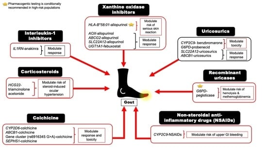
Figure 1. Regulation and handling of uric acid.
Futurepharmacol 02 00011 g001
Table 1. Pharmacogenetics Summary of Gout Treatment and CPIC Guideline Level of Evidence.
DrugMapped GenesEffectClinical OutcomesCPIC Guideline Level of Evidence aReferences
Xanthine oxidase inhibitors (XO)
Allopurinol or OxypurinolHLA-BSafetyHLA-B*58:01 allele significantly increases the risk of allopurinol-induced serious cutaneous reactionA[5,28]
AOXResponsers3731722 A>G is associated with a better response to the standard dose of allopurinol (300 mg/day) vs. non-carriersNA[29]
ABCG2Response/PKrs2231142 C>A (Q141K) is associated with poor response to allopurinolNA[30]
SLC22A12Response/PKrs505802 C>T may influence the response to allopurinol and the PK of oxypurinol as they are substrates for the URAT1NA[11,31,32]
FebuxostatUGT1A1Response/PKrs34650714 C>T is associated with lower doses of febuxostatNA[29]
Uricosuric Agents
ProbenecidSLC22A12ResponseHomozygous or heterozygous for the mutant allele (G774A) have impaired response to loading tests of probenecidNA[33,34]
ABCB1PKrs1045642 C>T could influence the PK effect of probenecid as an inhibitor when co-administered with Beta-lactamNA[35]
G6PDSafetyPossible hematologic adverse reactions in G6PD deficient patientsB[36]
BenzbromaroneCYP2C9SafetyCarriers of the no-function allele (CYP2C9*3) have reduced metabolic activity leading to prolonged exposure to benzbromarone relative to normal metabolizersNA[37,38]
Recombinant Uricase
PegloticaseG6PDSafetyRisk of hemolysis or methemoglobinemia in G6PD deficient patientsB[39]
Non-steroidal anti-inflammatory drugs (NSAIDs)
Ibuprofen, celecoxib, and other NSAIDsCYP2C9Safety/PKIncreased risk of NSAID-related GI bleeding in no-function allele (*3) carriers relative to normal function, as well as reduced metabolism and prolonged exposure to ibuprofen and celecoxib in CYP2C9 poor metabolizersA (ibuprofen and celecoxib); C (indomethacin, diclofenac, naproxen)[40,41]
Anti-inflammatory
ColchicineCYP2D6ResponseDiminished response to colchicine in CYP2D6*4 variant carriersNA[42]
ABCB1Inconsistent evidence wherein one study indicates good response in the T allele carriers of the SNP rs10455642 C>T, while another study suggests no response with the T alleleNA[43,44]
SEPHS1SafetyThe risk allele G of rs74795203 A>G significantly increases the risk of gastrointestinal adverse events by 2.5-fold with using colchicineNA[45]
KIF13A, RNU6-793PbSafetyThe risk allele A of rs6916345 G>A (intergenic) was significantly associated with a ~2-fold increased risk of gastrointestinal adverse events with colchicine compared with the G alleleNA[45]
Corticosteroids
Injectable triamcinolone acetonideHCG22SafetyThe G and T alleles of rs3873352 C>G and rs2523864 C>T, respectively, increase the risk of steroid-induced ocular hypertensionNA[46]
IL-1 inhibitor
AnakinraIL1RNResponse SNP cluster in strong linkage disequilibrium associated with poor response to anakinraNA[47]
PK, Pharmacokinetics; SNP, Single nucleotide polymorphism; CPIC Clinical Pharmacogenomic Implementation Consortium; HLA-B, Human leukocyte antigen B; AOX, Aldehyde oxidase; ABCG2, ATP-binding cassette transporter G2; UGT1 (members A1 and A3-10), Uridine diphosphate (UDP) glucoronosyl-transferase family 1; SLC22A12, Solute carrier family 22 member 12; ABCB1, Human adenosine triphosphate (ATP)-binding cassette subfamily B member 1; G6PD, Glucose-6-phosphate-dehydrogenase; CYP2C9, Cytochrome P450 2C9; CYP2D6, Cytochrome P450 2D6; SEPHS1, selenophosphate synthase 1; HCG22, HLA complex group 22; IL-1RN, Interleukin-1 receptor antagonist. Bolded letters indicate the risk allele. a CPIC level in the clinical context for rapid interpretation by clinicians includes A: Genetic information should be used to change prescribing of the affected drug (quality of evidence is high and in favor of changing prescribing); B: genetic information could be used to change prescribing of the affected drug, but alternative drugs are as effective and safe as non-genetically based dosing (optional change in prescribing); C: evidence levels vary and no prescribing actions recommended; and D: evidence is weak and conflicting, and no prescribing actions recommended. b The intergenic SNP rs6916345 G>A is in a candidate gene region spanning KIF13A, RNU6-793P located on chromosome 6 [48].
Publisher’s Note: MDPI stays neutral with regard to jurisdictional claims in published maps and institutional affiliations.

