Review

The Past, Present, Future: Pathophysiology, Diagnosis, and Treatment of Human Skin Diseases

by
Niki Ebrahimnejad
1,
Duaa Jaafar
2 and
Heidi Goodarzi
3,*
1
Department of Biology, University of California, Berkeley, CA 94720, USA
2
Children’s Dermatology of Orange County, Orange, CA 92868, USA
3
Children’s Hospital of Orange County, Orange, CA 92868, USA
*
Author to whom correspondence should be addressed.
Physiologia 2024, 4(1), 81-99; https://doi.org/10.3390/physiologia4010005
Submission received: 27 November 2023 / Revised: 29 January 2024 / Accepted: 30 January 2024 / Published: 8 February 2024
(This article belongs to the Special Issue Feature Papers in Human Physiology–2nd Edition)

Abstract

:
When thinking of skin disease, cancer comes up almost immediately as an example. While the American Cancer Society lists 6 major cancer types, the National Institute of Arthritis and Musculoskeletal and Skin Diseases identifies 13 significant benign skin disorders, reflecting the diversity of skin conditions in dermatology. This topical review aims to provide an overview of the pathophysiology of these major skin cancers and disorders and to summarize conventional diagnostic methods and current treatment approaches.

1. Introduction

A healthy person may dismiss sunscreen use on a summer’s day, but skin protection and skin care more broadly confer essential maintenance to the body’s largest organ beyond our own natural mechanisms. The skin plays a vital role in maintaining homeostasis, offering protection, and containing our organs and fluids [1]. When the skin is afflicted by disease, these essential functions are disrupted, putting the entire body at risk. One disease that easily comes to mind is skin cancer. Statistics from Center for Disease Control and Prevention reveal that approximately 6 million people in the U.S. receive treatment for various skin cancers annually, with an annual expenditure of nearly USD 9 billion on skin cancer treatment [2]. To be sure, there are over 2000 different dermatological diseases that merit in-depth review and investigation. Instead, this review provides broad coverage of major skin cancers and common skin disorders and conditions.
Certainly, cancer is far from the only manifestation of skin disease that people should be aware of. The National Institute of Arthritis and Musculoskeletal and Skin Diseases identifies 13 significant benign skin disorders [3]. While not inherently cancerous, these major disorders still negatively impact the quality of life of individuals and can even transform into life-threatening conditions if infected or neglected.
Furthermore, social stigma toward irregular skin conditions can place undue psychological stress on the individual through isolation and low self-worth. Even in mild cases, it is still possible for the affected individual to be forcefully exempted from fully participating in otherwise expected society roles due to the sick role phenomenon [4]. It is also worth noting that all demographics can develop skin cancer: age, sex, race, and ethnicity show differences in severity and incidence, but no group can claim immunity [5,6,7]. All things considered, it is imperative to emphasize the importance of awareness and advancements in dermatology, especially given the recent surge in diagnoses and evolving treatment options fueled by technological progress.
In exploring this landscape, this topical review aims to cover key topics discussed in the field of dermatology as an introduction. Publications from PubMed and Google Scholar and reports from research centers and governmental organizations focused on skin disease were pulled, evaluated, and used to summarize key features of the pathophysiology of major skin cancers and disorders and to explore conventional diagnostic methods and current treatment approaches.

2. Overview of Human Skin Diseases

2.1. Skin Cancers

This subsection aims to provide a brief description of the disease, its risk factors, and its incidence. Basal and squamous cell cancer also have some diagnosis and conventional treatment options.

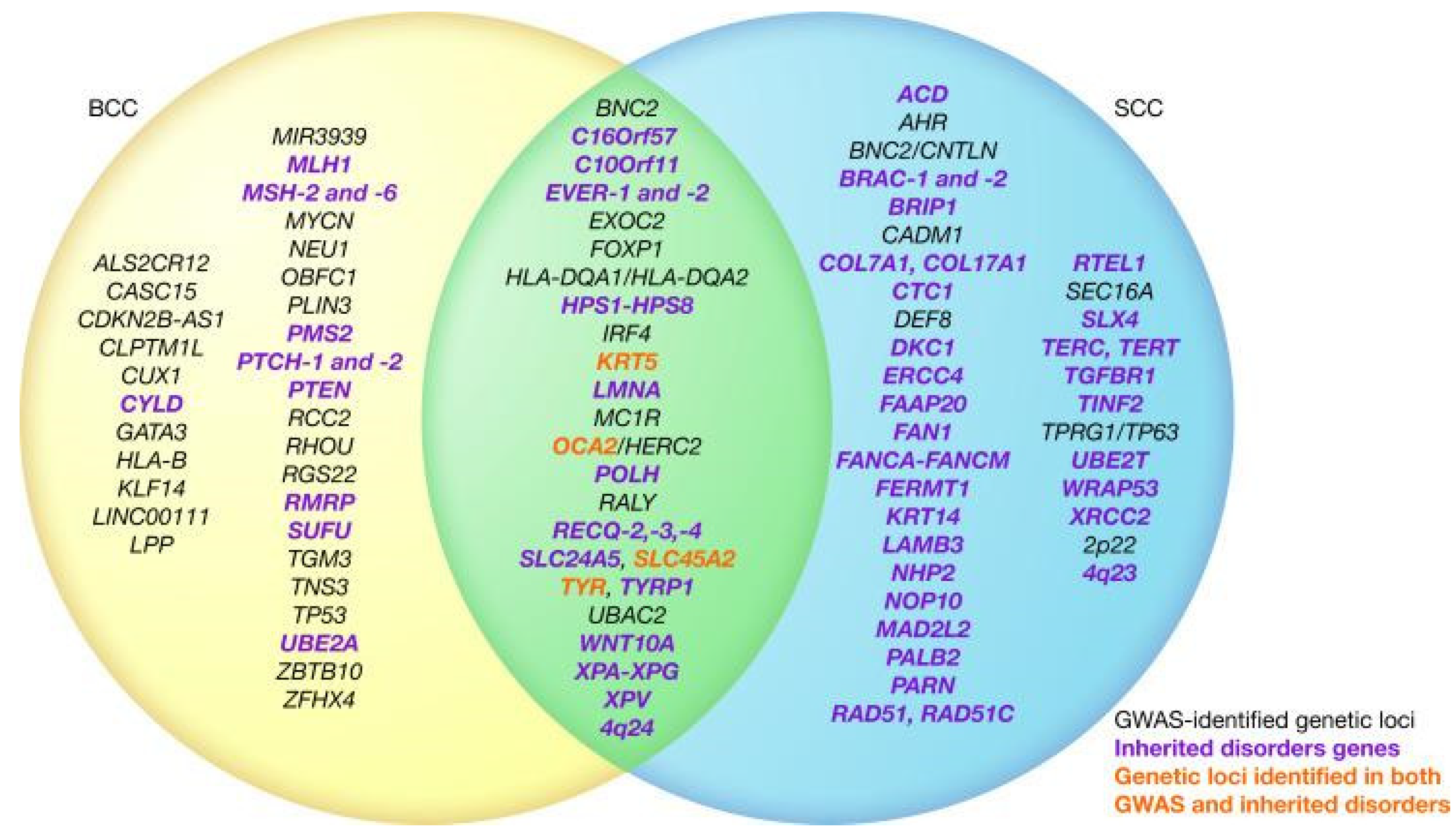
2.1.1. Basal Cell Cancer

The most common head and neck skin cancer, it is slow-growing, with low metastatic potential, and is locally invasive. The main risk factor is ultraviolet (UV) radiation. Diagnosis relies on clinical examination as well as skin biopsy for confirmation and to assess the recurrence risk. Treatment options include surgical excision, Mohs surgery, cryosurgery, electrodesiccation and curettage, imiquimod or fluorouracil, photodynamic therapy, and radiation therapy [8,9,10]. Despite being so common, basal cell cancer and squamous cell skin cancer cases are difficult to estimate because most cancer registries do not require and do not receive records of cases [7].

2.1.2. Squamous Cell Cancer

The second most prevalent skin cancer in the U.S., it often emerges from precursor lesions and has the potential to metastasize. UV radiation is the primary risk factor. Surgical excision is the primary treatment, with Mohs micrographic surgery preferred for head and neck cases or those with high-risk characteristics. Radiation therapy is an option for older patients or those unable to undergo surgery. Immunosuppression increases lifetime risk and metastasis [8,11].

2.1.3. Melanoma Skin Cancer

This is a metastasizing cancer that originates in melanocytes and can appear in various colors, from brown to white. It often starts on the trunk in men and the legs in women, with darker skin offering some protection, but not immunity. Risk factors include strong evidence of exposure to ultraviolet radiation, moles (large moles, over 50 moles, atypically shaped moles), family history (genetic inheritance of melanoma), weight, and fair phenotype (light skin that burns or freckles easily, naturally blonde or red hair, blue or green eyes). Statistically, American men under 50 years have seen incidence rates decline by 1% per year, while men over 50 years have stabilized rates. American women, however, see stable rates for those under 50 years, but a 1% increase per year for those over 50 years [7]. Trauma and chronic inflammation are associated with special site melanoma, particularly acral melanoma. Although melanoma is less common than some skin cancers, it has a higher tendency to metastasize if not diagnosed and treated [12].

2.1.4. Merkel Cell Skin Cancer

A rare, metastasizing skin cancer primarily typically affecting individuals over 50 years old. Risk factors include the Merkel cell polyomavirus, UV exposure, and immunosuppression. Diagnosis involves clinical examination, tissue biopsy, and cytokeratin-20 immunohistochemistry [13]. There is scant data to generate national incidence and prevalence rates; however, the largest dataset in Europe of this cancer type reports that men on average had it more often than women, with incidence rates increasing by 3.9% per year during 2004–2018 [14].

2.1.5. Lymphoma

Originating in the lymphatic system, lymphoma is the result of either T or B lymphocytes becoming cancerous. Risk factors for lymphoma include certain viral infections, exposure to certain chemicals and pesticides, family history, and a weakened immune system. Common methods for diagnosing lymphoma include blood tests, imaging studies like CT scans and PET scans, and lymph node biopsy [15,16,17]. The incidence of non-Hodgkin lymphoma decreased by about 1% per year from 2015 to 2019 [7].

2.1.6. Kaposi Sarcoma

Kaposi sarcoma results from Kaposi sarcoma-associated herpesvirus (KSHV) infections in the blood and lymphatic vessel cells. Risk factors include KSHV infection, which is more prevalent among those with weakened immune systems. Diagnosis relies on recognizing characteristic skin lesions [17,18]. Global declines in incidence in Europe, Latin America, the U.S., and Africa are said to be matched with declines in HIV incidence and prevalence due to safe sex practices, public awareness, and viral therapies [19,20].

2.2. Benign Skin Disorders

This subsection aims to provide a brief description of the disorder and its primary risk factors.

2.2.1. Acne

Acne is a condition caused when blocked skin follicles form a plug caused by oil from glands, bacteria, and dead cells which clump together and swell [3]. There are many environmental and behavioral contributors to the development of acne, including, but not limited to, air pollution, certain skincare products, medications, hormonal issues, and, more recently, diet and stress [21,22]. In addition, genetic inheritance of specific polymorphisms has been observed to yield the acne phenotype more prominently for certain ethnic groups [23,24,25,26].

2.2.2. Alopecia Areata

A condition causing hair to fall out in small, round patches [3]. Risk factors include, but are not limited to, a history of atopic dermatitis for the individual and family, inheritance of various single-nucleotide polymorphisms, pre-existing autoimmune thyroid diseases, and psychological stress [27,28,29,30].

2.2.3. Atopic Dermatitis

A skin disease characterized by itchiness, redness, swelling, cracking, and scaling [3]. It is closely associated with asthma, with several studies observing comorbidity of the two conditions [31]. Risk factors include, but are not limited to, sex, existing allergies, family history of allergens, and air pollution [31,32,33,34].

2.2.4. Epidermolysis Bullosa

A group of genetic connective tissue diseases causing painful blisters to form on the skin and scar [3]. Unlike the previous disorders, the consequences of severe epidermolysis bullosa can result in organ damage and failure. The most common form is epidermolysis bullosa simplex, which is restricted to the epidermis of the skin with minimal scarring. Both mutations to modifier genes and epigenetic mechanisms contribute to the loss of tissue integrity [35,36]. Non-genetic risk factors may include expression of inflammatory bowel disease reported in children and SARS-CoV-2 infection [37,38].

2.2.5. Hidradenitis Suppurativa

A chronic inflammatory condition characterized by pimple-like bumps, boils, and tunnels under the skin [3]. It is associated with bacterial infection of the apocrine sweat glands. Risk factors include, but are not limited to family history as it can be inherited, age (between adolescence and 40 years), sex (females more likely), weight, stress, and even early squamous cell cancer and psoriasis. It remains unclear whether race is a risk factor, with various studies in support of this, such as Mokos et al. (2023), and others that remain skeptical, such as Bryd et al. (2023) [39,40,41,42,43,44].

2.2.6. Ichthyosis

A rare autosomal recessive congenital disorder causing dry, thickened skin that resembles fish scales [3]. Reported genetic causes of this disorder are severe mutations in the adenosine-triphosphate-binding cassette A12 gene, resulting in an extreme form of ichthyosis, and the Vitamin D receptor gene (polymorphism), resulting in a milder form [43,44]. In the case of the latter, age and raised serum ALP levels have been suggested to be risk factors [45,46].

2.2.7. Pachyonychia Congenita

A rare congenital disorder causing thick nails, painful calluses, and other symptoms [3]. It is caused by variations in keratin genes, especially KRT6A and KRT6B [47].

2.2.8. Pemphigus

A disease where the immune system attacks healthy skin cells, resulting in blisters [3]. Having pemphigus may lead to osteopenia, osteoporosis, and pathologic fractures, although further investigation is needed [48,49,50]. One case report describes an interesting case of induced autoimmunity preventing pemphigus despite the patient having Hodgkin’s lymphoma, a risk factor for pemphigus [50]. Other suggested risk factors are sex thymic diseases, P wave dispersion increments, and diet diversity [51,52,53,54].

2.2.9. Psoriasis

A skin disease that causes red, scaly, painful, swollen skin [3]. While specific risk factors are still unclear for psoriasis, it is known to be associated with cardiovascular mortality, myocardial infarction, and stroke in its moderate to severe forms [55].

2.2.10. Raynaud’s Phenomenon

A condition shunting blood vessels and causing insufficient blood flow to the hands and feet [3]. Risk factors include migraine headaches, rheumatologic disease, vaso-occlusive diseases, hematologic disorders, physical injury, viral infection, and carpal tunnel syndrome [56]. In one case report, there was an association of Raynaud’s phenomenon with long-term silica exposure [57].

2.2.11. Rosacea

A long-term disease leading to reddened skin, pimples, and skin thickening, often affecting the face [3]. Risk factors include high temperatures, Demodex mites, overuse of aggressive face cleansers, and cardiovascular diseases [58,59,60,61]. There is a suggestion that alcohol consumption may also be a risk factor; however, follow-up studies of this scenario are required [62].

2.2.12. Scleroderma

A condition causing tight, hard skin and potential harm to blood vessels and organs [3]. In systemic sclerosis patients, ACE inhibitors with concomitant arterial hypertension are a risk factor, as well as high levels of RNA polymerase III antibodies [63,64]. Smoking, interestingly, has not been shown to be a risk factor [65].

2.2.13. Vitiligo

A common disorder leading to white patches of skin due to the destruction of melanocytes [3]. It may manifest in childhood or adulthood. There is some correlation between Hepatitis C virus presence and adult onset of vitiligo [66]. Polymorphisms in tumor necrosis factor-α and -β have been reported to be a risk factor, albeit only in one population [67]. Arguably, one of the biggest difficulties of vitiligo is the stigma it carries and how those with it may be othered, shunned, and ostracized for it. Studies have compared various psychological and psychosocial parameters between those with vitiligo and those without to find that, overall, there is a higher incidence of anxiety and depression associated with vitiligo that is consistent across different ethnic communities [68,69,70,71].

3. Pathophysiology of Human Skin Diseases

3.1. Common Pathophysiological Factors

This subsection aims to describe typically shared traits of skin diseases.

3.1.1. Inflammation

Inflammation is a natural immune response to protect against harmful stimuli, such as pathogens or tissue injury. In the context of skin diseases, when the skin is exposed to irritants, allergens, infections, or autoimmune reactions, the immune system triggers an inflammatory response [72]. This response can manifest as redness, swelling, heat, and pain. The issue arises with chronic or excessive inflammation contributing to skin diseases. TRPV-ion channels trigger the release of pro-inflammatory mediators such as calcitonin gene-related peptide and substance P, perpetuating conditions like psoriasis, atopic dermatitis, prurigo, and rosacea. Immune cells within the skin, including mononuclear cells, dendritic cells, and mast cells, also express TRPV1, further amplifying inflammation by releasing cytokines and neuropeptides [73]. This amplification can cause abnormal cell growth in the case of wound repair, forming a healing tissue niche resembling the tumor stroma if hijacked, a gateway toward skin cancer [74].

3.1.2. Immune System Dysregulation

Because the immune system triggers inflammation as an innate response to foreign particles, there is an overlap between the consequences of irregularity in the immune system and the inflammatory response. For instance, T cell populations on the skin, even rare ones like γδ T cells, have a specific role in allergic skin inflammation and in maintaining tissue homeostasis [75,76]. Effector T cells and memory T cells are accompanied by natural killer cells and T cells expressing MHCR on the skin. This impressive ensemble of specific immunity can collapse with interference in signaling or antigen expression. The classic case is HIV, which puts individuals at a higher risk of developing non-melanoma skin cancers (NMSCs) compared to the general population [77]. Human papillomaviruses have a similar impact, promoting squamous cell carcinoma [78].

3.1.3. Environmental Triggers

The top trigger for skin cancer is UV radiation, followed by ionizing radiation, typically from the sun. Due to the high energy of UV waves, they cause DNA damage and genetic mutations, especially by mutating pyrimidines into cyclobutene pyrimidine dimers that nucleotide substitution mechanisms try and fail to fix. UV radiation (and also ionizing radiation) levels are impacted by the ozone (or lack thereof), latitude, altitude, and weather conditions [79,80]. It is worth noting that UV radiation also contributes to speeding up skin aging, an otherwise natural part of aging and the human condition.

3.2. Biomarkers by Skin Disease

Major advances in elucidating gene pathways that may trigger skin diseases have been achieved thanks to the fields of computational biology and genomics. Because of the large volume gene pathways involved in skin diseases, the following sections are not meant to be taken as comprehensive.

3.2.1. Skin Cancers (Basal Cell Cancer, Squamous Cell Carcinoma, Melanoma)

Melanoma

For melanoma, the risk factors are primarily genetic over environmental, with telomere genes as a common source of mutations. The typical biomarkers of melanoma are BRAF, NRAS, PI3K-AKT/PTEN, p53, CDK4/CDKN2A, c-KIT, MC1R, and cadherin [81]. With the overlap of melanoma with other cancer families, several genes have stood out to be shared between melanoma and at least one other cancer type: CDKN2A/p16, CDK4, BRCA2, POT1, MITF, RB1, P53, BAP1, PTEN, and CHEK2. Many melanoma genes are not just shared with other cancers, but are also shared with neurological diseases. PLA2G6, BAP1, DCC, ERBB4, KIT, MAPK2, MITF, PTEN, and TP53 have all been implicated in both melanoma and Parkinson’s [82]. Turning over to non-coding DNA markers, telomeres have long been established to be mutated in melanoma, especially in the TERT promoter, although the exact degree across subtypes of melanoma and the relationship with different mutations is still being investigated [83].

Basal Cell Cancer

For basal cell cancer specifically, the Hedgehog pathway has mutations in multiple genes, promoting this type of cancer directly just as effectively as UV overexposure. While PTCH1 has been reported as the primary driver for basal cell cancer when bound excessively to the Hedgehog ligand, many secondary drivers exist that affect downstream components of the pathway, such as MYCN, PPPC, SK19, LATS1, ERBB2, PIK23C, N-RAS, K-RAS, H-RAS, PTPN14, RB1, and FBX7, and the IGF-PI3K-AKT, EGFR-MEK-ERK, and Hippo pathways [84,85].

Squamous Cell Carcinoma

For squamous cell carcinoma, which has the highest mutational burden of all cancers, many possible mutations can occur. First, the EGFR-MAPK pathway is overexpressed, resulting in downstream effects via RAS/RAS GTPase. Second, RAS adaptor proteins themselves may also be mutated, yielding metastasizing tumors. Third, RAF proteins (serine/threonine kinases activated by RAS), if activated and heterodimerized, then activate kinase MEK, which promotes new tumor formation. Fourth, NOTCH1 coactivated by MAML1, if knocked out, can no longer fulfill its tumor suppressor role. Fifth, TGF beta receptors are somewhat more complicated to target due to the opposite roles the SMAD proteins play downstream and upstream. Droll and Bao (2021) go into depth on the alterations in the aforementioned pathways that contribute to this cancer type. They also describe what effectors are associated with squamous cell carcinoma: p53, p63, p73, cyclin D1, p14, p16, and MYC [86].
The epigenetics of this cancer are also worth looking at. DNA hypomethylation, histone hypomethylation, and BAF complex loss of function have been reported.
As far as similarities go, both squamous cell carcinoma (right) and basal cell carcinoma (left) share several genetic factors, as summarized in Figure 1 [79].

3.2.2. Benign Skin Disorders (Atopic Dermatitis, Pemphigus Vulgaris, Psoriasis, Rosacea, Vitiligo)

Atopic Dermatitis

While over 30 genetic loci have been linked to atopic dermatitis, a loss of function mutation in filaggrin has been reported to be the strongest known genetic risk factor. Other genes that confer atopic dermatitis are interleukin-4 and -13 and their respective receptors. GWAS studies and other multi-omics investigations have found more candidate genes [87]. For elderly individuals, the rise in immunosenescence of the skin can be traced by a decrease in T-cell surface markers globally, a decrease in Langerhans cells expressing HBD3, an increase in IgG and IgA levels, and a decrease in IgM levels [88].

Pemphigus Vulgaris

Desmoglein-3, a Ca2+-dependent cell adhesion molecule, is considered the primary autoantigen. Additionally, proteomic studies have revealed several other autoantibodies, including desmocollins 1 and 3, muscarinic and nicotinic acetylcholine receptor subtypes, mitochondrial proteins, human leukocyte antigen molecules, thyroid peroxidase, and hSPCA1 [89,90]. These autoantibodies play various roles in keratinocyte physiology and cell adhesion.

Psoriasis

Psoriasis is characterized by the cytokines TNF-α, IFN-γ, IL-10, IL-12, IL17, IL-22, and IL-23, and the small molecules MAPK inhibitors, PDE-4 inhibitors, and JAK inhibitors [91]. Cytokines typically have varying structures, modes of detection (surface bound receptor, serum levels), sites of production, specific function, and effects, all of which are succinctly described by Parab and Doshi (2022). In addition, MAPK and p38 are overexpressed in psoriasis and directly contribute to cytokine production. Blocking these proteins, the cAMP pathway via the PDE enzyme and the JAK-STAT system via JAK1 and STAT3 are ideal approaches for treatment. In the presence of IL-1β and TNF-α, Langerhans cells fail to migrate and induce CD4+ T cell proliferation during the inflammatory response.
On the other hand, the IL-23/IL-17 axis with antimicrobial peptide HBD3, when activated, leads to the development of psoriasis [91,92]. Another axis of players that can influence psoriasis is the HPA axis. Deregulation of the cross talk between the varying levels of signaling in the HPA axis has been reported to contribute to psoriasis, and psoriasis itself can feed into the feed-forward mechanism of the HPA axis by expressing pro-inflammatory cytokines and hormones in response to stress [93].

Rosacea

Upregulation of TLR2 promotes the expression of the antimicrobial peptide cathelicidin, which is converted to LL-37, a promoter of angiogenesis and skin redness. TLR2 also activates NLRP3, leading to pustule formation and vascular reactivity. PAR2 activation results in inflammation, pruritus, and physical pain. Temperature changes, exercise, UV exposure, spicy food, and alcohol may also cause the TRP ankyrin and vanilloid subfamilies to release vasoactive neuropeptides like substance P and calcitonin gene-related peptide [94,95]. These genes, when upregulated, collectively contribute to the pathogenesis of rosacea.

Vitiligo

The pathogenesis of vitiligo involves several genes and molecular processes, with a focus on the IFN-γ-CXCL9/10-CXCR3 axis and its connection to the JAK/STAT pathway. Melanocytes in vitiligo patients have reduced adhesiveness and heightened susceptibility to oxidative stress [96].
Polymorphisms in HLA-A confer the most significant genetic risk, followed by antigen presentation genes (HLA-DRB1/DQA1, CPVL). Other biomarkers that have been suggested are those that mediate immune target cell lysis (GZMB, FASLG), regulate adaptive immunity (FOXP3, CTLA4, IL2RA, BACH2), and drive innate immunity (TICAM1, IFIH1, CD80), in addition to regulate melanocytes (TYR, PMEL, MC1R, OCA2-HERC2, IRF4) [97].
Various studies have also demonstrated the significance of the IFN-γ-CXCL9/10-CXCR3 axis in vitiligo. This axis inhibits melanogenesis, induces apoptosis of melanocytes, and recruits T cells to the skin. These processes are interconnected with the JAK/STAT pathway. Cytokines such as HSP70i, IL-15, IL-17/23, and TNF are recruited. The WNT signaling pathway and regulatory T cells are also implicated in vitiligo [96,97,98].

4. Diagnosis of Human Skin Diseases

4.1. Clinical Examination Methods

This subsection aims to describe common examination methods used by clinicians in evaluating skin.
The clinician, when interacting with the patient, can inquire about any history or recent changes in lifestyle and assess all skin with real-time communication. Additionally, there may be evaluation with a dermatoscope, skin scrapings for evaluation under microscope, skin culture, wood light. For hard-to-see features, such as pigment distribution and depth, or identifying of small parasites, dermatoscopes are very useful. Even so, there are limitations to superficial assessments, calling for more sophisticated tools.
Typically, additional clinical measures to identify and assess skin disease include taking a biopsy or scraping for culturing to determine the cell type. For cases of fungi or bacterial infection, a black or wood light is used to shine UV light in a dark room. For viral infections, a Tzanck test is performed to obtain fluid from blisters for analysis. When the medical practitioner wishes to observe how the skin responds to a potential allergen, there are four skin tests: use test, patch test, prick test, and intradermal test. The last two may prompt anaphylaxis and are used with caution [99]. For hard-to-see lesions such as scabies burrows, dermatoscopes are helpful, but their main role is to help distinguish malignant and benign pigmented lesions [100,101].

4.2. Histopathology

This subsection aims to describe common methods used to further characterize skin samples at a microscopic level.
Generally, histology slides of representative tissue are first assessed with the naked eye, then with a microscope at low power, and finally with a microscope at high power. There are many staining techniques and markers available in dermatopathology, with the four below as the most common:
  • Hematoxylin and eosin;
  • Immunohistochemistry;
  • Immunofluorescence (direct and indirect);
  • In situ hybridization and fluorescence in situ hybridization.
There are also 17 special stains for specific targets such as fungi, bacteria, mast cells, and various molecules. See page 155 from Ladoyanni (2022) for details [100].

5. Standard Treatment Approaches for Skin Diseases

Lists of skin disease treatments, including topical skin medications approved in the U.S., can be found in the Orange Book of the Food and Drug Administration (FDA) at https://www.fda.gov/drugs/resources-information-approved-drugs/approved-drug-products-therapeutic-equivalence-evaluations-orange-book (accessed on 20 December 2023).

5.1. Topical and Oral Medications

This subsection aims to cover the various categories of skin disease treatments.

5.1.1. Topical Preparations Overview

Preparations tend to consist of an active drug mixed with an inactive ingredient, known as the vehicle. The choice of vehicle affects the consistency of the product and how the drug interacts with the skin (Table 1) [102]. Other considerations are any side effects on other systems. For example, corticosteroids reduce inflammation by suppressing the immune response; therefore, they pose a risk for heightened infection and should be used strategically or with antifungal, antibacterial, or antiviral supplementary agents.
Some vehicles are better suited for certain functions than others and have differing sensations when applied. For instance, solutions are ideal cleansing agents because water is a powerful solvent, while ointments and powders are better protective agents. Sunscreen is a common example. There are eight categories of topical drugs: (1) cleansing, (2) protective, (3) moisturizing, (4) drying, (5) anti-itch, (6) anti-inflammatory, (7) anti-infective, and (8) keratolytic.

5.1.2. Oral Preparations Overview

Oral medications for skin diseases offer systemic treatment advantages such as consistent dosages, ensuring precise drug delivery to the bloodstream. This convenience is especially beneficial for individuals with extensive affected areas. However, oral medications can affect organs beyond the skin, and usually require monitoring, as it is essential to assess effectiveness and detect potential side effects. Some traditional formulations of oral medications are tablets (chewable, effervescent, buccal, sublingual), capsules, liquids, suspensions, and powders.

5.1.3. Topical and Oral Antibiotics

Topical antibiotics are available in various forms, such as creams, ointments, gels, lotions, and powders, making them suitable for treating a wide range of skin and mucosal infections. Dallo et al. (2023) summarized common topical antibiotics used in dermatology (Table 2) [103].
Some well-known oral antibiotics for many of the skin disorders discussed are doxycycline, minocycline, cephalexin, erythromycin, sarecycline, and azithromycin [104,105].
Recent research has uncovered the importance of the skin microbiome in maintaining skin health, calling for more careful prescription of topical and oral antibiotics. Antibiotic-resistant super-strains of bacteria are real concerns of unintended side effects, especially of oral antibiotics [106]. A case study of this danger is the discontinuation of early tetracycline oral antibiotics to treat acne [107]. Additionally, the skin has its own natural microbiome for healthy maintenance. Dréno et al. (2020) recognized a shift in understanding: rather than Cutibacterium acnes hyperproliferation, it is the loss of balance between the different C. acnes phylotypes and a collapse of the skin microbiome that results in acne development [108].

5.1.4. Topical and Oral Retinoids

Topical retinoids are dermatological medications derived from vitamin A that work by promoting skin cell turnover, unclogging pores, and stimulating collagen production. While effective, they can also cause skin irritation, dryness, and increased sensitivity to sunlight. Furthermore, they are contraindicated in pregnancy due to the developmental role of retinoic acid in embryogenesis.
There are four main approved topical retinoids on the U.S. (FDA) and Canadian markets (Health Canada): tretinoin, tazarotene, adapalene, and trifarotene. All have been used for acne vulgaris, and tazarotene is also applicable to plaque psoriasis [109].

5.1.5. Topical and Oral Antifungals

Topical antifungals are designed to treat fungal infections of the skin, nails, and mucous membranes. They work by inhibiting the growth and reproduction of fungi. These antifungal creams, ointments, or solutions are used for conditions like athlete’s foot, ringworm, and fungal nail infections. The two dominating causes are tinea pedis and onychomycosis [107]. For the latter condition, there are oral antifungal alternatives; however, they tend to have stronger side effects and more contraindications than topical antifungals, despite faster recoveries. Efinaconazole, tavaborole, ciclopirox, and amorolfine are some common topical antifungals, while terbinafine, itraconazole, and fluconazole are common oral options with rising alternative therapies, as well as posaconazole, fosravuconazole, voriconazole, and oteseconazole [110,111,112].

5.1.6. Topical and Oral Corticosteroids

Corticosteroids help to reduce skin inflammation, itching, and redness associated with conditions such as eczema, psoriasis, and dermatitis. They are available in ointments, creams, lotions, gels, solutions, foams, and shampoos. Topical corticosteroids are applied directly to the affected skin, while oral forms may be prescribed for more severe cases. Long-term or improper use of corticosteroids can lead to side effects like skin thinning, adrenal suppression, and even rosacea and dermatitis [113].
Betamethasone, clobetasol, fluocinonide, flurandrenolide, halobetasol, amcinonide, desoximetsaone, halcinonide, fluticasone, hydrocortisone, triamcinolone, and desonide are some established topical corticosteroids that vary by potency and generally treat eczema, psoriasis, dermatitis [114]. Pemphigus can be treated by corticosteroids, which are considered the first line of therapy [115].

5.1.7. Antiviral Medications

There are eight major viruses known for having significant consequences on skin with limitations in terms of available viral therapy (Table 3). The FDA lists profiles of approved vaccines at https://www.fda.gov/vaccines-blood-biologics/vaccines/vaccines-licensed-use-united-states (accessed on 2 December 2023). Despite the availability of vaccines for some viruses, the rapid evolution of viruses can still generate new strains unaffected by past inoculation efforts. For instance, in 2022, there was an outbreak in mpox in Europe when, historically, the mpox virus was endemic to Africa. Of greater concern is that the cases of mpox were transmitted via sexual intercourse, unlike past outbreaks [116,117].

5.1.8. Immunosuppressants and Immunotherapy

Medications used to treat cancer tend to address both the corrupted system and the immune system. They also offer an alternative to using corticosteroids, which are used as the first-line treatment for autoimmune cases such as pemphigus [118].
Immunosuppressants used in other therapies, such as solid organ transplantation, can put patients at risk for skin cancer. Thus, autoimmune diseases can be found as a comorbidity with skin diseases, primarily skin cancer. For example, prednisone and azathioprine suppress the inflammatory response and the formation of DNA in several types of lymphocytes, although alternatives like mycophenolate for azathioprine are viable options [119,120]. As far as comparisons go for these two popular immunosuppressants, azathioprine has better affordability, safety, and efficacy, while mycophenolate has fewer side effects and better tolerability for patients. Other high-risk immunosuppressants, like azathioprine, include calcineurin inhibitors and voriconazole. Low-risk options mainly consist of mTOR inhibitors. For patients with multiple skin cancers, acitretin or isotretinoin are used instead.
In contrast to immunosuppression, immunotherapy aims to stimulate and enhance the immune system to recognize and attack cancer cells. There are several types of immunotherapy: (1) drug-based blockage of inhibitory pathways with immune checkpoint inhibitors, (2) antibody treatment to trigger a natural immune response, and (3) CAR-T cell therapy. The first two categories have received much attention and advancement in the last few decades with the clinical success of regimes against CTLA4 and PD-1/PDL-1 across various cancers [121]. Zelin et al. (2022) [119] reported on the reported efficacies of immune checkpoint inhibitor regimes for basal, squamous, and Merkel cell carcinomas using the RECIST framework and careful consideration of how patient profiles factor into therapy success (Table 4). The majority of current immunotherapies rely on the anti-CTLA4 and anti-PD-1/PDL-1 axis; however, LAG-3, TIM-3, and TIGIT have received attention as viable immune checkpoint inhibitors against NMSC [122].
As far as cancer immunotherapies go, NMSC immunotherapy research is greatly outpaced by efforts for melanoma and other cancers, like non-small lung cancer and renal cancer. That said, some shared challenges of immunotherapies have been finding parameters that predict therapeutic efficacy and personalizing therapies, especially combination therapies, for such a complex and often heterogeneous disease. The rise of functional genomics in the omics sciences has greatly added to our understanding of cancer: whole-exome/genome sequencing can analyze cellular DNA and RNAseq; microarray techniques can show nuances in RNA forms and gene regulation; mass spectrometry and CyTOF can characterize folded and unfolded proteins; and loss- and gain-of-function screens (e.g., RNAi, CRISPR/Cas9) can aid in profiling the functions of genes [123]. To the last point, gene editing is still in its early days as a cancer therapy, with future research needed to effectively identify and target cancerous genetic mutations. Without a doubt, using a system like CRISPR/Cas9 to remove a deleterious sequence, add a missing key gene, or alter the copy number has huge potential to cure many disease types beyond just cancer.
The National Cancer Institute also lists FDA-approved skin cancer regimes at https://www.cancer.gov/about-cancer/treatment/drugs/skin (accessed on 2 October 2023).

5.2. Mechanical Therapy

This subsection describes therapies that utilize some physical mechanism in treating skin conditions: phototherapies, laser therapies, Mohs surgery, cryotherapy, and abrasion.

5.2.1. UV Light Therapy

Perhaps surprisingly, light can be turned around as a therapy for skin diseases, even though UV light and other forms of radiation are common sources of skin damage. Phototherapy has been reported to induce apoptosis and T-cell activation in some diseases, like psoriasis and vitiligo. Phototherapy is popular because of its wide availability, ease of administration, affordability, and therapeutic efficacy [124]. Vieyra-Garcia and Wolf (2021) summarized the leading phototherapies in three categories: psoralen plus UV A (PUVA), narrow-band UV B (NB-UVB), and UV A1. These can then be further divided into oral or topical administration.
PUVA utilizes psoralens and UV A light to treat skin diseases such as psoriasis. Psoralens are naturally occurring substances found in plants like figs, celery, and parsley, and have been used in medicine for thousands of years. In modern PUVA therapy, psoralens, such as 8-methoxypsoralen (8-MOP) or 5-methoxypsoralen (5-MOP), are administered either orally or topically before exposure to UVA light (320–400 nm). This treatment is particularly effective for conditions like psoriasis and cutaneous T-cell lymphoma. With UVA light, psoralens activate and interact with cellular components, including DNA, to induce the p53 pathway for cell cycle arrest and eventual apoptosis. While 8-MOP is the standard psoralen in PUVA therapy, 5-MOP is a safe alternative with less erythema but more melanogenic properties, making it suitable for specific skin conditions. It has, since the 1970s, been expanded to be combined with other treatments like oral retinoids, methotrexate, or vitamin D derivatives. Part of PUVA’s effectiveness comes from how deeply it can reach skin tissue: UVA has a longer wavelength than UV B, and so can reach the dermis [125].
UVB phototherapy follows in popularity, with a shorter but more intense impact region. Compared to broad-band UVB, which utilizes the entire UVB fraction (280–320 nm), NB-UVB was later found to be more effective than broad-band UVB in most cases because it covers wavelengths of 311–312 nm; thus, similarly to PUVA, the longer wavelength aids more in the body’s response to psoriasis and to biologics and induce apoptosis in keratinocytes, contributing to rapid plaque resolution. That said, NB-UVB can also cause levels of chemokines associated with acute epidermal injury to rise, a concern for patients with new Koebner response lesions or those in whom patches of diseased skin form at the “injury” site. Transcriptomic studies have revealed that NB-UVB exposure can shift the expression of numerous genes and induce the secretion of antimicrobial peptides and immune-modulating factors. Despite not deeply penetrating the dermis, NB-UVB indirectly influences the local environment, contributing to its therapeutic effects.
UVA1 therapy is the most recent of the three, and came about to treat skin conditions like atopic dermatitis and scleroderma. It was designed to reduce the risk of sunburn, with relatively longer exposure times than UVB and PUVA therapies, which require much shorter exposures. These high doses can reduce immune cell and mast cell populations by triggering apoptosis, leading to clinical improvement. UVA1 contributes to the generation of superoxide anions, the release of cytochrome c, the activation of apoptosis-initiating factor, and the cleavage of caspase 3, resulting in apoptosis in lymphocytes and immature mast cells [126].

5.2.2. Laser Therapy

Since the 1960s, laser therapy for skin diseases has served as a non-invasive treatment that utilizes specialized medical lasers to target and address various skin conditions. Different types of lasers are employed, each tailored to specific skin issues (Table 5). These lasers emit focused beams of intense light, which are absorbed by targeted pigments or tissues in the skin. Ablative lasers, for example, remove the epidermis and stimulate collagen production by heating up the dermis [127]. Different wavelengths, durations, and coverage have varying effects on skin damage and collagen production, depending on the condition being treated. The procedure also includes the application of a numbing cream or local anesthesia for comfort, and protective eyewear is used to shield the eyes from the laser light.
Laser therapy can effectively address acne scars, wrinkles, pigmentation disorders like melasma, vascular conditions such as rosacea, unwanted hair, and even tattoo removal. A related category, laser-assisted drug delivery, has been used in adults and adolescents for squamous cell carcinoma. As for infections, it is not recommended for antibiotic or antifungal cases, although it is antiviral when treating the face and genitalia, and antifungal prophylaxis is not recommended [133].

5.2.3. Mohs Micrographic Surgery

This technique was developed by Dr. Frederic Mohs in the 1930s as a means to fix and excise cutaneous tumors, and has been used to remove basal cell and squamous cell carcinomas [128,134,135]. Since then, it has been refined and expanded to more skin cancers, with established practices for post-Mohs surgical wounds. In its current state, the fresh tissue technique and the fixed tissue technique are used, with five-year recurrence rates as low as 1% for basal cell carcinomas and 3–5% for squamous cell carcinomas [129].
There has been extended use of this technique with melanoma, despite the prevailing wide excision method. Even though Dr. Mohs established its use in melanoma patients, the American Academy of Dermatology’s Melanoma Guidelines recommend wide excision but suggest Mohs’ as an alternative for anatomically constrained sites [130]. The wide margin of normal skin is needed because visual inspection of margins prior to excision is inherently inaccurate and often fails to detect subclinical tumor extension, yet even wide excision does not typically have established safe margins that are agreed upon. The concern with using Mohs’ surgery is that the 10 to 20 mm margins of excision that are recommended for wide excision are rarely achieved, and so the narrower margins of Mohs’ surgery stand to miss malignant tumors.
Brodland (2023) argues that this argumentation is not actually supported by clinical trials for wide excision versus Mohs’ surgery, and present-day use of Mohs’s surgery to remove melanoma has proven effective regardless. Improvements in immunostaining for melanoma antigens have helped in this aspect [132,136]. Other studies have reviewed case uses of Mohs’ surgery and noted little to no significant impact on patient survival and/or melanoma recurrence [131,137]. Another reason why Mohs’ surgery is not as common as may be expected is due to the lack of local access to a Mohs surgeon. A survey of 402 general dermatologists reported that a lack of local access to a Mohs surgeon was the most common deterring reason for melanoma in situ and malignant melanoma referral for Mohs’s surgery [138].

5.2.4. Cryotherapy

Cryotherapy involves the application of extreme cold to the skin’s surface for the management of various skin conditions. Commonly employed for dermatological purposes, cryotherapy is especially effective in warts, actinic keratosis, and certain precancerous lesions. During the procedure, a cryogen, often liquid nitrogen, is used to freeze the targeted skin lesions, causing the affected cells to undergo necrosis and eventual sloughing. The freezing process promotes vasoconstriction, reducing blood flow and minimizing inflammation, thereby facilitating the removal of abnormal skin growths [139]. Alternatives to cryotherapy include laser therapy and drug regimes; however, cryotherapy is a relatively quick and minimally invasive outpatient procedure, often requiring no anesthesia, making it a well-tolerated option for patients. However, it may cause temporary discomfort, redness, or blistering at the treatment site, and thus is not the best option for pain-intolerant patients.
One subtle benefit of cryotherapy compared to traditional excision methods is enhanced immunity. In the case of cryosurgery, the antigens present on the dead malignant cells are retained, allowing for a host immune response to develop [140,141,142].

5.2.5. Dermabrasion

Dermabrasion is utilized to enhance skin appearance by precisely removing its outer layers, similar to laser resurfacing. This non-surgical approach is effective in addressing fine lines, scars, sun damage, and other nuanced skin irregularities. The procedure involves using a high-speed rotating instrument, such as a wire brush or diamond wheel, to gently exfoliate and resurface the skin. Dermabrasion encourages skin regeneration, resulting in a smoother complexion. While generally safe, dermabrasion may cause temporary redness, swelling, and sensitivity. Dermabrasion has been used in combination with other skincare methods in the treatment of acne and has been shown to be more effective than when used in isolation [143].

6. Conclusions

Dermatology remains an ever-expanding field with the rise of new fields like computational pathology and functional genomics, bringing precision medicine closer to reality for more patients who are living with skin diseases and disorders. Developing technologies like gene editing will likely reach more milestones in the near future and, in turn, enhance our understanding of cellular and protein interactions to yield mechanistic explanations for normal and abnormal biological processes. Such information will greatly assist in the development of effective drug-based therapies and regimes, saving both patients’ lives and the time and effort expended in finding the best solution.

Author Contributions

Conceptualization, H.G.; investigation, N.E.; writing—original draft preparation, N.E.; writing—review and editing, H.G.; writing—review and editing, D.J. All authors have read and agreed to the published version of the manuscript.

Funding

This research received no external funding.

Conflicts of Interest

The authors declare no conflicts of interest.

References

  1. Yousef, H.; Alhajj, M.; Sharma, S. Anatomy, Skin (Integument), Epidermis; StatPearls Publishing: Treasure Island, FL, USA, 2022; Available online: https://www.ncbi.nlm.nih.gov/books/NBK470464/ (accessed on 25 September 2023).
  2. National Center for Chronic Disease Prevention and Health Promotion. Health and Economic Benefits of Skin Cancer Interventions. Center for Disease Control and Prevention. 2022. Available online: https://www.cdc.gov/chronicdisease/programs-impact/pop/skin-cancer.htm (accessed on 27 September 2023).
  3. National Institute of Arthritis and Musculoskeletal and Skin Diseases. Skin Diseases, National Institutes of Health. 2023. Available online: https://www.niams.nih.gov/health-topics/skin-diseases (accessed on 27 September 2023).
  4. Segall, A. The Sick Role Concept: Understanding Illness Behavior. J. Health Soc. Behav. 1976, 17, 162–169. [Google Scholar] [CrossRef]
  5. US Department of Health and Human Services. The Surgeon General’s Call to Action to Prevent Skin Cancer; Office of the Surgeon General (US): Washington, DC, USA, 2014. Available online: https://www.ncbi.nlm.nih.gov/books/NBK247178/ (accessed on 4 November 2023).
  6. Laughter, M.R.; Maymone, M.B.C.; Karimkhani, C. The Burden of Skin and Subcutaneous Diseases in the United States From 1990 to 2017. JAMA Dermatol. 2020, 156, 874–881. [Google Scholar] [CrossRef]
  7. Siegel, R.L.; Miller, K.D.; Wagle, N.S.; Jemal, A. Cancer statistics, 2023. CA Cancer J. Clin. 2023, 73, 17–48. [Google Scholar] [CrossRef]
  8. Madan, V.; Lear, J.T.; Szeimies, R.M. Non-melanoma skin cancer. Lancet 2010, 375, 673–685. [Google Scholar] [CrossRef] [PubMed]
  9. McDaniel, B.; Badri, T.; Steele, R.B. Basal Cell Carcinoma; StatPearls Publishing: Treasure Island, FL, USA, 2022. [Google Scholar]
  10. Tanese, K. Diagnosis and Management of Basal Cell Carcinoma. Curr. Treat. Options Oncol. 2019, 20, 13. [Google Scholar] [CrossRef] [PubMed]
  11. Howell, J.Y.; Ramsey, M.L. Squamous Cell Skin Cancer; StatPearls Publishing: Treasure Island, FL, USA, 2023. Available online: https://www.ncbi.nlm.nih.gov/books/NBK441939/ (accessed on 4 November 2023).
  12. Guo, W.; Wang, H.; Li, C. Signal pathways of melanoma and targeted therapy. Signal Transduct. Target. Ther. 2021, 6, 424. [Google Scholar] [CrossRef] [PubMed]
  13. Patel, P.; Hussain, K. Merkel cell carcinoma. Clin. Exp. Dermatol. 2021, 46, 814–819. [Google Scholar] [CrossRef] [PubMed]
  14. Mistry, K.; Levell, N.J.; Hollestein, L. Trends in incidence, treatment and survival of Merkel cell carcinoma in England 2004–2018: A cohort study. Br. J. Dermatol. 2023, 188, 228–236. [Google Scholar] [CrossRef] [PubMed]
  15. Dummer, R.; Vermeer, M.H.; Scarisbrick, J.J. Cutaneous T cell lymphoma. Nat. Rev. Dis. Primers 2021, 7, 61. [Google Scholar] [CrossRef] [PubMed]
  16. Goodlad, J.R.; Cerroni, L.; Swerdlow, S.H. Recent advances in cutaneous lymphoma-implications for current and future classifications. Virchows Arch. 2023, 482, 281–298. [Google Scholar] [CrossRef] [PubMed]
  17. Goyal, A.; LeBlanc, R.E.; Carter, J.B. Cutaneous B-Cell Lymphoma. Hematol. Oncol. Clin. N. Am. 2019, 33, 149–161. [Google Scholar] [CrossRef]
  18. Agaimy, A.; Mueller, S.K.; Harrer, T.; Bauer, S.; Thompson, L.D.R. Head and Neck Kaposi Sarcoma: Clinicopathological Analysis of 11 Cases. Head Neck Pathol. 2018, 12, 511–516. [Google Scholar] [CrossRef]
  19. Carrilho, C.; Lunet, N. Global trends in Kaposi sarcoma incidence and mortality: The need for action to reduce inequalities. Lancet Glob. Health 2023, 11, e1479. [Google Scholar] [CrossRef]
  20. Hussein, H.A.M.; Okafor, I.B.; Walker, L.R.; Abdel-Raouf, U.M.; Akula, S.M. Cellular and viral oncogenes: The key to unlocking unknowns of Kaposi’s sarcoma-associated herpesvirus pathogenesis. Arch. Virol. 2018, 163, 2633–2643. [Google Scholar] [CrossRef]
  21. Claudel, J.P.; Auffret, N.; Leccia, M.T.; Poli, F.; Dréno, B. Acne and nutrition: Hypotheses, myths and facts. J. Eur. Acad. Dermatol. Venereol. 2018, 32, 1631–1637. [Google Scholar] [CrossRef]
  22. Karadağ, A.S.; Balta, İ.; Saricaoğlu, H. The effect of personal, familial, and environmental characteristics on acne vulgaris: A prospective, multicenter, case controlled study. G. Ital. Dermatol. Venereol. 2019, 154, 177–185. [Google Scholar] [CrossRef]
  23. Bakry, O.; Shoeib, M.; Soliman, S.; Kamal, L. Neutrophil Cytosolic Factor-1 Genotyping in Acne Vulgaris. Skin Pharmacol. Physiol. 2021, 34, 51–56. [Google Scholar] [CrossRef]
  24. Heng, A.H.S.; Say, Y.H.; Sio, Y.Y.; Ng, Y.T.; Chew, F.T. Gene variants associated with acne vulgaris presentation and severity: A systematic review and meta-analysis. BMC Med. Genom. 2021, 14, 103. [Google Scholar] [CrossRef] [PubMed]
  25. Ibrahim, A.A.; Salem, R.M.; El-Shimi, O.S.; Baghdady, S.M.A.; Hussein, S. IL1A (-889) gene polymorphism is associated with the effect of diet as a risk factor in Acne Vulgaris. J. Cosmet. Dermatol. 2019, 18, 333–336. [Google Scholar] [CrossRef] [PubMed]
  26. Teder-Laving, M.; Kals, M.; Reigo, A. Genome-wide meta-analysis identifies novel loci conferring risk of acne vulgaris. Eur. J. Hum. Genet. 2023, 31, 1–8, Erratum in Eur. J. Hum. Genet. 2023. [Google Scholar] [CrossRef]
  27. Fujii, H.; Endo, Y.; Dainichi, T. Predictive factors of response to pulse methylprednisolone therapy in patients with alopecia areata: A follow-up study of 105 Japanese patients. J. Dermatol. 2019, 46, 522–525. [Google Scholar] [CrossRef]
  28. Jacobsen, E.W.; Pedersen, O.B.; Andorsdóttir, G.; Jemec, G.B.E.; Bryld, L.E. Family recurrence risk of alopecia areata in the Faroe Islands. Clin. Exp. Dermatol. 2019, 44, e224–e229. [Google Scholar] [CrossRef]
  29. Moravvej, H.; Tabatabaei-Panah, P.S.; Abgoon, R. Genetic variant association of PTPN22, CTLA4, IL2RA, as well as HLA frequencies in susceptibility to alopecia areata. Immunol. Investig. 2018, 47, 666–679. [Google Scholar] [CrossRef]
  30. Kinoshita-Ise, M.; Martinez-Cabriales, S.A.; Alhusayen, R. Chronological association between alopecia areata and autoimmune thyroid diseases: A systematic review and meta-analysis. J. Dermatol. 2019, 46, 702–709. [Google Scholar] [CrossRef]
  31. Tsakok, T.; Woolf, R.; Smith, C.H.; Weidinger, S.; Flohr, C. Atopic dermatitis: The skin barrier and beyond. Br. J. Dermatol. 2019, 180, 464–474. [Google Scholar] [CrossRef] [PubMed]
  32. Ho, C.L.; Chang, L.I.; Wu, W.F. The prevalence and risk factors of atopic dermatitis in 6-8 year-old first graders in Taipei. Pediatr. Neonatol. 2019, 60, 166–171. [Google Scholar] [CrossRef] [PubMed]
  33. Belugina, I.N.; Yagovdik, N.Z.; Belugina, O.S.; Belugin, S.N. Outdoor environment, ozone, radionuclide-associated aerosols and incidences of infantile eczema in Minsk, Belarus. J. Eur. Acad. Dermatol. Venereol. 2018, 32, 1977–1985. [Google Scholar] [CrossRef]
  34. Nishijima, H.; Suzuki, S.; Kondo, K.; Yamasoba, T.; Yanagimoto, S. Environmental factors associated with allergic rhinitis symptoms in Japanese university students: A cross-sectional. Auris Nasus Larynx 2018, 45, 1006–1013, Erratum in Auris Nasus Larynx 2019, 46, 485. [Google Scholar] [CrossRef]
  35. Prodinger, C.; Bauer, J.W.; Laimer, M. Translational perspectives to treat Epidermolysis bullosa-Where do we stand? Exp. Dermatol. 2020, 29, 1112–1122. [Google Scholar] [CrossRef]
  36. Yenamandra, V.K.; Vellarikkal, S.K.; Chowdhury, M.R. Genotype-Phenotype Correlations of Dystrophic Epidermolysis Bullosa in India: Experience from a Tertiary Care Centre. Acta Derm. Venereol. 2018, 98, 873–879. [Google Scholar] [CrossRef] [PubMed]
  37. Diaconescu, S.; Strat, S.; Balan, G.G. Dermatological Manifestations in Pediatric Inflammatory Bowel Disease. Medicina 2020, 56, 425. [Google Scholar] [CrossRef] [PubMed]
  38. Abdollahimajd, F.; Youssefian, L.; Pourani, M.R.; Vahidnezhad, H.; Uitto, J. Coronavirus disease 2019 and epidermolysis bullosa: Report of three cases. Dermatol. Ther. 2020, 33, e14194. [Google Scholar] [CrossRef] [PubMed]
  39. Byrd, A.S.; Rosenberg, A.Z.; Shipman, W.D. Hidradenitis suppurativa in Black and White patients—A clinical study. Eur. Rev. Med. Pharmacol. Sci. 2023, 27 (Suppl. S3), 92–98. [Google Scholar] [CrossRef] [PubMed]
  40. De, D.R.; Rick, J.W.; Shih, T.; Hsiao, J.L.; Hamzavi, I.; Shi, V.Y. COVID-19 Infection in Hidradenitis Suppurativa Patients: A Retrospective Study. Skin Appendage Disord. 2023, 9, 203–206. [Google Scholar] [CrossRef]
  41. Gierek, M.; Niemiec, P.; Szyluk, K.; Ochala-Gierek, G.; Bergler-Czop, B. Hidradenitis suppurativa and squamous cell carcinoma: A systematic review of the literature. Postepy Dermatol. Alergol. 2023, 40, 350–354. [Google Scholar] [CrossRef]
  42. Mokos, Z.B.; Čagalj, A.M.; Marinović, B. Epidemiology of Hidradenitis Suppurativa. Clin Dermatol 2023. ahead of print. [Google Scholar] [CrossRef]
  43. Lee, J.H.; Kwon, H.S.; Jung, H.M.; Kim, G.M.; Bae, J.M. Prevalence and comorbidities associated with hidradenitis suppurativa in Korea: A nationwide population-based study. J. Eur. Acad. Dermatol. Venereol. 2018, 32, 1784–1790. [Google Scholar] [CrossRef] [PubMed]
  44. Garg, A.; Wertenteil, S.; Baltz, R.; Strunk, A.; Finelt, N. Prevalence Estimates for Hidradenitis Suppurativa among Children and Adolescents in the United States: A Gender- and Age-Adjusted Population Analysis. J. Investig. Dermatol. 2018, 138, 2152–2156. [Google Scholar] [CrossRef]
  45. Shrestha, A.B.; Biswas, P.; Shrestha, S. Harlequin ichthyosis: A case report and literature review. Clin. Case Rep. 2022, 10, e6709. [Google Scholar] [CrossRef]
  46. Kaushik, H.; Mahajan, R.; Dabas, G. A cross-sectional study to find association of VDR gene polymorphism with non-syndromic congenital ichthyosis and with vitamin D deficiency. Arch. Dermatol. Res. 2023, 315, 551–557. [Google Scholar] [CrossRef]
  47. Smith, F.J.D.; Hansen, C.D.; Hull, P.R.; Kaspar, R.L.; McLean, I.; O’Toole, E.; Sprecher, E. Pachyonychia Congenita. In GeneReviews®; Adam, M.P., Mirzaa, G.M., Pagon, R.A., Eds.; University of Washington: Seattle, WA, USA, 2006; pp. 1993–2023. Available online: https://www.ncbi.nlm.nih.gov/books/NBK1280/ (accessed on 15 October 2023).
  48. Chovatiya, R.; Silverberg, J.I. Association of pemphigus and pemphigoid with osteoporosis and pathological fractures. Arch. Dermatol. Res. 2020, 312, 263–271. [Google Scholar] [CrossRef]
  49. Kang, M.; Bilgic, A.; Radjenovic, M.; Murrell, D.F. Osteoporosis and bone health in autoimmune blistering skin disease-an evidenced based review. J. Eur. Acad. Dermatol. Venereol. 2020, 34, 2745–2756. [Google Scholar] [CrossRef]
  50. De Medeiros, V.L.S.; Monteiro-Neto, A.U.; França, D.D.T.; Castelo Branco, R.; de Miranda Coelho, É.O.; Takano, D.M. Pemphigus Vulgaris After COVID-19: A Case of Induced Autoimmunity. SN Compr. Clin. Med. 2021, 3, 1768–1772. [Google Scholar] [CrossRef]
  51. Siddig, O.; Mustafa, M.B.; Kordofani, Y.; Gibson, J.; Suleiman, A.M. The epidemiology of autoimmune bullous diseases in Sudan between 2000 and 2016. PLoS ONE 2021, 16, e0254634. [Google Scholar] [CrossRef]
  52. Lin, N.; Li, X.; Lang, Y.; Han, J. Case Report: Pemphigus in Young Patients With Thymic Anomalies. Front. Med. 2022, 9, 844223. [Google Scholar] [CrossRef] [PubMed]
  53. Seifollahi, A.; Fazl, M.R.; Setayesh, L. The Association Between Dietary Diversity Score and Cardiovascular Risk Factors Among Patients With Pemphigus Vulgaris: A Cross Sectional Study. Clin. Nutr. Res. 2022, 11, 289–301. [Google Scholar] [CrossRef] [PubMed]
  54. Namazi, N.; Ariaeenejad, S.; Azad, M.E.; Pishgahi, M. Risk of Atrial Fibrillation in Pemphigus Vulgaris. Indian J. Dermatol. 2022, 67, 639–644. [Google Scholar] [CrossRef] [PubMed]
  55. Cozzani, E.; Rosa, G.M.; Burlando, M.; Parodi, A. Psoriasis as a cardiovascular risk factor: Updates and algorithmic approach. G. Ital. Dermatol. Venereol. 2018, 153, 659–665. [Google Scholar] [CrossRef] [PubMed]
  56. Kadian-Dodov, D. Cold Hands or Feet: Is It Raynaud’s or Not? Med. Clin. N. Am. 2023, 107, 829–844. [Google Scholar] [CrossRef] [PubMed]
  57. Lomanta, J.M.J.; Atienza, M.A.; Gonzales, J.R.M. Erasmus Syndrome: A Case Report and Literature Review. Am. J. Case Rep. 2022, 23, e937061. [Google Scholar] [CrossRef] [PubMed]
  58. Nobeyama, Y.; Aihara, Y.; Asahina, A. Characteristics of Rosacea and Similar Diseases in Patients Wearing Face Masks. Skin Appendage Disord. 2022, 8, 462–468. [Google Scholar] [CrossRef]
  59. Li, G.; Wang, B.; Zhao, Z. Excessive cleansing: An underestimating risk factor of rosacea in Chinese population. Arch. Dermatol. Res. 2021, 313, 225–234. [Google Scholar] [CrossRef]
  60. Tsai, T.Y.; Chiang, Y.Y.; Huang, Y.C. Cardiovascular Risk and Comorbidities in Patients with Rosacea: A Systematic Review and Meta-analysis. Acta Derm. Venereol. 2020, 100, adv00300. [Google Scholar] [CrossRef]
  61. Zhang, J.; Yan, Y.; Jiang, P. Association between rosacea and cardiovascular disease: A systematic review and meta-analysis. J. Cosmet. Dermatol. 2021, 20, 2715–2722. [Google Scholar] [CrossRef] [PubMed]
  62. Liu, L.; Xue, Y.; Chen, Y. Alcohol consumption and the risk of rosacea: A systematic review and meta-analysis. J. Cosmet. Dermatol. 2022, 21, 2954–2961. [Google Scholar] [CrossRef]
  63. Bütikofer, L.; Varisco, P.A.; Distler, O. ACE inhibitors in SSc patients display a risk factor for scleroderma renal crisis-a EUSTAR analysis. Arthritis Res. Ther. 2020, 22, 59. [Google Scholar] [CrossRef] [PubMed]
  64. Hesselstrand, R.; Scheja, A.; Wuttge, D.M. Scleroderma renal crisis in a Swedish systemic sclerosis cohort: Survival, renal outcome, and RNA polymerase III antibodies as a risk factor. Scand. J. Rheumatol. 2012, 41, 39–43. [Google Scholar] [CrossRef] [PubMed]
  65. Chaudhary, P.; Chen, X.; Assassi, S. Cigarette smoking is not a risk factor for systemic sclerosis. Arthritis Rheum. 2011, 63, 3098–3102. [Google Scholar] [CrossRef] [PubMed]
  66. Fawzy, M.M.; Hammad, N.M.; Sharaf, A.L.; Khattab, F. Hepatitis C virus infection could be a risk factor for adult-onset vitiligo in Egyptian patients: A cross-sectional study. J. Cosmet. Dermatol. 2022, 21, 4983–4989. [Google Scholar] [CrossRef] [PubMed]
  67. Al-Harthi, F.; Zouman, A.; Arfin, M.; Tariq, M.; Al-Asmari, A. Tumor necrosis factor-α and -β genetic polymorphisms as a risk factor in Saudi patients with vitiligo. Genet. Mol. Res. 2013, 12, 2196–2204. [Google Scholar] [CrossRef] [PubMed]
  68. Thompson, A.R.; Eleftheriadou, V.; Nesnas, J. The mental health associations of vitiligo: UK population-based cohort study. BJPsych. Open 2022, 8, e190. [Google Scholar] [CrossRef]
  69. Kussainova, A.; Kassym, L.; Akhmetova, A.; Glushkova, N.; Sabirov, U.; Adilgozhina, S.; Tuleutayeva, R.; Semenova, Y. Vitiligo and anxiety: A systematic review and meta-analysis. PLoS ONE 2020, 15, e0241445. [Google Scholar] [CrossRef]
  70. Yang, Y.T.; Hsu, C.H.; Wang, Y.F.; Chang, Y.J.; Yang, H.J.; Ko, J.L.; Yang, K.C. Worsening Quality of Life in Young Adult, Highly Educated, and Married Female Patients with Vitiligo: A Hospital-Based Case Control Study in Taiwan. Int. J. Environ. Res. Public Health 2022, 19, 6741. [Google Scholar] [CrossRef]
  71. Parsad, D.; Dogra, S.; Kanwar, A.J. Quality of life in patients with vitiligo. Health Qual. Life Outcomes 2003, 1, 58. [Google Scholar] [CrossRef]
  72. Naik, S.; Fuchs, E. Inflammatory memory and tissue adaptation in sickness and in health. Nature 2022, 607, 249–255. [Google Scholar] [CrossRef]
  73. Marek-Jozefowicz, L.; Nedoszytko, B.; Grochocka, M. Molecular Mechanisms of Neurogenic Inflammation of the Skin. Int. J. Mol. Sci. 2023, 24, 5001. [Google Scholar] [CrossRef]
  74. Jakovija, A.; Chtanova, T. Skin immunity in wound healing and cancer. Front. Immunol. 2023, 14, 1060258. [Google Scholar] [CrossRef]
  75. Novak, N.; Tordesillas, L.; Cabanillas, B. Diversity of T cells in the skin: Novel insights. Int. Rev. Immunol. 2023, 42, 185–198. [Google Scholar] [CrossRef]
  76. Castillo-González, R.; Cibrian, D.; Sánchez-Madrid, F. Dissecting the complexity of γδ T-cell subsets in skin homeostasis, inflammation, and malignancy. J. Allergy Clin. Immunol. 2021, 147, 2030–2042. [Google Scholar] [CrossRef]
  77. Venanzi Rullo, E.; Maimone, M.G.; Fiorica, F. Non-Melanoma Skin Cancer in People Living with HIV: From Epidemiology to Clinical Management. Front. Oncol. 2021, 11, 689789. [Google Scholar] [CrossRef]
  78. Hasche, D.; Akgül, B. Prevention and Treatment of HPV-Induced Skin Tumors. Cancers 2023, 15, 1709. [Google Scholar] [CrossRef]
  79. Choquet, H.; Ashrafzadeh, S.; Kim, Y.; Asgari, M.M.; Jorgenson, E. Genetic and environmental factors underlying keratinocyte carcinoma risk. JCI Insight 2020, 5, e134783. [Google Scholar] [CrossRef]
  80. Narayanan, D.L.; Saladi, R.N.; Fox, J.L. Ultraviolet radiation and skin cancer. Int. J. Dermatol. 2010, 49, 978–986. [Google Scholar] [CrossRef]
  81. Liu, Y.; Sheikh, M.S. Melanoma: Molecular Pathogenesis and Therapeutic Management. Mol. Cell. Pharmacol. 2014, 6, 228. [Google Scholar]
  82. Bataille, V. It’s Not All Sunshine: Non-sun-related Melanoma Risk-factors. Acta Derm. Venereol. 2020, 100, adv00137. [Google Scholar] [CrossRef]
  83. Newton-Bishop, J.; Bishop, D.T.; Harland, M. Melanoma Genomics. Acta Derm. Venereol. 2020, 100, adv00138. [Google Scholar] [CrossRef]
  84. Krakowski, A.C.; Hafeez, F.; Westheim, A.; Pan, E.Y.; Wilson, M. Advanced basal cell carcinoma: What dermatologists need to know about diagnosis. J. Am. Acad. Dermatol. 2022, 86, S1–S13. [Google Scholar] [CrossRef]
  85. Basset-Seguin, N.; Herms, F. Update in the Management of Basal Cell Carcinoma. Acta Derm. Venereol. 2020, 100, adv00140. [Google Scholar] [CrossRef]
  86. Droll, S.; Bao, X. Oh, the Mutations You’ll Acquire! A Systematic Overview of Cutaneous Squamous Cell Carcinoma. Cell Physiol. Biochem. 2021, 55, 89–119. [Google Scholar] [CrossRef]
  87. Løset, M.; Brown, S.J.; Saunes, M.; Hveem, K. Genetics of Atopic Dermatitis: From DNA Sequence to Clinical Relevance. Dermatology 2019, 235, 355–364. [Google Scholar] [CrossRef]
  88. Bocheva, G.S.; Slominski, R.M.; Slominski, A.T. Immunological Aspects of Skin Aging in Atopic Dermatitis. Int. J. Mol. Sci. 2021, 22, 5729. [Google Scholar] [CrossRef]
  89. Malik, A.M.; Tupchong, S.; Huang, S.; Are, A.; Hsu, S.; Motaparthi, K. An Updated Review of Pemphigus Diseases. Medicina 2021, 57, 1080. [Google Scholar] [CrossRef]
  90. Amber, K.T.; Valdebran, M.; Grando, S.A. Non-Desmoglein Antibodies in Patients With Pemphigus Vulgaris. Front. Immunol. 2018, 9, 1190. [Google Scholar] [CrossRef]
  91. Parab, S.; Doshi, G. An update on emerging immunological targets and their inhibitors in the treatment of psoriasis. Int. Immunopharmacol. 2022, 113 Pt A, 109341. [Google Scholar] [CrossRef]
  92. Yan, B.; Liu, N.; Li, J. The role of Langerhans cells in epidermal homeostasis and pathogenesis of psoriasis. J. Cell. Mol. Med. 2020, 24, 11646–11655. [Google Scholar] [CrossRef]
  93. Marek-Jozefowicz, L.; Czajkowski, R.; Borkowska, A. The Brain-Skin Axis in Psoriasis-Psychological, Psychiatric, Hormonal, and Dermatological Aspects. Int. J. Mol. Sci. 2022, 23, 669. [Google Scholar] [CrossRef]
  94. Daou, H.; Paradiso, M.; Hennessy, K.; Seminario-Vidal, L. Rosacea and the Microbiome: A Systematic Review. Dermatol. Ther. 2021, 11, 1–12. [Google Scholar] [CrossRef]
  95. Van Zuuren, E.J.; Arents, B.W.M.; van der Linden, M.M.D.; Vermeulen, S.; Fedorowicz, Z.; Tan, J. Rosacea: New Concepts in Classification and Treatment. Am. J. Clin. Dermatol. 2021, 22, 457–465. [Google Scholar] [CrossRef]
  96. Bergqvist, C.; Ezzedine, K. Vitiligo: A Review. Dermatology 2020, 236, 571–592. [Google Scholar] [CrossRef]
  97. Frisoli, M.L.; Essien, K.; Harris, J.E. Vitiligo: Mechanisms of Pathogenesis and Treatment. Annu. Rev. Immunol. 2020, 38, 621–648. [Google Scholar] [CrossRef]
  98. Feng, Y.; Lu, Y. Advances in vitiligo: Update on therapeutic targets. Front. Immunol. 2022, 13, 986918. [Google Scholar] [CrossRef]
  99. Benedetti, J. Diagnosis of Skin Disorders. In MSD Manual Consumer Version; Merck & Co., Inc.: Rahway, NJ, USA, 2022; Available online: https://www.msdmanuals.com/home/skin-disorders/biology-of-the-skin/diagnosis-of-skin-disorders (accessed on 1 December 2023).
  100. Ladoyanni, E. Histopathology of the Skin: General Principles. In Atlas of Dermatology, Dermatopathology and Venereology; Smoller, B., Bagherani, N., Eds.; Springer Nature: Cham, Switzerland, 2022; pp. 145–160. [Google Scholar]
  101. Schneider, S.L.; Kohli, I.; Hamzavi, I.H.; Council, M.L.; Rossi, A.M.; Ozog, D.M. Emerging imaging technologies in dermatology: Part I: Basic principles. J. Am. Acad. Dermatol. 2019, 80, 1114–1120. [Google Scholar] [CrossRef]
  102. Keri, J.E. Treatment of Skin Disorders. In Merck Manual Consumer Version; Merck & Co., Inc.: Rahway, NJ, USA, 2022; Available online: https://www.merckmanuals.com/home/skin-disorders/treatment-of-skin-disorders/treatment-of-skin-disorders (accessed on 1 December 2023).
  103. Dallo, M.; Patel, K.; Hebert, A.A. Topical Antibiotic Treatment in Dermatology. Antibiotics 2023, 12, 188. [Google Scholar] [CrossRef]
  104. Jo, J.H.; Harkins, C.P.; Schwardt, N.H. Alterations of human skin microbiome and expansion of antimicrobial resistance after systemic antibiotics. Sci. Transl. Med. 2021, 13, eabd8077. [Google Scholar] [CrossRef]
  105. Nagler, A.R.; Del Rosso, J. The Use of Oral Antibiotics in the Management of Rosacea. J. Drugs Dermatol. 2019, 18, 506. [Google Scholar]
  106. Baldwin, H. Oral Antibiotic Treatment Options for Acne Vulgaris. J. Clin. Aesthet. Dermatol. 2020, 13, 26–32. [Google Scholar]
  107. Rotta, I.; Sanchez, A.; Gonçalves, P.R.; Otuki, M.F.; Correr, C.J. Efficacy and safety of topical antifungals in the treatment of dermatomycosis: A systematic review. Br. J. Dermatol. 2012, 166, 927–933. [Google Scholar] [CrossRef]
  108. Dréno, B.; Dagnelie, M.A.; Khammari, A.; Corvec, S. The Skin Microbiome: A New Actor in Inflammatory Acne. Am. J. Clin. Dermatol. 2020, 21 (Suppl. S1), 18–24. [Google Scholar] [CrossRef]
  109. Callender, V.D.; Baldwin, H.; Cook-Bolden, F.E.; Alexis, A.F.; Stein Gold, L.; Guenin, E. Effects of Topical Retinoids on Acne and Post-inflammatory Hyperpigmentation in Patients with Skin of Color: A Clinical Review and Implications for Practice. Am. J. Clin. Dermatol. 2022, 23, 69–81. [Google Scholar] [CrossRef]
  110. Gupta, A.K.; Stec, N.; Summerbell, R.C. Onychomycosis: A review. J. Eur. Acad. Dermatol. Venereol. 2020, 34, 1972–1990. [Google Scholar] [CrossRef]
  111. Kovitwanichkanont, T.; Chong, A.H. Superficial fungal infections. Aust. J. Gen. Pract. 2019, 48, 706–711. [Google Scholar] [CrossRef]
  112. Gupta, A.K.; Talukder, M.; Venkataraman, M. Review of the alternative therapies for onychomycosis and superficial fungal infections: Posaconazole, fosravuconazole, voriconazole, oteseconazole. Int. J. Dermatol. 2022, 61, 1431–1441. [Google Scholar] [CrossRef]
  113. Goa, K.L. Clinical pharmacology and pharmacokinetic properties of topically applied corticosteroids. A review. Drugs 1998, 36 (Suppl. S5), 51–61. [Google Scholar] [CrossRef]
  114. Stacey, S.K.; McEleney, M. Topical Corticosteroids: Choice and Application. Am. Fam. Physician. 2021, 103, 337–343. [Google Scholar]
  115. Zhao, W.; Wang, J.; Zhu, H.; Pan, M. Comparison of Guidelines for Management of Pemphigus: A Review of Systemic Corticosteroids, Rituximab, and Other Immunosuppressive Therapies. Clin. Rev. Allergy Immunol. 2021, 61, 351–362. [Google Scholar] [CrossRef]
  116. Aromolo, I.F.; Maronese, C.A.; Avallone, G.; Beretta, A.; Boggio, F.L.; Murgia, G.; Marletta, D.A.; Barei, F.; Carrera, C.G.; Ramoni, S.; et al. Clinical spectrum of human monkeypox: An Italian single-centre case series. J. Eur. Acad. Dermatol. Venereol. 2023, 37, e368–e371. [Google Scholar] [CrossRef]
  117. Maronese, C.A.; Avallone, G.; Aromolo, I.F.; Spigariolo, C.B.; Quattri, E.; Ramoni, S.; Carrera, C.G.; Marzano, A.V. Mpox: An updated review of dermatological manifestations in the current outbreak. Br. J. Dermatol. 2023, 189, 260–270. [Google Scholar] [CrossRef]
  118. Koelzer, V.H.; Sirinukunwattana, K.; Rittscher, J.; Mertz, K.D. Precision immunoprofiling by image analysis and artificial intelligence. Virchows Arch. 2019, 474, 511–522. [Google Scholar] [CrossRef]
  119. Zelin, E.; Maronese, C.A.; Dri, A.; Toffoli, L.; Di Meo, N.; Nazzaro, G.; Zalaudek, I. Identifying Candidates for Immunotherapy among Patients with Non-Melanoma Skin Cancer: A Review of the Potential Predictors of Response. J. Clin. Med. 2022, 11, 3364. [Google Scholar] [CrossRef]
  120. Ajina, R.; Zamalin, D.; Weiner, L.M. Functional genomics: Paving the way for more successful cancer immunotherapy. Brief. Funct. Genom. 2019, 18, 86–98. [Google Scholar] [CrossRef]
  121. Wang, L.L.; Lin, S.K.; Stull, C.M. Cutaneous Oncology in the Immunosuppressed. Dermatol. Clin. 2023, 41, 141–162. [Google Scholar] [CrossRef]
  122. Griffith, C.F. Skin cancer in immunosuppressed patients. JAAPA 2022, 35, 19–27. [Google Scholar] [CrossRef]
  123. Kreher, M.A.; Konda, S.; Noland, M.M.B.; Longo, M.I.; Valdes-Rodriguez, R. Risk of melanoma and nonmelanoma skin cancer with immunosuppressants, part II: Methotrexate, alkylating agents, biologics, and small molecule inhibitors. J. Am. Acad. Dermatol. 2023, 88, 534–542. [Google Scholar] [CrossRef]
  124. Vieyra-Garcia, P.A.; Wolf, P. A deep dive into UV-based phototherapy: Mechanisms of action and emerging molecular targets in inflammation and cancer. Pharmacol. Ther. 2021, 222, 107784. [Google Scholar] [CrossRef] [PubMed]
  125. Mayo Clinic. Available online: https://www.mayoclinic.org/tests-procedures/laser-resurfacing/about/pac-20385114 (accessed on 20 October 2023).
  126. Braun, S.A.; Schrumpf, H.; Buhren, B.A.; Homey, B.; Gerber, P.A. Laser-assisted drug delivery: Mode of action and use in daily clinical practice. J. Dtsch. Dermatol. Ges. 2016, 14, 480–488. [Google Scholar] [CrossRef] [PubMed]
  127. Stanford Healthcare. Available online: https://stanfordhealthcare.org/medical-treatments/p/pulsed-dye-laser-treatment.html (accessed on 15 November 2023).
  128. Labadie, J.G.; Ibrahim, S.A.; Worley, B. Evidence-Based Clinical Practice Guidelines for Laser-Assisted Drug Delivery. JAMA Dermatol. 2022, 158, 1193–1201. [Google Scholar] [CrossRef] [PubMed]
  129. Swanson, N.A. Mohs Surgery: Technique, Indications, Applications, and the Future. Arch. Dermatol. 1983, 119, 761–773. [Google Scholar] [CrossRef] [PubMed]
  130. Robins, P.; Ebede, T.L.; Hale, E.K. The Evolution of Mohs Surgery. Available online: https://www.skincancer.org/treatment-resources/mohs-surgery/history-of-mohs/ (accessed on 15 November 2023).
  131. Elgash, M.; Young, J.; White, K.; Leitenberger, J.; Bar, A. An Update and Review of Clinical Outcomes Using Immunohistochemical Stains in Mohs Micrographic Surgery for Melanoma. Dermatol. Surg 2023. ahead of print. [Google Scholar] [CrossRef] [PubMed]
  132. Swetter, S.M.; Tsao, H.; Bichakjian, C.K.; Curiel-Lewandrowski, C.; Elder, D.E.; Gershenwald, J.E.; Guild, V.; Grant-Kels, J.M.; Halpern, A.C.; Johnson, T.M.; et al. Guidelines of care for the management of primary cutaneous melanoma. J. Am. Acad. Dermatol. 2019, 80, 208–250. [Google Scholar] [CrossRef]
  133. Ibrahim, A.M.; Omar, G.A.B.; Hamdino, M. Long-pulsed Nd: YAG laser (1064 nm) versus intralesional botulinum toxin type (A) in acne vulgaris therapy: A split face study. Int. J. Dermatol. 2023, 62, 822–830. [Google Scholar] [CrossRef] [PubMed]
  134. Mallat, F.; Chaaya, C.; Aoun, M.; Soutou, B.; Helou, J. Adverse Events of Light-Assisted Hair Removal: An Updated Review. J. Cutan. Med. Surg. 2023, 27, 375–387. [Google Scholar] [CrossRef]
  135. Kao, Y.C.; Lin, D.Z.; Kang, Y.N.; Chang, C.J.; Chiu, W.K.; Chen, C. Efficacy of Laser in Hair Removal: A Network Meta-analysis. J. Cosmet. Laser Ther. 2023, 25, 7–19. [Google Scholar] [CrossRef] [PubMed]
  136. Brodland, D.G. Mohs Micrographic Surgery for Melanoma: Evidence, Controversy, and a Critical Review of Excisional Margin Guidelines. Dermatol. Clin. 2023, 41, 79–88. [Google Scholar] [CrossRef]
  137. Crum, O.M.; Campbell, E.H.; Chelf, C.J.; Demer, A.M.; Brewer, J.D. Disease-specific survival of malignant melanoma after Mohs micrographic surgery is not impacted by initial margins: A systematic review and meta-analysis. JAAD Int. 2023, 13, 140–149. [Google Scholar] [CrossRef]
  138. Beal, B.T.; Udkoff, J.; Aizman, L.; Etzkorn, J.; Zitelli, J.A.; Miller, C.J.; Shin, T.M.; Sobanko, J.F.; Brodland, D.G. Outcomes of invasive melanoma of the head and neck treated with Mohs micrographic surgery—A multicenter study. J. Am. Acad. Dermatol. 2023, 89, 544–550. [Google Scholar] [CrossRef] [PubMed]
  139. Neill, B.C.; Siscos, S.M.; Bar, A.A.; Seger, E.W.; Latour, E.; Tolkachjov, S.N. Factors Influencing General Dermatologists When Referring Patients with Head and Neck Melanoma for Mohs Micrographic Surgery: A Nationwide Cross-Sectional Survey. Dermatol. Surg. 2023, 49, 451–455. [Google Scholar] [CrossRef]
  140. Ashique, K.T.; Kaliyadan, F.; Jayasree, P. Cryotherapy: Tips and Tricks. J. Cutan. Aesthet. Surg. 2021, 14, 244–247. [Google Scholar] [CrossRef] [PubMed]
  141. Sabel, M.S. Cryo-immunology: A review of the literature and proposed mechanisms for stimulatory versus suppressive immune responses. Cryobiology 2009, 58, 1–11. [Google Scholar] [CrossRef] [PubMed]
  142. Liao, Y.; Chen, Y.; Liu, S.; Wang, W.; Fu, S.; Wu, J. Low-dose total body irradiation enhances systemic anti-tumor immunity induced by local cryotherapy. J. Cancer Res. Clin. Oncol. 2023, 149, 10053–10063. [Google Scholar] [CrossRef]
  143. Goberdhan, L.T.; Schneider, K.; Makino, E.T.; Mehta, R.C. Combining Diamond-Tip Dermabrasion Treatments and Topical Skincare in Participants with Dry, Hyperpigmented, Photodamaged or Acne-Prone/Oily Facial Skin: A Clinical Usage Study. Clin. Cosmet. Investig. Dermatol. 2023, 16, 2645–2657. [Google Scholar] [CrossRef]
Figure 1. Venn Diagram of two most common cancer type genes from Choquet et al. (2020) [79].
Physiologia 04 00005 g001
Table 1. Comparison of topical vehicle types.
VehicleBenefitsDrawbacks
OintmentsStronger drug delivery, low irritancyGreasy feel and difficult to wash off
CreamsEasy to apply, relatively nonirritatingEvaporate easily, resulting in weak moisture and skin barrier formation; water content can allow for microbial growth
LotionsEasy applicationLow drug delivery, see cream drawbacks
FoamsAbsorbed rapidly, can be used in hairy areasCan leave skin dry due to alcohol and drying agents
SolutionsEasy to applyRequires additional agents to ease irritation caused by solvent
PowdersEffective at drying skinTricky application, inhalation danger, limited effectiveness with oily skin types, cannot form protective barrier on skin
GelsEasy application, useful for cleaning debris that cannot be washed with waterLow moisturization capability
Table 2. List of common topical antibiotics from Dallo et al. (2023) [103].
Skin DiseaseCommon Antibiotics
RosaceaAzelaic acid, minocycline, metronidazole
Acne vulgarisAzelaic acid, benzoyl peroxide, clindamycin, dapsone, minocycline
Hidradenitis suppurativaDoxycycline, clindamycin, TNF Inhibitors
Inflammatory skin conditionsImmunosuppresents or immunomodulating therapies such steroids
Skin and soft-tissue infectionAmikacin, clindamycin, fusidic acid, mupirocin, ozenoxacin, retapamulicin
Table 3. Viruses and state of antiviral treatments.
VirusSkin Disease StateVaccine Availability
Herpes simplex virusHerpes labialis, herpetic whitlowNo, No
Varicella–zoster virusChickenpox, shinglesYes, Yes
Human papillomavirus virusCommon warts, plantar warts, genital wartsNo, No, Yes
Measles virusMeaslesYes
CoxsackievirusHand, foot, and mouth diseaseNo
Human immunodeficiency virusInduced rashNo
Molluscum contagiosum virusWartsNo
Dengue virusDengue feverYes
Mpox virusMpoxYes
Table 4. Highlighted immunotherapies and relevant clinical trials from Zelin et al. (2022) [119]. SCC—squamous cell carcinoma, BCC—basal cell carcinoma, MCC—Merkel cell carcinoma.
AntibodySkin Cancer TargetClinical Trials
IpilimumabSCC, BCCNCT03521830
CemiplimabSCC, BCCNCT04428671, NCT03565783,
NCT03969004, NCT04428671, NCT04632433, NCT031326
Pembrolizumab SCC, MCCNCT02883556, NCT02721732
NCT02964559, NCT03082534
NCT03833167, NCT02690948
NCT04323202, NCT03712605
Nivolumab Melanoma, neck SCC, BCC, MCCNCT03834233, NCT04204837, NCT02978625, NCT04620200
NCT03521830, NCT03816332, NCT02834013, NCT04570683,
NCT03071406, NCT02978625,
NCT02196961, NCT03798639
AvelumabMCC, SCCNCT02155647, NCT02584829
NCT04393753, NCT04261855
NCT03853317, NCT03271372, NCT04291885, NCT03944941
NCT03737721
Table 5. Common types of lasers.
LaserTargetReference
CO2 laserScars, warts, wrinkles, skin outgrowths (benign and cancerous)Mayo Clinic [128], Braun et al. (2016) [129]
Erbium YAG laserSee aboveBraun et al. (2016) [129], Ibrahim (2023) [126]
Pulsed Dye laserVascular skin conditions like rosacea, scar tissue, hemangiomaStanford Healthcare [130], Mallat et al. (2023) [35]
Alexandrite and diode laserUnwanted hair and pigmentation disordersKao et al. (2023) [131], Mallat et al. (2023) [132]
Disclaimer/Publisher’s Note: The statements, opinions and data contained in all publications are solely those of the individual author(s) and contributor(s) and not of MDPI and/or the editor(s). MDPI and/or the editor(s) disclaim responsibility for any injury to people or property resulting from any ideas, methods, instructions or products referred to in the content.

