Review

Ecology Meets Physiology: Phenotypic Plasticity and the Ability of Animals to Adjust to Changing Environmental Conditions

by
Jan-Peter Hildebrandt
Department of Biology, Animal Physiology and Biochemistry, University of Greifswald, Loitzer Strasse 26, D-17489 Greifswald, Germany
Physiologia 2023, 3(2), 366-380; https://doi.org/10.3390/physiologia3020025
Submission received: 24 April 2023 / Revised: 25 May 2023 / Accepted: 6 June 2023 / Published: 8 June 2023

Abstract

:
Hyperplasia and hypertrophy, or their counterparts hypoplasia and hypotrophy, are elements of the adjustment of organ size and function in animals according to their needs under altered environmental conditions. As such processes are costly in terms of energy and biomaterials, it is assumed that they are beneficial for the survival of the individual. The ability of animals to perform such adjustments and the limitations in the scope of the adjustments are considered to be adaptive genetic traits which enable individual animals to survive regularly occurring changes in the environmental conditions in their habitats as long as such changes stay within critical limits. The restructuring of mono-functional glands in ducklings, which serve the animals in getting rid of excess amounts of ingested salt from the body, is presented as an example of complex plastic changes in organ structure. Phenotypic adjustments in these salt glands encompass both reversible processes, when environmental conditions switch back to the original state (‘phenotypic elasticity’), and irreversible ones (‘phenotypic plasticity’ in the narrow sense). As more information on genomes or transcriptomes of non-model animal species becomes available, we will better understand the biological significance of such phenotypic adjustments in animals in their natural environments and the underlying molecular mechanisms.

Graphical Abstract

1. Introduction

Every individual in an animal population differs from the other members in morphology, physiology, and behavior. Much of this diversity is due to genetic variability in the members of a population, i.e., subtle genetic differences between the individuals [1,2,3,4]. Genetic variability in animal populations is the prerequisite for evolutionary processes [5]. However, even in clonal organisms, which are genetically fully identical, differences in trait patterns occur; these are products of either coincidence [6,7] or differences in the environmental conditions during the embryonic development or other periods of an animal’s life span [8]. Furthermore, alterations in environmental conditions during the adult life of an animal may affect the structure and function of its cells and organs [9]. ‘Phenotypic plasticity’ is the ability of an animal to adjust the individual phenotype in response to certain stimuli from the environment or from endogenous sources in the absence of any changes in its genome [10,11]. Examples of this include the processes of skin tanning by sunlight (environmentally induced plasticity) or the increase in muscle mass in response to muscular work in humans (functionally induced plasticity).
In most cases, phenotypic plasticity is of adaptive value and increases the individual’s fitness under unstable environmental conditions. In such cases, the bandwidth of phenotypic plasticity with respect to a given trait is genetically defined and is itself subject to selection [12,13,14,15,16,17,18,19,20,21]. However, in some cases the environmentally induced phenotypic changes may be merely passive consequences of changes in environmental parameters (e.g., reduced growth rate when food is scarce and the animals are undernourished). In such cases, there is no adaptive value of phenotypic changes resulting from environmental change [22,23,24,25,26]. To judge the ecological significance of observed cases of phenotypic plasticity, it is necessary to determine whether individual fitness is maintained or increased (beneficial acclimation hypothesis) or not (detrimental acclimation hypothesis) [27]. The current ecological models consider these possibilities [28,29].

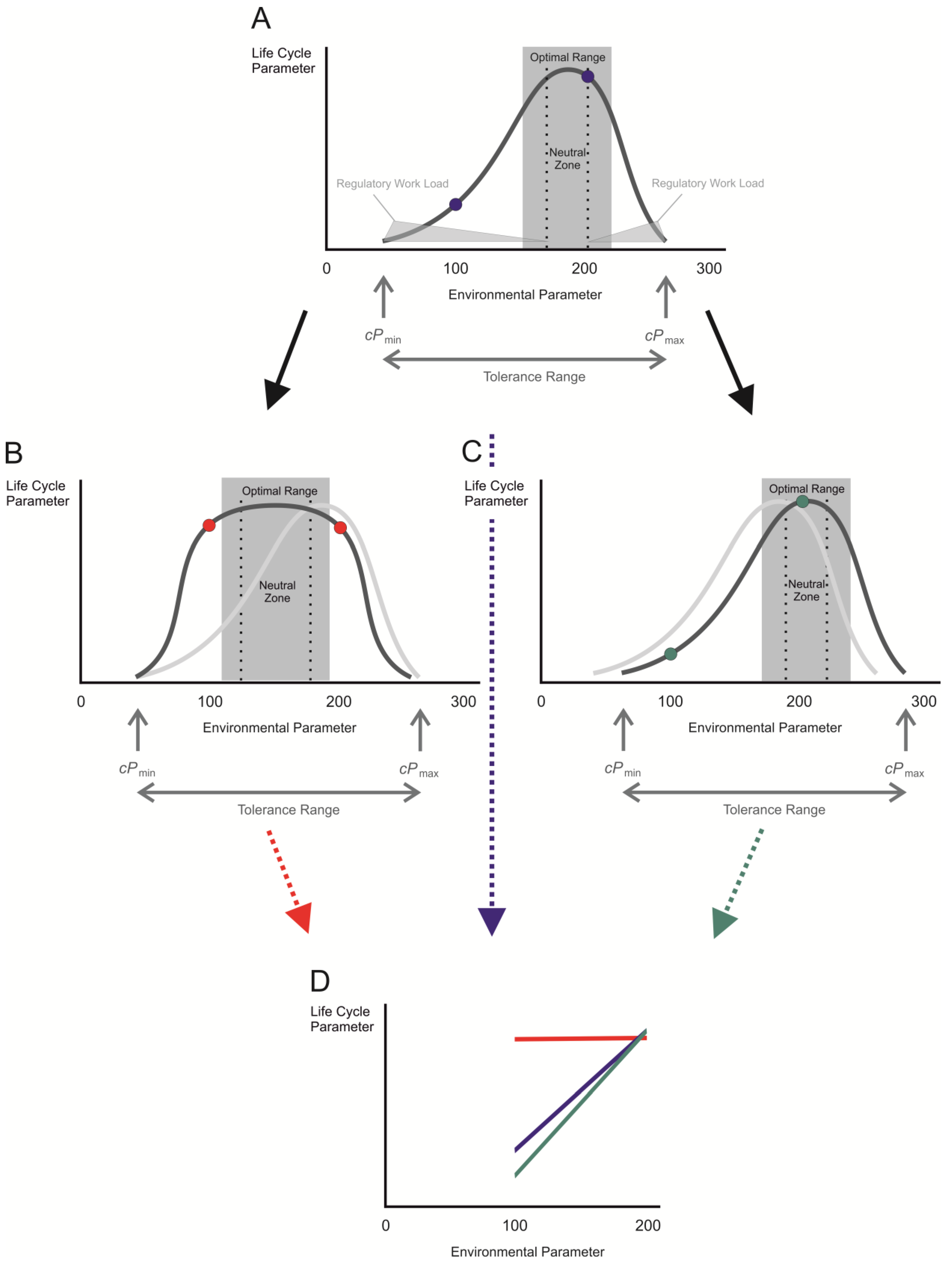
2. Measure for Plasticity in Response to Environmental Change: The Reaction Norm
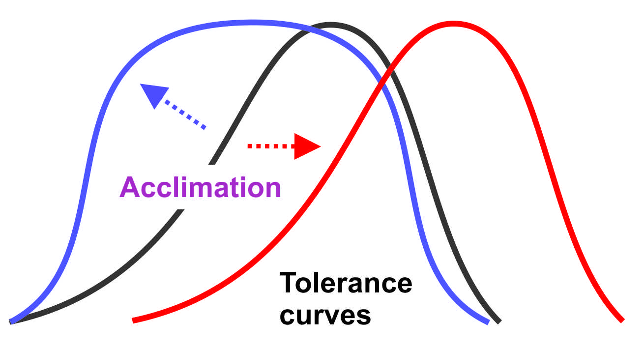
Most animal species are able to survive and produce offspring in environments which undergo changes in various abiotic and biotic parameters (salinity or pH of the surrounding medium, environmental temperature, food availability, etc.). The course and the extent to which a certain trait is changed in response to such shifts in environmental conditions are described by ‘reaction norms’ [1,30,31]. Comparisons of reaction norms enable ecologists to quantitatively describe imminent changes in life cycle parameters in different animals (body size, motility, fertility, etc.) and their dependency on defined shifts in environmental conditions (Figure 1).
There is a strong dependence of virtually every life cycle parameter in an individual animal on the exact value of a given environmental condition. The actual values of this life cycle parameter over the range of tolerable environmental conditions are represented by an optimum curve, the ‘tolerance curve’. The ‘optimal range’ is the interval of values around the maximum of the tolerance curve. In this range, the animal needs to divert only a little energy or no energy from other bodily functions towards maintaining its internal conditions, while keeping the life cycle parameter of interest at its maximum. Within the optimal range, the ‘neutral zone’ marks an interval of values of the external conditions at which the organism is in equilibrium with its environment. Under these conditions, the internal parameters do not to have to be defended against the external ones due to a lack in the gradients between the internal space and the environment or in disturbances of the physiological conditions within the body. The amount of energy that has to be invested to keep internal homeostasis, the regulatory workload, is at its minimum within this neutral zone (Figure 1).
However, if the environmental conditions lie outside the optimal range, increasing amounts of work have to be invested in homeostatic functions. The energy required for this purpose is then diverted away from the sustaining of the life cycle parameter of interest so that its value becomes smaller as we approach the upper or lower critical limits of the environmental condition (cPmax or cPmin, respectively). Beyond these limits, the amount of energy required for maintaining internal homeostasis would become so large that sustained life would become impossible. These limits define the ‘tolerance range’ of an individual.
Theoretically, the reaction norm of an animal for a given life cycle parameter (e.g., the growth rate) is defined by two or several points within the tolerance curve. Using only two points of different environmental parameter values results in a linear reaction norm with a certain slope (Figure 1A,D, blue dots or line, respectively). This type of reaction norm is only valid for a given interval of the environmental parameter and may be used to compare the behavior of different individuals from the same or from different populations in their responses to this particular environmental change. However, the linear reaction norm derived from the two points on the tolerance curve provides only simplified information on the response of an animal to environmental change and may miss important alterations in life cycle parameters when the interval of the environmental parameters has been selected in an unfavorable manner. The determination of a reaction norm using several different values of environmental parameters within the tolerance range is more informative. The best way of understanding the responsive behavior of an animal to environmental change is by the construction of a non-linear reaction norm which forms an approximation to the tolerance curve. This requires measurements of different values of the life cycle parameter of interest in small intervals of the environmental parameter within the tolerance range; thus, a lot more experimental work is needed.
An animal existing under environmental conditions outside of the optimal range but still within the critical limits experiences ‘physiological stress’ [32]. The regulatory work that this animal has to invest to stay alive is larger than that required when the animal’s environmental condition remains within the optimal range. If energetic resources are not limited, the animal may live under such unfavorable conditions for a long time. It is noteworthy that the responses of an animal living under the stress conditions represented by the left portion of the tolerance curve are entirely different from those that are required if the animal is exposed to conditions represented by the right portion of the curve.
As energy is usually a limiting factor in an animal’s life, it is unlikely that an animal will maintain high metabolic rates for sustained periods to cope with physiological stress. Instead, the animal will activate gene regulatory, biochemical, or physiological mechanisms [33] to reduce metabolic demands, to protect organs and tissues from damage, or to optimize the functions of regulatory organs to match the increasing regulatory demands [34,35,36,37,38]. These environmentally induced measures in an animal are summed up as ‘acclimation’ (in cases when animals adjust to new environmental conditions induced by an experimenter in the laboratory) or as ‘acclimatization’ (when wild animals adjust to changes in their natural environments) [25,39].

3. Acclimation

The exposure of an animal to physiological stress generally alters both the tolerance limits and the reaction norms for the relevant physiological traits. During acclimation, the tolerance curve may be broadened (Figure 1B) or shifted on the x-axis (Figure 1C). Such acclimation processes may result in changes in the position and/or the slope of the reaction norm of relevant physiological or other life cycle parameters (Figure 1D) when those of naïve (Figure 1A) or of acclimated animals are compared (Figure 1B,C).
Environmentally induced alterations in gene transcription, in the translation rates of certain transcripts, in the posttranslational modifications of proteins, or in the epigenetic mechanisms may be involved in such responses of animals to environmental stress [33,40,41,42]. This usually requires the sensing of the new environmental condition, the systemic processing of information [34,43,44,45,46,47,48,49], and, at the cellular level, the initiation of signal transduction and activation of transcription factors [50]. In many cases, processes such as cell differentiation or cell proliferation in tissues or organs are required to enable an animal to successfully cope with the stressful conditions [51].
The experience of stressful environmental conditions may not only result in acute changes but may also create an elevated level of resilience which enables animals to respond quicker or more economically to further periods of environmental stress once they have successfully dealt with such stress conditions before. These ‘hormetic’ effects, which may last for different periods of time (’memory´), may play important roles in hardening animals against further periods of environmental stress [52].
Animal physiologists use the term ‘physiological adaptation’ to differentiate this type of adjustment in single individuals from ‘genetic adaptation’, which is an evolutionary process involving several generations of animals, genetic recombination, and selection. Ecologists, however, avoid the term ‘adaptation’ when discussing environmentally induced alterations in individual animals and prefer to use the term ‘adaptive phenotypic plasticity’ [53,54]. When such plastic processes in response to changing environmental conditions occur during ontogenesis (e.g., during embryonic development), the alterations in the phenotype are subsumed under the term ‘developmental plasticity’ [1,55,56].

4. Reversibility of Plastic Changes

Whether adaptive phenotypic changes are reversible or not depends on the biological limitations of the respective organ systems in the animals and on the nature of the environmental condition. The organ system that responds to environmental change must have the ability to respond in different directions, and the direction should be correlated with the actual environmental alteration. However, the patterning of a certain type of environmental alteration (frequent or infrequent events) also affects the kind of response in the animal.
Chronic changes in an environmental condition or the expectation that an acute change may prevail or at least occur frequently in the future may trigger irreversible changes in cell and organ functions that are maintained for the rest of the animal’s lifetime. Such cases represent ‘phenotypic plasticity in the narrow sense’.
However, there are also cases in which environmentally induced plastic changes are reversible once the environmental conditions have returned to the original ones. In such cases, the plastic changes may be induced and reversed many times during the remaining lifespan of an animal. Examples include the seasonal changes occurring in inhabitants of the temperate zones [57], e.g., in hibernators [58] or in animals that change fur thickness in preparation for summer or winter, respectively. When the environmental alterations occur in such a way that the animal is affected unexpectedly or if transient changes in such conditions occur from time to time without a clear rhythm (fluctuation) or take place only rarely throughout an animal´s lifetime, it is especially important for an animal to be able to respond to such alterations in a reversible manner. In such cases, it is more economical for an organism to react acutely by investing in molecular and cellular reorganization or organ optimization and to reverse these processes as soon as their effects are not needed any more, rather than maintaining the altered phenotype even when conditions have normalized again. This helps to save energy which would otherwise have to be spent on the maintenance of obsolete traits for long periods.
Some authors call the ability of organisms to reverse phenotypic changes in response to environmental stimuli ‘phenotypic flexibility’ [6,59,60,61]. However, this term is rather general and does not reflect the fact that it needs biological work and energy to reach a new steady state in an altered environment and to reverse it after the environmental alteration which had triggered the response is terminated. Thus, we suggest that the term ‘phenotypic elasticity’ is better suited for describing reversible adjustments in animals to changes in environmental conditions. This term allows the clear differentiation between reversible alterations of traits in animals to transient changes in the environmental conditions and those that can only be induced once in the lifetime of an organism (‘phenotypic plasticity in the narrow sense’) [62]. In addition, this term allows the differentiation between phenotypic changes that are advantageous (albeit energetically costly) for an individual (and thus are of adaptive value for the organism) and all others.

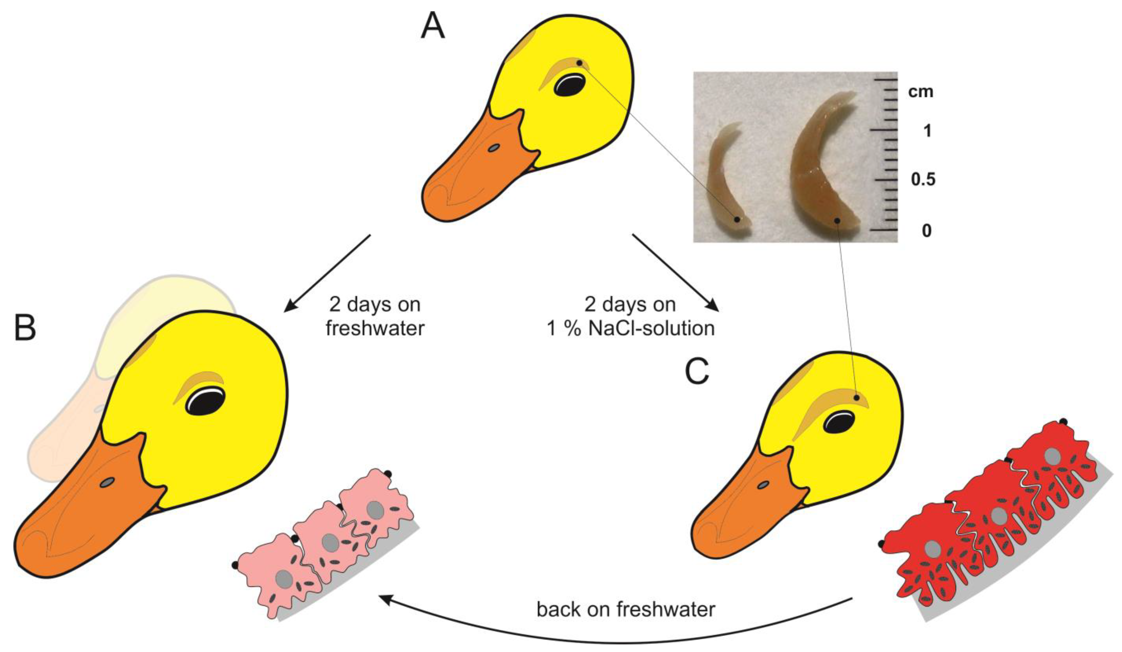
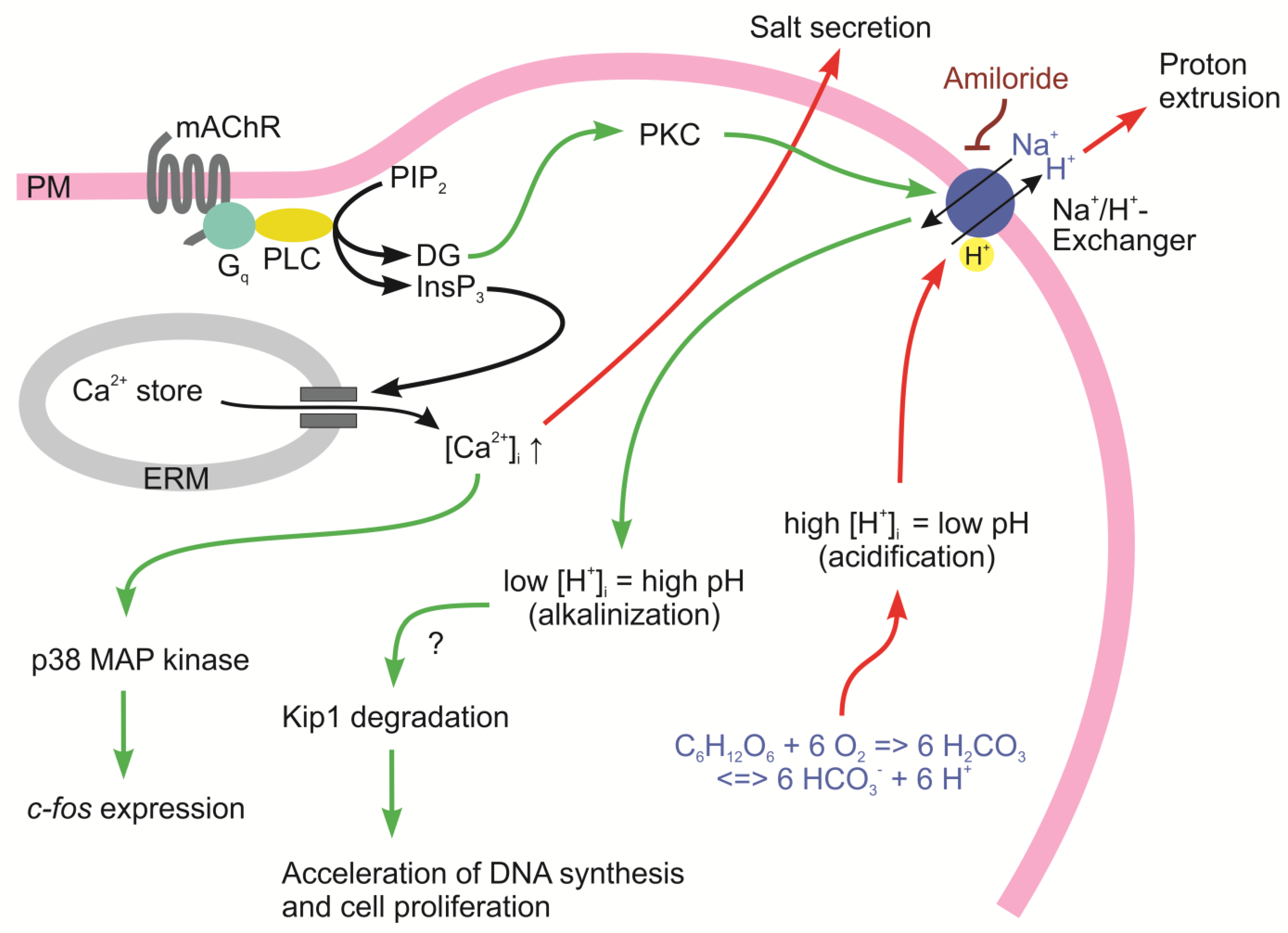
5. The Avian Salt Gland as an Example of Environmentally Induced Irreversible and Reversible Phenotypic Changes

Vertebrates living in dry or marine environments or consuming salty food face the challenge of maintaining osmotic, ionic, and volume balances in their body fluids [63]. Excess salt in the body fluids of animals compromises cellular functions. Thus, if a surplus of ions occurs in the body water of these animals it has to be excreted as rapidly as possible while retaining as much water as possible in the body. While most mammals eliminate excess sodium chloride (NaCl) in the form of concentrated urine via the kidneys when water is available [64], non-mammalian vertebrates living in arid or marine habitats with limited access to fresh water use extrarenal salt excretory organs to rid themselves of excess salt in the body. In birds, monofunctional salt glands (Figure 2) excrete a highly concentrated NaCl solution from the body while saving body water [65,66]. In ducklings (Anas platyrhynchos), these multi-tubular glands sit alongside the supraorbital bones on both sides of the skull and drain their products into the nasal cavity next to the nostrils. When the animals are salt-loaded, these glands produce a sodium chloride-rich fluid that may be up to four times more concentrated than the blood plasma [65]. The secretory cells lining the gland tubules produce this fluid by ‘secondary active chloride secretion’ [67].
During the initial exposure of such birds to osmotic stress, the salt glands undergo a period of cell growth and cell differentiation which results in organ maturation. In geese and ducklings, these maturation processes encompass the amplification of the number and deepness of the basolateral plasma membrane infoldings and the upregulation of the expression and/or activity of enzymes involved in energy metabolism and ion transport, as well as increases in the number of mitochondria per cell and the aerobic capacity of tissues and organs [68,69,70,71,72]. This is accompanied by increases in cell size in the secretory cells and in an increase in organ weight (Figure 2), a process that is called ‘hypertrophy’. The branching pattern of the secretory tubules gets more complex, and the tubule portions that contain fully differentiated salt gland cells get larger (Figure 2C) when compared with control animals of the same age which have not received salt loads (Figure 2B). Cellular differentiation in the secretory cells by exposing the animals to osmotic stress results in increases in protein abundance and the activity of the sodium/potassium-ATPase, indicating that the salt secretory potency of gland tissue gets upregulated [72]. The structural and functional changes occurring in the salt gland upon the initial salt-loading of ducklings optimize the salt-secretory capacity of the glands and allow the animals to survive for unlimited periods under unfavorable environmental conditions. Although these adjustments are costly in terms of building material and energy, they can be considered beneficial [73].
Those adjustments that are associated with cellular differentiation in the glands are reversible when salt loading is terminated or when the animals have unlimited access to fresh water. This allows the conclusion that environmentally mediated cell differentiation in the salt glands falls into the category of ‘phenotypic elasticity’. The most likely reason why the cellular differentiation can be rapidly up- and downregulated within one or two days according to the momentary needs of an animal is that the sustained maintenance of a highly differentiated state of cells and tissues in the gland in addition to salt secretion is energetically highly demanding [74]. This was illustrated by comparisons of the general growth rates in individual ducklings at 1 week of age upon hatching for another 48 h. Osmotically stressed animals (replacement of drinking water by a 1% NaCl solution) had an overall growth rate of only 50–70% of that of their unstressed (‘naïve’) siblings, despite being offered food ad libitum (unpublished observation). However, as shown in the inset of Figure 2, the salt glands of the stressed animals were 2 to 3 times bigger than those of the naïve animals.
When a duckling is exposed to salt stress for the first time in its life, the secretory tubuli in its glands are elongated and show increases in branching [70,75]. These alterations in the structure of the salt gland are based on increases in the cell number per gland. The amplification of the cellular material is a process that is termed ‘hyperplasia’ [76,77,78]. In preparation for cell proliferation, partially differentiated cells at the terminals of the secretory tubules start synthesizing DNA after the animals have been exposed to osmotic stress for at least 12 h [76]. The elevated rates of DNA synthesis are maintained for a period of just 12 h (i.e., between 12 and 24 h after the onset of osmotic stress) while the initial stimulus for DNA synthesis and cell proliferation in the gland, the osmotic burden in the animal, still prevails. When this period of hyperplasia is terminated, the cell number per gland will have at least doubled. This contributes substantially to the increase in overall organ size (see insert in Figure 2). The cell number per gland is then stable and cannot be reduced by removing the osmotic stimulus or feeding fresh water ad libitum to the animals. Thus, there is no way of inducing hyperplastic growth in the salt gland more than once in the lifetime of a duck. This indicates that this aspect of the environmentally driven growth phenomenon is an irreversible plastic feature of the gland (including in the narrow sense of the term).
The combination of hypertrophy and hyperplasia in response to salt stress in the duckling results in a final organ size that is 2 to 3 times larger than that in naïve animals of the same age (Figure 2). Such an increase in organ size, which is especially impressive in comparison with the lagging overall growth of the animals under salt stress (see above), indicates that it aims at optimization of the salt-secreting capacity of the gland.
There is still only limited knowledge on the molecular mechanisms mediating and regulating cell differentiation and cell proliferation in the gland. There are definitely differences in transmembrane and intracellular signaling in the salt gland cells of naïve and salt-stressed animals, respectively [79]. The salt glands are innervated by the parasympathetic nervous system, and the activation of muscarinic acetylcholine receptors (mAChRs) on the surface of the salt gland cells (Figure 3) is required not only for triggering salt secretion in the differentiated gland, but also for inducing cell differentiation and proliferation in the salt glands of naïve animals [80]. Comparisons of the cell surface densities of muscarinic acetylcholine receptors in isolated salt gland cells of the ducklings revealed that cells from naïve animals express 3 times more mAChRs compared with the cells isolated from the animals which had recently experienced salt stress (stressed animals) [81]. However, the ligand-binding properties of these receptors were identical and resembled those of the mammalian M3 subtype of mAChRs (coupling to phospholipase C and calcium signaling via a Gq/11-type heterotrimeric G-protein [82]). Activation of these receptors in isolated salt gland cells using submaximal concentrations of the stable mAChR agonist carbachol resulted in substantial accelerations in inositol lipid and inositol phosphate turnover, with 5 times higher inositol phosphate accumulation rates in cells from naïve ducklings compared with cells from stressed animals [83]. Calcium signaling was elicited upon receptor activation in both cell types. These signals were composed of initial peaks of the cytosolic free calcium concentration ([Ca2+]i) from the basal level of approximately 100 nmol/L to 400–500 nmol/L, followed by a sustained phase of elevated [Ca2+]i of approximately 200–300 nmol/L that was stable as long as the receptor activation was maintained. While the initial transient peak of [Ca2+]i was due to the release of calcium ions from intracellular stores, the sustained plateau resulted from ‘capacitative calcium entry’ from the extracellular space through the plasma membrane [84]. Such elevations in [Ca2+]i had previously been identified as key signals in the triggering of salt secretion from the gland [85].
The observation that full calcium signals could be elicited by activation of a relatively low number of activated mAChRs (and low rates of inositol phosphate accumulation) in cells isolated from stress-experienced ducklings raised the question whether the much higher receptor density and the higher turnover rates in the phosphoinositide metabolism in muscarinically activated cells of naïve ducklings may play roles in mediating the adaptive responses in the glands, specifically cell proliferation and cell differentiation. We raised the hypothesis that the same type of signals may induce entirely different cellular effects if applied in different intensities or to cell systems with different properties [86].
These considerations prompted further investigations on the downstream effects of mAChR activation and calcium signaling in the signal transduction pathways and cell physiology of naïve salt gland cells (Figure 3). The screening of extracts of the salt gland tissue of naïve animals acutely exposed to salt stress for immediate early gene expression revealed that the protooncogene c-fos was transiently active. The Fos protein could be detected between 1 and 12 h after the onset of salt stress in the salt-secretory cells, while the Jun protein was constitutively present and quantitatively unaffected by salt stress [87]. Fos expression could also be elicited by mAChR activation of the salt gland cells in vitro, indicating that Fos expression was dependent on inositide and calcium signaling. Generally, dimers of Fos and Jun form the transcription factor AP-1 [88], which controls the transcription rate of many of the genes which are important for growth and development. In search of the missing link in the signaling pathway between sustained elevation in [Ca2+]i and c-fos gene expression, we identified p38 MAP kinase as an essential component [89]. It was activated in mAChR-stimulated naïve salt gland cells, and Fos expression was suppressed in the presence of the inhibitors of this kinase.
However, the increase in cell number by activation of the cell proliferation in partially differentiated peripheral cells in the salt glands of the naïve animals was found to depend on the downregulation of the cell cycle inhibitor p27Kip1 [90]. p27Kip1 is the regulatory subunit of a cyclin-dependent kinase (CDK) and arrests cell cycle progression in proliferation-competent cells at the G0/G1-S checkpoint [91,92]. It is known that Kip1 undergoes site-specific phosphorylation in cells receiving mitotic stimuli (e.g., by growth factor signaling), marking the respective molecules for polyubiquitinylation and proteasomal degradation [93]. Western blot studies using extracts of salt gland tissue revealed that p27Kip1 was abundantly expressed in the glandular tissue of naïve ducklings. It was downregulated within 5 to 8 h after initially feeding saline to the animals instead of fresh water [90]. The downregulation of the Kip1 protein was not accompanied by changes in the mRNA levels, indicating that Kip1 was regulated mainly at the translational (protein synthesis) or posttranslational levels (protein degradation). In cultured nasal gland tissue, Kip1 expression was downregulated by activation of the muscarinic acetylcholine receptor, which indicates that mAChR signaling plays a role in the re-entry of quiescent gland cells into the cell cycle.
A previous observation might shed some light on a potential mechanism that supports or mediates Kip1 ubiquitinylation and degradation in mAChR-stimulated naïve salt gland cells. Exposing salt gland cells to the muscarinic receptor agonist carbachol elicits responses in cytosolic pH in these cells. The cytosol of cells isolated from salt-stressed animals undergoes slight and transient acidification, most likely resulting from the production of additional protons in the activated oxidative carbohydrate metabolism and a short lag period in the activation of proton extrusion by the sodium/proton exchanger (NHE) (Figure 3). However, when the cells from naïve animals were treated in the same way, they showed a sustained cytosolic alkalinization of 0.1 pH units above the previous level [94]. Such prolonged alkalinization of the cytosol has already been described in other cell types which had been mitotically activated by growth factors [95,96]. Plasma membrane receptors coupled to the activation of phospholipase C generally mediate the production of two types of second messengers in the respective cells: inositol phosphates and diacylglycerol [97]. The latter stays in the cytosolic leaflet of the plasma membrane and activates protein kinase C (PKC). PKC, in turn, phosphorylates an amino acid residue in the C-terminal region of the NHE, which increases the binding affinity of a regulatory proton binding site next to the transport site in this transport molecule. This results in sustained transport activity and cytosolic alkalinization in activated cells [98]. Several authors postulated a causal relationship between cytosolic alkalinization and the initiation of DNA synthesis in preparation for cell proliferation in such cells [95,96,98]. Unfortunately, these reports did not receive much attention. We were able to confirm the relationship of cytosolic alkalinization and the onset of DNA synthesis using mAChR-activated naïve salt gland cells [76]. The pretreatment of cultured salt gland tissue with the NHE inhibitor amiloride suppressed the mAChR-mediated cytosolic alkalinization as well as the increase in the DNA synthesis rate. These results support the hypothesis that mitogenic stimulation of cells and the associated cytosolic alkalinization may have a permissive or a signaling function for the initiation of DNA replication in preparation for cell proliferation, at least in proliferation-competent cells.
Other than the exocrine glands in mammals (e.g., lacrimal, salivary, sweat, or mammary glands), the avian salt gland is monofunctional, i.e., it secretes a highly concentrated sodium chloride solution on demand. All other glands secrete proteins or mucus in addition to ions and water [99,100], which complicates investigations on signal transduction because the different secretory processes are often independently controlled and mediated by different cell types. Most of the mammalian glands are acinar glands, which have club-like terminal structures containing the secretory cells at the ends of branched tubules, whereas the avian salt gland is purely tubular with the secretory cells lining these tubules. Despite these differences, it may be rewarding to consider some findings on signaling and gene regulation obtained in developing mammalian glands as guidelines for further studies on the control of cell proliferation and cell differentiation during environmentally induced organ maturation in the avian salt gland. To mention just one example, the induction of exocrine gland development and the proliferation of epithelial progenitor cells in mammals is mediated by fibroblast growth factor-10 (FGF-10) [101,102,103,104,105,106,107,108]. FGF-10 binds to and activates the FGF receptor 2b (FGFR2b), which induces the expression of the transcription factors Sox9 and Sox10. These transcription factors are responsible for expanding the population of epithelial progenitor cells [109] and direct the development of their progeny toward the secretory phenotype [110]. Moreover, they mediate the elongation of glandular tubules during gland maturation [111].
As a chromosome-level genome [112] and a transcriptome [113] of the domestic duck have been sequenced, interested researchers may now follow these lines of investigation of signaling and transcription regulation in the avian salt gland by comparing the transcriptomes of salt gland tissues from naïve and salt-stressed animals. This may improve our understanding of the adaptive responses of this organ to salt stress in the animal and may reveal the molecular and mechanistic details of the elastic and plastic responses in the gland to environmental stress. As ducklings are easy to maintain and the induction of these processes is easy to achieve without major animal manipulation, the salt gland of the duckling will remain a model system and will help to create a better understanding of environmentally induced cellular growth and differentiation processes at the tissue and organ levels of vertebrate organisms in general.

6. Conclusions

The mechanisms underlying the regulation of cell sizes and cell numbers in animal tissues and organs are largely unknown [51]. Whether environmentally driven hyper-/hypotrophy and hyper-/hypoplasia are really necessary for animals to cope with changing environmental conditions is still unclear in many cases of elastic or plastic changes. That such changes are energetically costly may indicate—but not prove—that they are beneficial for the animal. To decide this, survival or performance tests have to be conducted in animals under stressful conditions, allowing acclimation processes to occur in one group while suppressing them in the other.
In addition to those phenotypic adjustments that affect the salt/water relations of animals, other physiological parameters, e.g., thermal acclimation, metabolic responses to hypoxia, or exercise-dependent plasticity in skeletal muscle [114,115,116] have been investigated. While we are beginning to understand the evolutionary processes of local adaptation [117], there are only some initial studies dealing with the molecular mechanisms underlying physiological adaptation or acclimation (sensing environmental parameters, signal transduction, gene transcription, RNA processing, protein synthesis, post-translational modifications of proteins, etc.) [114,117,118,119,120,121], including epigenetic phenomena [8,33,122,123,124]. These studies are especially important in times of climate and environmental changes as knowledge about these processes and their limitations may shed light on the ability of individual animals and animal populations to survive such changes.

Funding

Financial support was provided by the Deutsche Forschungsgemeinschaft (DFG) (RTG 2010, RESPONSE).

Institutional Review Board Statement

Not applicable.

Informed Consent Statement

Not applicable.

Data Availability Statement

The data referred to in this review are published and publicly available in databases specified in the original publications.

Acknowledgments

I thank all members of the Animal Physiology group of the University of Greifswald for their input.

Conflicts of Interest

The author declares no conflict of interest.

References

  1. Pigliucci, M.; Schlichting, C.D.; Jones, C.S.; Schwenk, K. Developmental reaction norms: The interactions among allometry, ontogeny and plasticity. Plant Species Biol. 1996, 11, 69–85. [Google Scholar] [CrossRef] [Green Version]
  2. Weinkove, D.; Leevers, S.J. The genetic control of organ growth: Insights from Drosophila. Curr. Opin. Genet. Dev. 2000, 10, 75–80. [Google Scholar] [CrossRef] [PubMed]
  3. Ferenci, T.; Maharjan, R. Mutational heterogeneity: A key ingredient of bet-hedging and evolutionary divergence? Bioessays 2015, 37, 123–130. [Google Scholar] [CrossRef] [PubMed]
  4. Hoban, S.; Kelley, J.L.; Lotterhos, K.E.; Antolin, M.F.; Bradburd, G.; Lowry, D.B.; Poss, M.L.; Reed, L.K.; Storfer, A.; Whitlock, M.C. Finding the genomic basis of local adaptation: Pitfalls, practical solutions, and future directions. Am. Nat. 2016, 188, 379–397. [Google Scholar] [CrossRef] [Green Version]
  5. Charmantier, A.; Garant, D. Environmental quality and evolutionary potential: Lessons from wild populations. Proc. Biol. Sci. 2005, 272, 1415–1425. [Google Scholar] [CrossRef] [Green Version]
  6. Woods, H.A. Mosaic physiology from developmental noise: Within-organism physiological diversity as an alternative to phenotypic plasticity and phenotypic flexibility. J. Exp. Biol. 2014, 217, 35–45. [Google Scholar] [CrossRef] [Green Version]
  7. de Villemereuil, P.; Gaggiotti, O.E.; Mouterde, M.; Till-Bottraud, I. Common garden experiments in the genomic era: New perspectives and opportunities. Heredity 2016, 116, 249–254. [Google Scholar] [CrossRef] [Green Version]
  8. Tejedor, J.R.; Fraga, M.F. Interindividual epigenetic variability: Sound or noise? Bioessays 2017, 39, 1700055. [Google Scholar] [CrossRef]
  9. Cossins, A.; Fraser, J.; Hughes, M.; Gracey, A. Post-genomic approaches to understanding the mechanisms of environmentally induced phenotypic plasticity. J. Exp. Biol. 2006, 209, 2328–2336. [Google Scholar] [CrossRef] [Green Version]
  10. Pigliucci, M.; Murren, C.J.; Schlichting, C.D. Phenotypic plasticity and evolution by genetic assimilation. J. Exp. Biol. 2006, 209, 2362–2367. [Google Scholar] [CrossRef] [Green Version]
  11. Fusco, G.; Minelli, A. Phenotypic plasticity in development and evolution: Facts and concepts. Philos. Trans. R. Soc. B 2010, 365, 547–556. [Google Scholar] [CrossRef] [PubMed] [Green Version]
  12. Beaman, J.E.; White, C.R.; Seebacher, F. Evolution of plasticity: Mechanistic link between development and reversible acclimation. Trends Ecol. Evol. 2016, 31, 237–249. [Google Scholar] [CrossRef] [PubMed]
  13. Chevin, L.-M.; Collins, S.; Lefèvre, F. Phenotypic plasticity and evolutionary demographic responses to climate change: Taking theory out to the field. Funct. Ecol. 2013, 27, 967–979. [Google Scholar] [CrossRef] [Green Version]
  14. Minelli, A.; Fusco, G. Developmental plasticity and the evolution of animal complex life cycles. Philos. Trans. R. Soc. B 2010, 365, 631–640. [Google Scholar] [CrossRef] [Green Version]
  15. Moczek, A.P.; Sultan, S.; Foster, S.; Ledon-Rettig, C.; Dworkin, I.; Nijhout, H.F.; Abouheif, E.; Pfennig, D.W. The role of developmental plasticity in evolutionary innovation. Proc. Biol. Sci. 2011, 278, 2705–2713. [Google Scholar] [CrossRef] [Green Version]
  16. Peirson, B.R.E. Plasticity, stability, and yield: The origins of Anthony David Bradshaw’s model of adaptive phenotypic plasticity. Stud. Hist. Philos. Sci. Part C Stud. Hist. Philos. Biol. Biomed. Sci. 2015, 50, 51–66. [Google Scholar] [CrossRef]
  17. Pfennig, D.W.; McGee, M. Resource polyphenism increases species richness: A test of the hypothesis. Philos. Trans. R. Soc. B 2010, 365, 577–591. [Google Scholar] [CrossRef] [Green Version]
  18. Schlichting, C.D.; Smith, H. Phenotypic plasticity: Linking molecular mechanisms with evolutionary outcomes. Evol. Ecol. 2002, 16, 189–211. [Google Scholar] [CrossRef]
  19. Sommer, R.J. Phenotypic plasticity: From theory and genetics to current and future challenges. Genetics 2020, 215, 1–13. [Google Scholar] [CrossRef]
  20. Joschinski, J.; Bonte, D. Transgenerational plasticity and bet-hedging: A framework for reaction norm evolution. Front. Ecol. Evol. 2020, 8, 517183. [Google Scholar] [CrossRef]
  21. Waldvogel, A.-M.; Feldmeyer, B.; Rolshausen, G.; Exposito-Alonso, M.; Rellstab, C.; Kofler, R.; Mock, T.; Schmid, K.; Schmitt, I.; Bataillon, T.; et al. Evolutionary genomics can improve prediction of species’ responses to climate change. Evol. Lett. 2020, 4, 4–18. [Google Scholar] [CrossRef] [PubMed] [Green Version]
  22. Leroi, A.M.; Bennett, A.F.; Lenski, R.E. Temperature acclimation and competitive fitness: An experimental test of the beneficial acclimation assumption. Proc. Natl. Acad. Sci. USA 1994, 91, 1917–1921. [Google Scholar] [CrossRef] [PubMed] [Green Version]
  23. Palacio-Lopez, K.; Beckage, B.; Scheiner, S.; Molofsky, J. The ubiquity of phenotypic plasticity in plants: A synthesis. Ecol. Evol. 2015, 5, 3389–3400. [Google Scholar] [CrossRef] [PubMed]
  24. van Kleunen, M.; Fischer, M. Constraints on the evolution of adaptive phenotypic plasticity in plants. New Phytol. 2005, 166, 49–60. [Google Scholar] [CrossRef] [PubMed]
  25. Wilson, R.S.; Franklin, C.E. The detrimental acclimation hypothesis—Response. Trends Ecol. Evol. 2002, 17, 408. [Google Scholar] [CrossRef]
  26. Acasuso-Rivero, C.; Murren, C.J.; Schlichting, C.D.; Steiner, U.K. Adaptive phenotypic plasticity for life-history and less fitness-related traits. Proc. Biol. Sci. 2019, 286, 20190653. [Google Scholar] [CrossRef] [Green Version]
  27. Loeschcke, V.; Hoffmann, A.A. The detrimental acclimation hypothesis. Trends Ecol. Evol. 2002, 17, 407–408. [Google Scholar] [CrossRef]
  28. Lalejini, A.; Ferguson, A.J.; Grant, N.A.; Ofria, C. Adaptive phenotypic plasticity stabilizes evolution in fluctuating environments. Front. Ecol. Evol. 2021, 9, 715381. [Google Scholar] [CrossRef]
  29. Romero-Mujalli, D.; Rochow, M.; Kahl, S.; Paraskevopoulou, S.; Folkertsma, R.; Jeltsch, F.; Tiedemann, R. Adaptive and nonadaptive plasticity in changing environments: Implications for sexual species with different life history strategies. Ecol. Evol. 2021, 11, 6341–6357. [Google Scholar] [CrossRef]
  30. Gabriel, W.; Lynch, M. The selective advantage of reaction norms for environmental tolerance. J. Evol. Biol. 1992, 5, 41–59. [Google Scholar] [CrossRef] [Green Version]
  31. Scharloo, W. Developmental and physiological aspects of reaction norms—Drosophila data link genetic variation and phenotypic response to the environment. BioScience 1989, 39, 465–471. [Google Scholar] [CrossRef]
  32. Schulte, P.M. What is environmental stress Insights from fish living in a variable environment. J. Exp. Biol. 2014, 217, 23–34. [Google Scholar] [CrossRef] [PubMed] [Green Version]
  33. Ecker, S.; Pancaldi, V.; Valencia, A.; Beck, S.; Paul, D.S. Epigenetic and transcriptional variability shape phenotypic plasticity. Bioessays 2018, 40, 1700148. [Google Scholar] [CrossRef]
  34. Conlon, I.; Raff, M. Size control in animal development. Cell 1999, 96, 235–244. [Google Scholar] [CrossRef] [PubMed] [Green Version]
  35. Costanzo, J.P.; Lee, R.E. Avoidance and tolerance of freezing in ectothermic vertebrates. J. Exp. Biol. 2013, 216, 1961–1967. [Google Scholar] [CrossRef] [Green Version]
  36. Duman, J.G. Animal ice-binding (antifreeze) proteins and glycolipids: An overview with emphasis on physiological function. J. Exp. Biol. 2015, 218, 1846–1855. [Google Scholar] [CrossRef] [Green Version]
  37. Kültz, D. Molecular and evolutionary basis of the cellular stress response. Annu. Rev. Physiol. 2005, 67, 225–257. [Google Scholar] [CrossRef]
  38. Mymrikov, E.V.; Seit-Nebi, A.S.; Gusev, N.B. Large potentials of small heat shock proteins. Physiol. Rev. 2011, 91, 1123–1159. [Google Scholar] [CrossRef] [Green Version]
  39. Prosser, C.L. Adaptational Biology: Molecules to Organisms; Wiley: New York, NY, USA, 1986. [Google Scholar]
  40. Bradshaw, W.E.; Holzapfel, C.M. Light, time, and the physiology of biotic response to rapid climate change in animals. Annu. Rev. Physiol. 2010, 72, 147–166. [Google Scholar] [CrossRef] [Green Version]
  41. Rosenthal, J.J.C. The emerging role of RNA editing in plasticity. J. Exp. Biol. 2015, 218, 1812–1821. [Google Scholar] [CrossRef] [Green Version]
  42. Lamka, G.F.; Harder, A.M.; Sundaram, M.; Schwartz, T.S.; Christie, M.R.; DeWoody, J.A.; Willoughby, J.R. Epigenetics in ecology, evolution, and conservation. Front. Ecol. Evol. 2022, 10, 871791. [Google Scholar] [CrossRef]
  43. Bryant, P.J.; Simpson, P. Intrinsic and extrinsic control of growth in developing organs. Q. Rev. Biol. 1984, 59, 387–415. [Google Scholar] [CrossRef]
  44. Crickmore, M.A.; Mann, R.S. The control of size in animals: Insights from selector genes. Bioessays 2008, 30, 843–853. [Google Scholar] [CrossRef] [PubMed] [Green Version]
  45. Hafen, E.; Stocker, H. How are the sizes of cells, organs, and bodies controlled? PLoS Biol. 2003, 1, e86. [Google Scholar] [CrossRef] [Green Version]
  46. Nijhout, H.F.; Riddiford, L.M.; Mirth, C.; Shingleton, A.W.; Suzuki, Y.; Callier, V. The developmental control of size in insects. Wiley Interdiscip. Rev. Dev. Biol. 2014, 3, 113–134. [Google Scholar] [CrossRef] [PubMed]
  47. Parker, J. Morphogens, nutrients, and the basis of organ scaling. Evol. Dev. 2011, 13, 304–314. [Google Scholar] [CrossRef]
  48. Potter, C.J.; Xu, T. Mechanisms of size control. Curr. Opin. Genet. Dev. 2001, 11, 279–286. [Google Scholar] [CrossRef]
  49. Yang, X.; Xu, T. Molecular mechanism of size control in development and human diseases. Cell Res. 2011, 21, 715–729. [Google Scholar] [CrossRef]
  50. Lessells, C.K. Neuroendocrine control of life histories: What do we need to know to understand the evolution of phenotypic plasticity? Philos. Trans. R. Soc. B 2008, 363, 1589–1598. [Google Scholar] [CrossRef] [Green Version]
  51. Miettinen, T.P.; Caldez, M.J.; Kaldis, P.; Björklund, M. Cell size control—A mechanism for maintaining fitness and function. Bioessays 2017, 39, 1700058. [Google Scholar] [CrossRef] [Green Version]
  52. Costantini, D.; Metcalfe, N.B.; Monaghan, P. Ecological processes in a hormetic framework. Ecol. Lett. 2010, 13, 1435–1447. [Google Scholar] [CrossRef] [PubMed]
  53. Fischer, K.; Karl, I. Exploring plastic and genetic responses to temperature variation using copper butterflies. Clim. Res. 2010, 43, 17–30. [Google Scholar] [CrossRef]
  54. DeWitt, T.J.; Sih, A.; Wilson, D.S. Costs and limits of phenotypic plasticity. Trends Ecol. Evol. 1998, 13, 77–81. [Google Scholar] [CrossRef]
  55. Fischer, K.; Eenhoorn, E.; Bot, A.N.M.; Brakefield, P.M.; Zwaan, B.J. Cooler butterflies lay larger eggs: Developmental plasticity versus acclimation. Proc. R. Soc. Lond. B Biol. Sci. 2003, 270, 2051–2056. [Google Scholar] [CrossRef]
  56. Nijhout, H.F. Development and evolution of adaptive polyphenisms. Evol. Dev. 2003, 5, 9–18. [Google Scholar] [CrossRef] [PubMed]
  57. Aubry, L.M.; Williams, C.T. Vertebrate phenological plasticity: From molecular mechanisms to ecological and evolutionary implications. Integr. Comp. Biol. 2022, 62, 958–971. [Google Scholar] [CrossRef]
  58. Fu, R.; Gillen, A.E.; Grabek, K.R.; Riemondy, K.A.; Epperson, L.E.; Bustamante, C.D.; Hesselberth, J.R.; Martin, S.L. Dynamic RNA regulation in the brain underlies physiological plasticity in a hibernating mammal. Front. Physiol. 2021, 11, 624677. [Google Scholar] [CrossRef]
  59. Piersma, T.; Drent, J. Phenotypic flexibility and the evolution of organismal design. Trends Ecol. Evol. 2003, 18, 228–233. [Google Scholar] [CrossRef] [Green Version]
  60. Piersma, T.; Lindström, A. Rapid reversible changes in organ size as a component of adaptive behaviour. Trends Ecol. Evol. 1997, 12, 134–138. [Google Scholar] [CrossRef] [Green Version]
  61. Piersma, T.; van Gils, J.A. The Flexible Phenotype—A Body-Centered Integration of Ecology, Physiology, and Behaviour; Oxford University Press: Oxford, NY, USA, 2011. [Google Scholar]
  62. Hildebrandt, J.-P.; Wiesenthal, A.A.; Müller, C. Phenotypic plasticity in animals exposed to osmotic stress—Is it always adaptive? Bioessays 2018, 40, e1800069. [Google Scholar] [CrossRef]
  63. Schmidt-Nielsen, K. Animal Physiology: Adaptation and Environment; Cambridge University Press: Cambridge, MA, USA, 1997. [Google Scholar]
  64. Dantzler, W.H. Comparative Physiology of the Vertebrate Kidney; Springer: Berlin/Heidelberg, Germany, 1989. [Google Scholar]
  65. Schmidt-Nielsen, K. The salt-secreting gland of marine birds. Circulation 1960, 21, 955–967. [Google Scholar] [CrossRef] [PubMed] [Green Version]
  66. Technau, G. Die Nasendrüse der Vögel. J. Ornithol. 1936, 84, 511–617. [Google Scholar] [CrossRef]
  67. Frizzell, R.A.; Hanrahan, J.W. Physiology of epithelial chloride and fluid secretion. Cold Spring Harb. Perspect. Med. 2012, 2, a009563. [Google Scholar] [CrossRef] [PubMed] [Green Version]
  68. Barrnett, R.J.; Mazurkiewicz, J.E.; Addis, J.S. Avian salt gland: A model for the study of membrane biogenesis. Methods Enzymol. 1983, 96, 627–659. [Google Scholar] [CrossRef] [PubMed]
  69. Ellis, R.A.; Goertemiller, C.C.; DeLellis, R.A.; Kablotsky, Y.H. The effect of a salt water regimen on the development of the salt glands of domestic ducklings. Dev. Biol. 1963, 8, 286–308. [Google Scholar] [CrossRef]
  70. Ernst, S.A.; Ellis, R.A. The development of surface specialization in the secretory epithelium of the avian salt gland in response to osmotic stress. J. Cell. Biol. 1969, 40, 305–321. [Google Scholar] [CrossRef]
  71. Fletcher, G.L.; Stainer, I.M.; Holmes, W.N. Sequential changes in the adenosinetriphosphatase activity and the electrolyte excretory capacity of the nasal glands of the duck (Anas platyrhynchos) during the period of adaptation to hypertonic saline. J. Exp. Biol. 1967, 47, 375–391. [Google Scholar] [CrossRef]
  72. Hildebrandt, J.-P. Changes in Na+/K+-ATPase expression during adaptive cell differentiation in avian nasal salt gland. J. Exp. Biol. 1997, 200, 1895–1904. [Google Scholar] [CrossRef]
  73. Fänge, R.; Schmidt-Nielsen, K.; Robinson, M. Control of secretion from the avian salt gland. Am. J. Physiol. 1958, 195, 321–326. [Google Scholar] [CrossRef]
  74. Krohn, M.; Hildebrandt, J.-P. Cross-talk of phosphoinositide- and cyclic nucleotide-dependent signaling pathways in differentiating avian nasal gland cells. J. Comp. Physiol. B Biochem. Syst. Environ. Physiol. 2004, 174, 461–470. [Google Scholar] [CrossRef]
  75. Müller, C.; Hildebrandt, J.-P. Salt glands—The perfect way to get rid of too much sodium chloride. Biologist 2003, 50, 255–258. [Google Scholar]
  76. Bentz, C.; Schwarz, M.; Hildebrandt, J.-P. Cytosolic pH affects DNA-synthesis in nasal gland cells of osmotically stressed ducklings, Anas platyrhynchos. Zoology 1999, 102, 10–17. [Google Scholar]
  77. Hanwell, A.; Peaker, M. The control of adaptive hypertrophy in the salt glands of geese and ducks. J. Physiol. 1975, 248, 193–205. [Google Scholar] [CrossRef] [PubMed] [Green Version]
  78. Holmes, W.N.; Stewart, D.J. Changes in the nucleic acid and protein composition of the nasal glands from the duck (Anas platyrhynchos) during the period of adaptation to hypertonic saline. J. Exp. Biol. 1968, 48, 509–519. [Google Scholar] [CrossRef] [PubMed]
  79. Hildebrandt, J.-P. Coping with excess salt: Adaptive functions of extrarenal osmoregulatory organs in vertebrates. Zoology 2001, 104, 209–220. [Google Scholar] [CrossRef]
  80. Peaker, M. Salt glands in marine birds: What triggers secretion and what makes them grow? In Comparative Physiology: Water, Ions and Fluid Mechanics; Schmidt-Nielsen, K., Bolis, L., Maddrell, S.H.P., Eds.; Cambridge University Press: Cambridge, MA, USA, 1978; pp. 207–212. [Google Scholar]
  81. Hildebrandt, J.-P.; Shuttleworth, T.J. Muscarinic receptor characterization in differentiating avian exocrine cells. Am. J. Physiol. Regul. Integr. Comp. Physiol. 1994, 266, R674–R681. [Google Scholar] [CrossRef] [Green Version]
  82. Hildebrandt, J.-P.; Shuttleworth, T.J. A Gq-type G protein couples muscarinic receptors to inositol phosphate and calcium signaling in exocrine cells from the avian salt gland. J. Membr. Biol. 1993, 133, 183–190. [Google Scholar] [CrossRef]
  83. Hildebrandt, J.-P.; Shuttleworth, T.J. Inositol phosphates and [Ca2+]i signals in a differentiating exocrine cell. Am. J. Physiol. Cell Physiol. 1991, 261, C210–C217. [Google Scholar] [CrossRef]
  84. Shuttleworth, T.J. Temporal relationships between Ca2+ store mobilization and Ca2+ entry in an exocrine cell. Cell Calcium 1994, 15, 457–466. [Google Scholar] [CrossRef]
  85. Lowy, R.J.; Dawson, D.C.; Ernst, S.A. Primary culture of duck salt gland. II. Neurohormonal stimulation of active transport. Am. J. Physiol. 1985, 249, C41–C47. [Google Scholar] [CrossRef]
  86. Shuttleworth, T.J.; Hildebrandt, J.-P. Vertebrate salt glands—Short- and long-term regulation of function. J. Exp. Zool. 1999, 283, 689–701. [Google Scholar] [CrossRef]
  87. Hildebrandt, J.-P.; Gerstberger, R.; Schwarz, M. In vivo and in vitro induction of c-fos in avian exocrine salt gland cells. Am. J. Physiol. Cell Physiol. 1998, 275, C951–C957. [Google Scholar] [CrossRef] [PubMed]
  88. Dalhäusser, A.K.; Rössler, O.G.; Thiel, G. Regulation of c-fos gene transcription by stimulus-responsive protein kinases. Gene 2022, 821, 146284. [Google Scholar] [CrossRef] [PubMed]
  89. Hildebrandt, J.-P.; Prowald, A. Ca2+ and p38 MAP kinase regulate mAChR-mediated c-Fos expression in avian exocrine cells. Am. J. Physiol. Cell Physiol. 2000, 278, C879–C884. [Google Scholar] [CrossRef] [Green Version]
  90. Rohlfing, A.-K.; Schill, T.; Müller, C.; Hildebrandt, P.; Prowald, A.; Hildebrandt, J.-P. Attenuation of cell cycle regulator p27(Kip1) expression in vertebrate epithelial cells mediated by extracellular signals in vivo and in vitro. J. Comp. Physiol. B Biochem. Syst. Environ. Physiol. 2005, 175, 511–522. [Google Scholar] [CrossRef]
  91. Bencivenga, D.; Stampone, E.; Roberti, D.; Della Ragione, F.; Borriello, A. p27(Kip1), an intrinsically unstructured protein with scaffold properties. Cells 2021, 10, 2254. [Google Scholar] [CrossRef]
  92. Bendris, N.; Lemmers, B.; Blanchard, J.M. Cell cycle, cytoskeleton dynamics and beyond: The many functions of cyclins and CDK inhibitors. Cell Cycle 2015, 14, 1786–1798. [Google Scholar] [CrossRef] [Green Version]
  93. Lu, Z.; Hunter, T. Ubiquitylation and proteasomal degradation of the p21(Cip1), p27(Kip1) and p57(Kip2) CDK inhibitors. Cell Cycle 2010, 9, 2342–2352. [Google Scholar] [CrossRef] [Green Version]
  94. Bentz, C.; Hildebrandt, J.-P. Adaptive differentiation of avian exocrine cells alters their pHi response to mAChR activation. Am. J. Physiol. Cell Physiol. 1995, 268, C944–C951. [Google Scholar] [CrossRef]
  95. Moolenaar, W.H.; Defize, L.H.; De Laat, S.W. Ionic signalling by growth factor receptors. J. Exp. Biol. 1986, 124, 359–373. [Google Scholar] [CrossRef]
  96. Grinstein, S.; Rotin, D.; Mason, M.J. Na+/H+ exchange and growth factor-induced cytosolic pH changes. Role in cellular proliferation. Biochim. Biophys. Acta 1989, 988, 73–97. [Google Scholar] [CrossRef] [PubMed]
  97. Newton, A.C. Protein kinase C: Perfectly balanced. Crit. Rev. Biochem. Mol. Biol. 2018, 53, 208–230. [Google Scholar] [CrossRef] [PubMed] [Green Version]
  98. Wakabayashi, S.; Fafournoux, P.; Sardet, C.; Pouyssegur, J. The Na+/H+ antiporter cytoplasmatic domain mediates growth factor signals and controls “H+-sensing”. Proc. Natl. Acad. Sci. USA 1992, 89, 2424–2428. [Google Scholar] [CrossRef] [Green Version]
  99. Freeman, S.C.; Malik, A.; Basit, H. Physiology, Exocrine Gland; StatPearls: Treasure Island, FL, USA, 2022. Available online: https://www.ncbi.nlm.nih.gov/books/NBK542322/ (accessed on 19 April 2023).
  100. Khan, S.; Fitch, S.; Knox, S.; Arora, R. Exocrine gland structure-function relationships. Development 2022, 149, dev197657. [Google Scholar] [CrossRef]
  101. Govindarajan, V.; Ito, M.; Makarenkova, H.P.; Lang, R.A.; Overbeek, P.A. Endogenous and ectopic gland induction by FGF-10. Dev. Biol. 2000, 225, 188–200. [Google Scholar] [CrossRef] [Green Version]
  102. Bhushan, A.; Itoh, N.; Kato, S.; Thiery, J.P.; Czernichow, P.; Bellusci, S.; Scharfmann, R. FGF10 is essential for maintaining the proliferative capacity of epithelial progenitor cells during early pancreatic organogenesis. Development 2001, 128, 5109–5117. [Google Scholar] [CrossRef] [PubMed]
  103. Entesarian, M.; Matsson, H.; Klar, J.; Bergendal, B.; Olson, L.; Arakaki, R.; Hayashi, Y.; Ohuchi, H.; Falahat, B.; Bolstad, A.I.; et al. Mutations in the gene encoding fibroblast growth factor 10 are associated with aplasia of lacrimal and salivary glands. Nat. Genet. 2005, 37, 125–128. [Google Scholar] [CrossRef]
  104. Knosp, W.M.; Knox, S.M.; Hoffman, M.P. Salivary gland organogenesis. WIREs Dev. Biol. 2012, 1, 69–82. [Google Scholar] [CrossRef]
  105. Chen, Z.; Huang, J.; Liu, Y.; Dattilo, L.K.; Huh, S.-H.; Ornitz, D.; Beebe, D.C. FGF signaling activates a Sox9-Sox10 pathway for the formation and branching morphogenesis of mouse ocular glands. Development 2014, 141, 2691–2701. [Google Scholar] [CrossRef] [Green Version]
  106. Chatzeli, L.; Gaete, M.; Tucker, A.S. FGF10 and SOX9 are essential for the establishment of distal progenitor cells during mouse salivary gland development. Development 2017, 144, 2294–2305. [Google Scholar] [CrossRef] [Green Version]
  107. Paine, I.S.; Lewis, M.T. The terminal end bud: The little engine that could. J. Mammary Gland Biol. Neopl. 2017, 22, 93–108. [Google Scholar] [CrossRef] [PubMed] [Green Version]
  108. Rivetti, S.; Chen, C.; Chen, C.; Bellusci, S. FGF10/FGFR2b signaling in mammary gland development, homeostasis, and cancer. Front. Cell Dev. Biol. 2020, 8, 415. [Google Scholar] [CrossRef] [PubMed]
  109. Seymour, P.A.; Freude, K.K.; Tran, M.N.; Mayes, E.E.; Jensen, J.; Kist, R.; Scherer, G.; Sander, M. SOX9 is required for maintenance of the pancreatic progenitor cell pool. Proc. Nat. Acad. Sci. USA 2007, 104, 1865–1870. [Google Scholar] [CrossRef] [PubMed] [Green Version]
  110. Athwal, H.K.; Murphy, G.; Tibbs, E.; Cornett, A.; Hill, E.; Yeoh, K.; Berenstein, E.; Hoffman, M.P.; Lombaert, I.M.A. Sox10 regulates plasticity of epithelial progenitors toward secretory units of exocrine glands. Stem Cell Rep. 2019, 12, 366–380. [Google Scholar] [CrossRef] [Green Version]
  111. Garg, A.; Zhang, X. Lacrimal gland development: From signaling interactions to regenerative medicine. Dev. Dyn. 2017, 246, 970–980. [Google Scholar] [CrossRef] [PubMed] [Green Version]
  112. Li, J.; Zhang, J.; Liu, J.; Zhou, Y.; Cai, C.; Xu, L.; Dai, X.; Feng, S.; Guo, C.; Rao, J.; et al. A new duck genome reveals conserved and convergently evolved chromosome architectures of birds and mammals. Gigascience 2021, 10, giaa142. [Google Scholar] [CrossRef]
  113. Huang, Y.; Li, Y.; Burt, D.W.; Chen, H.; Zhang, Y.; Qian, W.; Kim, H.; Gan, S.; Zha, Y.; Li, J.; et al. The duck genome and transcriptome provide insight into an avian influenza virus reservoir species. Nat. Genet. 2013, 45, 776–783. [Google Scholar] [CrossRef] [Green Version]
  114. McClelland, G.B.; Scott, G.R. Muscle plasticity. In The Physiology of Fishes, 4th ed.; Evans, D.H., Claiborne, J.B., Currie, S., Eds.; CRC Press: Boca Raton, FL, USA, 2014; pp. 1–31. [Google Scholar]
  115. Pörtner, H.-O. Oxygen- and capacity-limitation of thermal tolerance: A matrix for integrating climate-related stressor effects in marine ecosystems. J. Exp. Biol. 2010, 213, 881–893. [Google Scholar] [CrossRef] [Green Version]
  116. Somero, G.N. Unity in diversity: A perspective on the methods, contributions, and future of comparative physiology. Ann. Rev. Physiol. 2000, 62, 927–937. [Google Scholar] [CrossRef] [Green Version]
  117. Fields, P.A.; Dong, Y.; Meng, X.; Somero, G.N. Adaptations of protein structure and function to temperature: There is more than one way to ‘skin a cat’. J. Exp. Biol. 2015, 218, 1801–1811. [Google Scholar] [CrossRef] [Green Version]
  118. Ho, D.H.; Burggren, W.W. Epigenetics and transgenerational transfer: A physiological perspective. J. Exp. Biol. 2010, 213, 3–16. [Google Scholar] [CrossRef] [PubMed] [Green Version]
  119. Lockwood, B.L.; Somero, G.N. Transcriptomic responses to salinity stress in invasive and native blue mussels (genus Mytilus). Mol. Ecol. 2011, 20, 517–529. [Google Scholar] [CrossRef] [PubMed]
  120. Storey, K.B. Regulation of hypometabolism: Insights into epigenetic controls. J. Exp. Biol. 2015, 218, 150–159. [Google Scholar] [CrossRef] [Green Version]
  121. Tomanek, L.; Somero, G.N. Interspecific- and acclimation-induced variation in levels of heat-shock proteins 70 (hsp 70) and 90 (hsp 90) and heat-shock transcription factor-1 (HSF1) in congeneric marine snails (genus Tegula): Implications for regulation of hsp gene expression. J. Exp. Biol. 2002, 205, 677–685. [Google Scholar] [CrossRef] [PubMed]
  122. Burggren, W. Epigenetic inheritance and its role in evolutionary biology: Re-evaluation and new perspectives. Biology 2016, 5, 24. [Google Scholar] [CrossRef] [PubMed] [Green Version]
  123. Chapelle, V.; Silvestre, F. Population epigenetics: The extent of DNA methylation variation in wild animal populations. Epigenomes 2022, 6, 31. [Google Scholar] [CrossRef] [PubMed]
  124. Duncan, E.J.; Cunningham, C.B.; Dearden, P.K. Phenotypic plasticity: What has DNA methylation got to do with it? Insects 2022, 13, 110. [Google Scholar] [CrossRef]
Figure 1. Dependence of a life cycle parameter in an animal on the condition of its environment and the potential adjustments during acclimation. The original tolerance curve (A) may change in width (B) or position on the x-axis (C) (original is a grey shadow for comparison) during acclimation processes. Such changes result in alterations in the slopes of reaction norms (D) as exemplified by comparing the resulting reaction norms within the limits of the colored spots in A, B, and C, which represent the same pair of environmental conditions. cPmin—critical minimum of the environmental parameter; cPmax—critical maximum of the environmental parameter. Further explanations in the text.
Physiologia 03 00025 g001
Figure 2. Reversible and irreversible plastic changes in salt glands of ducklings under salt stress. One-week-old ducklings reared on fresh water upon hatching have slim salt glands (A). Due to fast growth at early age, the head of the duckling gets substantially bigger (compared with the initial situation: shadow in the back of the duck head) when the animal is further maintained with unlimited access to fresh water for another 2 days (B). The salt gland is quiescent and contains many partially differentiated secretory cells lining the gland tubules (B). When the animal is reared on a 1% NaCl solution instead of fresh water for these two days, the general growth of the duckling is severely impaired, but the salt gland grows by a factor of 2–3 in tissue mass (insert, right); this is accompanied by hyperplasia (increase in cell number per gland) as well as hypertrophy and cell differentiation of the tubular epithelial cells (larger cells, amplification of basolateral infoldings, and increase in the number of mitochondria) (C). While the traits associated with cell differentiation are reversible when animals are set back on fresh water (phenotypic elasticity), the number of cells in the gland does not decrease upon switching the animals back to drinking fresh water. Thus, the process of hyperplastic growth is irreversible (phenotypic plasticity in the narrow sense).
Physiologia 03 00025 g002
Figure 3. Scheme of the signal transduction pathways and cell physiological processes in naïve (green arrows) or stressed (red arrows) avian salt gland cells activated by the muscarinic acetylcholine receptor. The question mark indicates that a causal link between these elements has not yet been experimentally established. Further explanations are in the text. [Ca2+]i—Cytosolic (intracellular) free calcium ion concentration; DG—Diacylglycerol; ERM—Endoplasmic reticulum membrane; Gq—Heterotrimeric G-protein of the Gq/11-type; [H+]i—Cytosolic (intracellular) free proton concentration; InsP3—Inositol-1,4,5-trisphosphate; mAChR—Muscarinic acetylcholine receptor; PIP2—Phosphatidylinositol-4,5-bisphosphate; PKC—Protein kinase C; PLC—Phospholipase Cβ; PM—Plasma membrane.
Physiologia 03 00025 g003
Disclaimer/Publisher’s Note: The statements, opinions and data contained in all publications are solely those of the individual author(s) and contributor(s) and not of MDPI and/or the editor(s). MDPI and/or the editor(s) disclaim responsibility for any injury to people or property resulting from any ideas, methods, instructions or products referred to in the content.

