Article

Enhance Trial: Effects of NAD3® on Hallmarks of Aging and Clinical Endpoints of Health in Middle Aged Adults: A Subset Analysis Focused on Blood Cell NAD+ Concentrations and Lipid Metabolism

1
School of Kinesiology, Auburn University, Auburn, AL 36849, USA
2
The Center for Applied Health Sciences, Canfield, OH 44406, USA
*
Author to whom correspondence should be addressed.
Physiologia 2022, 2(1), 20-31; https://doi.org/10.3390/physiologia2010002
Submission received: 11 January 2022 / Revised: 18 February 2022 / Accepted: 2 March 2022 / Published: 21 March 2022

Abstract

:
Limited pre-clinical and clinical data suggest theacrine or theacrine-based supplements modulate biological processes associated with lipid metabolism and aging. Herein, we sought to examine if 12 weeks of daily supplementation with a theacrine-based supplement (termed NAD3®; 312 mg of combined Wasabia japonica freeze-dried rhizome standardized for isothicyantes, theacrine, and copper (I)niacin chelate) altered serum lipids as well as select nicotinamide adenine dinucleotide (NAD+)-associated metabolites in peripheral blood mononuclear cells (PBMCs). Twenty-eight participants (12 males, 16 females) were randomly assigned to receive either NAD3 (n = 13; age: 52 ± 7 years old, body mass index: 29.0 ± 5.0 kg/m2) or a cellulose placebo (n = 15; age: 51 ± 5 years old, body mass index: 28.3 ± 3.9 kg/m2). Blood samples were obtained in mornings following overnight fasts prior to supplementation (Pre) and following the 12-week intervention (Post). PBMCs were freshly isolated and prepared for targeted NAD+ metabolomics, and serum as well as whole blood was assayed for blood lipids and other safety markers through a commercial laboratory. Significant interactions (p < 0.05) were observed for total cholesterol, LDL cholesterol, and LDL: HDL ratio and post hoc analyses indicated these biomarkers significantly decreased with NAD3 supplementation (Pre-to-Post percent decreases were 11.1, 15.2, and −18.9%, respectively). A significant interaction was also observed for PBMC NAD+: NADH values, where levels trended downward from Pre to Post in the CTL group (p = 0.081) and values at Post were greater in NAD3 versus CTL (p = 0.023). No interactions were observed for systolic/diastolic blood pressure, body mass, or blood markers indicative of clinical safety. Although participant numbers were limited, these first-in-human data demonstrate a theacrine-based NAD3 supplement can favorably alter biomarkers of lipid metabolism and cellular NAD+ status. However, the latter data are limited to targeted NAD+ metabolites, and the effects of supplementation on other cellular metabolites or mechanisms related to the observed outcomes need to be further explored.

1. Introduction

Aging is associated with cellular senescence and metabolic dysfunction in the liver, and this can result in low-grade inflammation as well as dyslipidemia [1]. Peripheral blood mononuclear cells (PBMCs) produce higher amounts of proinflammatory cytokines with aging [2], and this phenomenon has been termed “inflamm-aging”. Nutritional provision to promote healthy aging has gained mainstream popularity over the recent years. In this regard, several nutritional supplements that purportedly mitigate hallmarks of aging, such as metabolic and nutrient-sensing dysfunction, inflammation, altered proteostasis, genomic instability, telomere and epigenetic dysregulation, stem cell exhaustion and cellular senescence exist. These supplements include, but are not limited to, creatine monohydrate [3], omega-3 fatty acids [4], NAD+ precursors such as Niacin, Niacinamide, NMN (nictotinamide mononucleotide) and NR (nicotinamide riboside) [5], and various plant-based polyphenols [6].
Theacrine (1,3,7,9-tetramethyluric acid) is a purine alkaloid present in wild tea plants and is structurally similar to caffeine [7]. Various pre-clinical and clinical studies have shown theacrine or theacrine-based supplements can reduce inflammation [8,9], act as an antioxidant [7], and reduce blood lipid levels [10]. Additionally, our laboratory recently reported that a theacrine-containing supplement (NAD3) was capable of increasing myocellular nicotinamide adenine dinucleotide (NAD+) concentrations in vitro [11]. From an aging perspective, these data were provocative given that age-associated declines in tissue levels of NAD+ have been reported from inflammatory, metabolic, and DNA insults [12]. The NAD+: NADH ratio plays a critical role in regulating the overall redox state of the nucleus and mitochondria [13]. Moreover, the NAD+: NADH ratio serves as a proxy of mitochondrial electron transport chain activity and overall oxidative metabolic capacity [14], and aging has consistently been shown to promote the decline of nuclear and mitochondrial NAD+: NADH ratio values [15]. Thus, there is mounting evidence to suggest theacrine-containing supplements improve lipid metabolism while also potentially offsetting some of the physiological detriments of aging. However, beyond one of the studies cited above [10], human data are limited regarding how theacrine-based supplements affects biomarkers related to metabolism and health.
Wasabi (Wasabia japonica) is a member of the Brassicaceae family of plants that includes broccoli, watercress, brussels sprouts, cabbage, and cauliflower, and is rich in glucosinolates and multiple isothiocyanates (ITCs). Over 20 ITCs have been characterized in wasabi, but the long-chain 6-, 7-, and 8-methylsulfinylhexyl/heptyl/octyl ITCs are mainly responsible for flavor, odor, and bioactivity of the Brassica vegetables, and appear to be particularly unique to wasabi [16]. These unique long-chain ITCs have been demonstrated to impart anti-inflammatory, cell-cycle regulating, cytoprotective, and cellular resilience benefits in pre-clinical model systems via multiple mechanisms including, but not limited to: (i) lipoxygenase, cyclooxygenase, cAMP and cGMP phosphodiesterase inhibition, (ii) Nrf2-mediated gene expression, and (iii) altered histone deacetylase (HDAC) and DNA methyltransferase (DNMT) activities [17,18,19,20,21].
Cuprous niacin chelate (Cu(I)NA2) provides copper in the reduced +1 valance state, and is used as an essential cofactor in the antioxidant enzymes superoxide dismutase (SOD1 and SOD3) to neutralize superoxide (O2) radicals located in the cytoplasm and extracellular compartments, respectively [22]. Moreover, copper is also a critical cofactor in cytochrome c oxidase, also known as the terminal complex IV of the electron transport chain, and this complex plays a vital role in every cell utilizing oxidative phosphorylation for meeting bioenergetic needs [23]. Notably, aging has been linked to mitochondrial dysfunction, reduced cytochrome c oxidase activity, and SOD3 activity [24,25,26].
The purpose of the current study was to examine how 12 weeks of daily supplementation with a theacrine-based supplement (termed NAD3®; 312 mg of combined Wasabia japonica, theacrine, and cuprous niacin) altered NAD+ metabolites in PBMCs as well as serum lipids in humans. Given the preliminary data discussed above, we hypothesized that NAD3 supplementation would favorably alter the blood lipid levels and increase NAD+ concentrations and/or favorably alter the NAD+: NADH ratio in PBMCs.

2. Methods

2.1. Ethical Approval and Recruitment

All procedures were approved by the Institutional Review Board at Integreview, Inc. (Austin, TX, USA; Protocol #CS-01-2020), and this study conformed to the standards set by the latest revision of the Declaration of Helsinki. Eligible participants had to be between the ages of 40 and 60 years, have a BMI between 18.5 and 34.9 kg/m2, and had to be free from cardio-metabolic diseases (e.g., morbid obesity, type II diabetes, severe hypertension) as determined via questionnaires. This age range was chosen given that, while we were not interested in studying a diseased population, the processes of metabolic dysfunction and cellular aging begin during middle adulthood [27]. The BMI range was chosen to capture normal to class I obese BMI values. Additionally, eligible participants could not regularly consume (i.e., at least five days/week) resveratrol, quercetin, pterostilbene, coQ10, grapefruit, nicotinamide riboside, probiotics, prebiotic fiber, green tea, niacin (vitamin B3), multivitamin/multimineral, or products meant to promote “healthy aging” or “anti-aging” or “longevity” products in the two weeks prior to screening as well as throughout the study. Eligible participants could also not consume greater than two alcoholic drinks per day, have a known sensitivity to any ingredient in the test formulations, or could not have been in another research study in 30 days prior to enrollment. Female participants could not be pregnant or attempt to become pregnant during the course of the study. Interested participants provided verbal and written consent to participate prior to data collection procedures outlined below.

2.2. Testing Sessions and Supplementation

Baseline (Pre) testing occurred following an overnight fast. During this test, participants had their body mass obtained using a digital column scale (Seca 769; Hanover, MD, USA) with height and body mass collected to the nearest 0.5 cm and 0.1 kg, respectively. Thereafter, participants were seated, and blood pressure was obtained from the right arm following a five-minute rest period using an automated sphygmomanometer (Omron HEM-780). Venous blood was then obtained by a research nurse in a 5 mL serum separator tube (BD Vacutainer, Franklin Lakes, NJ, USA). Approximately 30 min following collection, tubes were centrifuged at 3500× g for 10 min at room temperature. Serum aliquots were placed in 1.7 mL polypropylene tubes and stored at −80 °C until batch-processing for serum analyses. A second 8 mL tube of blood (CPT Cell Preparation Tube; BD Vacutainer) was collected for the fresh isolation of PBMCs. Briefly, upon blood collection into these tubes, tubes were inverted ~8 times and set to incubate at room temperature for approximately 30 min. Thereafter, tubes were centrifuged at 1500× g for 20 min at room temperature. Buffy coat aliquots were obtained following centrifugation, placed in 1.7 mL polypropylene tubes, and stored at −80 °C until batch-processing for PBMC analyses.
Following Pre testing, participants were matched for age, sex, and body mass index prior to being randomized into the control group (2 cellulose-containing vegetarian capsules) or NAD3 group (per 2 vegetarian capsules: 312 mg of combined Wasabia japonica, theacrine, and cuprous niacin). Each placebo vegetarian capsule contained 500 mg of food-grade, mass, and color-matched microcrystalline cellulose, where bulk powder was sieved through 60-mesh sieve, blended for content uniformity, and loaded into Hypromellose (vegetarian) capsules utilizing a contract manufacturer in the United States of America, following 21 CFR part 111 dietary supplement current good manufacturing practice rule (DS cGMP). Similarly, each NAD3 treatment capsule was mass and color-matched with 344 mg microcrystalline cellulose and 156 mg of a proprietary blend of Wasabia japonica (freeze-dried rhizome) cultivar, standardized to NLT 12,000 ppm isothiocyanates + ≥97.0% Theacrine + Copper Nicotinic Acid chelated complex (17–20% Copper by ICP-Mass spectroscopy). NAD3 treatment capsules were blended as bulk powder and sieved through 60-mesh sieve for content uniformity and loaded into Hypromellose capsules manufactured for Compound Solutions, Inc. (Carlsbad, CA, USA) in the USA, following 21 CFR part 111 DS cGMP. All constituents are Generally Recognized As Safe (GRAS) substances per US FDA GRAS Final Rule with doses recognized by qualified experts to have reasonable certainty of causing no harm under conditions of intended use.
Participants completed daily supplement check off sheets that were turned in at the end of the study. Moreover, pill counts were performed to at the end of the study to confirm at least 90% compliance to the dosing regimen. Those that did not comply were removed from analysis, and this is discussed below. Participants and investigators were blinded to the treatments, and participants were instructed to consume two capsules per day with breakfast. Following 12 weeks of supplementation, participants returned to the laboratory following an overnight fast for Post testing, which replicated Pre testing procedures outlined above.
Finally, participants were instructed to maintain their usual and customary diet and physical activity patterns. In this regard, 3-day diet records and the IPAQ questionnaires were collected and assessed pre and post. Notably, no significant changes were noted in macronutrient intakes, micronutrient intakes, or physical activity from pre to post testing in either supplementation group.

2.3. Serum and Targeted PBMC NAD Metabolome Analyses

Serum markers were analyzed in a blinded fashion through LabCorp (Dublin, OH, USA). Targeted NAD+ metabolomics was performed on PBMC lysates in a blinded fashion by a commercial vendor (Creative Proteomics; Shirley, NY, USA). Standard substances of three targeted compounds (NAD+, NADH, NAAD) were used to prepare a stock standard solution freshly in a 13C10-GTP internal standard-Tris buffer solution. This solution was serially diluted to prepare calibration solutions. The concentration range was 0.0025 to 40 nM for each compound. After thawing PBMC lysates on ice, the volume of the liquid in each sample was topped to 100 μL with Tris buffer. Then, 400 μL of deoxygenated ethanol was added to each tube. Samples were vortexed for 1 min and sonicated for 30 s, followed by centrifugal clarification for 10 min at 5 °C. The pellets were used for protein assays in order to normalize values. Then, 150 μL of the clear supernatant of each sample was dried at room temperature under a nitrogen gas flow in the dark. The residue was reconstituted in 100 μL of the internal standard solution and 20-μL aliquots of the resultant sample solutions and the calibration solutions were injected for liquid chromatography-mass spectrometric multiple reaction monitoring (LC-MS/MRM) on a Waters Acquity UPLC coupled to a Sciex QTRAP 6500 Plus mass spectrometer with (−) ion detection. A reversed-phase C18 column (2.1 × 100 mm, 1.8 μm) was used for LC separation, with a tributylamine buffer (solvent A) and methanol (solvent B) as the mobile phase for binarysolvent gradient elution of 5% to 90% B over 25 min at 50 °C and 0.25 mL/min. Calibration curves of individual metabolites were constructed with internal calibration. Concentrations of individual metabolites detected in each sample were calculated by interpolating the calibration curves with the peak area ratios measured from injections of sample solutions.
Notably, PBMC NAD+ metabolites were of interest herein given that we have previously shown that NAD3 increases cellular nicotinamide phosphoribosyltransferase (NAMPT) protein levels in vitro [11]. Cellular NAD+ biosynthesis can be catalyzed through the salvage/recycling pathway and NAMPT is the rate-limiting enzyme in this pathway [28]. Hence, we contend that NAD3 may alter cellular concentrations of NAD+ or the NAD+: NADH ratio through affecting NAMPT or other salvage and synthesis enzymes, rather than increasing serum NAD+ concentrations and uptake into cells.

2.4. A priori Sample Size Calculations and Statistical Analysis

Based on our previous related study [10], our a priori power analyses using a range of power (0.8–0.9) and effect sizes (0.55–0.7), a sample size of approximately 18 finishers in each group was chosen to detect a statistically significant difference in the primary outcome variables between groups. Notably n = 17 CTL and n = 17 NAD3 participants began the trial. However, n = 2 CTL and n = 4 NAD3 participants were excluded from analysis due to non-compliance. Thus, n = 15 CTL and n = 13 NAD3 participants that completed the trial herein.
All statistical analyses were performed using SPSS v25.0 (IBM Corp, Armonk, NY, USA). Dependent variables were analyzed using two-way repeated measures ANOVAs. When a significant interaction was evident, post hocs were performed using dependent samples t-tests within groups from Pre to Post, and between-samples t-tests between each group at the Pre and Post time points. All data are presented in figures and tables as means ± standard deviation (SD) values, and statistical significance was established as p < 0.05.

3. Results

3.1. General Outcomes

Table 1 contains general demographics between groups at baseline, and these are data for the n = 15 CTL and n = 13 NAD3 participants that completed the trial.
Table 2 contains body mass and blood pressure outcomes. In summary, no significant main effects or interactions were evident.

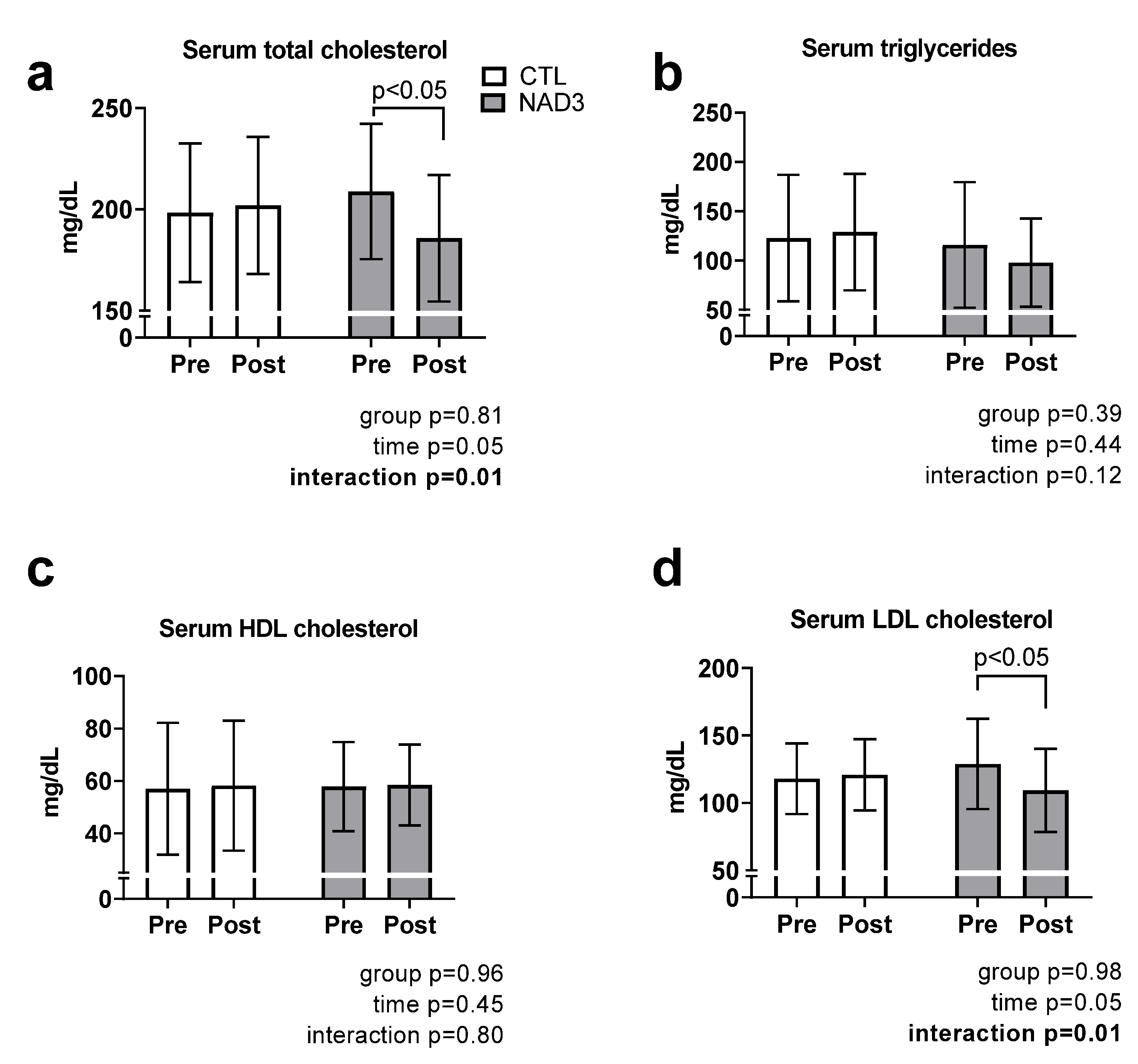
3.2. Effects of Supplementation on Serum Lipids

Figure 1 contains data regarding the effects of supplementation on serum lipids. Significant interactions were observed for total cholesterol (interaction p = 0.010; Figure 1a) and LDL cholesterol concentrations (interaction p = 0.010; Figure 1d), and post hoc analyses indicated a significant decrease with NAD3 supplementation (−11.1% for total cholesterol and −15.2% for LDL cholesterol, p < 0.05 for both variables). No significant interactions were observed for serum triglycerides (interaction p = 0.120; Figure 1b) or HDL cholesterol levels (interaction p = 0.820; Figure 1c).

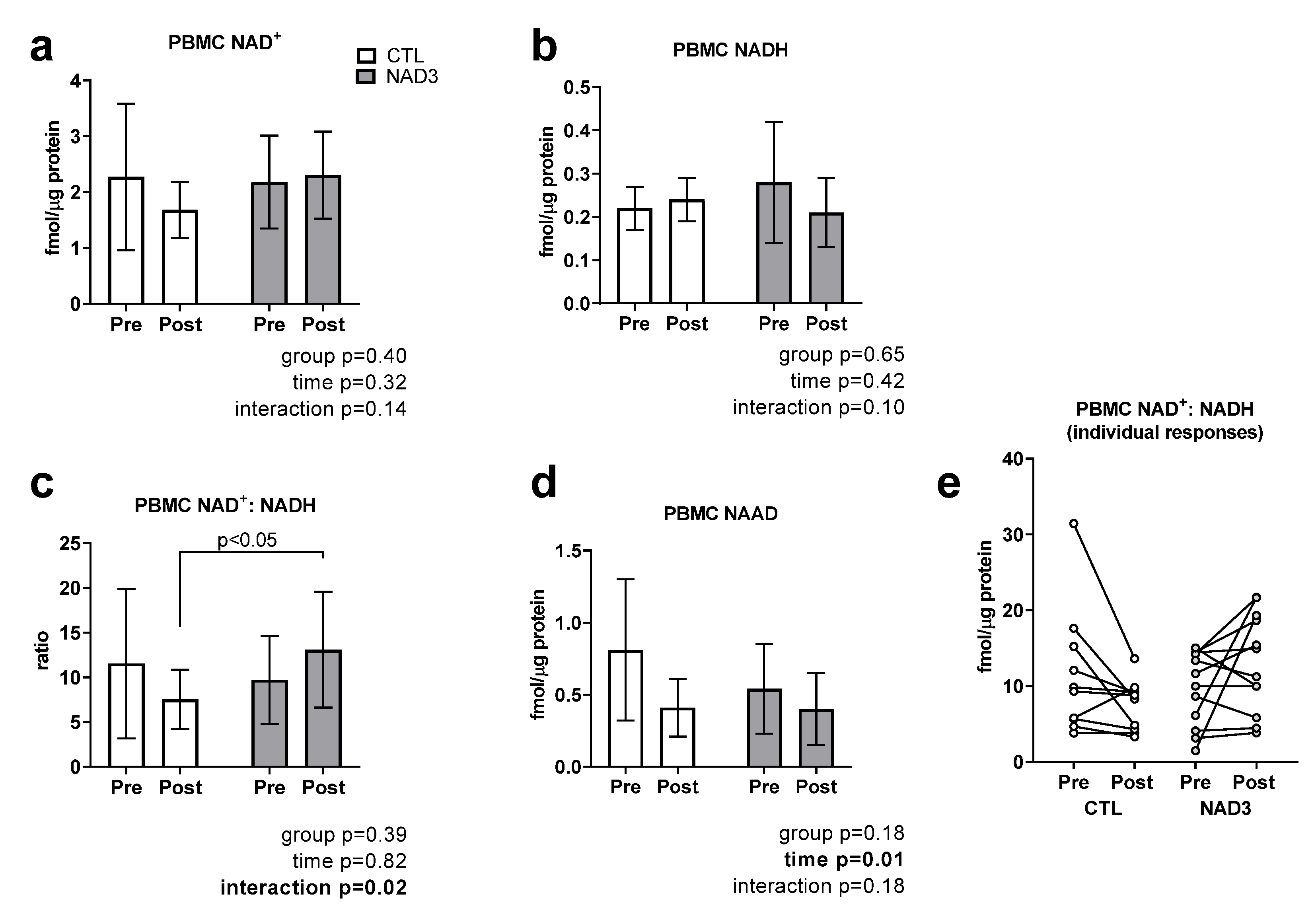
3.3. Effects of Supplementation on PBMC NAD+, NADH, NAD+/NADH, and NAAD

Due to limited PBMC lysate yields, only 10 control participants and 12 NAD3 participants were analyzed for targeted NAD+ metabolomics at Pre and Post. No significant interactions were observed for PBMC NAD+ concentrations (interaction p = 0.144; Figure 2a), NADH concentrations (interaction p = 0.096; Figure 2b), or nicotinic acid adenine dinucleotide (NAAD) (interaction p = 0.183; Figure 2d). A significant interaction (p = 0.020) was observed for PBMC NAD+/NADH values (CTL Pre: 11.5 ± 8.4, Post: 7.5 ± 3.3; NAD3 Pre: 9.7 ± 4.9, Post: 13.1 ± 6.5), where values trended downward from Pre to Post in the CTL group (p = 0.081) and values at Post were greater in NAD3 versus CTL (p = 0.023; Figure 2c). Individual responses for data in Figure 2c are presented in Figure 2e.

3.4. Effects of Supplementation on Blood Markers Indicative of Clinical Safety

Table 3 contains whole blood and serum markers related to clinical safety. No significant interactions were evident with the exception of serum creatinine levels (interaction p = 0.020). Post hoc analysis indicated this value increased with NAD3 supplementation only. However, in spite of this increase with NAD3 supplementation, values were below the typical reference range (adult men: 0.74–1.35 mg/dL, adult women: 0.59–1.04 mg/dL).

4. Discussion

We sought to examine if 12 weeks of daily NAD3 supplementation altered select NAD+-associated metabolites in PBMCs as well as serum lipids. Significant interactions (p < 0.05) were observed for total and LDL cholesterol, where values significantly decreased with NAD3 supplementation. Additionally, a significant interaction was observed for PBMC NAD+/NADH values, where values trended downward from Pre to Post in the CTL group (p = 0.081) and values at Post were greater in NAD3 versus CTL (p = 0.023). These data collectively demonstrate a theacrine-based NAD3 supplement can favorably alter biomarkers of lipid metabolism and cellular NAD+ status. While promising, our n-sizes per group were limited. Additionally, these data are limited to targeted NAD+ metabolites. In this regard, the downstream effects of these observations remain to be determined. Moreover, more comprehensive omics-based profiling needs to be performed in order to examine how supplementation affects other cellular metabolites and mechanisms.
As with our previous report showing eight weeks of theacrine supplementation (300 mg/d) reduced serum total and LDL cholesterol concentrations [10], the current study demonstrates similar findings over a 12-week supplementation period, except with a much smaller dose of theacrine as part of the NAD3 complex. This physiological effect of NAD3 supplementation could be due to multiple mechanisms. First, theacrine exerts some of its physiological effects through ligand binding with adenosine and dopamine receptors [29]. An elegant mouse knockout model has demonstrated that A2b adenosine receptor (A2bAR) signaling is involved in the maintenance of blood lipid levels [30]; specifically, A2bAR knockout mice exhibit hypercholesterolemia. In lieu of our findings, this raises the possibility that NAD3 supplementation enhances A2bAR signaling in the liver, which in turn, lowers cholesterol biosynthesis. Additionally, theacrine has been shown to reduce nonalcoholic fatty liver disease in rodents through activating SIRT3 and increasing fatty acid oxidation [9], and we have reported that NAD3 increases myocellular global sirtuin activity in vitro [11]. Hence, the activation of SIRT3 via NAD3 may be an involved mechanism leading to the observations herein on NAD3-induced improvements in serum lipids.
Wasabia japonica extracts from either leaf or root fractions rich in ITCs have also been demonstrated to have a hypolipidemic effect, along with xanthine oxidase inhibition in rats fed a normal diet [31]. Allyl and phenethyl isothiocyante treatment, such as those found in wasabi root extract, has been shown to decrease total cholesterol, triglycerides, hepatic lipid accumulation, and atherosclerotic plaque formation in high fat diet-induced obesity C57BL/6 mice [32]. ITCs have also been shown to influence multiple genes along the reverse cholesterol lipid metabolism and hepatosteatosis pathway such as peroxisome proliferator activated receptor-gamma (PPARγ), liver-X-receptor α (LXR-α), AMP-activated protein kinase (AMPK), sterol regulatory element-binding protein-1 (SREBP1), acetyl-CoA carboxylase, and ATP-binding cassette subfamily A member 1 (ABCA1) [33,34]. Theacrine has been shown to reduce plasma AST and ALT, hepatic steatosis, liver inflammation, and histologic damage, and also improves energy expenditure in HFD-fed and restraint stress liver damage mice models by reducing IL1-beta, TNF-alpha, and IL-6 gene expression, ameliorating dysfunctional acylcarnitine metabolism, and promoting the SIRT3/LCAD signaling pathway [9,35]. These mechanistic hepatic data on theacrine and ITCs are intriguing in light of plasma AST an ALT levels trending lower (p = 0.11 and p = 0.07, respectively) in the NAD3 supplemented group. These collective data support the notion that the combination of theacrine and ITCs from NAD3 supplementation may have provided multiple mechanisms responsible for the reduced total and LDL cholesterol findings observed in our study.
The potential for NAD3 to maintain PBMC NAD+/NADH levels relative to the control group is intriguing for a variety of reasons. First, there is enthusiasm surrounding the role that tissue NAD+ concentrations play in the aging process [12], and some research suggests that the age-associated loss in tissue NAD+ levels contributes to loss of cellular resilience to metabolic and inflammatory stress, epigenetic drift, aging, and cellular senescence [36]. The de novo formation of NAD+ from the amino acid tryptophan occurs via the kynurenine pathway [37], albeit NAD+ biosynthesis can also occur via the salvage/recycling pathway. NAMPT is the rate-limiting enzyme in the latter of these two pathways [38]. Nicotinamide riboside (NR) supplementation has gained recent notoriety for being capable of increasing blood and tissue NAD+ levels [39,40], and this is due to NR being a precursor substrate in the NAD+ salvage pathway. Indeed, NAD3 contains microgram amounts of niacin representing only 5% of the Daily Value for niacin (vitamin B3) developed by the U.S. Food and Drug Administration. Although niacin is a substrate used for NAD+ biosynthesis [41], our observations of NAD3 supplementation being able to maintain PBMC NAD+/NADH levels are not likely to be related to the insignificant dose of niacin that is used to chelate and stabilize the copper in +1 valence state. Other human data published on the use of NAD+ precursors to increase blood NAD+ levels demonstrate that between 100 mg to 1000 mg daily of NR, NMN, niacin, or niacinamide have been required to significantly increase PBMC levels of NAD+, which are over 100 to 1000-fold greater than the dose of niacin included in the NAD3 supplement [5,13,42,43]. As NAD+ status links cellular metabolism to signaling and transcriptional events that are central to cytoprotection, multiple hallmarks of aging and cardiometabolic health, additional mechanistic insight and clinical data are needed to better ascertain the therapeutic potential for NAD3. We have previously shown that NAD3 upregulates myocellular NAD+ concentrations in vitro [11], and this coincided with an upregulation in NAMPT enzyme levels. While promising, we lack the mechanistic data explaining how NAD3 upregulates NAMPT and/or if this occurs through increased adenosine or dopamine receptor signaling. While cellular NAD+/NADH levels are seemingly a readout of biological aging [44], the outcomes of our study and whether this led to improvements in PBMC function remain to be determined. Thus, these data along with our in vitro data suggesting NAD3 positively affects cellular NAD+ levels warrant additional human research with regard to potential physiological outcomes.
A last topic of deliberation involves whether the supplementation interaction observed with PBMC NAD+: NADH extends to other cell types (e.g., vascular endothelial cells, muscle cells, liver cells), and whether this potential affect affects other cellular outcomes. While speculative, it may be possible that the observed improvements in blood lipids could have also been due to NAD3 supplementation positively affecting hepatocyte NAD+ concentrations. In this regard, others have shown that treating hepatocytes with different fatty acids increases cellular NAD+: NADH values, and this leads to positive alterations in lipid metabolism [45]. However, we are very cautious with this hypothesis given that liver tissue was not obtained from human subjects herein.

5. Conclusions

These data continue to demonstrate a theacrine and ITC-based supplement can positively alter molecular markers related to lipid metabolism, cardiometabolic health and hallmarks of aging. Moreover, these are the first human data to suggest a theacrine and ITC-based supplement can positively affect the NAD+ metabolome in human blood cells. Given the limited data in this area, more research is warranted in other human tissues (e.g., skeletal muscle, skin, etc.).

Author Contributions

Conceptualization, H.L.L., T.N.Z., M.D.R.; methodology, all authors; investigation, all authors; data curation, all authors; writing—original draft preparation, H.L.L., T.N.Z., M.D.R.; writing—review and editing, all authors; funding acquisition, H.L.L., T.N.Z. All authors have read and agreed to the published version of the manuscript.

Funding

Costs involved with executing the study and assays were provided by JUVN3 Holdings.

Institutional Review Board Statement

All procedures were approved by the Institutional Review Board at Integreview, Inc. (Austin, TX, USA; Protocol #CS-01-2020), and this study conformed to the standards set by the latest revision of the Declaration of Helsinki.

Informed Consent Statement

Informed consent was obtained from all subjects involved in the study.

Data Availability Statement

The raw data will be provided with undue reservation by the corresponding author (mdr0024@auburn.edu) upon reasonable request.

Acknowledgments

We thank the participants for volunteering for this study.

Conflicts of Interest

M.D.R. has been a paid consultant in the past by the funding entity for scientific writing endeavors. Additionally, the funding entities have committed a multiple-year laboratory donation to the laboratory of M.D.R. for graduate student stipends, graduate student projects, and assay development. However, none of said donations have been used for salary support for M.D.R., H.L.L. and T.N.Z. have received grants and contracts to conduct research on dietary supplements, and both are involved with multiple patents (including, NAD3, the topic of this manuscript). However, neither investigator was involved with the third-party analyses of dependent variables herein, statistical analyses, or primary drafting of this manuscript.

References

  1. Hunt, N.J.; Kang, S.W.S.; Lockwood, G.P.; Le Couteur, D.G.; Cogger, V.C. Hallmarks of Aging in the Liver. Comput. Struct. Biotechnol. J. 2019, 17, 1151–1161. [Google Scholar] [CrossRef]
  2. Desai, A.; Grolleau-Julius, A.; Yung, R. Leukocyte function in the aging immune system. J. Leukoc. Biol. 2010, 87, 1001–1009. [Google Scholar] [CrossRef]
  3. Gualano, B.; Rawson, E.S.; Candow, D.G.; Chilibeck, P.D. Creatine supplementation in the aging population: Effects on skeletal muscle, bone and brain. Amino Acids 2016, 48, 1793–1805. [Google Scholar] [CrossRef]
  4. Troesch, B.; Eggersdorfer, M.; Laviano, A.; Rolland, Y.; Smith, A.D.; Warnke, I.; Weimann, A.; Calder, P.C. Expert Opinion on Benefits of Long-Chain Omega-3 Fatty Acids (DHA and EPA) in Aging and Clinical Nutrition. Nutrients 2020, 12, 2555. [Google Scholar] [CrossRef]
  5. Martens, C.R.; Denman, B.A.; Mazzo, M.R.; Armstrong, M.L.; Reisdorph, N.; McQueen, M.B.; Chonchol, M.; Seals, D.R. Chronic nicotinamide riboside supplementation is well-tolerated and elevates NAD(+) in healthy middle-aged and older adults. Nat. Commun. 2018, 9, 1286. [Google Scholar] [CrossRef]
  6. Serino, A.; Salazar, G. Protective Role of Polyphenols against Vascular Inflammation, Aging and Cardiovascular Disease. Nutrients 2018, 11, 53. [Google Scholar] [CrossRef] [Green Version]
  7. Sheng, Y.Y.; Xiang, J.; Wang, Z.S.; Jin, J.; Wang, Y.Q.; Li, Q.S.; Li, D.; Fang, Z.T.; Lu, J.L.; Ye, J.H.; et al. Theacrine from Camellia kucha and Its Health Beneficial Effects. Front. Nutr. 2020, 7, 596823. [Google Scholar] [CrossRef]
  8. Gao, M.; Zheng, J.; Zheng, C.; Huang, Z.; Huang, Q. Theacrine alleviates chronic inflammation by enhancing TGF-beta-mediated shifts via TGF-beta/SMAD pathway in Freund’s incomplete adjuvant-induced rats. Biochem. Biophys. Res. Commun. 2020, 522, 743–748. [Google Scholar] [CrossRef]
  9. Wang, G.E.; Li, Y.F.; Zhai, Y.J.; Gong, L.; Tian, J.Y.; Hong, M.; Yao, N.; Wu, Y.P.; Kurihara, H.; He, R.R. Theacrine protects against nonalcoholic fatty liver disease by regulating acylcarnitine metabolism. Metabolism 2018, 85, 227–239. [Google Scholar] [CrossRef]
  10. Taylor, L.; Mumford, P.; Roberts, M.; Hayward, S.; Mullins, J.; Urbina, S.; Wilborn, C. Safety of TeaCrine(R), a non-habituating, naturally-occurring purine alkaloid over eight weeks of continuous use. J. Int. Soc. Sports Nutr. 2016, 13, 2. [Google Scholar] [CrossRef] [Green Version]
  11. Mumford, P.W.; Osburn, S.C.; Fox, C.D.; Godwin, J.S.; Roberts, M.D. A Theacrine-Based Supplement Increases Cellular NAD(+) Levels and Affects Biomarkers Related to Sirtuin Activity in C2C12 Muscle Cells In Vitro. Nutrients 2020, 12, 3727. [Google Scholar] [CrossRef] [PubMed]
  12. Verdin, E. NAD(+) in aging, metabolism, and neurodegeneration. Science 2015, 350, 1208–1213. [Google Scholar] [CrossRef] [PubMed]
  13. Pirinen, E.; Auranen, M.; Khan, N.A.; Brilhante, V.; Urho, N.; Pessia, A.; Hakkarainen, A.; Kuula, J.; Heinonen, U.; Schmidt, M.S.; et al. Niacin Cures Systemic NAD(+) Deficiency and Improves Muscle Performance in Adult-Onset Mitochondrial Myopathy. Cell Metab. 2020, 31, 1078–1090.e1075. [Google Scholar] [CrossRef] [PubMed]
  14. Sauve, A.A. NAD+ and vitamin B3: From metabolism to therapies. J. Pharmacol. Exp. Ther. 2008, 324, 883–893. [Google Scholar] [CrossRef] [Green Version]
  15. Gomes, A.P.; Price, N.L.; Ling, A.J.; Moslehi, J.J.; Montgomery, M.K.; Rajman, L.; White, J.P.; Teodoro, J.S.; Wrann, C.D.; Hubbard, B.P.; et al. Declining NAD(+) induces a pseudohypoxic state disrupting nuclear-mitochondrial communication during aging. Cell 2013, 155, 1624–1638. [Google Scholar] [CrossRef] [PubMed] [Green Version]
  16. Shin, S.W.; Ghimeray, A.K.; Park, C.H. Investigation of total phenolic, total flavonoid, antioxidant and allyl isothiocyanate content in the different organs of Wasabi japonica grown in an organic system. Afr. J. Tradit. Complement. Altern. Med. 2014, 11, 38–45. [Google Scholar] [CrossRef] [Green Version]
  17. Uto, T.; Hou, D.X.; Morinaga, O.; Shoyama, Y. Molecular Mechanisms Underlying Anti-Inflammatory Actions of 6-(Methylsulfinyl)hexyl Isothiocyanate Derived from Wasabi (Wasabia japonica). Adv. Pharm. Sci. 2012, 2012, 614046. [Google Scholar] [CrossRef] [Green Version]
  18. Greaney, A.J.; Maier, N.K.; Leppla, S.H.; Moayeri, M. Sulforaphane inhibits multiple inflammasomes through an Nrf2-independent mechanism. J. Leukoc. Biol. 2016, 99, 189–199. [Google Scholar] [CrossRef] [Green Version]
  19. Wagner, A.E.; Boesch-Saadatmandi, C.; Dose, J.; Schultheiss, G.; Rimbach, G. Anti-inflammatory potential of allyl-isothiocyanate--role of Nrf2, NF-(kappa) B and microRNA-155. J. Cell. Mol. Med. 2012, 16, 836–843. [Google Scholar] [CrossRef]
  20. Dashwood, R.H.; Ho, E. Dietary agents as histone deacetylase inhibitors: Sulforaphane and structurally related isothiocyanates. Nutr. Rev. 2008, 66 (Suppl. S1), S36–S38. [Google Scholar] [CrossRef] [Green Version]
  21. Qu, X.; Proll, M.; Neuhoff, C.; Zhang, R.; Cinar, M.U.; Hossain, M.M.; Tesfaye, D.; Grosse-Brinkhaus, C.; Salilew-Wondim, D.; Tholen, E.; et al. Sulforaphane epigenetically regulates innate immune responses of porcine monocyte-derived dendritic cells induced with lipopolysaccharide. PLoS ONE 2015, 10, e0121574. [Google Scholar] [CrossRef] [PubMed]
  22. Arredondo, M.; Munoz, P.; Mura, C.V.; Nunez, M.T. DMT1, a physiologically relevant apical Cu1+ transporter of intestinal cells. Am. J. Physiol. Cell Physiol. 2003, 284, C1525–C1530. [Google Scholar] [CrossRef] [PubMed]
  23. Sugiura, A.; McLelland, G.L.; Fon, E.A.; McBride, H.M. A new pathway for mitochondrial quality control: Mitochondrial-derived vesicles. EMBO J. 2014, 33, 2142–2156. [Google Scholar] [CrossRef] [Green Version]
  24. Gonzalez-Freire, M.; de Cabo, R.; Bernier, M.; Sollott, S.J.; Fabbri, E.; Navas, P.; Ferrucci, L. Reconsidering the Role of Mitochondria in Aging. J. Gerontol. A Biol. Sci. Med. Sci. 2015, 70, 1334–1342. [Google Scholar] [CrossRef] [PubMed] [Green Version]
  25. Jang, J.Y.; Blum, A.; Liu, J.; Finkel, T. The role of mitochondria in aging. J. Clin. Investig. 2018, 128, 3662–3670. [Google Scholar] [CrossRef] [PubMed] [Green Version]
  26. Sasaki, T.; Abe, Y.; Takayama, M.; Adachi, T.; Okano, H.; Hirose, N.; Arai, Y. Association among extracellular superoxide dismutase genotype, plasma concentration, and comorbidity in the very old and centenarians. Sci. Rep. 2021, 11, 8539. [Google Scholar] [CrossRef]
  27. Barzilai, N.; Huffman, D.M.; Muzumdar, R.H.; Bartke, A. The critical role of metabolic pathways in aging. Diabetes 2012, 61, 1315–1322. [Google Scholar] [CrossRef] [Green Version]
  28. Lamb, D.A.; Moore, J.H.; Mesquita, P.H.C.; Smith, M.A.; Vann, C.G.; Osburn, S.C.; Fox, C.D.; Lopez, H.L.; Ziegenfuss, T.N.; Huggins, K.W.; et al. Resistance training increases muscle NAD(+) and NADH concentrations as well as NAMPT protein levels and global sirtuin activity in middle-aged, overweight, untrained individuals. Aging (Albany NY) 2020, 12, 9447–9460. [Google Scholar] [CrossRef]
  29. Feduccia, A.A.; Wang, Y.; Simms, J.A.; Yi, H.Y.; Li, R.; Bjeldanes, L.; Ye, C.; Bartlett, S.E. Locomotor activation by theacrine, a purine alkaloid structurally similar to caffeine: Involvement of adenosine and dopamine receptors. Pharm. Biochem. Behav. 2012, 102, 241–248. [Google Scholar] [CrossRef]
  30. Koupenova, M.; Johnston-Cox, H.; Vezeridis, A.; Gavras, H.; Yang, D.; Zannis, V.; Ravid, K. A2b adenosine receptor regulates hyperlipidemia and atherosclerosis. Circulation 2012, 125, 354–363. [Google Scholar] [CrossRef] [Green Version]
  31. Lee, Y.S.; Yang, J.H.; Bae, M.J.; Yoo, W.K.; Ye, S.; Xue, C.C.; Li, C.G. Anti-oxidant and Anti-hypercholesterolemic Activities of Wasabia japonica. Evid. Based Complement. Altern. Med. 2010, 7, 459–464. [Google Scholar] [CrossRef] [PubMed] [Green Version]
  32. Gwon, M.H.; Im, Y.S.; Seo, A.R.; Kim, K.Y.; Moon, H.R.; Yun, J.M. Phenethyl Isothiocyanate Protects against High Fat/Cholesterol Diet-Induced Obesity and Atherosclerosis in C57BL/6 Mice. Nutrients 2020, 12, 3657. [Google Scholar] [CrossRef] [PubMed]
  33. Gwon, M.H.; Yun, J.M. Phenethyl Isothiocyanate Improves Lipid Metabolism and Inflammation via mTOR/PPARgamma/AMPK Signaling in the Adipose Tissue of Obese Mice. J. Med. Food 2021, 24, 666–669. [Google Scholar] [CrossRef] [PubMed]
  34. Chuang, W.T.; Liu, Y.T.; Huang, C.S.; Lo, C.W.; Yao, H.T.; Chen, H.W.; Lii, C.K. Benzyl Isothiocyanate and Phenethyl Isothiocyanate Inhibit Adipogenesis and Hepatosteatosis in Mice with Obesity Induced by a High-Fat Diet. J. Agric. Food Chem. 2019, 67, 7136–7146. [Google Scholar] [CrossRef] [PubMed]
  35. Li, W.X.; Li, Y.F.; Zhai, Y.J.; Chen, W.M.; Kurihara, H.; He, R.R. Theacrine, a purine alkaloid obtained from Camellia assamica var. kucha, attenuates restraint stress-provoked liver damage in mice. J. Agric. Food Chem. 2013, 61, 6328–6335. [Google Scholar] [CrossRef] [PubMed]
  36. Goody, M.F.; Henry, C.A. A need for NAD+ in muscle development, homeostasis, and aging. Skelet. Muscle 2018, 8, 9. [Google Scholar] [CrossRef] [PubMed] [Green Version]
  37. Rongvaux, A.; Andris, F.; Van Gool, F.; Leo, O. Reconstructing eukaryotic NAD metabolism. Bioessays 2003, 25, 683–690. [Google Scholar] [CrossRef]
  38. Revollo, J.R.; Grimm, A.A.; Imai, S. The NAD biosynthesis pathway mediated by nicotinamide phosphoribosyltransferase regulates Sir2 activity in mammalian cells. J. Biol. Chem. 2004, 279, 50754–50763. [Google Scholar] [CrossRef] [Green Version]
  39. Airhart, S.E.; Shireman, L.M.; Risler, L.J.; Anderson, G.D.; Nagana Gowda, G.A.; Raftery, D.; Tian, R.; Shen, D.D.; O’Brien, K.D. An open-label, non-randomized study of the pharmacokinetics of the nutritional supplement nicotinamide riboside (NR) and its effects on blood NAD+ levels in healthy volunteers. PLoS ONE 2017, 12, e0186459. [Google Scholar] [CrossRef]
  40. Trammell, S.A.; Schmidt, M.S.; Weidemann, B.J.; Redpath, P.; Jaksch, F.; Dellinger, R.W.; Li, Z.; Abel, E.D.; Migaud, M.E.; Brenner, C. Nicotinamide riboside is uniquely and orally bioavailable in mice and humans. Nat. Commun. 2016, 7, 12948. [Google Scholar] [CrossRef]
  41. Makarov, M.V.; Trammell, S.A.J.; Migaud, M.E. The chemistry of the vitamin B3 metabolome. Biochem. Soc. Trans. 2019, 47, 131–147. [Google Scholar] [CrossRef] [PubMed]
  42. Conze, D.; Brenner, C.; Kruger, C.L. Safety and Metabolism of Long-term Administration of NIAGEN (Nicotinamide Riboside Chloride) in a Randomized, Double-Blind, Placebo-controlled Clinical Trial of Healthy Overweight Adults. Sci. Rep. 2019, 9, 9772. [Google Scholar] [CrossRef] [PubMed]
  43. Ito, T.K.; Sato, T.; Takanashi, Y.; Tamannaa, Z.; Kitamoto, T.; Odagiri, K.; Setou, M. A single oral supplementation of nicotinamide within the daily tolerable upper level increases blood NAD+ levels in healthy subjects. Transl. Med. Aging 2021, 5, 43–51. [Google Scholar] [CrossRef]
  44. Imai, S.; Guarente, L. NAD+ and sirtuins in aging and disease. Trends Cell Biol. 2014, 24, 464–471. [Google Scholar] [CrossRef] [PubMed]
  45. Chen, G.C.; Su, H.M.; Lin, Y.S.; Tsou, P.Y.; Chyuan, J.H.; Chao, P.M. A conjugated fatty acid present at high levels in bitter melon seed favorably affects lipid metabolism in hepatocytes by increasing NAD(+)/NADH ratio and activating PPARalpha, AMPK and SIRT1 signaling pathway. J. Nutr. Biochem. 2016, 33, 28–35. [Google Scholar] [CrossRef] [PubMed]
Figure 1. Effects of supplementation on serum lipids. Data are presented as means ± standard deviation values for 15 CTL and 13 NAD3 participants. Abbreviations: CTL, control group; NAD3, supplementation group.
Physiologia 02 00002 g001
Figure 2. Targeted NAD+ metabolome data. Data are presented as means ± standard deviation values for 15 CTL and 13 NAD3 participants. Abbreviations: CTL, control group; NAD3, supplementation group.
Physiologia 02 00002 g002
Table 1. Baseline characteristics.
VariableCTL (n = 15)NAD3 (n = 13)p-Value
Gender6 M/9 F6 M/7 F-
Age (years)51 ± 552 ± 60.57
BMI (kg/m2)28.3 ± 3.929.0 ± 50.68
Legend: data are presented as means ± standard deviation values for 15 CTL and 13 NAD3 participants. Abbreviations: CTL, control group; NAD3, supplementation group.
Table 2. Body mass and blood pressure outcomes.
VariableGroupPrePost2 × 2 ANOVA p-Values
Body Mass (kg)CTL
NAD3
80.0 ± 15.2
87.2 ± 20.9
80.5 ± 15.7
87.6 ± 21.9
Group
Time
G × T
0.70
0.13
0.77
Systolic Blood Pressure (mm Hg)CTL
NAD3
128 ± 15
127 ± 11
127 ± 9
129 ± 8
Group
Time
G × T
0.88
0.83
0.52
Diastolic Blood Pressure (mm Hg)CTL
NAD3
80 ± 7
78 ± 10
80 ± 8
80 ± 7
Group
Time
G × T
0.72
0.38
0.66
Legend: data are presented as means ± standard deviation values for 15 CTL and 13 NAD3 participants. Abbreviations: CTL, control group; NAD3, supplementation group; G × T, group-by-time interaction p-value.
Table 3. Blood markers of clinical safety.
VariableGroupPrePost2 × 2 ANOVA
WBCs
(cells × 103/μL)
CTL5.46 ± 1.225.39 ± 1.17Group0.20
NAD36.31 ± 2.696.51 ± 2.72Time0.86
G × T0.69
RBCs
(cells × 103/μL)
CTL4.53 ± 0.544.57 ± 0.52Group0.78
NAD34.44 ± 0.414.55 ± 0.39Time0.04
G × T0.28
Hemoglobin
(g/dL)
CTL13.7 ± 1.913.9 ± 1.7Group0.66
NAD314.0 ± 1.414.2 ± 1.3Time0.33
G × T0.80
Hematocrit
(%)
CTL40.3 ± 4.641.5 ± 4.4Group0.82
NAD340.6 ± 3.742.0 ± 2.9Time0.005
G × T0.82
Platelets (×103/uL)CTL258 ± 83247 ± 58Group0.23
NAD3282 ± 68289 ± 62Time0.69
G × T0.18
Neutrophils (%)CTL56.0 ± 6.955.5 ± 10.6Group0.61
NAD352.6 ± 9.455.6 ± 10.6Time0.40
G × T0.25
Lymphocytes (%)CTL32.3 ± 6.432.5 ± 7.0Group0.78
NAD334.3 ± 8.432.1 ± 9.0Time0.40
G × T0.34
Monocytes (%)CTL8.85 ± 1.689.08 ± 2.33Group0.36
NAD38.25 ± 2.268.00 ± 3.49Time0.98
G × T0.61
Eosinophils (%)CTL2.39 ± 1.042.39 ± 1.33Group0.11
NAD34.00 ± 2.923.58 ± 2.91Time0.42
G × T0.42
Basophils (%)CTL0.39 ± 0.650.46 ± 0.52Group0.17
NAD30.75 ± 0.620.75 ± 0.75Time0.73
G × T0.73
Glucose (mg/dL)CTL91.9 ± 8.597.2 ± 13.2Group0.20
NAD396.3 ± 11.896.7 ± 14.2Time0.86
G × T0.69
BUN (mg/dL)CTL12.8 ± 2.013.5 ± 2.6Group0.47
NAD313.3 ± 2.214.2 ± 2.7Time0.18
G × T0.84
Creatinine (mg/dL)CTL0.81 ± 0.190.84 ± 0.19Group0.33
NAD30.86 ± 0.210.95 ± 0.22 §Time0.001
G × T0.02
BUN:CreatinineCTL16.6 ± 5.216.6 ± 4.8Group0.56
NAD316.2 ± 4.115.2 ± 3.1Time0.47
G × T0.47
Sodium (mEq/mL)CTL139 ± 3139 ± 2Group0.69
NAD3139 ± 3139 ± 3Time0.87
G × T0.63
Potassium (mEq/mL)CTL4.25 ± 0.234.28 ± 0.24Group0.96
NAD34.27 ± 0.184.26 ± 0.28Time0.82
G × T0.70
Chloride (mEq/mL)CTL102 ± 3103 ± 2Group0.49
NAD3104 ± 3103 ± 3Time0.48
G × T0.33
Calcium (mg/dL)CTL9.35 ± 0.289.36 ± 0.36Group0.33
NAD39.42 ± 0.319.52 ± 0.32Time0.41
G × T0.48
Total Protein
(g/dL)
CTL6.92 ± 0.446.89 ± 0.43Group0.52
NAD36.75 ± 0.406.86 ± 0.40Time0.54
G × T0.35
Albumin (g/dL)CTL4.37 ± 0.294.35 ± 0.28Group0.44
NAD34.42 ± 0.184.44 ± 0.25Time0.92
G × T0.62
Bilirubin
(g/dL)
CTL0.67 ± 0.250.58 ± 0.17Group1.00
NAD30.59 ± 0.260.55 ± 0.19Time0.10
G × T0.67
Alkaline Phosphatase
(U/L)
CTL75.5 ± 20.778.7 ± 22.5Group0.48
NAD370.2 ± 16.572.6 ± 21.8Time0.08
G × T0.78
AST (U/L)CTL26.4 ± 18.626.4 ± 16.8Group0.46
NAD324.2 ± 9.020.6 ± 6.7Time0.11
G × T0.11
ALT (U/L)CTL30.8 ± 24.231.0 ± 24.2Group0.42
NAD327.2 ± 11.022.8 ± 8.7Time0.11
G × T0.07
Legend: data are presented as means ± standard deviation values for 15 CTL and 13 NAD3 participants. Abbreviations: CTL, control group; NAD3, supplementation group; G × T, group-by-time interaction p-value.
Publisher’s Note: MDPI stays neutral with regard to jurisdictional claims in published maps and institutional affiliations.

