Article

Characterization of an Acidogenic Bacterial Consortium as Probiotic and Its Effect on Rumen Fermentation In Vitro and In Vivo

by
Carolina Robles-Rodríguez
1,
Diego Cardoso-Carmona
1,
Laura González-Dávalos
2,
Carlos Lozano-Flores
3,
Allan Páez-Trejo
4,
Armando Shimada
2 and
Ofelia Mora
2,*
1
Posgrado en Ciencias de la Producción y de la Salud Animal, Universidad Nacional Autónoma de México (UNAM), Ciudad de México 04510, Mexico
2
Laboratorio de Rumiología y Metabolismo Nutricional, Facultad de Estudios Superiores Cuautitlán, UNAM, Juriquilla 76230, Mexico
3
Instituto de Neurobiología, Universidad Nacional Autónoma de México (UNAM), Juriquilla 76230, Mexico
4
Centro de Enseñanza, Investigación y Extensión en Producción Animal en Altiplano (CEIEPAA), Facultad de Medicina Veterinaria y Zootecnia, Universidad Nacional Autónoma de México (UNAM), Tequisquiapan 76794, Mexico
*
Author to whom correspondence should be addressed.
Ruminants 2023, 3(4), 324-346; https://doi.org/10.3390/ruminants3040028
Submission received: 22 August 2023 / Revised: 10 October 2023 / Accepted: 12 October 2023 / Published: 16 October 2023

Abstract

:
Probiotics are live microorganisms that promote host health through microbiota balance and immune modulation. We assessed an acidogenic bacterial consortium (ABC) with promising probiotic properties, focusing on its resilience during transit through the digestive tract in ruminants and determining its optimal in vitro dosage. The ABC exhibited antibiotic resistance, thrived at pH levels between 5 and 7 for 24 and 48 h, and showed a 77% survival rate in artificial gastric juice. Moreover, it not only endured bile salt exposure but also multiplied. The ABC exhibited 10.74% of coaggregation capabilities against E. coli. Optimal dosage determination revealed that 4 × 10⁸ was the ideal concentration, as higher doses did not yield significant differences in dry matter digestion. In the in vivo trial with Limousin Heifers, the ABC led to enhanced total volatile fatty acid (VFA) production, increased daily weight gains, and improved feed conversion rates compared to the control group. These findings underscore the potential of the ABC as a probiotic to boost animal productivity and overall health.

1. Introduction

Ruminant nutritionists have developed many feed supplementation methods, such as antibiotic growth promoters, to enhance production by limiting the effects of pathogenic infections on ruminant productivity [1]. Maintaining a good balance in the rumen microbiota confers the correct growth for the host animal and greater production of animal food products. The rumen is an efficient animal–microbe mutualism system that benefits both participants [2], where rumen microbiota provide the main energy source for ruminants, the end-products of fermentation, volatile fatty acids (VFAs, mainly acetate, propionate, and butyrate), with the propionate being the only gluconeogenic VFA used by the host animal.
For decades, antibiotics have played a dual role in animal production as therapeutic agents and growth-promoting additives, with the aim of increasing productivity. However, the widespread use of antibiotics has led to severe concerns over microbial resistance and the exacerbation of residual effects in food intended for human consumption [3]. Consequently, several governments, including those in the European Union, the United States, and Mexico, have significantly reduced or even banned the use of antibiotics as growth promoters.
Antibiotic resistance is a major public health issue that must be prevented and controlled, emphasizing the importance of research-driven initiatives to develop effective alternatives for use in animal production [4,5]. The introduction of alternative additives, such as plant-based products, prebiotics, and probiotics, was necessary, which are utilized in the prophylaxis and treatment of gastrointestinal diseases.
Probiotics are microorganisms that confer health benefits on their host animal when given in adequate doses due to their nutritional, immunological, bacteriostatic, and bactericidal effects [6,7,8], and they serve as a viable alternative for the prevention and treatment of certain calf pathologies, as well as enhancing their productivity [9]. Several studies reported the benefits of the oral administration of probiotics to ruminants, such as regulating and balancing the gut microbiota, reducing diarrhea, protecting against infections and diseases, and promoting the development and growth of animals [10].
Probiotics or directly fed microorganisms (DFM) can be (1) monostrain, containing one strain of one species, (2) multistrain, containing more than one strain of the same species or closely related species, for example, Lactobacillus acidophilus and Lactobacillus casei, and (3) multispecies, containing strains of different species belonging to one or preferably more genera [11]. The latter are known as bacterial consortia, which are a set of various species of microorganisms that interact in synchrony for mutual benefit within a community and are characterized by communication between themselves either through substance exchange or molecular signals, division of different tasks, and performing multi-step functions [12,13,14].
The probiotics demonstrated the ability to improve the rumen environment by consuming oxygen in the organ, modifying the ruminal environment, benefiting the growth of strictly anaerobic microbes such as cellulolytic bacteria, enhancing fiber digestibility, modifying the pH, and competing for adhesion sites on the rumen wall and intestinal mucosa. Furthermore, they have exhibited the capacity to regulate the profiles of VFA production, particularly propionate, which is the primary gluconeogenic VFA; this capability arises from specific bacteria present in bacterial consortia that can compete with methanogenic bacteria for hydrogen. This competition leads to increased volatile fatty acids and reduced methane production by efficient cow microbiota, aligning with a greater energy efficiency [13,14], and the production of antagonistic compounds such as bacteriocins A and B that affect the development of E. coli and Salmonella spp. [15,16,17,18].
However, the beneficial response to probiotic supplementation in ruminants needs to be more consistent, which is dependent on the microbial strain selected, combination of strains, dose, time, and frequency of supplementation. Thus, it is advisable to source potential target host probiotic strains from the target host intestine to recognize the nature of the microbiota of the target host. Furthermore, these microorganisms must be well identified and evaluated for their potential probiotic activities, such as tolerance to temperature and pH, gastric digestibility conditions, and antibiotic resistance, to be considered Generally Recognized as Safe (GRAS, FDA) [19]. In our previous work [20], we evaluated the effect of oral administration of an acidogenic bacterial consortium (ABC) on changes in VFA proportions in ruminant sheep, resulting in the capacity to alter ruminal fermentation and increasing the production of short-chain fatty acids (SCFAs) [20,21].
Therefore, this study aimed to characterize an ABC as a potential probiotic, evaluating different in vitro probiotic tests (susceptibility to antibiotics, tolerance to temperature and pH, antagonist effects, and simulated gastric and intestine conditions), as well as its in vivo effects on VFA production, changes in rumen microbiota, and productivity parameters such as daily weight gain and blood glucose levels in Limousin heifers.

2. Materials and Methods

In this section, we provide an overview of the materials and methods employed in our study to assess the safety and efficacy of our new probiotic candidate. The primary objective of our research is to determine whether this probiotic meets the stringent criteria for GRAS status, ensuring its safety for consumption. Also, we conducted in vivo trials involving Limousine heifers to assess the effects of our probiotic on productive parameters within the livestock industry, to evaluate the probiotic’s influence on factors such as weight gain, feed efficiency, and ruminal microbial populations. Our methodology is designed to evaluate the ABC safety profile, probiotic properties, and potential benefits.

2.1. Broth and Agar Nutrient Medium Preparation

A highly acidogenic bacterial consortium (ABC) obtained from a brewery’s waste was used [21]. It was genetically characterized, and the results are shown in Table 1. For their maintenance and preservation, the microorganisms were grown at 37 °C under anaerobic conditions in a nutrient medium (NM) containing (per liter) glucose, 2 g; yeast extract, 1 g; tryptone, 2 g; KH2PO4, 1 g; K2HPO4, 1.66 g; (NH4)2SO4, 0.5 g; CaCl2, 0.1 g; NaCl, 0.1 g; MgSO4, 0.1 g; NaHCO3, 3.5 g; L-cysteine, 0.5 g; vitamin solution, 1.25%; and mineral solution, 1.25%. The medium was gassed with CO2, and pH was adjusted to 6.5 with HCl, before autoclaving for 15 min at 121 °C [21].
The mineral solution was composed of (g/L) FeCl2-4H2O, 2 g; CoCl2-6H2O, 0.05 g; EDTA, 0.5 g; MnCl2-4H2O, 0.5 g; NiCl2-6H2O, 0. 05 g; Na2SeO3, 0.1 g; AlCl3-6H2O, 0.05 g; H3BO3, 0.05 g; ZnCl2, 0.05 g; (NH4)6Mo7O24-4H2O, 0.05 g; and CuCl2-2H2O, 0.05 g.
The vitamin solution was composed of (mg/L) biotin, 2; folic acid, 2; pyridoxine-HCl, 10; riboflavin, 5; thiamine, 5; nicotinic acid, 5; pantothenic acid, 5; vitamin B12, 0.1; p-aminobenzoic acid, 5; and lipoic acid, 5.
Both solutions were added once the autoclaving process was completed. To prepare the inoculum, 49 mL of this NM solution was transferred to a 50 mL conical tube, subsequently, 0.5 mL of the ABC glycerol stock culture was inoculated into the tube, and the inoculated NM solution was then incubated anaerobically at 37 °C. The bacterial growth curve and generation time were measured every 2 h until an OD 600 nm of 0.5–0.6 was reached.
For the agar plates, 23 g of agar (A1296, Sigma-Aldrich, Taufkirchen, Germany) was placed in the NM and sterilized. After this, like the culture broth, the vitamin and mineral solution were added and poured into sterile Petri dishes before it solidified, ensuring an adequate amount (approximately 12 mL) to cover the entire surface of the plate.
To create and maintain anaerobic conditions in the culture media, GasPak™ EZ Anaerobe Systems (BD, Sparks, MD, USA) were utilized.

2.2. Acidogenic Bacterial Consortia Characterization and Culture Conditions

The ABC obtained from a brewery’s waste was used [20]. It was genetically characterized, and the microbiota compositions are shown in Table 1. For their maintenance and preservation, the microorganisms were grown at 37 °C under anaerobic conditions in a nutrient medium (NM) broth.
The bacterial growth curve and generation time were measured every 2 h until an OD 600 nm of 0.5–0.6 was reached.
For the agar plates, 23 g of agar (A1296, Sigma-Aldrich, Taufkirchen, Germany) was placed in the NM and sterilized. After this, like the culture broth, the vitamin and mineral solutions were added. Then, the growing ABC was added and poured into sterile agar Petri dishes before it solidified, ensuring an adequate amount (approximately 12 mL) to cover the entire surface of the plate.
To prepare agar NM and maintain incubation in anaerobic conditions in the culture media, GasPak™ EZ Anaerobe Systems (BD, Sparks, MD, USA) were utilized at 37 °C for 24 h.

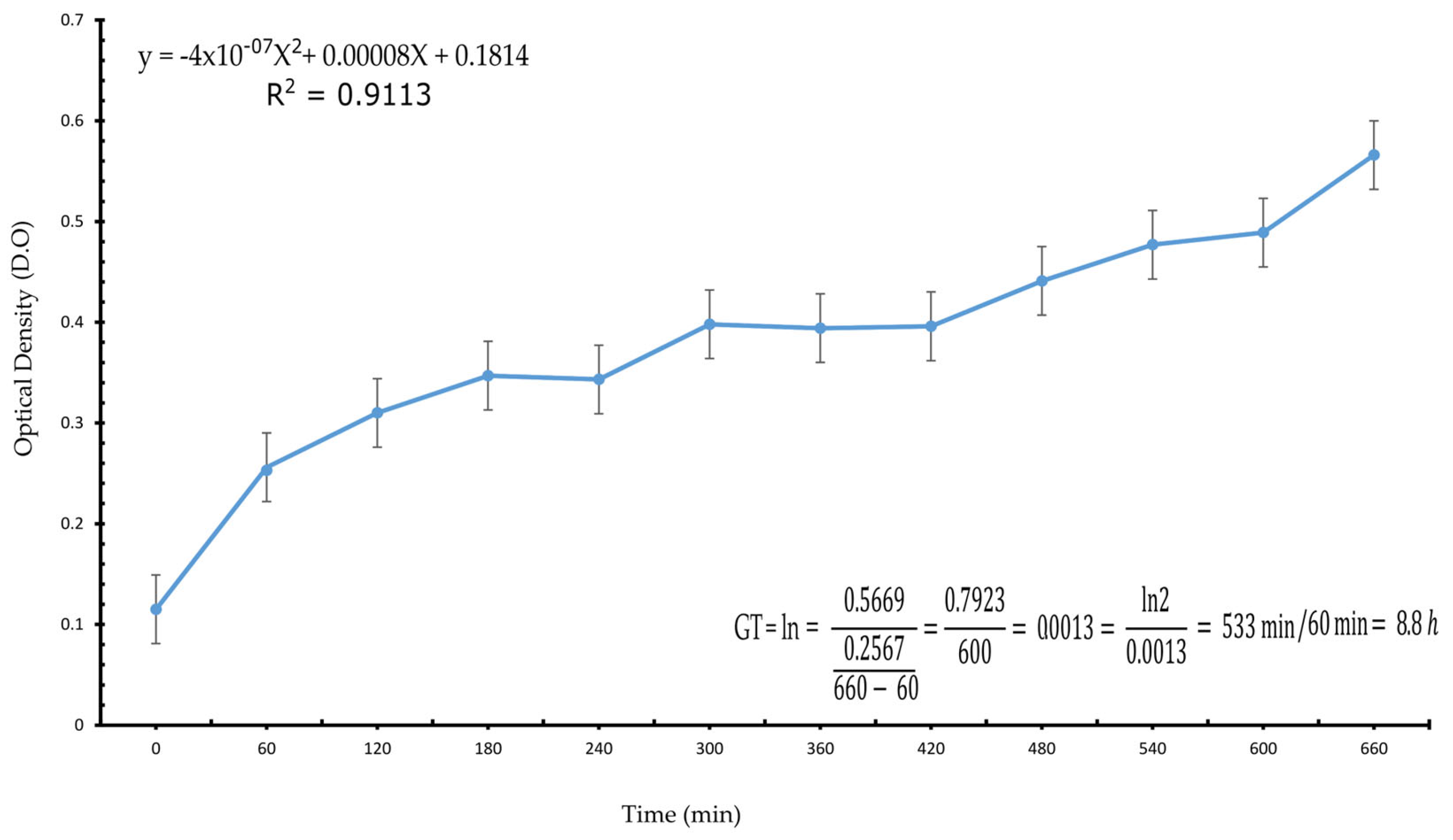
Generation Time of the Bacterial Consortium

Generation time refers to the time required for a cell to divide or a population to double. To determine the generation time of the consortium, the Ratkowsky equation [22] was used:
TG = ln2 / µ
where TG = generation time and μ = the growth rate value, which is determined by ln of T/t, where T = final biomass time and t = initial biomass time. The generation time of ABC was determined based on bacterial growth from a culture with a 1:10 ratio over 12 h in MN at 37 °C. From this culture, the required amount was taken to inoculate a new culture to achieve an OD 600 nm of 0.1. Measurements were taken every hour using the UV-Vis spectrophotometer HP/Agilent 8453 (SpectraLab Scientific Inc., Markham, ON, Canada) until reaching the maximum logarithmic phase.

2.3. Probiotic Tests

2.3.1. Susceptibility to Antibiotics

To assess antibiotic susceptibility, the ABC was subjected to an antibiogram test which comprises a profile or pattern of results from antibiotic susceptibility testing specific to a bacterial strain or species, offering insights into the susceptibility or resistance of bacteria to various antibiotics. It is desirable for a probiotic not to present antibiotic resistance.
One hundred microliters of ABC culture (OD 600 nm = 0.5–0.6) was seeded on NM agar plates. Once the ABC was seeded, it was left to dry for 3–5 min, and then, the following multidiscs with antibiotics were added and incubated anaerobically at 37 °C for 24 h: Gram (−): AK Amikacin (30 µg), AM Ampicillin (10 µg), CB Carbenicillin (100 µg), CL Chloramphenicol (30 µg), NET Netilmicin (30 µg), NF Nitrofurantoin (300 µg), and NOF Norfloxacin (10 µg). Gram (+): DC Dicloxacillin (1 µg), CLM Clindamicin (30 µg), E Erythromycin (15 µg), PE Penicillin (10 U), VA Vancomycin (30 µg), and TE Tetracycline (30 µg). Gram (−) and (+): AM Ampicillin (30 and 10 µg, respectively), CF Cephalothin (30 µg), CFX Cefotaxime (30 µg), CPF Ciprofloxacin (5 µg), GE Gentamicin (10 µg), and SXT Sulfamethoxazole/Trimethoprim (25 µg) (MULTIBAC-ID, Investigación Diagnostica, Iztapala, CDMX, MEX). The diameter of the inhibition zone (mm) was measured, and the results were expressed as resistant (R), intermediate (I), and susceptible (S) according to the manufacturer’s instructions.

2.3.2. Determination of Tetracycline Resistance Genes Tet M, Tet K, and Tet W

To verify antimicrobial safety and determine whether tetracycline resistance was found in genomic or plasmid DNA, PCR tests for the resistance genes Tet M (F: GTGGACAAAGGTACAACGAG; R: CGGTAAAGTTCGTCACACAC), Tet K (F: TTATGGTGGTTGTAGCTAGAAA; R: AAAGGGTTAGAAACTCTTGAAA), and Tet W (F: GAGAGCCTGCTATATGCCAGC; R: GGGCGTATCCACAATGTTAAC) were performed. PCR amplification for the Tet M, Tet K, and Tet W genes was carried out from genomic and plasmid DNA previously extracted from the ABC. The PCR conditions were 95 °C—5 min; 95 °C—30 s, 64 °C—30 s, 72 °C—30 s; 30 cycles and a final alignment of 72 °C—7 min.

2.3.3. Resistance to pH

One hundred microliters of ABC culture (OD 600 nm = 0.5–0.6) was inoculated in NM broth at different pH values of 2, 3, 4, 5, 5.5, 6, 7, and a control at pH = 6.5 was used. Then, it was incubated anaerobically at 37 °C for 24 and 48 h. After incubation, six 10-fold serial dilutions (v/v, ratio) were made starting at 1:1 × 101 (v/v, ratio). One hundred microliters of the dilution was taken and plated on NM agar and grown anaerobically at 37 °C for 24 h. Results were expressed as survival in percentage of CFU/mL.

2.3.4. Gastric Juice Resistance Test

Five 15 mL tubes were inoculated with 1 mL of ABC in 10 mL of artificial gastric juice (2 g NaCl and 3.2 g pepsin/L at pH 2.5); they were incubated at 37 °C for 30, 60, 90, and 120 min, extracting one tube at each time. From each tube extracted, 100 μL of the sample was taken, seeded in plates with NM agar, and incubated at 37 °C for 24 h; then, colonies were counted. Resistance was estimated by comparing the viable cell counts with and without artificial gastric juice, using the formula
R = (CFC / gj) / (CFU / wogj) × 100
where R is the resistance to gastric juice, CFU is the number of colony forming units, gj is milliliters of medium with gastric juice, and wogj is the milliliters of medium without gastric juice.

2.3.5. Resistance to Bile Salts

One hundred microliters of ABC culture (OD 600 nm = 0.5–0.6) were inoculated in NM broth at pH 6.5 with or without 0.3% ox-bile salt and grown anaerobically at 37 °C. The growth curve of ABC culture was monitored by measuring OD 600 nm every 2 h up to 6 h. The culture was then diluted by 10-fold serial dilutions to 1:1 × 10⁷ (v/v, ratio). One hundred microliters of the dilution was taken and plated on NM agar and incubated anaerobically at 37 °C for 24 h. The survival rate was expressed as the percentage of CFU/mL.

2.3.6. Coaggregation Capacity

Coaggregation capacity plays a crucial role in elucidating how pathogens adhere to host tissues, serving as a pivotal factor in the initiation of infections. In the context of probiotic tests, it also provides valuable insights into guiding antimicrobial strategies. This knowledge offers potential avenues to disrupt detrimental bacterial interactions, ultimately contributing to the promotion of health and well-being.
For the evaluation of coaggregation capacity, ABC was grown in NM until a value of 0.5 at OD 600 nm was achieved. An aliquot of 750 μL of the ABC was mixed with the same volume of a suspension of the coaggregation partner E. coli (INCQS 00219, origin ATCC 8739) and then vortexed for 30 s. The OD value of 600 nm was determined at time 0 (baseline, just after mixing the suspensions) and after 60 min of incubation at 37 °C. Coaggregation was measured using the equation
% coaggregation = [(OD0 − OD60) / OD0] × 100,
where
OD0 refers to the initial OD value determined at time 0 and OD60 refers to the OD value of the supernatant after incubation for 60 min.

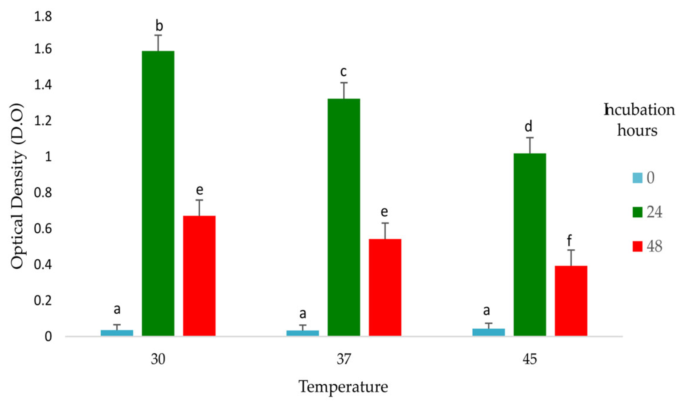
2.3.7. Tolerance to Temperature

One hundred microliters of ABC culture (OD 600 nm = 0.5–0.6) was inoculated in NM broth and incubated anaerobically at 30, 37, and 45 °C, with OD 600 nm measured at 24 and 48 h. Survival results were expressed as the percentage of CFU/mL.

2.3.8. Antagonistic Activity

Bacterial antagonism was evaluated using the disc diffusion method [23] with modifications, employing two pathogenic strains (S. typhimurium ATCC 19028 and E. coli ATCC 11229, donated by the Facultad de Medicina, Universidad Autónoma de Quéretaro). The ABC cultures were incubated anaerobically in NM broth and the pathogenic strains in LB broth at 37 °C overnight. One hundred microliters of pathogens were spread on Mueller–Hinton agar plates. ABC cultures were centrifuged at 3000 rpm for 5 min, and after that, ABC cultures supernatants were added to sterile paper discs and placed on the Mueller–Hinton agar plates. After incubation at 37 °C for 24 h, the growth inhibition zone (mm) was measured. The antagonistic activity was considered positive when the average growth inhibition zone diameter was ≥10 mm.

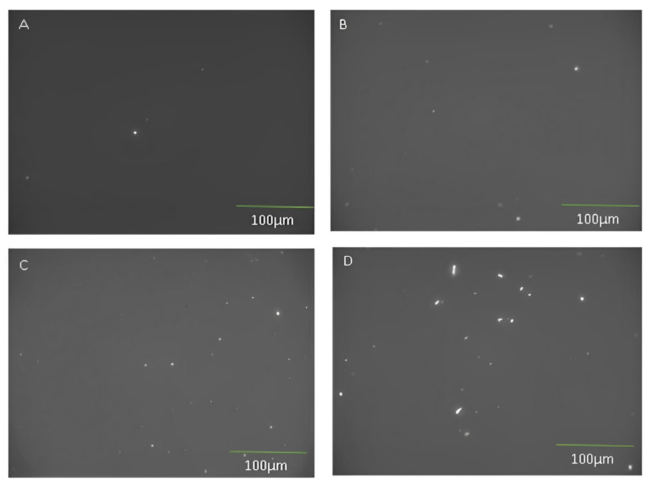
2.3.9. Adhesion to Mucus

Sheep small intestinal mucin was prepared according to the methods described by Vigil et al. [24] and used to assess the adhesion of ABC. Briefly, ABC was labeled by incubation with 250 µL of 4,6-dichlorotriazinyl aminofluorescein (DTAF) in 500 µL of PBS at 60 °C for 2 h and washed three times by resuspension in PBS and collected by centrifugation at 10,000× g 10 min at 4 °C. Ten µL of labeled ABC was mixed with 200 µL of ovine mucin and incubated at 37 °C for 2 h, washed three times with PBS and diluted 1:1000 and 1:100,000, and fixed with paraformaldehyde (PFA 4%) on microscope slides. Adherence was expressed as CFU/mL. Viable bacteria adhered to the mucus were observed under a microscope. ImageJ was used to quantitate viable bacteria. For microscopic analysis, the MosaiX module for the APOTOME system with the 40×/1.30 DIC (UV) VIS-IR M27 Plan-Apochromat oil immersion objective was used to obtain a full mosaic image (1 mm2). Three individual image stacks were collected and assembled using the MosaiX system (Carl Zeiss, Jena, Germany) for each histological slide. ImageJ64 and FIJI were used for the analysis of the 2D images. Quantitation of particles was performed using Yen’s image thresholding method [25].

2.4. In Vitro Disappearance of Dry Matter and VFA Production

To assess the ABC’s capacity to modify volatile fatty acid profiles in vitro and to evaluate its potential as a fiber digestibility enhancer, we conducted in vitro experiments to measure the dry matter disappearance and VFA production. These analysis results provide information about the possible effects of the ABC administration in vivo.
To measure the volatile fatty acid (VFA) production and the in vitro dry matter disappearance (IVDMD) by the ABC in rumen liquid, a pre-inoculum was prepared with the ABC in NM broth and allowed to grow for 8 h under anaerobic conditions at 37 °C. Subsequently, an inoculum was prepared by transferring a volume of 1 mL of the pre-inoculum to a 50 mL tube with 49 mL of NM broth. The inoculum was incubated at the same temperature and anaerobic conditions as the pre-inoculum until an OD 600 nm of 0.5, corresponding to the exponential growth phase of the ABC, was reached. Once the desired OD was reached, the inoculum was centrifuged at 5000 rpm for 15 min to separate a cell pellet from the liquid culture medium. The cell pellet was later added to the Daisy jars, in different doses (4 × 10⁸; 4 × 10¹⁰; 4 × 10¹²) to find the optimal dose; this optimal dose shows the minimum amount of CFU/mL in which ABC improves fiber digestibility at the rumen level. The results of this test were used in the in vivo test.
The rumen fluid was obtained from Brown Swiss bulls (using the same bull for each sampling to minimize individual variation). the extraction procedure consisted of introducing a probe into the rumen through the cannula, filtered with clean gauze and collected in a clean container with an airtight lid, and gassed with CO2. It was kept at 39 °C during transfer to the laboratory, where upon arrival, it was mixed with artificial saliva buffer solution [21].
The IVDMD technique at 24 and 48 h was performed following the protocol recommended by the manufacturer of the Daisy II incubator (ANKOM Technology, Fairport, NY, USA) using FN-57 bags with a pore size of 25 μm.
The substrate for the ruminal microorganisms was the same diet consumed by the Brown Swiss bulls from which ruminal liquid was obtained, and this was previously ground to 2 mm before being added to the bags.
The bags were previously washed with acetone for five min and completely air-dried, then weighed individually, and 0.5 g of ground feed was placed inside each bag. They were heat-sealed and stored at room temperature. In each of the 4 jars in the Daisy II incubator, we placed 10 FN-57 bags. Subsequently, we added artificial saliva buffer to the jars, which was prepared by combining buffer A and buffer B in a 5:1 ratio. Buffer solution A comprised the following per liter: K2HPO4, 10 g; MgSO4-7H2O, 0.5 g; NaCl, 0.5 g; CaCl2.2H2O, 0.1 g; urea, 0.5 g. Buffer solution B contained the following per liter: Na2CO3, 15 g; Na2S-9H2O, 1 g. The resulting mixture was adjusted to achieve a final pH of 6.8 and maintained at a temperature of 39 °C until it was incorporated into 400 mL of rumen fluid [21]. Additionally, 5 mL of the ABC culture (OD 600 nm = 0.5–0.6) was introduced into the mixture.
The nylon bags with the substrate (diet) were recovered. They were washed with running water. After washing, the bags were dried in an oven at 60 °C for 12 h and were weighed. The following formula was used to calculate the percentage of IVDMD:
% Disappearance = 100 − (P3 − (P1 × C)) × 100 / P2
where
P1 = Original weight of the bag;
P2 = Weight of the sample;
P3 = Final weight of the bag (after in vitro fermentations);
C = Correction factor (final weight of the blank bag/original weight of the blank bag).
For the quantification of volatile fatty acids (VFAs; acetate, propionate, and butyrate) at three distinct time points (0, 24, and 48 h), two samples from each jar were taken and placed in separate 15 mL conical tubes, with approximately 10 mL of ruminal fluid per tube for VFA extraction, and each collected sample was read in duplicate in the gas chromatograph.
The sample preparation for gas chromatograph was as follows: 1.5 mL of the ruminal fluid collected was centrifuged at 3500× g for 10 min at 4 °C to sediment the cell pellet, then 1200 μL of supernatant was recovered in a clean Eppendorf tube, and 240 μL of 25% metaphosphoric acid was added to obtain a 5:1 ratio. The tubes were incubated on ice for 30 min to sediment the proteins and then centrifuged at 12,000× g for 15 min at 4 °C. The supernatants were finally filtered with glass fiber membranes and stored in 1:5 dilution at −20 °C until analysis.
An Agilent 7890B gas chromatograph was used. The chromatography conditions were as follows: 170 °C oven, 190 °C injector, 210 °C detector, a column with a flow rate of 2.5 mL/min, and an FID (Flame Ionization Detector).

2.5. Effect of the Addition of Fresh ABC to Limousin Heifers

The in vivo experiment was carried out at the Centro de Enseñanza, Investigación y Extensión en Producción Animal en el Altiplano (CEIEPAA-UNAM), located in Tequisquiapan, Querétaro.
The protocol was approved by CICUAE-UNAM (CICUAE.DC-2019/4.2, UNAM). Fourteen Limousin heifers (n = 7), weighing approximately 200 kg at the beginning of the experiment and with an age of 9 months, were used. These heifers had a BCS of 3–3.5. The animals were housed in individual pens and fed a diet based on forage (alfalfa dry) and concentrate Nucalf 2, NUTEC® (1.5 kg/animal/day).
Two treatments were tested: the ABC (4 × 10⁸ CFU) treatment, which was offered daily to the animals mixed with 50 g of ground corn, and the control treatment with 50 g of ground corn without probiotics. The animals were assigned in pairs to either treatment using a pair-feeding statistical design. Every 28 days (on 2 consecutive days), the animals were weighed, and blood samples were collected to measure blood glucose levels, while ruminal fluid samples were obtained to assess whether ABC alters the microbiota composition and VFA quantification.
For the growth of the ABC, the reagents described in the in vitro experiment were used in the process. Subsequently, an inoculum was prepared by transferring a volume of 1 mL of the pre-inoculum to a 50 mL Erlenmeyer flask with 49 mL of NM broth. The inoculum was incubated at the same temperature and anaerobic conditions as the pre-inoculum until an OD 600 nm = 0.5, corresponding to the exponential growth phase of the microbial consortium, was reached. Once the desired OD was reached, the inoculum was centrifuged at 5000 rpm for 20 min to separate the cell pellet from the liquid culture medium.
Blood was taken from the jugular vein. Following the aseptic procedures for obtaining the sample, it was collected in vacutainer tubes without anticoagulant; the blood sample was centrifuged to obtain the serum and from this, the concentration of glucose per animal was determined [26], measuring the absorbance in a spectrophotometer at 540 nm.
Rumen fluid was obtained using the oral probing technique [27]. Two samples were collected per animal, these were placed in 15 mL conical tubes, in an approximate amount of 10 mL of ruminal fluid per tube. One of the samples was used for DNA extraction and subsequent analysis of the microbial species present in the rumen, and the other for VFA quantification using the same procedure described in Section 2.4.

2.5.1. DNA Extraction and Microbiota rRNA 16s Sequencing

To verify if the ABC modified the microbial population in the rumen samples present in our study, genomic DNA was used to determine the bacterial diversity. DNA extraction was performed according to the RBB+C method [28]. DNA concentration was measured with a Qubit 3.0 fluorometer (Life Technologies, Carlsbad, CA, USA), and DNA integrity was verified by agarose gel electrophoresis (1%). The purified DNA was subject to partial 16S DNA gene sequencing to confirm bacteria identity. Sequencing of the rumen microbiome was performed by Research and Testing Laboratory (RTL) Genomics (Lubbock, TX, USA) with Illumina Myseq sequencer. Illumina produces FASTQ files with a phred offset of +33. FASTQ files contain all the raw sequence data generated by the sequencer; they may contain information regarding the primer for amplicon sequencing. To allow for more run flexibility, RTL genomics used a 2-step PCR process for Illumina sequencing that uses universal adapters and sequences the forward and reverse primers. FASTQ files generated by Illumina sequencer come in 2 forms depending on the method used: paired or single end. In this case, we used pair-end sequence.
Results were expressed as relative abundance (%). Data quality control and analyses were mostly performed using the USEARCH V.11 pipeline. The FASTQ forward and reverse files were merged into a single FASTQ file per sample, quality control and processing included removing adapters and cutting the sequences to length-based filtering of 400 bp (reads smaller than 200 bp were excluded from the analysis). The resulting read files were then aligned to RDP V.16 to define operational taxonomic units (OTUs) for taxonomic assignment; the OTUs table was generated at 97% identity. The Uclust method was used to cluster the reads into OTUs [29].

2.5.2. Relative Quantification of Lactobacillus spp. and Propionibacterium spp.

In order to verify the microbial population in ABC obtained from 16S sequencing, qPCR was performed. DNA extraction was performed according to the RBB+C method [22]. A total of 50 ng of DNA measured in a Nanodrop 1000 spectrophotometer were analyzed in a StepOne Real-Time PCR System (Applied Biosystems, Foster City, CA, USA). The primers used were universal primers: BACT1369 F: CGGTGAATACGTTCYCGG; PROK1492 R: GGWTACCTTGTTACGACTT [23]; Lactobacillus F: AGCAGTAGGGAATCTTCCA; Lactobacillus R: CACCGCTACACATGGAG [24]; Propionibacterium F: ACGGGAGGCAGAGTGGG; and Propionibacterium R: TCTCCTACDMKCYCTTTAC. Conditions of PCR: 40 cycles; 95 °C 10 min, 95 °C 10 s, 56 °C 15 s, 72 °C 15 s. Melting curve: 56 °C 1 min, increasing temperature up to 95 °C. Samples were analyzed in duplicate. Relative abundance of the DNA target was calculated using the Bacchetti de Gregoris et al. [25] method.

2.6. Statistical Analysis

Data were obtained in triplicate (antibiotic susceptibility, bile salts resistance, resistance to pH, temperature tolerance, antagonistic activity, and relative quantification of Lactobacillus spp. and Propionibacterium spp.) or duplicate (in vitro adhesion assay, disappearance of dry matter, and VFA production in vitro) and expressed as standard error of the mean (SEM); differences between samples were analyzed by one-way analysis of variance (ANOVA) and Tukey’s Kramer tests, and the differences with p  <  0.05 were considered significant. Data were analyzed using the SAS version 9.3 software (SAS Institute, Cary, NC, USA).
For the evaluation of the effect of the probiotic on the productive variables, as well as the quantification of blood glucose and VFA production, a pair feeding analysis was performed thus resulting in 7 pairs, and subsequently, a Student’s t-test for paired samples was used for data analysis.
t = X D μ 0 S D   / n
For this equation, the difference D between all the pairs has to be calculated:
XD is the mean of the differences;
SD is the standard deviation of the differences;
µ0 is the constant different from 0;
n is the sample size.
Taxonomic assignment was performed using the RDP V.16 reference database with 97% identity. Data were analyzed in the R platform (3.6) with the Phyloseq library.

3. Results

3.1. Growth Rate

According to the ABC growth curve and the analysis of the equation according to [30], the ABC generation time (GT) was determined to be 8.8 h, calculated based on the equation GT = ln2/µ [22]. The growth curve is shown in Figure 1.

3.2. Probiotic Tests

3.2.1. Susceptibility to Antibiotics

The results of the antibiograms are presented in Table 2. The ABC recorded susceptibility to the antibiotic’s sulfamethoxazole + trimethoprim, vancomycin, ampicillin (commonly used for digestive disorders), and cephalothin; however, the ABC recorded resistance against gentamicin and tetracycline.

3.2.2. Tetracycline Resistance Genes Tet R Tet M and Tet W

Genomic and plasmid DNA extraction of ABC was performed for PCR and determined where tetracycline resistance was present.
Tet R and Tet W genes were not found in ABC, and in the case of the Tet M gene, it was found in the genomic DNA.

3.2.3. Resistance to pH

Table 3 shows the number of CFUs from the resistance test of the ABC at different pH for 24 and 48 h. For 24 and 48 h at pH 6.5 and 7, there is a higher CFU growth, in contrast to acidic pH 2 and 3, which did not record growth at 48 h. No differences were found in the number of CFUs at pH 5.5 at 48 h and pH 6 at 24 h (2.6 × 10¹⁰ and 1.9 × 10¹⁰, respectively), and similarly, there was no difference in CFUs at pH 5.5 and 6 incubated for 48 h compared to CFUs at pH 7 incubated for 24 h (2.6 × 10¹⁰, 3.0 × 10¹⁰ and 3.5 × 10¹⁰, respectively). The highest number of CFUs is observed when the ABC is incubated at pH 6.5 and 7 for 24 and 48 h (3.7 × 10¹⁰, 4.1 × 10¹⁰, 3.5 × 10¹⁰, and 4.0 × 10¹⁰, respectively).

3.2.4. Gastric Juice Resistance Test

The pH value in the abomasum varies from 2 to 4 depending on the diet. Therefore, ABC was challenged at pH 2, 3, and 4 for 2 h which is the average time the digesta remains in the abomasum. Table 4 shows the results of the CFU after the ABC was incubated at different pH. At pH 4, the highest number of CFUs was observed (5.0 × 10⁵) compared to pH 2 and 3 (3.6 × 10⁴ and 9.0 × 10⁴, respectively). Table 5 shows the survival results of the ABC. The ABC has a survival of 100% at 60 min of incubation and 76.7% at 150 min of incubation, meaning that ABC is able to withstand the acidity and the presence of pepsin.

3.2.5. Resistance to Bile Salts

Table 6 shows the results of the challenge to bile salts. In the ABC, no differences were found between the control bacteria (grown in NM) and the treatment (NM enriched with bile salts). The ABC was found to be resistant to bile salts.

3.2.6. Tolerance to Temperature

Figure 2 shows the results of tolerance to the different temperatures at 24 and 28 h. At 24 h, a higher DO was observed compared to at 48 h. Growth was better at 30 °C, then 37 °C, and finally, at 45 °C (p < 0.05).

3.2.7. Coaggregation Capacity

The ABC demonstrated a coaggregation capacity of 10.74% against E. coli during the 60 min test.

3.2.8. Antagonistic Activity

ABC showed positive inhibition against E. coli because they had a zone of inhibition of 10.5 ± 0.5 mm. No inhibition was observed for S. typhimurium (8 ± 1.5 mm).

3.2.9. Adhesion to Mucus

Figure 3 shows the results of the adhesion to mucins, was much higher for the ABC compared to P. acidipropionici counts. In contrast, no viable bacteria were found in the negative control. These results suggest the potential for PCB adherence and colonization of the intestinal tract.

3.3. Disappearance of Dry Matter and VFA Production In Vitro

Table 7 shows the results of IVDMD. As can be seen at 24 h, there was no difference between treatments, but at 48 h, there was a better DMD in the treatments in which the ABC was added (51.73%, 51.43%, and 51.97% for treatments 4 × 10⁸, 4 × 10¹⁰ and 4 × 10¹², respectively); however, no difference was found between them.
The dose of 4 × 10⁸ was selected for further experiments.
Table 8 shows the effect of ABC addition on VFA production in vitro. Increases of 72.7, 74.8, and 48.3% were observed with the addition of the ABC for acetate, propionate, and butyrate, respectively. For total VFAs, the increase was 72.7% (p < 0.05).

3.4. Effect of the Addition of Fresh ABC to Limousin Heifers

The animals were weighed on day 1 and then every 28 days for two consecutive days, to record their growth and development. When analyzing the data, a difference (p ≤ 0.001) between treatments can be observed (Table 9), showing an improvement in the weight gain of up to 10 kg more in the animals that were supplemented with the ABC.
This was also demonstrated in the daily weight gain (DWG) (Table 9) of the animals where the difference between treatments (p < 0.001) was greater for the animals that received the ABC, with a DWG of 200 g more than the control group, which may demonstrate that the ABC has a beneficial effect on the animals’ DWG.
Table 9 shows the effect of the ABC on the dry matter intake (DMI), daily weight gain (DWG), and feed conversion (FC) of Limousin heifers (p < 0.001).

3.4.1. Serum Glucose Determination

Glucose quantification was performed to determine the effect that increased VFA could have on circulating blood glucose. When analyzing the data by paired means, this was significant (p = 0.05) for the ABC group. Statistical analysis of each sample showed that the ABC group at day 85 had a significant increase in glucose, 90.52 (SD = 2.6) vs. 81.69 (SEM = 3.02) for the ABC and control groups, respectively (Table 10).

3.4.2. ABC Effects on VFA Production

Table 11 shows the effect of probiotic addition on the molar concentrations of VFA in the rumen fluid. The concentration of acetate produced an increase of 14.5%, and there was an increase of 19% for propionate (p < 0.05). Butyrate was not different (p > 0.05). The total VFA difference was 15.4% (p < 0.05).

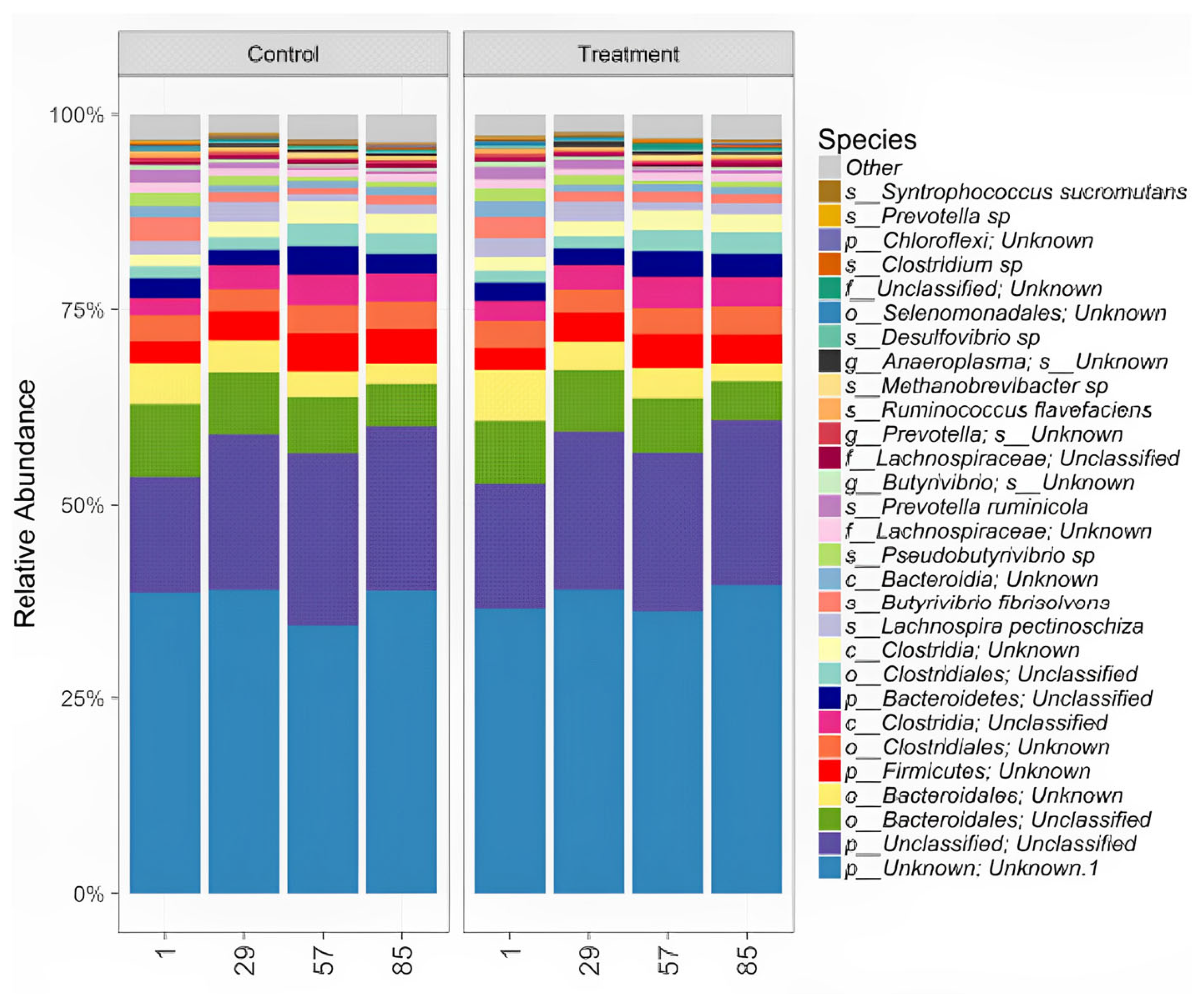
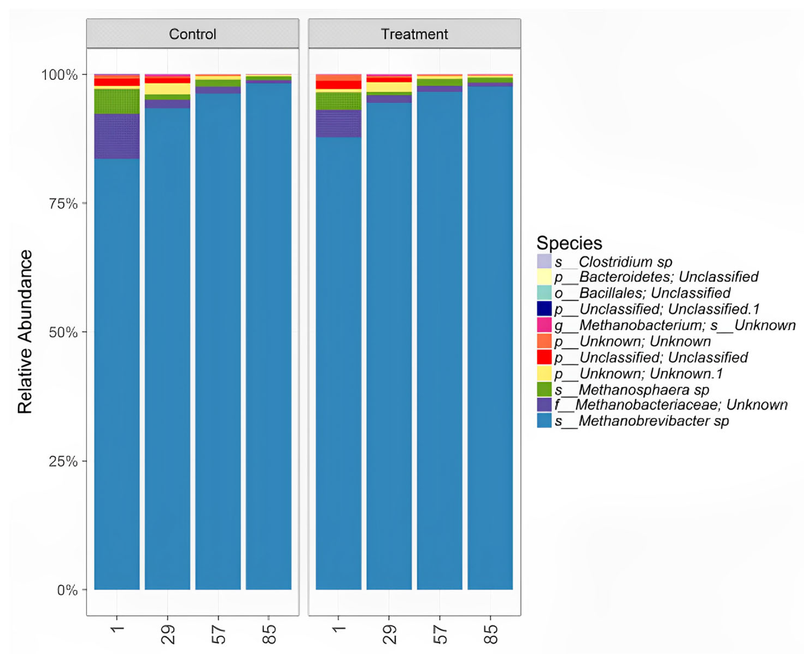
3.4.3. ABC Effects on Ruminal Microbiota

The possible changes in bacterial and archaeal populations in the rumen liquid after ABC administration are shown in Figure 4 and Figure 5. No different changes in the bacterial population at the species level were found between the ABC treatment and the control group (Figure 4). However, upon analysis of Archaea species, a decrease in methane-producing species was found (Figure 5, p < 0.05).

3.4.4. Relative Quantification of Lactobacillus spp. and Propionibacterium spp.

The relative abundance of Lactobacillus spp. in the ABC was 0.091%, whereas the relative abundance of Propionibacterium spp. was 0.013%.

4. Discussion

4.1. Probiotic Tests

The use of probiotics is subject to various laws to ensure their safety for both human and animal consumption. In the USA, microorganisms used for consumer purposes must have Generally Regarded as Safe (GRAS) status, regulated by the Food and Drug Administration (FDA). In Europe, the European Food Safety Authority (EFSA) introduced the term Qualified Presumption of Safety (QPS). The EFSA considers that bacterial strains carrying antibiotic resistance related to acquired genetic determinants have a high potential for horizontal spread and should not be incorporated into food. Otherwise, it considers that bacterial strains possessing antibiotic resistance by intrinsic or chromosomal mutation have a minimal or low potential for horizontal resistance spread and can generally be incorporated into food [31,32].
Resistance to gentamicin, erythromycin, and tetracycline was reported within the ABC; the bacterial genera present were Lactobacillus spp., Enterococcus spp. and Bacillus spp. Albuquerque et al. [33] and Argyri et al. [34], who worked with different strains of Lactobacillus spp., found intrinsic resistance to gentamicin, erythromycin, tetracycline, and kanamycin. Baccouri et al. [35], working with Enterococcus faecalis OB14, reported resistance to gentamicin and erythromycin. Enterococcus spp. resistance to gentamicin has been described as intrinsic, non-transmissible, and widely distributed [36,37]. This erythromycin resistance in Lactobacillus spp. has been associated with a single mutation in the 23S rRNA gene [38]. An important point and one to be taken with caution is the observed tetracycline resistance; some studies have described the tetracycline resistance genes tet (M), tet (L), tet (K), and tet (S) as chromosomal in lactic acid bacteria [38,39], while others have shown tet (M) as localized on plasmids [40], which can commonly move between bacteria. For the case of this ABC, the only gene of resistance to tetracycline was the Tet M, which is in the genomic DNA and does not represent a risk factor to transmit this resistance according to what has been established by the EFSA; when resistance is found in the chromosomal part of the consortium, there is a minimal factor for the spread of horizontal resistance, and it can be incorporated into food [31,32].
Probiotics exert their beneficial effects on the host by modifying the composition and activity of the intestinal microbiota [41]. To achieve this, the bacterial strains used as probiotics must survive challenges in the host’s digestive tract (DT) [42] including variations in pH throughout the ruminant DT compartments (reticulum–rumen, omasum, abomasum, small intestine, and colon), as well as resistance to bile in the intestine [43]. Our ABC exhibits robust survivability, particularly at low pH levels, with optimal survival at pH 6 and above, and it also demonstrates resistance to bile salts.
The pH is probably the most important factor affecting the microbial population and its activities. The rumen fermentation products, particularly acetate, propionate, lactate, and CH4, are strongly affected by ruminal pH, mediated largely by the effect of pH on microbes [44]. The sensitivity of ruminal bacteria to pH is dictated by the pH gradient across their cell membranes and their ability to regulate intracellular pH. These physiolo gical pH ranges in the rumen and other parts of the DT are influenced by dietary factors. For instance, Kern et al. [45] measured the pH of the digestive tract of cattle fed with hay at a daily rate of 2% of body weight for 30 d. The DT pH values were 6.9, 3.1, 7.3, 7.0, and 7.2 for the rumen, abomasum, ileum, cecum, and terminal colon, respectively. Wheeler et al. [46] collected pH data from cattle fed with concentrates at 48.5 and 59.6% of the ration, reporting pH of 5.38 to 6.58, 2.27 to 4.16, 5.75 to 6.78, and 5.0 to 6.8 for the rumen, abomasum, ileum, and colon, respectively.
Ruminal pH greatly influences microbial activity, with the magnitude and duration of pH levels being key factors. The ABC thrived optimally at pH 6.5 and 7, both at 24 and 48 h, as shown by CFU counts (Table 3), similar to those reported by Maldonado et al. [47] who worked with different strains of Lactobacillus spp. obtained from the rumen of Bradford and Brangus cattle and found that Lactobacillus spp. tolerated acid pH (3.0) differently by the tested strains. Only Lactobacillus fermentum were able to grow at pH 3.0, which agrees with our results obtained for pH 2.0, 3.0, and 4.0 even though at 24 h there were still CFUs. The intolerance of Enterococcus spp. and Weissella spp. to highly acidic conditions is consistent with their inability to adapt to acid stress, as previously reported [48]. However, physiologically, ruminal pH does not reach values as low as 2.0, 3.0, 4.0, and 5.0 (without presenting any lethal pathology).
The ABC used in this work exhibited significant survival across a simulated gastrointestinal tract. Since probiotics should survive in the stomach (in this case the abomasum) where gastric juice is present, tolerance to acid pH is an important selection criterion for probiotics, with the pH of the abomasum being in the range of 2 to 4 depending on the type of diet [45,46] and a residence time of about 2 h.
Normally, this low pH causes strong decreases in bacterial counts as reported by Albuquerque et al. [33], who worked with several strains of Lactobacillus spp. Only one strain in Lactobacillus fermentum 296 was able to survive after 2 and 3 h at pH of 2.0, which agrees with our results, even though at pH of 4.0, there was a significantly higher amount of CFUs. Although there is survival of the ABC at this pH for 2 h, in vivo in the abomasum, probiotics are not necessarily challenged at pH values as low as 2.0 and 3.0 because the abomasal environment can be buffered by food components.
Bile comprises a complex mixture of components, encompassing proteins, ions, pigments, cholesterol, and diverse bile salts, which have demonstrated protective properties against pathogenic bacteria. The precise mechanisms underlying how bile triggers cell death remain poorly understood, and it remains uncertain whether cell death arises from membrane damage or from damage at the DNA-level, or both [49].
The ABC is resistant to bile salts at 0.3% and our result is like that reported by Sánchez et al. [50], who observed resistance to bile salts in four strains of Lactobacillus spp. isolated from calves. A factor that may favor the survival of these microorganisms in vivo in the duodenum is the presence of feed, since the bacteria may not be exposed to bile salts, and thus toxicity on the membranes could be avoided.
One of the relevant properties of some probiotics is the ability to coaggregate with certain pathogens and, consequently, prevent their access to the mucous membranes. We demonstrated that the bacterial consortium presents a 10.74% coaggregation capacity against Escherichia coli. This result is similar to those mentioned by Albuquerque et al. [33], who studied nine strains of Lactobacillus spp. as potential probiotics; they reported that the coaggregation capacity of their strains ranged between 2.4 and 41.3%. It is postulated that the coaggregation capacity depends on the bacterial species involved, being specific among strains and affected by the environmental conditions in which this interaction takes place. The pH and salt concentration of the medium modify this characteristic, probably because of the influence on the surface charges of the bacteria; thus, low pH and high electrolyte concentration favor it [51].
Another important characteristic of a probiotic is its ability to promote bacterial colonization of the intestinal mucosa [52]. In general, we observed good adhesion of ABC to mucus; in Video S1, we could observe the CFUs of the acidogenic bacterial consortium (ABC) adhered to mucins. Cueto-Vigil et al. [24] showed LAB had high adhesion to mucus, while López and Espinoza [53] found that Lactobacillus plantarum can adhere to and colonize intestinal cells in vitro. The ability of probiotics to reduce pathogenic microorganisms in the digestive tract may be due to the production of bacteriocin and exclusion due to competition by adherence to the intestinal epithelium. Indeed, some strains of Lactobacillus spp. and Bifidobacterium spp. have hydrophobic surface proteins that promote non-specific adhesion to animal cells, cover receptor binding sites, and prevent pathogenic microorganisms from binding to intestinal epithelium [53,54].
Among the benefits of various probiotics is the ability to improve the digestibility of feedstuffs and it is thought that, consequently, animal performance is improved. As can be seen in Table 7, dry matter disappearance was not affected by the treatments at 24 h. This was also reported by Ghorbani et al. [55] who worked with two probiotic bacterial strains and their combination (Propionibacterium P15 1 × 10⁹ CFU/g and Propionibacterium P15 plus Enterococcus faecium 1 × 10⁹ CFU/g) with a dosage of 10 g/animal/d in cannulated steers; they found no differences in DM digestion (in situ) on any of the substrates used. These findings have also been reported with other probiotic bacterial strains by authors such as Cagle et al. [56], with Saccharomyces cerevisae. Newbold et al. [57], also with Saccharomyces cerevisae, did not observe that degradation was significantly affected by treatment. Souza et al. [16], working with Bacillus subtilis spores, and Qiao et al. [58], also with a Bacillus subtilis, found no differences in dry matter digestibility.
Concerning VFA production, the ABC increases it by almost 15–19% in acetate and propionate, respectively. The use of probiotics in ruminants is recommended to improve intestinal health and productive variables in young animals. Different products have been used to manipulate ruminal fermentation [59]; our ABC has been shown to increase VFA production both in vitro and in vivo (Table 8 and Table 11), which makes it a product with great potential for use in both dairy and beef cattle.
It is necessary then to clarify that the disappearance of dry matter does not have to be paired with VFA production, and we could speculate that the increase in VFA is due to a greater efficiency in the utilization of energy substrates available to ruminal bacteria, which is a possible mechanism of action of the ABC. It could also be due to less wastage of substrates or less availability of nitrogen, since a forage was used as a substrate for the bacteria and since this was limited, the bacteria had more carbohydrates available for conversion to VFAs.

4.2. Effect of the Addition of Fresh ABC to Limousin Heifers

Comparing the results obtained in this work with the use of the probiotic where the feeding of the animals was based on forages, we can see the efficacy of its use on weight gain, where there was an increase in DWG (1 kg daily), going from an average of 283 kg to 371 kg of weight in 84 days. If this growth trend is maintained, 400 kg calves could be obtained in 112 days.
Krehbiel et al. [41] reported that probiotic supplementation to fattening cattle can produce an increase of approximately 2.5 to 5% in DWG and 2% in feed efficiency. Arantzamendi et al. [60] described the use of Bacillus toyoi (Toyocerin®), in three different farm units and locations, with beef cattle, showing improvements for DWG of 10% and 6.2% for the final gain. Dick et al. [61] and Vyas et al. [62], where Propionibacterium spp. were tested as probiotics in steers, observed no change in DWG between the control and treated groups, nor was a reduction in DMI observed.
Triphati [63] tested three different yeast strains Kluyveromyces marximanus, Saccharomyces cerevisiae, Saccharomyces uvarum, and a combination of the three in a 1:1:1 ratio. A difference was observed in the weight gain of the animals supplemented with Saccharomyces cerevisiae and with the yeast mixture, where weight gains were 21% and 16%, respectively.
Alvarez [64] used a mixture of Lactobacillus acidophilus, Bacillus thermophilum, Bacillus subtilis, and Saccharomyces cerevisiae, and found no effect on DWG. These data agree with those reported by Krehbiel et al. [41], who concluded that the response in dry matter intake in animals consuming probiotics has been inconsistent and is dependent on factors such as climate, sanitary condition of the farm, and the health of the animals.
When observing the molar concentrations (Table 11) for the total VFA produced, it is evident that with the addition of the ABC, there is an increase in the concentration of VFAs. The concentration of acetate produced had an increase of 15%, and 19% for propionate. Butyrate was not different. The capacity shown by the ABC to modify the rumen environment causing an increase in VFAs (Table 8 and Table 11) was notorious even with the use of forages, since diets with high forage content usually produce a lower amount of these; however, the acidogenic potential of the ABC could not be determined. The addition of concentrate or grain to the substrate may cause the expected impact on the propionic fraction, increasing the molar ratio of propionate at the expense of acetate causing a better acetate: propionate ratio [65].
Most of the work completed to study the effect of microbial additives on productive performance in ruminants has been carried out with growing and fattening animals or in production (dairy) [66].
Most studies report an increase in propionate and a decrease in the acetate: propionate ratio with supplementation of Propionibacterium acidipropionici [67,68,69] or propionate [70,71,72] in a variety of ruminant diets, suggesting that it is a useful tool to increase the glycogenic potential of the diet. Other researchers have reported no differences in total VFAs, but acetate was either decreased [67,69] or not altered [68] by supplementation with Propionibacterium acidipropionici. In the present study, an increase in total VFAs was observed for animals in the treated group, but there was no change in the acetate: propionate ratio.
Several studies have reported a successful establishment of products from one of the most important lactate-utilizing species in the rumen, Megasphaera elsdenii in sheep and cattle, where an increase in ruminal fermentation was seen to result in increased production of VFAs and helped to regulate ruminal pH preventing acidosis, although its mechanism of action is not fully elucidated [73,74].
These data on bacterial probiotics are consistent with the performance shown by the ABC, where it exerts an effect on ruminal fermentation, increasing weight gain and increasing VFAs of the animals that were supplemented with the consortium.

5. Conclusions

The evaluated ABC exhibits significant potential for use as a Direct-Fed Microbial (DFM) or probiotic due to several key attributes. Firstly, it demonstrates innocuousness, lacking antibiotic resistance and antibiotic resistance plasmids, it displays remarkable survivability under diverse digestive tract conditions encountered in ruminants. This includes tolerance to varying pH levels in the reticulum and rumen (ranging from 5.5 to 7.0), as well as robust survival in the abomasum, even in acidic environments. Furthermore, it exhibits resilience, with a 76.7% survival rate in the presence of HCl, pepsin, and pH 2.0, along with resistance to bile salts. Additionally, the ABC displays the ability to coaggregate with Escherichia coli (10.74%) and enhances in vitro dry matter digestion (IVDMD) at 48 h (51.73%).
The ABC contributes to improved total volatile fatty acid (VFA) production both in vitro and in vivo, resulting in enhanced daily weight gain (DWG) and feed conversion in Limousin beef cattle. These attributes collectively position it as a promising candidate for probiotic use, especially in animals consuming high-forage diets.

Supplementary Materials

The following supporting information can be downloaded at: https://www.mdpi.com/article/10.3390/ruminants3040028/s1, Video S1: Colony-forming units (CFUs) of the acidogenic bacterial consortium (ABC) adhered to mucins. Staining with (5-(4,6-Dichlorotriazinyl) Aminofluorescein) (DTAF identified by color green, Thermo Fisher Scientific, USA) for the bacterial membrane and HOECHST-33342 (Blue, Thermo Fisher Scientific, USA) for DNA. The microscopy was performed with CONFOCAL-780LSM (Zeiss) 63X. The visualization was performed with AMIRA software (Thermo Fisher Scientific, USA) in the CAVE VR System (automatic virtual environment, University of Illinois, Chicago, USA).

Author Contributions

Conceptualization, O.M. and A.S.; methodology, D.C.-C., C.R.-R. and A.P.-T.; software, C.R.-R.; validation, C.R.-R. and L.G.-D.; formal analysis, C.R.-R. and D.C.-C.; investigation, O.M., C.R.-R. and L.G.-D.; resources, A.S.; data curation, C.R.-R. and O.M.; writing—original draft preparation, C.R.-R., O.M. and L.G.-D.; writing—review and editing, O.M., L.G.-D. and A.S.; visualization, C.L.-F.; supervision, A.S.; project administration, L.G.-D.; funding acquisition, A.S. and O.M. All authors have read and agreed to the published version of the manuscript.

Funding

This research was funded by PAPIIT-UNAM, grant numbers IT200621, IG200818 to A.S., and IN213021 to O.M., Cátedras de Investigación FESC-UNAM CI2229 to A.S. and CONACYT 211559 to O.M.

Institutional Review Board Statement

The animal study protocol was approved by Comité Institucional para el Cuidado y Uso de Animales de Estudio CICUAE-UNAM (CICUAE.DC-2019/4.2, UNAM).

Informed Consent Statement

Not applicable.

Data Availability Statement

The data supporting the findings of this study are available upon request. Please contact the authors at ofemora66@unam.mx for access to the data.

Acknowledgments

We thank Nydia Hernández-Ríos from Microscopy Unit (INB-UNAM), Alejandro de León and Luis Aguilar (LAVIS-UNAM), Adolfo Kunio Yabuta-Osorio from CEIEPAA (FMVZ-UNAM), Jaime Pérez-Trevilla (LIPATA-UNAM), and Sarahí Rodríguez-González (FM-UAQ) for their technical support. Carolina Robles-Rodríguez and Diego Cardoso-Carmona thank CONACYT for the scholarship at PCiPSA-UNAM.

Conflicts of Interest

The authors declare no conflict of interest.

References

  1. Arowolo, M.A.; He, J. Use of probiotics and botanical extracts to improve ruminant production in the tropics: A review. Anim. Nutr. 2018, 4, 241–249. [Google Scholar] [CrossRef] [PubMed]
  2. Krause, D.O.; Denman, S.E.; Mackie, R.I.; Morrison, M.; Rae, A.L.; Attwood, G.T.; McSweeney, C.S. Opportunities to improve fiber degradation in the rumen: Microbiology, ecology, and genomics. FEMS Microbiol. Rev. 2003, 27, 663–693. [Google Scholar] [CrossRef]
  3. García-Sorrondegui, M. Empleo de Probióticos en los Animales. 2012. Available online: https://www.engormix.com/ganaderia/levaduras-ganado-engorde/empleo-probioticos-animales_a29474/ (accessed on 14 June 2023).
  4. F.A.O./W.H.O. Joint FAO/WHO (Food and Agriculture Organization/World Health Organization) Working Group Report on Drafting Guidelines for the Evaluation of Probiotics in Food; FAO: Rome, France; WHO: Geneva, Switzerland, 2002. [Google Scholar]
  5. Garcia, M.; Lopez, Y.; Carcasses, A. Empleo de Probióticos en Animales. Sitio Argentino de Producción Animal. 2012. Available online: http://www.produccion-animal.com.ar/ (accessed on 14 June 2023).
  6. F.A.O. Biotecnología Agrícola. Available online: http://www.fao.org/3/Y5160s/y5160s07.htm (accessed on 14 June 2023).
  7. Guarner, F.; Perdigon, G.; Corthier, G.; Salminen, S.; Koletzko, B.; Morelli, L. Should yoghurt cultures be considered probiotic? Br. J. Nutr. 2005, 93, 783–786. [Google Scholar] [CrossRef] [PubMed]
  8. Pineiro, M.; Stanton, C. Probiotic Bacteria: Legislative Framework—Requirements to Evidence Basis. J. Nutr. 2007, 137, 850S–853S. [Google Scholar] [CrossRef]
  9. Uyeno, Y.; Shigemori, S.; Shimosato, T. Effect of Probiotics/Prebiotics on Cattle Health and Productivity. Microbes Environ. 2015, 30, 126–132. [Google Scholar] [CrossRef] [PubMed]
  10. Reuben, R.C.; Elghandour, M.M.M.Y.; Alqaisi, O.; Cone, J.W.; Márquez, O.; Salem, A.Z.M. Influence of microbial probiotics on ruminant health and nutrition: Sources, mode of action and implications. J. Sci. Food Agric. 2022, 102, 1319–1340. [Google Scholar] [CrossRef]
  11. Ochoa Carreño, C.; Montoya Restrepo, D.A. Microbial Consortia: A Biological Metaphor Applied to Enterprise Association in Agricultural Production Chains. Rev. Fac. Cienc. Econ. 2010, 18, 55–74. [Google Scholar] [CrossRef]
  12. Timmerman, H.M.; Koning, C.J.M.; Mulder, L.; Rombouts, F.M.; Beynen, A.C. Monostrain, multistrain and multispecies probiotics. A comparison of functionality and efficacy. Int. J. Food Microbiol. 2004, 96, 219–233. [Google Scholar] [CrossRef]
  13. Li, Z.; Lei, X.; Chen, X.; Yin, Q.; Shen, J.; Yao, J. Long-term and combined effects of N-[2-(nitrooxy)ethyl]-3-pyridinecarboxamide and fumaric acid on methane production, rumen fermentation, and lactation performance in dairy goats. J. Anim. Sci. Biotechnol. 2021, 12, 125. [Google Scholar] [CrossRef]
  14. Ben Shabat, S.K.; Sasson, G.; Doron-Faigenboim, A.; Durman, T.; Yaacoby, S.; Miller, M.E.B.; White, B.A.; Shterzer, N.; Mizrahi, I. Specific microbiome-dependent mechanisms underlie the energy harvest efficiency of ruminants. ISME J. 2016, 10, 2958–2972. [Google Scholar] [CrossRef]
  15. Timmerman, H.; Mulder, L.; Everts, H.; van Espen, D.; van der Wal, E.; Klaassen, G.; Rouwers, S.; Hartemink, R.; Rombouts, F.; Beynen, A. Health and Growth of Veal Calves Fed Milk Replacers With or Without Probiotics. J. Dairy Sci. 2005, 88, 2154–2165. [Google Scholar] [CrossRef]
  16. Souza, V.; Lopes, N.; Zacaroni, O.; Silveira, V.; Pereira, R.; Freitas, J.; Almeida, R.; Salvati, G.; Pereira, M. Lactation performance and diet digestibility of dairy cows in response to the supplementation of Bacillus subtilis spores. Livest. Sci. 2017, 200, 35–39. [Google Scholar] [CrossRef]
  17. Chokeshaiusaha, K.; Sananmuang, T.; Puthier, D.; Nguyen, C. An innovative approach to predict immune-associated genes mutually targeted by cow and human milk microRNAs expression profiles. Vet. World 2018, 11, 1203–1209. [Google Scholar] [CrossRef] [PubMed]
  18. Gaggìa, F.; Mattarelli, P.; Biavati, B. Probiotics and prebiotics in animal feeding for safe food production. Int. J. Food Microbiol. 2010, 141, S15–S28. [Google Scholar] [CrossRef]
  19. Fuller, R. A review: Probiotics in man and animals. J. Appl. Bacteriol. 1989, 66, 365–378. [Google Scholar]
  20. Robles, C. Adición de un Consorcio Bacteriano Altamente Propiogénico al Líquido Ruminal Ovino y su Efecto Sobre la Relación Molar Acetato: Propionato. Bachelor’s Thesis, Universidad Veracruzana, Veracruz, Mexico, 2017. [Google Scholar]
  21. Aguilar, M. Desarrollo de una Celda Electroquímica Microbiana con Bacterias Propionogénicas Para Incrementar la Concentración de Propionato y Disminuir la Concentración de Metano en Líquido Ruminal in Vitro. Master’s Thesis, Universidad Autónoma de México, Mexico City, Mexico, 2016. [Google Scholar]
  22. Ratkowsky, D.A.; Olley, J.; McMeekin, T.A.; Ball, A. Relationship between temperature and growth rate of bacterial cultures. J. Bacteriol. 1982, 149, 1–5. [Google Scholar] [CrossRef] [PubMed]
  23. Bhunia, A.K.; Johnson, M.C.; Ray, B. Purification, characterization and antimicrobial spectrum of a bacteriocin produced by Pediococcus acidilactici. J. Appl. Bacteriol. 1988, 65, 261–268. [Google Scholar] [CrossRef] [PubMed]
  24. Cueto-Vigil, M.; Acuña-Monsalve, Y.; Valenzuela-Riaño, J. In Vitro Evaluation of Probiotic Potential of Lactic Bacteria Acid Isolated From Coastal Serum. Actual Biológicas 2010, 32, 129–138. [Google Scholar] [CrossRef]
  25. Yen, J.-C.; Chang, F.-J.; Chang, S. A new criterion for automatic multilevel thresholding. IEEE Trans. Image Process. 1995, 4, 370–378. [Google Scholar] [CrossRef]
  26. Robles, C. Aumento en la Síntesis de Propionato Ruminal Mediante el Diseño de Productos Biotecnológicos. Master’s Thesis, Universidad Autónoma de México, Mexico City, Mexico.
  27. Chai, Z.; Song, L.; Liu, J.; Wu, Y. Insertion depth of oral stomach tubes may affect the fermentation parameters of ruminal fluid collected in dairy cows. J. Dairy Sci. 2012, 95, 5978–5984. [Google Scholar] [CrossRef]
  28. Yu, Z.; Morrison, M. Improved extraction of PCR-quality community DNA from digesta and fecal samples. Biotechniques 2004, 36, 808–812. [Google Scholar] [CrossRef] [PubMed]
  29. Edgar, R.C. Search and clustering orders of magnitude faster than BLAST. Bioinformatics 2010, 26, 2460–2461. [Google Scholar] [CrossRef]
  30. Ratkowsky, D.A.; Ross, T. Modelling the bacterial growth/no growth interface. Appl. Env. Microbiol. 1998, 20, 29–33. [Google Scholar] [CrossRef]
  31. E.F.S.A European Food Safety Authority. Introduction of a Qualified Presumption of Safety (QPS) Approach for Assessment of Selected Microorganisms Referred to EFSA Opinion of the Scientific Committee. 2007, pp. 1–16. Available online: https://www.efsa.europa.eu/en/efsajournal/pub/587 (accessed on 8 October 2020).
  32. EFSA Panel on Additives and Products or Substances Used in Animal Feed (FEEDAP). EFSA J. 2018. Available online: https://www.efsa.europa.eu/en/science/scientific-committee-and-panels/feedap (accessed on 8 October 2020).
  33. Albuquerque, T.M.R.; Garcia, E.F.; Araújo, A.d.O.; Magnani, M.; Saarela, M.; de Souza, E.L. In Vitro Characterization of Lactobacillus Strains Isolated from Fruit Processing By-Products as Potential Probiotics. Probiotics Antimicrob. Proteins 2018, 10, 704–716. [Google Scholar] [CrossRef] [PubMed]
  34. Argyri, A.A.; Zoumpopoulou, G.; Karatzas, K.A.G.; Tsakalidou, E.; Nychas, G.-J.E.; Panagou, E.Z.; Tassou, C.C. Selection of potential probiotic lactic acid bacteria from fermented olives by in vitro tests. Int. J. Food Microbiol. 2013, 33, 282–291. [Google Scholar] [CrossRef]
  35. Baccouri, O.; Boukerb, A.M.; Farhat, L.B.; Zébré, A.; Zimmermann, K.; Domann, E.; Cambronel, M.; Barreau, M.; Maillot, O.; Rincé, I.; et al. Probiotic Potential and Safety Evaluation of Enterococcus faecalis OB14 and OB15, Isolated From Traditional Tunisian Testouri Cheese and Rigouta, Using Physiological and Genomic Analysis. Front. Microbiol. 2019, 10, 881. [Google Scholar] [CrossRef]
  36. Kristich, C.J.; Rice, L.B.; Arias, C.A. Enterococcal infection treatment and antibiotic resistance. In Enterococci: From Commensals to Leading Causes of Drug Resistant Infection; Gilmore, M.S., Clewell, D.B., Ike, Y., Shankar, N., Eds.; Massachusetts Eye and Ear Infirmary: Boston, MA, USA, 2014. [Google Scholar]
  37. Barbosa, J.; Ferreira, V.; Teixeira, P. Antibiotic susceptibility of enterococci isolated from traditional fermented meat products. Int. J. Food Microbiol. 2009, 26, 527–532. [Google Scholar] [CrossRef] [PubMed]
  38. Gueimonde, M.; Sánchez, B.; de Los Reyes-Gavilán, C.G.; Margolles, A. Antibiotic resistance in probiotic bacteria. Front. Microbiol. 2013, 4, 202. [Google Scholar] [CrossRef] [PubMed]
  39. Shao, Y.; Zhang, W.; Guo, H.; Pan, L.; Zhang, H.; Sun, T. Comparative studies on antibiotic resistance in Lactobacillus casei and Lactobacillus plantarum. Food Control. 2015, 50, 250–258. [Google Scholar] [CrossRef]
  40. Danielsen, M. Characterization of the tetracycline resistance plasmid pMD5057 from Lactobacillus plantarum 5057 reveals a composite structure. Plasmid 2002, 48, 98–103. [Google Scholar] [CrossRef]
  41. Krehbiel, C.R.; Rust, S.R.; Zhang, G.; Gilliland, S.E. Bacterial direct-fed microbials in ruminant diets: Performance response and mode of action. J. Anim. Sci. 2003, 81, 120–132. [Google Scholar] [CrossRef]
  42. Bunešová, V.; Vlková, E.; Geigerová, M.; Rada, V. Effect of rearing systems and diets composition on the survival of probiotic bifidobacteria in the digestive tract of calves. Livest. Sci. 2015, 178, 317–321. [Google Scholar] [CrossRef]
  43. Gueimonde, M.; Salminen, S. New methods for selecting and evaluating probiotics. Dig. Liver Dis. 2006, 38, S242–S247. [Google Scholar] [CrossRef]
  44. Lana, R.P.; Russell, J.B.; Van Amburgh, M.E. The role of pH in regulating ruminal methane and ammonia production. J. Anim. Sci. 1998, 76, 2190–2196. [Google Scholar] [CrossRef]
  45. Kern, D.L.; Slyter, L.L.; Leffel, E.C.; Weaver, J.M.; Oltjen, R.R. Ponies vs. Steers: Microbial and Chemical Characteristics of Intestinal Ingesta. J. Anim. Sci. 1974, 38, 559–564. [Google Scholar] [CrossRef]
  46. Wheeler, W.E.; Noller, C.H. Gastrointestinal Tract pH and Starch in Feces of Ruminants. J. Anim. Sci. 1977, 44, 131–135. [Google Scholar] [CrossRef] [PubMed]
  47. Maldonado, N.C.; Ficoseco, C.A.; Mansilla, F.I.; Melián, C.; Hébert, E.M.; Vignolo, G.M.; Nader-Macías, M.E.F. Identification, characterization and selection of autochthonous lactic acid bacteria as probiotic for feedlot cattle. Livest. Sci. 2018, 212, 99–110. [Google Scholar] [CrossRef]
  48. De Vuyst, L.; Vrancken, G.; Ravyts, F.; Rimaux, T.; Weckx, S. Biodiversity, ecological determinants, and metabolic exploitation of sourdough microbiota. Int. J. Food Microbiol. 2009, 26, 666–675. [Google Scholar] [CrossRef]
  49. Merritt, M.E.; Donaldson, J.R. Effect of bile salts on the DNA and membrane integrity of enteric bacteria. J. Med. Microbiol. 2009, 58, 1533–1541. [Google Scholar] [CrossRef]
  50. Sánchez, L.; Omura, M.; Lucas, A.; Pérez, T.; Llanes, M.; Ferreira, C. Cepas de Lactobacillus spp. con capacidades probióticas aisladas del tracto intestinal de terneros neonatos. Rev. Salud Anim. 2015, 37, 94–104. [Google Scholar]
  51. Vandevoorde, L.; Christiaens, H.; Verstraete, W. Prevalence of coaggregation reactions among chicken lactobacilli. J. Appl. Bacteriol. 1992, 72, 214–219. [Google Scholar] [CrossRef]
  52. Kolter, R.; Greenberg, E.P. Microbial sciences: The superficial life of microbes. Nature 2006, 441, 300–302. [Google Scholar] [CrossRef]
  53. López, A.; Espinoza, E. Evaluación del Efecto Probiótico (Pruebas de viabilidad y antagonismo in vitro) del L. plantarum en Queso Crema de Cabra Itle. Universidad de Guayaquil. 2017. Available online: http://repositorio.ug.edu.ec/handle/redug/22132 (accessed on 12 January 2020).
  54. Molina, A. Probióticos y su mecanismo de acción en alimentación animal. Agron Mesoam. 2019, 30, 601–611. [Google Scholar] [CrossRef]
  55. Ghorbani, G.R.; Morgavi, D.P.; Beauchemin, K.A.; Leedle, J.A.Z. Effects of bacterial direct-fed microbials on ruminal fermentation, blood variables, and the microbial populations of feedlot cattle. J. Anim. Sci. 2002, 80, 1977–1985. [Google Scholar] [CrossRef]
  56. Cagle, C.M.; Fonseca, M.A.; Callaway, T.R.; Runyan, C.A.; Cravey, M.D.; Tedeschi, L.O. Evaluation of the effects of live yeast on rumen parameters and in situ digestibility of dry matter and neutral detergent fiber in beef cattle fed growing and finishing diets. Appl. Anim. Sci. 2020, 36, 36–47. [Google Scholar] [CrossRef]
  57. Newbold, J. Probiotics for ruminants. Ann Zootec. 1996, 45, 329–335. [Google Scholar] [CrossRef]
  58. Qiao, G.H.; Shan, A.S.; Ma, N.; Ma, Q.Q.; Sun, Z.W. Effect of supplemental Bacillus cultures on rumen fermentation and milk yield in Chinese Holstein cows. J. Anim. Physiol. Anim. Nutr. 2010, 94, 429–436. [Google Scholar] [CrossRef]
  59. Bąkowski, M.; Kiczorowska, B. Probiotic microorganisms and herbs in ruminant nutrition as natural modulators of health and production efficiency—A review. Ann. Anim. Sci. 2021, 21, 3–28. [Google Scholar] [CrossRef]
  60. Arantzamendi, L.; Jiménez, G.; Blanch, A. Efecto del Probiótico Bacillus Toyoi Sobre los Parámetros Productivos de Vacuno de engorde. Rev. Ganad. 2005, 32, 30–32. [Google Scholar]
  61. Dick, K.; Duff, G.; Limesand, S.; Cuneo, S.; Knudson, D.; McMurphy, C.; Hall, L.; Bernal-Rigoli, J.; Marchello, J. Effects of a direct-fed microbial on digestive-tract morphology of Holstein bull calves and performance and carcass characteristics of Holstein steers. Anim. Sci. 2013, 29, 107–115. [Google Scholar] [CrossRef]
  62. Vyas, D.; McGeough, J.; McGinn, M.; McAllister, A.; Beauchemin, A. Effect of Propionibacterium spp. on ruminal fermentation, nutrient digestibility, and methane emissions in beef heifers fed a high-forage diet. J. Anim. Sci. 2014, 92, 2192–2201. [Google Scholar] [CrossRef]
  63. Tripathi, M.; Karim, S. Effect of individual and mixed live yeast culture feeding on growth performance, nutrient utilization and microbial crude protein synthesis in lambs. Anim. Feed. Sci. Technol. 2010, 155, 163–171. [Google Scholar] [CrossRef]
  64. Lvarez, M. Comportamiento Productivo y Digestibilidad de Nutrientes de Novillos en Engorda Suplementados con un Aditivo a Base de Probióticos y Enzimas Digestivas. Ph.D. Thesis, Universidad Autónoma de Chihuahua, Chihuahua, Mexico, 2017. [Google Scholar]
  65. Miyasaka, A.S. Nutrición Animal, 2nd ed; Trillas: Mexico City, Mexico, 2009. [Google Scholar]
  66. Carro, M.; Ranilla, M.; Tejido, M. Utilización de aditivos en la alimentación del ganado ovino y caprino. Sitio Argent. Prod Anim. 2006, 3, 26–37. [Google Scholar]
  67. Lehloenya, K.V.; Krehbiel, C.; Mertz, K.; Rehberger, T.; Spicer, L. Effects of Propionibacteria and Yeast Culture Fed to Steers on Nutrient Intake and Site and Extent of Digestion. J. Dairy Sci. 2008, 91, 653–662. [Google Scholar] [CrossRef]
  68. Stein, D.; Allen, D.; Perry, E.; Bruner, J.; Gates, K.; Rehberger, T.; Mertz, K.; Jones, D.; Spicer, L. Effects of Feeding Propionibacteria to Dairy Cows on Milk Yield, Milk Components, and Reproduction. J. Dairy Sci. 2006, 89, 111–125. [Google Scholar] [CrossRef] [PubMed]
  69. Weiss, W.; Wyatt, D.; McKelvey, T. Effect of Feeding Propionibacteria on Milk Production by Early Lactation Dairy Cows. J. Dairy Sci. 2008, 91, 646–652. [Google Scholar] [CrossRef]
  70. Van Houtert, M.F.J.; Leng, R.A. Protein, acetate and propionate for roughage-fed lambs. 1. Body and blood composition. Anim. Prod. 1993, 56, 359–368. [Google Scholar] [CrossRef]
  71. Van Houtert, M.F.J. The production and metabolism of volatile fatty acids by ruminants fed roughages: A review. Anim. Feed. Sci. Technol. 1993, 43, 189–225. [Google Scholar] [CrossRef]
  72. Wang, C.; Liu, Q.; Yang, W.Z.; Dong, Q.; Yang, X.M.; He, D.; Zhang, P.; Dong, K.; Huang, Y. Effects of selenium yeast on rumen fermentation, lactation performance and feed digestibilities in lactating dairy cows. Livest. Sci. 2009, 126, 239–244. [Google Scholar] [CrossRef]
  73. Henning, H.; Horn, H.; Leeuw, J.; Meissner, H.; Hagg, M. Effect of ruminal administration of the lactate-utilizing strain Megasphaera elsdenii (Me) NCIMB 41125 on abrupt or gradual transition from forage to concentrate diets. Anim. Feed. Sci. Technol. 2010, 157, 20–29. [Google Scholar] [CrossRef]
  74. Klieve, A.V.; Hennessy, D.; Ouwerkerk, D.; Forster, R.J.; Mackie, R.I.; Attwood, G.T. Establishing populations of Megasphaera elsdenii YE 34 and Butyrivibrio fibrisolvens YE 44 in the rumen of cattle fed high grain diets. J. Appl. Microbiol. 2003, 95, 621–630. [Google Scholar] [CrossRef] [PubMed]
Figure 1. Growth curve of ABC in NM broth. The bars represent the SEM. GT = generation time.
Ruminants 03 00028 g001
Figure 2. Temperature (°C) resistance of ABC at different hours of incubation. Different letters indicate significant differences. (p < 0.05, n = 3).
Ruminants 03 00028 g002
Figure 3. Adherence to mucins of ovine gut. (A) Negative control, (B) positive control P. acidipropionici + mucins, (C) bacterial consortium without mucins, (D) bacterial consortium + mucins.
Ruminants 03 00028 g003
Figure 4. Effect of ABC on the relative abundance of bacterial species inhabiting the rumen of Limousin heifers.
Ruminants 03 00028 g004
Figure 5. Effect of ABC on the relative abundance of Archaea species inhabiting the rumen of Limousin heifers.
Ruminants 03 00028 g005
Table 1. Microorganisms present in the acidogenic bacterial consortium (ABC).
PhylumFamily
ActinobacteriaPropionibacteriaceae
Nocardioidaceae
BacteroidetesRikenellaceae
Prophyromonadaceae
ChloriflexiAnaerolinaeceae
Caldilineacea
FirmicutesRuminococcaceae
Clostridiaceae
Veillonellaceae
Peptococcaceae
Enterococcaceae
NitrospiraNitrospiraceae
ProteobacteriaEnterobacteriaceae
Syntrophobacteraceae
Syntrophaceae
Methyloccystaceae
Desulfovibrionaceae
Vibrionaceae
SpirochaetesSpirochaetaceae
Table 2. Antibiograms for Gram-positive and Gram-negative bacteria tested in the ABC.
Gram-PositiveABCGram-NegativeABC
SXT (25 μg)SusceptibleAM (10 μg)Susceptible
PE (10 u)ResistantAK (30 μg)Susceptible
VA (30 μg)SusceptibleGE (10 μg)Susceptible
TE (30 μg)ResistantCF (30 μg)Susceptible
AM (10 μg)SusceptibleNET (30 μg)Susceptible
CPF (5 μg)IntermediateCL (30 μg)Intermediate
GEN (10 μg)ResistantSXT (25 μg)Intermediate
NF (300 μg)Susceptible
SXT = sulfamethoxazole + trimethroprim, PE = penicillin, VA = vancomycin, TE = Tetracycline, AM = ampicillin, CPF = ciprofloxacin, GEN = gentamicin, AK = amikacin, GE = gentamicin, CF = cephalotin, NET = netilmicin, CL = chloranmphenicol, NF = nitrofurantoin, ABC = acidogenic bacterial consortium.
Table 3. pH resistance of ABC at 24 and 48 h.
pH2.03.04.05.05.56.06.57SEM
24 h6.6 × 107 a,+3.3 × 10⁸ a,+5.0 × 10⁹ a,b,+6.0 × 10⁹ a,b,+8.7 × 10⁹ b,+1.9 × 10¹⁰ c,+3.7 × 10¹⁰ e,+3.5 × 10¹⁰ d,e,+2.7 × 10⁸
48 h0 a,+0 a,+3.8 × 10⁹ a,b,+9.8 × 10⁹ b,+2.6 × 10¹⁰ c,d,*3.0 × 10¹⁰ d,*4.1 × 10¹⁰ e,+4.0 × 10¹⁰ e,+2.7 × 10⁸
a–e Different superscripts in the same row indicate significant differences (p < 0.0001, n = 3). +,* Different superscripts in the same column indicate significant differences between time points (p < 0.0001, n = 3). ABC = acidogenic bacterial consortium, SEM = standard error of the mean.
Table 4. CFUs of ABC at 2 h incubated in NM at different pH.
2.03.04.0
CFUs3.0 × 107 a9.0 × 107 a5.0 × 108 b
SEM782,209.60782,209.60782,209.60
a,b Different superscripts in the same row indicate significant differences (p < 0.01, n = 3). CFUs = colony forming units, ABC = acidogenic bacterial consortium, SEM = standard error of the mean.
Table 5. Survival rate (%) of ABC on artificial gastric juice.
Bacteria30 min60 min90 min120 min150 min
ABC25.0100.083.365.076.7
ABC = acidogenic bacterial consortium.
Table 6. CFUs of ABC incubated for 6 h in NM and bile salts.
BacteriaIncubation BrothCFUsSEM
ABCNM1.6 × 106842,120.3
ABCNM + Bile salts1.4 × 106842,120.3
(p ˃ 0.05, n = 3) CFUs = colony forming units, ABC = acidogenic bacterial consortium, SEM = standard error of the mean, NM = nutrient medium.
Table 7. Dry matter disappearance (%) at 24 and 48 h.
Time (h)ControlABC
4 × 10⁸
ABC
4 × 10¹⁰
ABC
4 × 10¹²
SEM
2445.43 a,+44.32 a,+43.38 a,+45.17 a,+0.91
4848.86 b,*51.73 c,*51.43 c,*51.97 c,*0.91
a,b,c Different superscripts in the same row indicate significant differences (p < 0.01, n = 5). +,* Different superscripts in the same column indicate significant differences between time points (p < 0.0001, n = 3). ABC = acidogenic bacterial consortium; SEM = standard error of the mean.
Table 8. Effect of the addition of the ABC to the rumen fluid on the VFA production in 24 h of in vitro fermentation.
ControlABC
mMol/LMeanSEMMeanSEM
Acetate23.8 a5.741.6 b4.7
Propionate8.9 a1.313.2 b1.09
0.86
Butirate2.8 a1.06.5 b
Total VFAs35.5-61.3-
Acetate: Propionate ratio2.7:1 a0.113.2:1 b0.09
a,b Different superscripts between rows indicate statistical differences (p < 0.05, n = 3). ABC = acidogenic bacterial consortium; SEM = standard error of the mean; VFAs = volatile fatty acids.
Table 9. Effect of ABC on body weight (IW, FW), dry matter intake (DMI), daily weight gain (DWG), and feed conversion (FC) of Limousin heifers.
Productive Parameters (kg)ControlSEMABCSEM
IW274.9 a11.6283.0 b14.3
FW348.5 a7.1371.8 b5.5
DWG0.87 a0.041.06 b0.03
DMI11.9 a1.711.1 b0.97
FC11.4 a2.189.22 b1.80
a,b Different superscripts between columns indicate statistical differences (p ≤ 0.001, n = 7). IW = initial weight, FW = final weight, DMI = dry matter intake, DWG = daily weight gain, FC = feed conversion, ABC = acidogenic bacterial consortium, SEM = standard error of the mean.
Table 10. Effect of ABC addition on blood glucose.
DaysControl
(mg/dL)
SEMABC
(mg/dL)
SEM
Day 179.074.1480.464.43
Day 2978.274.1479.054.43
Day 5784.484.1488.864.78
Day 8581.69 b3.0290.52 a2.62
a,b Different superscripts between rows indicate statistical differences (p < 0.001, n = 7). ABC = acidogenic bacterial consortium, SEM = standard error of the mean.
Table 11. Effect of ABC consortium addition on feed over VFA production (mmol/L) at the rumen level.
ControlSEMABCSEM
Acetate94.87 a71.35108.46 b65.62
Propionate17.60 a10.120.98 b9.01
Butyrate7.98 a14.99.62 a15.8
Total VFAs120.45139.06
a,b Different superscripts between rows indicate statistical differences (p ≤ 0.05, n = 7). ABC = acidogenic bacterial consortium, SEM = standard error of the mean, VFAs = Volatile fatty acids.
Disclaimer/Publisher’s Note: The statements, opinions and data contained in all publications are solely those of the individual author(s) and contributor(s) and not of MDPI and/or the editor(s). MDPI and/or the editor(s) disclaim responsibility for any injury to people or property resulting from any ideas, methods, instructions or products referred to in the content.

