Review

Nutritional and Physiological Constraints Contributing to Limitations in Small Intestinal Starch Digestion and Glucose Absorption in Ruminants

by
Ronald J. Trotta
1,*,
David L. Harmon
1,
James C. Matthews
1 and
Kendall C. Swanson
2
1
Department of Animal and Food Sciences, University of Kentucky, Lexington, KY 40546, USA
2
Department of Animal Sciences, North Dakota State University, Fargo, ND 58108, USA
*
Author to whom correspondence should be addressed.
Ruminants 2022, 2(1), 1-26; https://doi.org/10.3390/ruminants2010001
Submission received: 25 October 2021 / Revised: 17 December 2021 / Accepted: 20 December 2021 / Published: 23 December 2021
(This article belongs to the Special Issue Feature Papers of Ruminants 2021-2022)

Abstract

:
Increased efficiency of nutrient utilization can potentially be gained with increased starch digestion in the small intestine in ruminants. However, ruminants have quantitative limits in the extent of starch disappearance in the small intestine. The objective is to explore the nutritional and physiological constraints that contribute to limitations of carbohydrate assimilation in the ruminant small intestine. Altered digesta composition and passage rate in the small intestine, insufficient pancreatic α-amylase and/or small intestinal carbohydrase activity, and reduced glucose absorption could all be potentially limiting factors of intestinal starch assimilation. The absence of intestinal sucrase activity in ruminants may be related to quantitative limits in small intestinal starch hydrolysis. Multiple sequence alignment of the sucrase-isomaltase complex gives insight into potential molecular mechanisms that may be associated with the absence of intestinal sucrase activity, reduced capacity for intestinal starch digestion, and limitations in the efficiency of feed utilization in cattle and sheep. Future research efforts in these areas will aid in our understanding of small intestinal starch digestion and glucose absorption to optimize feeding strategies for increased meat and milk production efficiency.

1. Introduction

Ruminants consume various types of carbohydrates at different stages of their production life cycle. This includes fructose (during fetal development) [1], lactose (from milk), galactose (from digestion of lactose), glucose (dietary origin) [2], sucrose (from high sugar feedstuffs), starch and starch-digestion products (maltose, isomaltose, and limit dextrins), and cellulose and hemicellulose (fiber). A common finding is that ruminants readily utilize glucose, galactose, and lactose but not sucrose, maltose, or starch [3,4]. Understanding the limitations in carbohydrate assimilation will identify biological processes that can be manipulated to improve nutrient utilization and the efficiency of meat and milk production.
Grain-based diets containing moderate to large proportions of starch are typically fed to increase the net energy concentrations of the diet allowing for more efficient growth and improved product quality. When grain-based diets are fed, up to 40% of dietary starch intake can escape ruminal fermentation and flow to the small intestine for potential enzymatic digestion [5]. Shifting the site of carbohydrate digestion and absorption from the rumen to the small intestine can provide energetic advantages because postruminal glucose can be used more efficiently and provide more net ATP production than ruminal glucose fermentation to short-chain fatty acids [6,7,8,9].
Several studies have demonstrated that small intestinal carbohydrate assimilation is functionally different in ruminants compared with nonruminant animals. The objective of this review is to explore the nutritional and physiological constraints that contribute to limitations of carbohydrate assimilation in the ruminant small intestine. This includes digesta composition and passage rate, the extent of small intestinal disappearance, regulation of digestive enzymes and absence of sucrase activity, and possible limitations in glucose absorption. Nutrient composition from dietary origin can be substantially altered through the process of ruminal fermentation. Small intestinal starch disappearance as a percentage of duodenal flow decreases with increasing intestinal starch flow but can be improved with increased postruminal protein or non-essential amino acid supply [10]. Nearly all of the carbohydrases present in the intestinal mucosa or pancreas of ruminants contain less activity compared with nonruminants [11,12,13,14,15,16]. It remains unknown why sucrase and palatinase activities are not present in the intestinal mucosa of ruminants [14]. The lack of an adaptive response of glucose transporters to dietary substrates [17,18] and the disproportional relationship between intestinal carbohydrate disappearance and portal glucose appearance [19] raise concerns about the capacity for glucose absorption in ruminants. To achieve energetic advantages with increased small intestinal starch digestion and glucose absorption [20], factors limiting these processes must be understood to identify potential strategies or solutions to increase the efficiency of starch utilization.

2. Small Intestinal Digesta Composition and Passage Rate

2.1. Small Intestinal Digesta Composition

Duodenal nutrient flows in ruminants can drastically differ in response to the composition of the diet because of pregastric fermentation of dietary components [21]. Therefore, duodenal digesta composition of ruminants can be substantially different from the composition of duodenal digesta in nonruminants [22]. Obvious factors such as dietary intake, diet composition, foregut retention time, and foregut anatomy and digestive function differentiate the ruminant from nonruminants. These factors contribute to the differences in digesta flow to the small intestine, which in turn, could affect how small intestinal function is coordinated.
The microbes of the reticulorumen contribute substantially to the animal’s metabolizable protein supply because large amounts of microbial protein flow to the small intestine for hydrolysis and absorption. With intestinal microbial protein flow, there are simultaneous flows of nucleic acids, as well as α-linked glucose from microbial polysaccharides [23].
Early comparative studies demonstrated that short-chain fatty acid concentrations in the small intestine were small across both ruminant and nonruminant species [24]. Dietary lipid composition is altered by ruminal microbial populations via lipolysis and biohydrogenation [25], which alters duodenal digesta by increasing the concentration of non-esterified saturated fatty acids. Indeed, approximately 90% of dietary lipids reach the duodenum as saturated fatty acids in ruminants [26]. Therefore, concentrations of unsaturated fatty acids in duodenal digesta are typically much greater in nonruminant species because they do not undergo ruminal biohydrogenation as in ruminants.
Ruminal fermentation of starch is largely affected by level of intake, grain processing, and rate of passage [27] and this can lead to large proportions of starch flowing to the small intestine for potential enzymatic digestion. Ruminal starch fermentation likely increases the amount of partially hydrolyzed starch and starch-digestion products that are present in duodenal digesta compared with nonruminants. Differences in duodenal carbohydrate, nitrogen, and lipid composition between ruminants and nonruminants could potentially contribute to changes in digestive and absorptive functions in the small intestine because regulation of digestion and absorption can be coordinated through luminal nutrient flows [28].

2.2. Small Intestinal Passage Rate

In general, the rate of ruminal passage typically increases with increasing dry matter or energy intake [29,30]. Other factors influencing ruminal passage rates include particle size (within a given diet) [31], rumination and ingestive behavior, specific gravity [32], forage quality [33] or treatment [34,35], forage:concentrate [36], protein supplementation [37], animal species and breed [38,39,40], physiological state [41,42], ambient temperature [43], and reticular motility [44,45]. Increased rates of ruminal passage can be associated with increased ruminal microbial protein synthesis and efficiency and flow of microbial protein to the small intestine [46,47]. Therefore, some factors influencing ruminal passage can also potentially influence passage to the small intestine.
However, the flow of digesta through the duodenum is essentially continuous in ruminants [21] while ileal flow is intermittent [48]. In mature cattle, continuous in- and outflow of digesta leads to a relatively constant and small abomasal capacity [49]. However, it should be recognized that abomasal emptying occurs in the milk-fed calf and has similarities to nonruminants [49]. Continuous fermentation and ruminal passage rate have a large influence on digesta flow to the abomasum and subsequently influences abomasal passage rate. Because of the nearly continuous duodenal flow, several other physiological processes seem relatively continuous in the ruminant, as well. For example, the near-continuous flow of digesta in the intestine has been thought to minimize diurnal variations in pancreatic exocrine secretion [21,50]. Furthermore, this may affect buffering capacity and in turn, digestive enzyme activity because digestive enzymes are pH-dependent. Although the optimal pH ranges for digestive enzymes are similar between ruminants and nonruminants, it should be noted that the intestinal contents of the ruminant small intestine remain acidic for an appreciable length (approximately 7 m in sheep) [51]. Subsequently, the lack of a postprandial glucose increase in response to feeding could also potentially be attributed to relatively constant metabolic processes in nutrient assimilation in ruminants. Because the abomasal and intestinal flow of digesta are essentially constant, it is unclear if regulatory mechanisms between splanchnic tissues, such as neural or hormonal signaling, are altered. More definitive studies are needed in ruminants to evaluate how near continuous abomasal flow affects autonomic control of digestion in the small intestine and if the lack of abomasal emptying could contribute to limitations in digestive enzyme production or secretion.
Owens et al. [7] suggested that intestinal retention time could potentially limit the extent of small intestinal starch disappearance. Digesta typically spends less than 3 h in the small intestine of steers [52] which is comparable with intestinal retention time in pigs [53]. Luminal nutrient composition in the distal intestine can influence hormonal secretion which may act to slow digesta passage to increase digestion in more proximal locations [54]. However, postruminal casein supply did not influence small intestinal transit time in steers duodenally infused with raw corn starch [55]. In milk-fed calves, casein did not influence the rate of abomasal emptying or intestinal transit time [56]. Even if passage rate limited the extent of intestinal carbohydrate disappearance, it is thought that this factor is not independent of the activity or amount of carbohydrases [10].

3. Small Intestinal Starch Disappearance

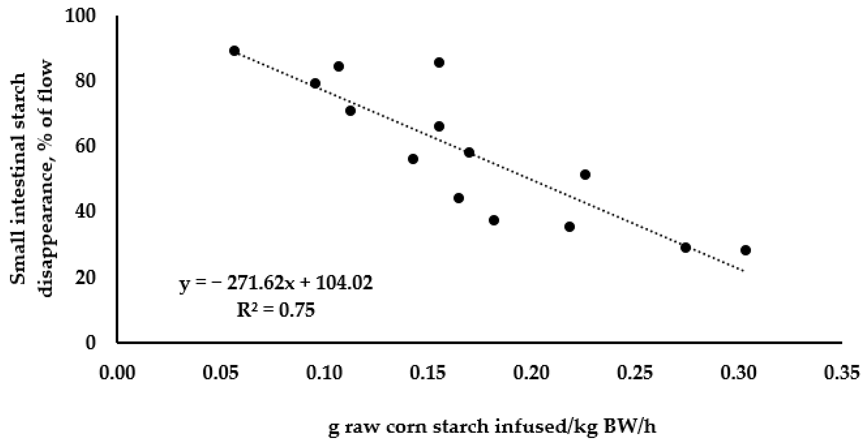
3.1. Small Intestinal Starch Disappearance: Linear Relationships

Early work using dietary [57,58] or abomasal infusion models [59] demonstrated that the extent of postruminal starch disappearance was much lower than in nonruminants. These authors concluded that the extent of starch disappearance in the small intestine was inadequate for optimum utilization. Indeed, summaries have indicated that the extents of small intestinal starch disappearance in beef cattle (55%) [7] and dairy cows (60%) [60] are inadequate to achieve potential energetic advantages over ruminal fermentation of starch [20]. Interestingly, the limitation of small intestinal starch disappearance is proportional to small intestinal starch flow instead of an absolute maximal value (i.e., plateau) [27,61]. Linear relationships between intestinal starch appearance and small intestinal starch disappearance were first suggested by Ørskov et al. [62], and a linear regression model was developed to predict small intestinal starch digestibility in lambs [61]. When Owens et al. [7] reviewed the literature, they found that there was a positive linear relationship between the amount of starch flowing to the small intestine (g/d) and small intestinal starch disappearance (g/d). Furthermore, there is a negative linear relationship between the amount of starch flowing to the small intestine (g/d) and small intestinal starch disappearance (%) [63]. These reviews were compiled data from site of digestion experiments with duodenal starch flows resulting from dietary starch intake. Using data from postruminal starch infusion models in cattle shows that as the amount of raw corn starch infused per hour relative to body weight (BW) increased, small intestinal starch disappearance decreased (Figure 1). Linear relationships are not normally expected in biology [8], including digestion, which typically conforms to a non-linear relationship because of Michaelis–Menten kinetics. It was previously calculated that at least a 70% digestibility of starch in the small intestine was necessary to avoid the inefficiencies associated with large intestinal starch digestion [20]. However, a plateau in efficiency may not be achievable under practical feeding conditions [8]. Collectively, these data demonstrate a linear relationship between intestinal starch supply and starch disappearance across dietary and infusion models in cattle and sheep.

3.2. Influence of Nitrogenous Compounds on Small Intestinal Starch Disappearance

Several studies have demonstrated that postruminal protein or amino acids can increase small intestinal starch disappearance in ruminants. Indirect evidence of increased pancreatic α-amylase [71] and increased net portal glucose flux [72] with abomasal casein infusion led to the speculation that postruminal protein flow could increase small intestinal starch disappearance. Increasing levels of postruminal casein (0 g/d to 200 g/d) infusion resulted in linear improvements in small intestinal starch disappearance (g/d and % of flow) in cattle abomasally infused with raw corn starch [65]. Similar results were obtained in sheep receiving abomasal casein infusions while consuming a dry-rolled sorghum grain diet [73] or a cracked-corn-based diet [74]. Later, Brake et al. [55] demonstrated that increasing levels of postruminal casein (0 g/d, 200 g/d, or 400 g/d) infusion could increase small intestinal starch disappearance in steers duodenally infused with raw corn starch within 6 d. In a follow-up study, amino acid treatments were used to represent similar proportions of amino acids to those found in casein. Non-essential amino acid infusions (similar to the profile of casein) increased small intestinal starch digestibility but essential amino acid infusions did not [66]. This observation was further supported when Glu or Glu + Phe + Trp + Met increased small intestinal starch digestibility but the Phe + Trp + Met treatment did not [66]. Furthermore, increasing supply (0 g/d, 60 g/d, and 120 g/d) of duodenal Glu increased small intestinal starch disappearance to a similar magnitude achieved with 400 g/d of casein [67]. Other trials with essential amino acids (Leu and Phe) have not found any effects on small intestinal starch disappearance in goats [75].

3.3. Influence of Grain Processing on Small Intestinal Starch Disappearance

The effects of grain processing on small intestinal or postruminal starch disappearance have been reviewed extensively [5,7,20,27,76,77]. The current nutrient requirements of beef cattle [78] use data from Sniffen et al. [79] and Owens and Zinn [80] to show the effects of grain type and degree of processing on postruminal starch disappearance. The postruminal starch disappearance coefficients reported are 30–40%, 65–70%, 80–90%, 85–95%, and 92–97% for whole, dry-rolled or cracked, meal, high-moisture, and steam-flaked methods of corn processing, respectively [78]. Owens et al. [77] developed linear models to predict postruminal starch disappearance in dairy and finishing diets. In general, starch digestibility of diets containing whole or rolled corn typically decreases linearly with increasing intestinal flow, as described previously. However, extensive methods of corn processing, such as high-moisture and steam-flaking, are typically not affected by increasing intestinal flow because they are highly digestible postruminally. It is not clear whether or not enhanced postruminal starch digestibility with extensive corn processing methods are directly related to increased small intestinal starch digestibility. The issues of variable duodenal flow, maintenance of ileal cannulas, and maintaining production levels of intake in cattle with ileal cannulas are largely why measurements of small intestinal disappearance with different grain processing methods have not been evaluated. A complicating factor is that extensive grain processing methods that increase postruminal digestibility also increase ruminal digestibility [81]. Therefore, steam-flaking and high-moisture processing methods result in greater ruminal starch disappearance and decreased intestinal starch flows relative to whole-shelled or dry-rolled processing methods. Because of this, comparisons of postruminal starch digestibility coefficients that were obtained using dietary models had differing intestinal starch flows. More studies in which duodenal flow is controlled are needed to clarify how grain processing methods affect the extent of small intestinal starch disappearance in ruminants. This will aid in understanding the limits of the extent of small intestinal starch disappearance in ruminants.

4. Pancreatic α-Amylase

4.1. Influence of Dry Matter and Energy Intake on Pancreatic α-Amylase Activity

The effects of nutrition on pancreatic exocrine function in ruminants have been reviewed previously [16,54,82,83,84,85,86,87]. In nonruminants, carbohydrase activities typically increase proportional to luminal substrate flow [88]. However, in ruminants, postruminal digestive enzymes respond differently to diet and luminal nutrient flows [16]. Russell et al. [89] evaluated the effects of diet and energy intake on pancreatic α-amylase activity in steers. They fed either an alfalfa hay diet (hay) or a corn and corn-silage-based diet (grain) at 1 × net energy of maintenance (NEm) or the grain diet at 2 × or 3 × NEm. At 1 × NEm intake, they found that steers consuming the grain diet had lower pancreatic α-amylase activity per gram protein than steers consuming the hay diet. Furthermore, increasing the energy intake of the grain diet from 1 to 2 × NEm increased pancreatic α-amylase activity per gram protein by two-fold, without any additional increases at 3 × NEm.
To further evaluate the effects of diet and energy intake on carbohydrase activities, Kreikemeier et al. [90] fed either a 90% forage (alfalfa hay) or 90% grain (sorghum and wheat) diet at 1 or 2 × the NEm requirement. In steers consuming the grain diet, pancreatic α-amylase concentration and total content was lower than steers consuming forage. Additionally, when energy intake increased from 1 to 2 × NEm, pancreatic α-amylase activity and total content increased with an increase in pancreatic mass. In contrast, previous studies demonstrated that increasing starch intake could increase pancreatic α-amylase activity [91,92]. However, these studies were confounded with energy intake. Results from Russell et al. [89] and Kreikemeier et al. [90] demonstrated that increasing energy intake up to 2× maintenance can increase pancreatic α-amylase activity. In addition, steers consuming starch-based diets had lower activity of pancreatic α-amylase. However, the diet effects on pancreatic α-amylase were less clear, as the alfalfa hay-based diets had greater crude protein levels. This led to the hypothesis that changes in luminal carbohydrate and protein flow could influence pancreatic α-amylase activity.
More recent studies have evaluated the effects of dietary intake restriction on pancreatic α-amylase activity in ruminants. Dietary intake restriction decreased pancreatic α-amylase activity in nonpregnant ewes [93], pregnant ewes [94,95], and pregnant beef cows [96,97]. Changes in pancreatic α-amylase activity in response to changes in dry matter or energy intake may be related to the abundance and activity of pancreatic proteins involved in energy metabolism. Increasing dry matter intake increased the abundance of ATP synthase, Na+/K+-ATPase, proliferating cell nuclear antigen, and ubiquitin in the pancreas of steers [98]. Dietary intake restriction of pregnant beef cows decreased ATP synthase abundance in the pancreas [99]. Proteomic analyses suggest that intracellular activity and abundance of proteins related to energy metabolism in the pancreas may be associated with pancreatic α-amylase activity [100].

4.2. Influence of Dietary or Luminal Carbohydrate on Pancreatic α-Amylase Activity

While pancreatic α-amylase activity in nonruminants increases in response to luminal starch flows [88], the response is opposite in ruminants. High levels of postruminal carbohydrate supply as starch, partially hydrolyzed starch, or glucose decreases pancreatic α-amylase activity when energy intake is controlled. Abomasal infusions of partially hydrolyzed starch decreased pancreatic α-amylase concentration, specific activity, and secretion in steers compared with steers ruminally infused with partially hydrolyzed starch or steers infused with water [50]. The same decrease in pancreatic α-amylase activity in response to abomasal partially hydrolyzed starch was observed with pancreatic tissue samples [101]. Similarly in wethers, abomasal infusions of raw corn starch decreased pancreatic α-amylase concentration and secretion compared with control wethers receiving abomasal infusion of water [71]. These studies demonstrated that luminal complex carbohydrate flow decreases pancreatic α-amylase activity in cattle. In a study by Swanson et al. [102], abomasal infusions of either glucose or partially hydrolyzed starch decreased pancreatic α-amylase concentration, specific activity, and secretion in steers. This study demonstrated that downregulation of pancreatic α-amylase is not due solely to luminal complex carbohydrate flow. However, it remains unclear whether luminal glucose concentration, absorbed glucose, or both regulate pancreatic α-amylase activity in ruminants. Increasing levels of ruminal glucose infusions did not affect plasma amylase concentrations in lambs fed a 50% concentrate diet [103]. In neonatal dairy calves, supplementing fructose at 2.2 g/kg of BW did not statistically increase pancreatic α-amylase activity; however, pancreatic α-amylase activity was 42% greater in fructose-fed calves [104]. This could partially result from an increase in metabolizable energy intake.

4.3. Influence of Dietary or Luminal Nitrogenous Compounds on Pancreatic α-Amylase Activity

As stated previously, studies by Russell et al. [89] and Kreikemeier et al. [90] demonstrated that pancreatic α-amylase activity was greater in steers fed an alfalfa hay diet compared with a grain-based diet. These authors speculated that differences in dietary crude protein (and therefore, rumen undegradable protein and metabolizable protein) contribute to differences in pancreatic α-amylase activity. In sheep, Wang and Taniguchi [71] abomasally infused water (control), raw corn starch, or raw corn starch + casein and measured pancreatic exocrine secretion. Pancreatic α-amylase activity was depressed with abomasal starch infusion; however, abomasal infusion of starch with casein restored α-amylase activity to the same level as the control. Similarly, increasing levels of abomasal casein supply (0 g/d, 60 g/d, 120 g/d, or 180 g/d) linearly increased pancreatic α-amylase concentration, specific activity, and secretion in steers postruminally infused with raw corn starch [105]. Feeding a 68.7% concentrate diet with supplemental casein to steers produced increases in duodenal α-amylase concentrations and serum cholecystokinin (CCK) concentrations [106].
More information is needed to understand how the association between luminal nutrient supply, hormones and neuropeptides, and enzyme activities are coordinated to influence intestinal starch disappearance in ruminants. The effects of individual amino acids on pancreatic exocrine function have been studied predominantly with preruminant calves and lambs [86]. Several amino acids including Arg, Leu, Ile, and Phe have been shown to influence pancreatic α-amylase activity in ruminants. Similarly, rumen-protected Trp supplementation to steers consuming a high-concentrate diet was associated with greater postruminal starch disappearance, increased luminal amylase activity in the duodenum, and increased serum CCK and melatonin [107]. We have found that melatonin supplementation to gestating ewes increased maternal pancreatic α-amylase activity [94] and small intestinal maltase, isomaltase, and glucoamylase activities [108]. Tryptophan and its metabolites are precursors to the synthesis of biogenic amines such as serotonin and melatonin.
Responses in pancreatic α-amylase activity to individual amino acids have varied with the type of amino acid, length of infusion, and animal species. Arginine administration through jugular blood did not influence pancreatic α-amylase activity in non-pregnant ewes [93]. Similarly, dietary rumen-protected Arg supplementation to ewes during mid- to late-gestation did not influence pancreatic α-amylase activity of lamb offspring at 54 d of age [109]. After 14 d of duodenally infusing increasing levels of Phe, Yu et al. [110] observed linear increases in pancreatic α-amylase specific activity, and a cubic response in α-amylase secretion in goats. In the short-term experiment (10 h), they found a quadratic response in pancreatic α-amylase secretion to increasing levels of Phe. Moreover, increasing levels of Leu linearly increased α-amylase concentration in pancreatic juice after 14 d of duodenal infusion [111]. In dairy heifers, duodenal infusions of 10 g/d Leu increased total pancreatic secretion, α-amylase concentration, and α-amylase secretion [112]. Increases in pancreatic α-amylase activity were observed with duodenal infusions or Leu (3 g/d or 9 g/d) and Phe (2 g/d) in goats [113]. However, when Leu (1.435 g/L milk), Phe (0.725 g/L milk), or a combination of Leu and Phe (1.435 g Leu/L milk and 0.725 g Phe/L milk) were fed to milk-fed calves, pancreatic α-amylase specific activity was not influenced [114]. Similarly, increasing levels of Leu supplementation to neonatal calves in milk replacer did not affect pancreatic α-amylase activity [115]. These data suggest that Leu can increase pancreatic α-amylase activity in post-weaning ruminants but not in milk-fed calves. Duodenal infusions of 20 g/d or 30 g/d of Ile have been shown to increase pancreatic α-amylase activity in dairy heifers after 12 h or 10 d of infusion [116]. In cell culture models using pancreatic acinar cells, amino acids such as Phe [117], Leu [118,119,120], and Ile [121] increased α-amylase release. Despite increases in small intestinal starch disappearance with Glu [66,67], it is unclear if these increases are related to increases in pancreatic α-amylase activity, as our recent experiment found duodenal glutamic acid infusion did not influence pancreatic α-amylase in steers [122].
Indeed, a few studies have begun to explore cellular and molecular mechanisms driving associations between increased pancreatic α-amylase activity in ruminants and amino acid supply. Phenylalanine increases α-amylase activity in dairy calves and the initiation of messenger ribonucleic acid (mRNA) translation through phosphorylation of ribosomal protein S6 kinase 1 (S6K1) and eukaryotic translation initiation factor 4E binding protein 1 (4EBP1) [118]. Leucine and Ile have been shown to increase α-amylase synthesis and phosphorylation of the mammalian target of rapamycin (mTOR) signaling pathway [118,121]. Proteomic analysis has suggested that Leu modulates increases in pancreatic α-amylase activity in dairy calves by increasing citrate synthase activity in the tricarboxylic acid cycle, ATPase activity and oxidative phosphorylation, and stimulating the general secretory signaling pathway in pancreatic acinar cells [123]. In these studies, Phe, Leu, and Ile were the only amino acids studied and future research is needed to evaluate how other amino acids could affect pancreatic exocrine function in ruminants.

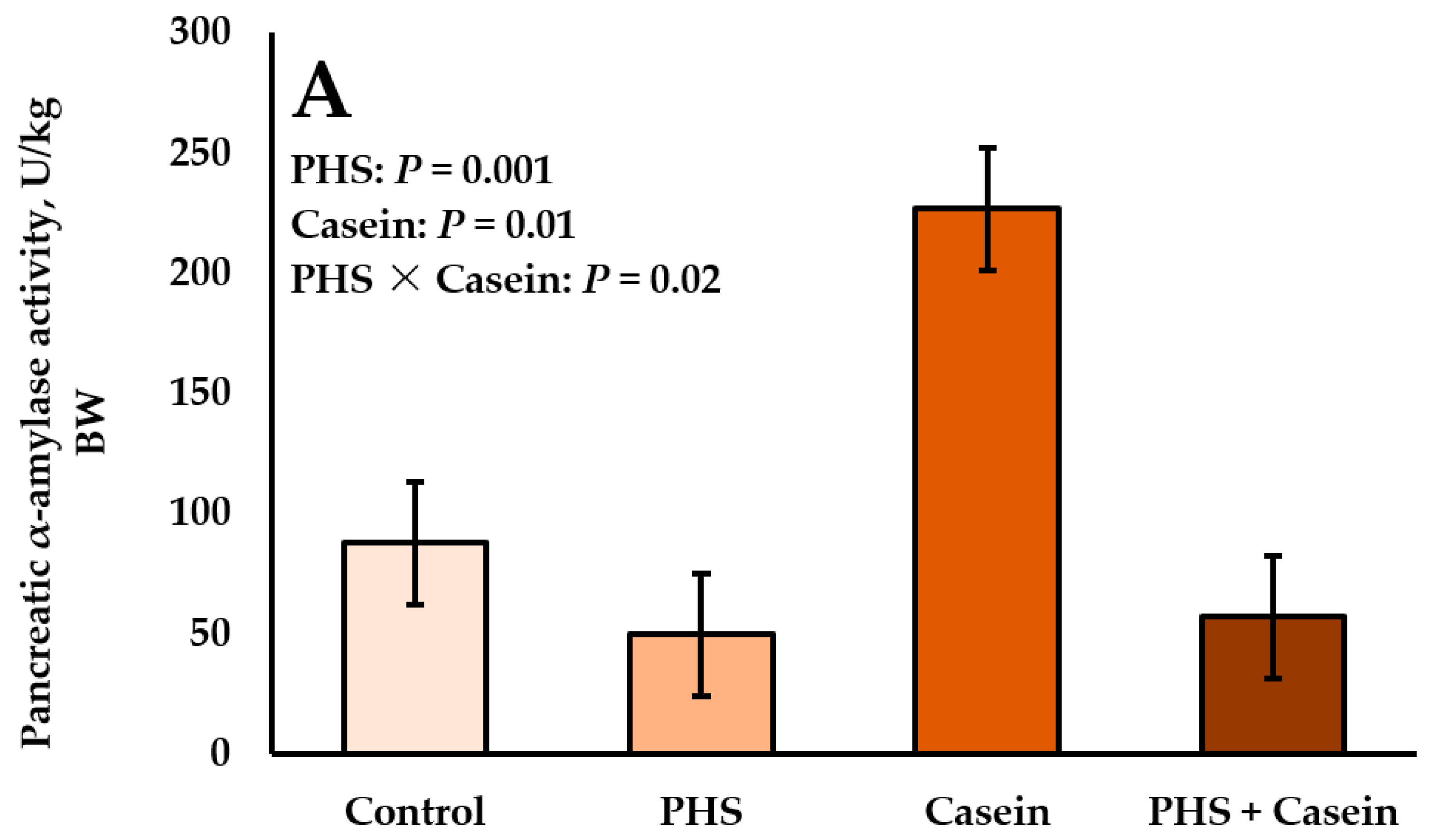
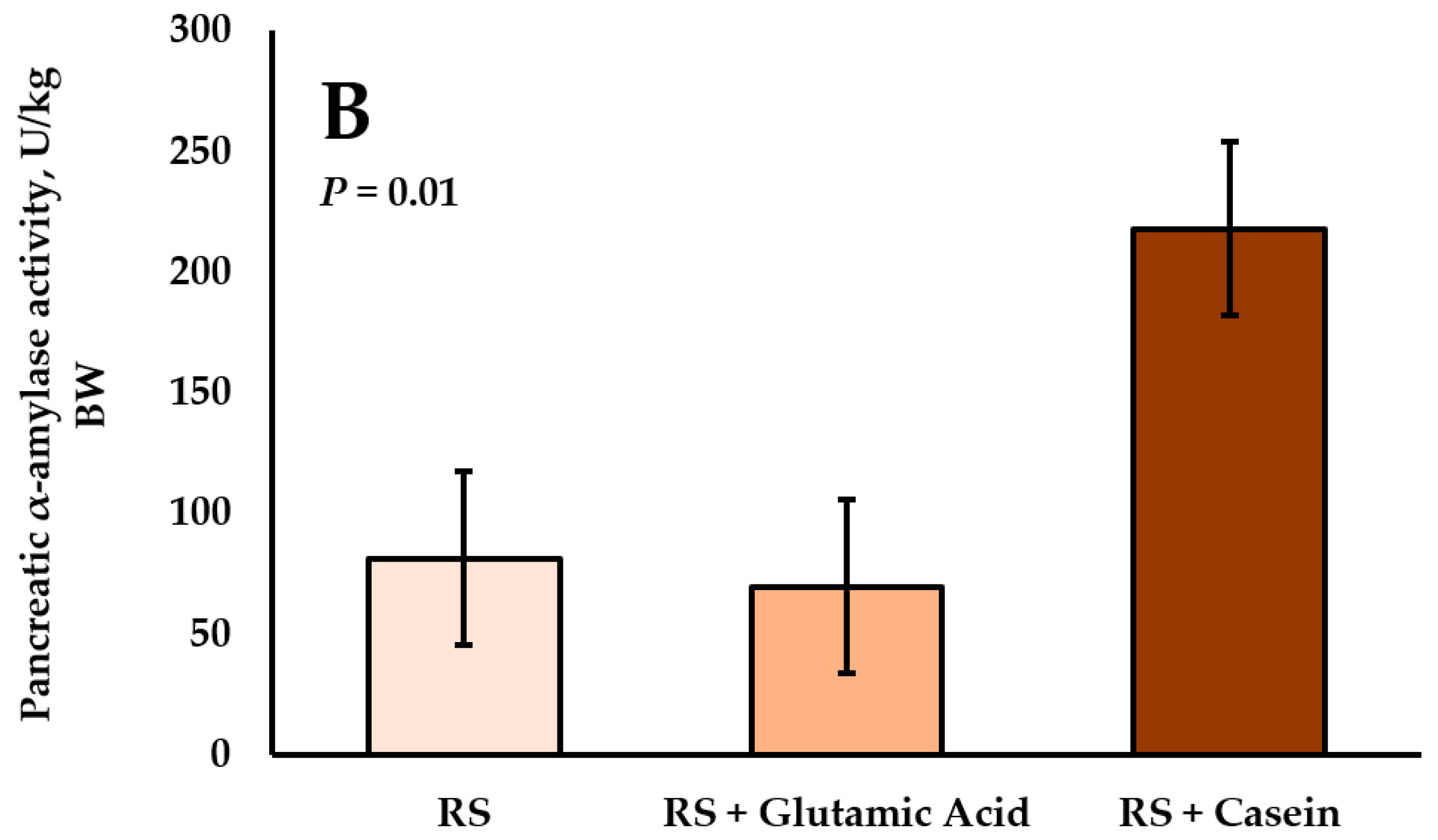
4.4. Interactions between Starch and Protein Supply on Pancreatic α-Amylase Activity

Complex interactions can occur with simultaneous starch and protein supply in the ruminant small intestine [84]. Interactions between starch and protein on pancreatic α-amylase were evaluated with the following treatments: (1) water, (2) partially hydrolyzed starch, (3) casein, and (4) partially hydrolyzed starch + casein. In Holstein calves, abomasal casein infusion for 7 d increased pancreatic α-amylase activity relative to BW (Figure 2) [101]. However, the combination of partially hydrolyzed starch and casein produced a response in pancreatic α-amylase that was less than casein and not different from partially hydrolyzed starch or control. In support of these results, Swanson et al. [124] and Swanson et al. [125] found similar responses in pancreatic α-amylase activity to infusion treatments in pancreatic explant and secretion models, respectively. The combined results suggest that the benefits of postruminal protein supply may be overridden by the presence of starch in the small intestine. In contrast, we recently demonstrated that duodenal infusion of raw corn starch with casein for 58 d increases pancreatic α-amylase activity by 290% compared with raw corn starch infusion alone [122]. Interestingly, the responses in pancreatic α-amylase activity relative to BW were numerically similar across studies (casein: 226 U/kg BW and raw starch + casein: 218 U/kg BW). Furthermore, activity of pancreatic α-amylase in response to starch treatments was similar, as well (partially hydrolyzed starch: 49.5 U/kg BW and raw starch: 81.3 U/kg BW). The results from Trotta et al. [122] may suggest that a longer adaptation to postruminal protein supply could overcome the negative effects of starch on pancreatic α-amylase activity.
Alternatively, partially hydrolyzed starch and raw corn starch may elicit different responses in pancreatic α-amylase activity. Branco et al. [126] reported that the coefficient of true small intestinal digestibility for partially hydrolyzed starch infused at 40 g/h was 88%. Therefore, the high digestibility of partially hydrolyzed starch may not require the amount of pancreatic α-amylase that raw corn starch does and thus, decreased activity of α-amylase with partially hydrolyzed starch flow might be a function of the starch source. The high small intestinal digestibility of partially hydrolyzed starch relative to raw corn starch likely allows for more glucose to be absorbed and it is still unclear if luminal or absorbed glucose regulates pancreatic α-amylase activity. No definitive studies have tested the influence of corn starch source (raw corn starch or partially hydrolyzed starch) with postruminal casein on pancreatic α-amylase activity.
Secretion of pancreatic α-amylase increases with increasing dietary starch intake in nonruminants [88], which is similar to responses in ruminants to increasing postruminal protein supply. Increasing dietary crude protein concentrations with ruminal escape soybean meal resulted in linear increases in pancreatic α-amylase activity [127]. However, at the same time, there were decreasing proportions of high-moisture corn in the ration. Complex interactions between luminal carbohydrate and protein flow on small intestinal starch digestion likely differ between ruminants and nonruminants. Increasing dry matter or energy intake results in increased microbial-N flow to the small intestine [128]. Moreover, ruminal starch supply increased duodenal bacterial-N and total-N flow compared with postruminal starch supply [129]. Synchrony of ruminal starch and protein fermentation increases ruminal bacterial-N outflow [76] and responses of the pancreas to luminal nutrients may be related to changes in the evolutionary control of pancreatic exocrine function.

4.5. Pancreatic α-Amylase Activity and Small Intestinal Starch Disappearance

Despite increases in pancreatic α-amylase activity in response to postruminal protein or amino acid flow, it is unclear if increases in pancreatic α-amylase are related to increases in small intestinal starch disappearance. Associations between increased small intestinal starch disappearance [65] and increased pancreatic α-amylase activity [105] with postruminal casein supply have led to speculation that pancreatic α-amylase could be the primary factor limiting the extent of small intestinal starch disappearance. However, in most studies where pancreatic α-amylase increased, small intestinal carbohydrases were not evaluated. Therefore, there may be confounding effects in determining the limiting factor if there are also simultaneous increases in small intestinal carbohydrase activities.
Furthermore, several enzyme infusion studies with exogenous α-amylase have failed to show a response in small intestinal starch disappearance in cattle. Remillard et al. [130] infused porcine pancreatic α-amylase or bicarbonate in a 2 × 2 factorial design into the jejunum and failed to see any effects on small intestinal starch disappearance in steers fed an 85% grain diet. Abomasal infusions of exogenous amylase (Bacillus subtilis origin) with 880 g/d of raw corn starch [68] or increasing levels of raw corn starch up to 1993 g/d [131] did not influence postruminal starch disappearance in heifers. Likewise, duodenal infusion of exogenous porcine pancreatic α-amylase in amounts equivalent to two, four, or five times the endogenous amount of bovine pancreatic α-amylase secretion (22 kU/h) did not result in increased small intestinal starch disappearance in steers [69].

5. Small Intestinal Carbohydrases

5.1. Influence of Energy Intake on Small Intestinal Maltase, Isomaltase, Glucoamylase Activity

Compared with pancreatic α-amylase, there is far less information describing the influence of diet and luminal nutrient supply on the regulation of small intestinal carbohydrases in ruminants. Early studies demonstrated that diet composition (forage vs. grain) and excess energy intake had little influence on small intestinal carbohydrase activities [89,90,92]. Increasing dietary intake from 1 to 2 × NEm did not influence intestinal maltase activity in steers [18]. Young bulls consuming a ground corn-based diet had greater duodenal maltase specific activity than young bulls consuming a whole shelled corn-based diet, and there was no diet effect in the jejunum [132]. In dairy calves, milk replacer intake and butyrate supplementation did not influence maltase activity or mRNA expression of maltase-glucoamylase or sucrase-isomaltase in the small intestine [133]. Sixty-percent nutrient restriction of ewes during mid- to late-gestation increased small intestinal maltase, isomaltase, and glucoamylase activities [95]. Overall, changes in energy intake status do not seem to have specific effects on small intestinal carbohydrase activities [16,18].

5.2. Influence of Macronutrients on Small Intestinal Maltase, Isomaltase, Glucoamylase Activity

Few studies have investigated the effects of specific nutrients on small intestinal carbohydrase activities in ruminants; most information is about starch or starch-digestion products. Abomasal infusions of partially hydrolyzed starch increased jejunal maltase activity in sheep but decreased jejunal maltase activity in cattle [134]. In another experiment, partially hydrolyzed starch infusions for 7 d did not influence maltase activity in any site of the small intestine in cattle [17]. Later, steers receiving abomasal infusions of glucose or partially hydrolyzed starch for 35 d had greater maltase specific activity than steers receiving ruminal starch infusions [18]. This may indicate that luminal substrate flow (maltose, isomaltose, and limit dextrins) can increase carbohydrase activities in the small intestine. In neonatal calves, 18% replacement of lactose with maltodextrin, maltodextrin with a high degree of α-1,6 branching, and maltose decreased jejunal maltase specific activity [135]. Furthermore, jejunal isomaltase-specific activity decreased in response to greater amounts of maltodextrin or maltodextrin with a high degree of α-1,6 branching. Additionally, steers infused with partially hydrolyzed starch into the abomasum had greater maltase specific activity than steers infused with casein, with partially hydrolyzed starch + casein being intermediate [136]. Dietary fructose differentially regulated small intestinal maltase-glucoamylase (increase) and sucrase-isomaltase (decrease) mRNA expression in neonatal calves [104]. In the same study, glucoamylase activity was found to increase with dietary fructose while maltase activity was unchanged [104].
In rats, 80% of apparent maltase activity is derived from sucrase-isomaltase and the remaining 20% is derived from maltase-glucoamylase [137,138]. The concept that maltase activity is derived from multiple enzyme subunits is supported by heat-inactivation studies by Coombe and Siddons [14]. Therefore, changes in maltase-glucoamylase and sucrase-isomaltase mRNA expression with dietary fructose may explain why glucoamylase and maltase activities were differentially regulated. Conflicting reports on the responses of carbohydrase activity in different animal models make interpretations difficult. A greater understanding of how luminal starch, starch-digestion intermediates, and glucose regulate small intestinal carbohydrase activities is needed.
There is limited information about how dietary or luminal amino acid supply influences small intestinal carbohydrase activity in ruminants. Increasing supplemental Leu (0 g/kg, 0.4 g/kg, 0.6 g/kg or 0.8 g/kg of BW) in milk replacer linearly decreased maltase and isomaltase activities of calves [115]. Similarly, feeding Leu at 2.9% of DM to neonatal lambs in milk replacer for 42 d decreased small intestinal maltase and isomaltase activity at slaughter after an 82 d finishing period [115]. Rumen-protected Arg supplementation to gestating ewes fed at 60% of nutrient requirements did not influence small intestinal digestive enzyme activities of lamb offspring at day 54 of age [109]. Although Glu has been shown to increase small intestinal starch disappearance in steers after 12 d [66,67], our recent study found only a small increase in duodenal maltase activity in steers duodenally infused with raw corn starch and Glu for 58 d [122].
Duodenal infusions of raw corn starch with casein increased jejunal maltase, isomaltase, and glucoamylase activities in steers [122]. It is unclear if increases in small intestinal carbohydrases with postruminal casein infusion are directly related to increased luminal protein flow because peptide hydrolysates and free amino acids from casein might influence neuroendocrine signaling to increase carbohydrase activity in the small intestine. Alternatively, increased luminal protein flow might cause increased carbohydrase activity indirectly. Increased flow of luminal substrates (maltose, isomaltose, and limit dextrins) as a result of greater hydrolysis of amylose and amylopectin in response to increases in pancreatic α-amylase activity might modulate increases in small intestinal carbohydrase activities. In Caco-2 cells, supply of maltose induced synthesis of a higher molecular weight sucrase-isomaltase immunoblot band compared with glucose, fructose, isomaltose, and fructose [139]. Using Caco-2 cells, Chegeni et al. [140] suggested there was a luminal maltose-sensing mechanism that increases apparent maltase activity by enhancing intracellular trafficking of sucrase-isomaltase to the apical membrane. These effects were associated with an increased mRNA expression of TAS1R2 and TAS1R3, the genes encoding the dimeric sweet taste receptor subunits T1R2-T1R3. These authors suggested that T1R2-T1R3 could potentially mediate effects of luminal maltose on sucrase-isomaltase activity [140].

6. Sucrase

6.1. Intestinal Sucrase Activity Is Absent in Multiple Ruminant Species

A remaining enigma of ruminant digestive physiology is the absence of sucrase activity in the small intestine. Several studies have investigated and characterized digestive enzyme activity along the small intestine with various ruminant species, ages, and diets. Yet, there has been a failure to detect active sucrase in the small intestine. This is in contrast to nonruminant species including the pig and human. Dollar and Porter [3] were the first to report the absence of sucrase activity in young calves. Furthermore, no measurable sucrase activity was detected in mucosa or small intestinal digesta contents from lambs [11]. Later reports by Huber et al. [141] and Siddons [13] corroborated the findings that sucrase activity is absent from the digestive tract of the young calf. With cattle ranging from 4 days of age up to 6 years of age, no detectable amounts of sucrase were found in the small intestine [13,90]. Shirazi-Beechey et al. [142] attempted to measure sucrase activity in isolated brush-border membrane vesicles (BBMV) from lamb intestine and also did not detect any sucrase activity. More recently, we have been unable to detect sucrase activity in intestinal mucosa samples from neonatal calves [104], growing steers [122], or fetal, neonatal, or gestating sheep [95,109].
The lack of sucrase activity seems to expand to a wider range of ruminants other than sheep and cattle and even some nonruminant foregut fermenters. Marine mammals such as whales and dolphins have similarities in the sucrase-isomaltase protein sequence to even-toed ungulates. Although it is unknown if dolphins or whales possess intestinal sucrase activity [143], the sea lion does not [144]. A comparative study demonstrated that sucrase activity was not detected in any ruminant species including sheep, goat, roe deer, and moose [145]. Although not considered a ruminant, the kangaroo is a foregut fermenter and does not possess intestinal sucrase activity [146]. In the pseudoruminant camel intestine, glucoamylase and maltase activities were two- and three-fold greater than sucrase activity [147].

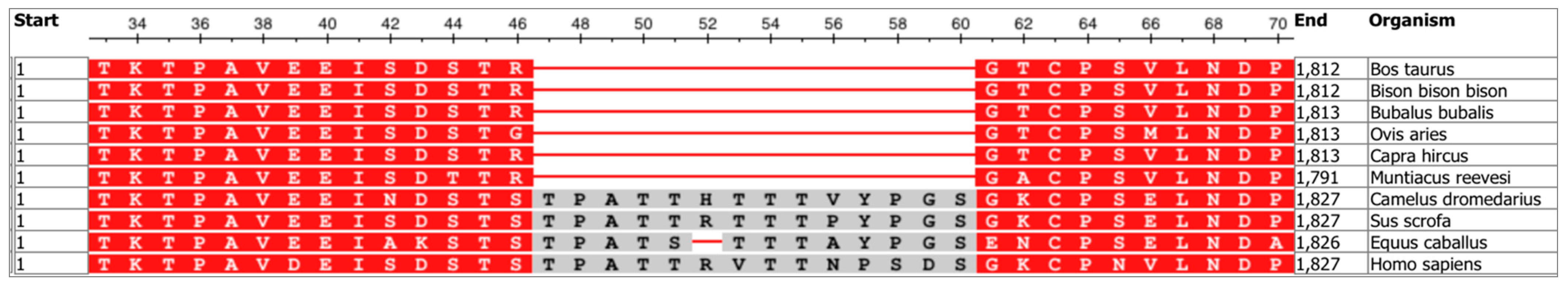
6.2. Congenital Sucrase-Isomaltase Deficiency and Multiple Sequence Alignment

Interestingly, the absence of sucrase activity in the small intestine of ruminants appears to be similar to congenital sucrase-isomaltase deficiency (CSID) in infants [14]. There are seven distinct phenotypes of CSID in humans with phenotypes I, II, and III resulting in completely inactive sucrase and isomaltase activities [148]. It would seem unlikely that mechanisms driving phenotypes I, II, or III of CSID would explain the inactivity of sucrase in ruminants because ruminants do have active isomaltase activity [14]. However, in phenotype V of CSID, it is characterized by the presence of isomaltase activity and absence of sucrase activity.
The primary structure of the sucrase-isomaltase complex was deduced in the rabbit intestine and a “stalk” region (amino acids 33–70) was identified as the connection between the transmembrane domain and N-terminal sucrase-isomaltase (isomaltase subunit) [149]. In phenotype V of CSID, pro-sucrase-isomaltase is cleaved intracellularly in the trans-Golgi network, and the sucrase subunit is subsequently degraded while the isomaltase subunit is transported to the apical membrane [148,150]. Later experiments determined that the signals for apical sorting were located in the O-glycosylated “stalk” region and membrane anchoring domain of the isomaltase subunit [151,152].
The stalk region of sucrase-isomaltase may be important to understanding the sucrase phenotype of ruminants. Using multiple sequence alignment, the sucrase-isomaltase amino acid sequence was compared between several ruminant species and the camel, pig, horse, and human (Figure 3). At amino acid 46 of the sequence, the selected ruminant species (cattle, bison, water buffalo, goat, and Reeve’s muntjac) all have an Arg residue, while sheep have a Gly residue. Furthermore, the camel, pig, horse, and human all contain a Ser residue at this position. Immediately following amino acid 46, the sequence from amino acids 47–60 is missing in the selected ruminant species. This region is enriched with Ser and Thr residues and has been suggested to be O-glycosylated to be protected from degradation from pancreatic proteases [149]. Indeed, bypassing the pancreatic duct in rats to prevent luminal pancreatic secretions resulted in a decreased rate of sucrase degradation and increased intestinal sucrase activity [153]. N- and O-glycosylation sites of sucrase-isomaltase are essential components for proper folding and intracellular trafficking to the apical membrane [151,154].
This missing region of the sucrase-isomaltase protein could perhaps explain why ruminants do not have intestinal sucrase activity. Using multiple sequence alignment, mutations in the coding regions of the sucrase or isomaltase subunits that result in other phenotypes of CSID [148] did not occur in the sucrase-isomaltase sequences of the selected ruminant species (data not shown). It should be noted that although the horse and the camel are herbivores that consume mostly forage-based diets, these species have been shown to have active intestinal sucrase activity [147,155] and the 47–60 amino acid sequence of sucrase-isomaltase. These comparative sequence findings provide evidence that the absence of sucrase activity in ruminants could potentially be similar to phenotype V of CSID due to the absence of the 47–60 amino acid sequence of the stalk region, which is important for intracellular sorting to the apical membrane. Cellular localization and molecular characterization of the sucrase-isomaltase complex in ruminants is warranted, with specific regard to phenotype V of CSID.

6.3. Nutritional Influences on Sucrase-Isomaltase mRNA Expression and Activity

Few studies have attempted to induce sucrase activity by nutritional methods in ruminants. Milk-feeding or dietary supplementation of sucrose to lambs did not induce sucrase activity and small intestinal disappearance of sucrose was small [156]. Because sucrase activity was not induced by its own substrate and small intestinal disappearance was low, those authors concluded that intestinal disappearance of sucrose was most likely due to microbial fermentation [156]. These conclusions were supported by increased cecal microbial counts and increased fecal N excretion with sucrose inclusion [156]. Likewise, abomasal infusions of sucrose did not induce sucrase activity in lambs [157].
In one case report in humans, dietary fructose supplementation increased sucrase activity by nearly four-fold in a patient with CSID [158]. Although the phenotype of the patient was not revealed, the patient had deficient, but not absent, activity of sucrase and isomaltase before beginning fructose treatment [158]. After treatment, sucrase activity levels were still approximately 18.5% of the amount of sucrase activity from the patient’s family members [158]. Fructose supplementation at 2.2 g/kg of BW did not induce sucrase activity in neonatal calves fed milk replacer [104]. However, dietary fructose decreased sucrase-isomaltase mRNA expression, suggesting that sucrase-isomaltase may be transcriptionally regulated by dietary fructose in the ruminant small intestine. These studies indicate that fructose supplementation is not effective at inducing or increasing sucrase activity in patients with CSID or in ruminants.

6.4. Impacts on Carbohydrate Digestion

Recent evidence from nonruminant studies suggests that the absence of sucrase can have other physiological consequences on carbohydrate digestion. Nichols et al. [159] demonstrated that the absence of sucrase activity leads to a reduction in starch digestion and postprandial glucose response with a sucrase-deficient shrew model. Furthermore, when supplemented with an oral glucoamylase enzyme, sucrase-deficient shrews had blood glucose concentrations that were similar to the control shrews (containing normal sucrase activity). These authors concluded that sucrase was the predominant mucosal enzyme involved in starch digestion because of its affinity towards multiple starch substrates [160]. In steers, duodenal infusions of exogenous glucoamylase increased small intestinal starch disappearance [69]. Collectively, these data suggest that ruminants have quantitative limits in carbohydrate digestion similar to humans with congenital sucrase-isomaltase deficiency and supplemental enzymes may replace missing intestinal hydrolytic activity to improve small intestinal carbohydrate digestibility.

7. Glucose Absorption

7.1. Sodium/Glucose Cotransporter-1 (SGLT1)

Sodium/glucose cotransporter-1 (SGLT1), glucose transporter 5 (GLUT5), and glucose transporter 2 (GLUT2) are thought to be the predominant carbohydrate transporters in the small intestine of ruminants [84]. Many studies in ruminants have concluded that SGLT1 activity and SGLT1 abundance were greatest in milk-fed lambs and declines with age [142,161,162,163]. Shirazi-Beechey et al. [163] demonstrated that duodenal infusions of a 30 mM glucose solution for 4 d in adult sheep increased the rate of glucose transport by 40- to 80-fold which was also accompanied by an increase in SGLT1 abundance. Furthermore, Dyer et al. [164] demonstrated that duodenal fructose infusions can increase jejunal SGLT1 activity in lambs. These authors concluded that luminal sugar is sensed in the intestine, independent of glucose metabolism, and that the inducing sugar does not need to be a substrate of SGLT1. However, duodenal infusion of raw corn starch did not influence SGLT1 activity in sheep [165].
Moreover, regulation of carbohydrate transport in ruminants has been suggested to be influenced by the presence of sweet taste receptors in the bovine and ovine small intestine (T1R2-T1R3) [166]. The sweet taste receptor signaling mechanism was proposed by Moran et al. [167], based on research with mice. Luminal sugar is sensed in the small intestine by T1R2-T1R3 and its associated G-protein, gustducin, which induces a signaling cascade, leading to a subsequent increase in glucagon-like peptide-2 secretion. Glucagon-like peptide-2 binds to its receptor on the submucosal plexus, eliciting a neuronal response to evoke the release of vasoactive intestinal peptide (VIP) or pituitary adenylate cyclase-activating peptide (PACAP) in absorptive enterocytes. Binding of either VIP or PACAP to its receptor on the basolateral membrane of absorptive enterocytes results in an increase in intracellular cyclic adenosine monophosphate (cAMP) levels, leading to an upregulation of SGLT1 [167].
However, there is an apparent difference between cattle and sheep in carbohydrate transport and their ability to respond to diet or luminal nutrient supply (Table 1). In companion studies, jejunal Na+-dependent glucose cotransport activity was determined in cattle and sheep ruminally or abomasally infused with partially hydrolyzed starch for 7 d [17,134]. Postruminal infusion of partially hydrolyzed starch increased SGLT1 activity by two-fold in both sheep and cattle compared with ruminal infusion of partially hydrolyzed starch [134]. In the next experiment, SGLT1 activity was evaluated in steers ruminally or abomasally infused with partially hydrolyzed starch for 7 d across multiple sites of the small intestine [17]. Abomasal infusion of partially hydrolyzed starch did not increase SGLT1 activity in any site of the small intestine [17]. Later, after increasing the adaptation length to 35 d, Rodriguez et al. [18] found that abomasal partially hydrolyzed starch or glucose infusions did not influence SGLT1 abundance or activity. It should be noted that increasing dietary energy intake to 2 × NEm also did not influence SGLT1 abundance or activity [18]. Similarly, Liao et al. [168] infused partially hydrolyzed starch ruminally or abomasally and found only tendencies to influence SGLT1 mRNA expression. They reported that ruminal partially hydrolyzed starch infusions tended to increase duodenal SGLT1 mRNA expression and that abomasal infusions of partially hydrolyzed starch tended to increase ileal SGLT1 mRNA expression [168]. Lohrenz et al. [169] reported that in lactating dairy cows fed a high starch (24% of DM) or low starch diet (12% of DM) there was no difference in duodenal or jejunal SGLT1 mRNA or protein expression. Moreover, duodenal or jejunal GLUT2 mRNA expression, protein amount on the apical membrane, or total protein amount was not influenced by diet [169]. Because of the interactions with luminal protein and carbohydrate in the ruminant small intestine on starch disappearance and enzyme activity, SGLT1 abundance and activity were evaluated [136]. Using the same treatments as Swanson et al. [101], abomasal partially hydrolyzed starch, casein, or their combination did not influence SGLT1 abundance or activity in steers [136]. In contrast, abomasal infusions of casein increased SGLT1 activity in the proximal jejunum and whole small intestine in lambs [74]. In goats, SGLT1 activity was greatest when corn- or wheat-based diets were fed but without any changes in transporter affinity or protein abundance [170]. Although Dyer et al. [164] determined that duodenal fructose infusions can increase jejunal SGLT1 activity and abundance in lambs, dietary fructose supplementation to neonatal calves did not influence SGLT1 mRNA expression [104]. Collectively, these data suggest that bovine nutrient transporters involved in small intestinal carbohydrate absorption are less sensitive to diet or luminal nutrient supply than sheep.

7.2. Glucose Transporter 5 (GLUT5)

Fructose is passively transported across the intestinal apical membrane by GLUT5 [171]. Dietary fructose supplementation has been shown to increase GLUT5 mRNA expression and enhance intestinal fructose transport in neonatal rats [172]. Zhao et al. [173] found that GLUT5 mRNA expression in the intestine is significantly lower than in the liver or kidney in cattle. In contrast, many authors have reported that the greatest amount of GLUT5 mRNA expression is found in the small intestine in humans, rats, mice, rabbits, chickens, and horses [174]. Nutritional regulation of GLUT5 by fructose requires luminal presence of fructose in the intestine [175] and GLUT5 mRNA expression is directly proportional to intestinal luminal fructose concentration in weaning rats [172]. In cattle, ruminal or abomasal infusions of partially hydrolyzed starch did not affect GLUT5 mRNA expression in the duodenum, jejunum, or ileum [168]. Dietary fructose supplementation to neonatal calves fed milk replacer did not influence GLUT5 mRNA expression in the small intestine [104].
Douard and Ferraris [174] discussed the complex relationships between age, luminal fructose supply, and induction of GLUT5 in neonatal (milk only), weaning (milk + solid feed), and post-weaning (solid feed only) rats. In general, GLUT5 expression is nutritionally regulated by luminal fructose during weaning (14–28 days of age) and post-weaning (>28 days of age) in rats [172,175,176,177,178,179]. However, in neonatal rats (<14 days of age), GLUT5 expression can increase with luminal fructose and glucocorticoid supply but not luminal fructose alone [180,181,182]. Therefore, nutritional regulation of GLUT5 by fructose is age-dependent in rats and this process could be similar in cattle.

7.3. Glucose Transporter 2 (GLUT2)

Glucose transporter 2 is thought to be the primary basolateral transporter of monosaccharides from intestinal enterocytes. The apical GLUT2 hypothesis [183] in which GLUT2 translocates to the apical membrane and contributes to apical (luminal) sugar transport has been controversial [171]. Whether or not GLUT2 translocation occurs in ruminants or contributes to apical sugar uptake under physiological substrate concentrations have not been adequately evaluated. Abundance of GLUT2 in the small intestine has been specifically evaluated in BBMV from lactating dairy cows [169], newborn calves [184], and lactating ewes [185]. Lohrenz et al. [169] quantified GLUT2 abundance in BBMV, as well as, crude cell membrane extracts (CCM) from duodenal and jejunal mucosal tissue of lactating dairy cows. They found that GLUT2 was present in duodenal and jejunal BBMV. Steinhoff-Wagner et al. [184] demonstrated that GLUT2 was present in BBMV prepared from mid-duodenal and proximal-, mid-, and distal-jejunal mucosa of newborn calves. Additionally, they used immunofluorescence to show localization of GLUT2 on the apical and basolateral membranes [184]. Their data showed that the apical:basolateral distribution of GLUT2 was positive [184], indicating greater abundance of GLUT2 on the apical membrane. A follow-up study demonstrated that feeding colostrum for 4 d after birth decreased basolateral GLUT2 fluorescence and increased apical GLUT2 fluorescence, suggesting an increase in GLUT2 translocation in small intestinal enterocytes of calves [186].
However, in sheep, jejunal BBMV did not express GLUT2, whereas jejunal CCM did [185]. Brush-border membrane vesicles can potentially be contaminated with increased basolateral enrichment, indicated by increased Na+/K+-ATPase activity or abundance compared with the homogenate. Contamination of BBMV with the basolateral membrane could artificially increase GLUT2 abundance estimates in the apical membrane [187]. Activity of SGLT1 is typically assessed using BBMV preparations and measuring glucose uptake in the presence or absence of Na+. Bauer et al. [17] measured Na+-independent glucose uptake in BBMV from cattle and found that at 200 µM luminal glucose, Na+-independent glucose uptake only contributed to 3% of total glucose uptake by BBMV. These data indicate that Na+-independent uptake activity at 200 µM luminal glucose in the apical membrane is unlikely to be a major route of glucose absorption in growing beef steers. Solvent drag [188], a phenomena where glucose is paracellularly absorbed across intracellular junctions, is also not thought to be a major route of glucose absorption under physiological conditions because passive diffusion of glucose is small in cattle [189]. Further evaluation is needed across different physiological states, intakes, and luminal glucose concentrations to better understand the contribution of GLUT2 to apical glucose transport in ruminants.
Studies evaluating effects of nutrition on GLUT2 mRNA expression or protein abundance have not been consistent in ruminants. Abomasal infusion of partially hydrolyzed starch tended to increase ileal GLUT2 mRNA expression in steers [168]. Duodenal or jejunal GLUT2 mRNA expression and protein abundance in BBMV or CCM were not influenced by feeding diets with differing starch concentrations to lactating dairy cows [169]. Klinger et al. [185] found that jejunal GLUT2 abundance was greater for lactating ewes compared with dried-off ewes. Dietary fructose supplementation to neonatal calves did not influence GLUT2 mRNA expression in the duodenum, jejunum, or ileum [104].

7.4. Portal Appearance of Glucose

There is a disproportional relationship between intestinal carbohydrate disappearance and portal glucose appearance in cattle. In mature ruminants, limited amounts of glucose appear in portal blood [190] which indicate that microbial fermentation and/or visceral metabolism of glucose are substantial. Short-chain fatty acid concentrations in digesta are typically used to evaluate the fermentability of a given diet or nutrient. Reductions in ileal pH and increased short-chain fatty acid concentrations in ileal digesta could suggest microbial activity in the small intestine [19,156,191]. In general, small intestinal short-chain fatty acid concentrations are far less than large intestinal concentrations in cattle [19] and pigs [192]. Huntington and Reynolds [193] abomasally infused glucose or raw corn starch in lactating dairy cows and beef heifers and measured net nutrient flux across the portal-drained viscera (PDV). They reported that approximately 65% of the infused glucose appeared in portal blood and this was similar between lactating dairy cows and beef heifers. However, only 35% and 8% (26% average) of the infused corn starch appeared in portal blood as glucose for the beef heifer and lactating cow, respectively. It should be noted that these calculations were based on the amount of carbohydrate infused, not disappearance of the carbohydrate.
Kreikemeier et al. [19] were the first to quantify small intestinal carbohydrate disappearance and net portal glucose absorption in cattle simultaneously. Holstein steers abomasally infused with glucose, corn dextrin, or corn starch at 60 g/h had 94% of glucose, 38% of corn starch, and 29% of corn dextrin disappearance in the small intestine that could be accounted for in portal blood [19]. These authors suggested that glucose could potentially be used as a substrate within the small intestine or that small intestine carbohydrate disappearance could be partially due to microbial fermentation. They concluded that approximately 35% of raw corn starch that disappears in the small intestine resulted in net portal glucose absorption. In a similar study, Holstein steers were abomasally infused with water, glucose, corn dextrin, or corn starch at 66 g/h [64]. A total of 73% of glucose, 60% of corn dextrin, and 57% of corn starch that disappeared in the small intestine could be accounted for as net portal glucose flux. Shifting the site of starch digestion from the rumen to the small intestine increased glucose utilization by PDV tissues (132%), PDV glucose flux (310%), and irreversible loss of glucose (59%) in growing beef steers infused with partially hydrolyzed starch [9]. In general, these studies collectively demonstrate that intestinal starch flow does result in an increase in net portal glucose flux; however, there is a large amount of glucose that is utilized by splanchnic tissues. When corrected for visceral metabolism, glucose uptake by the PDV was 77% of supply [9].
In beef steers, abomasal raw corn starch infusions with casein increased portal glucose appearance by 0.38 g per gram of casein infused [72]. However, when corn starch was infused ruminally and casein was infused abomasally, net portal glucose flux did not differ [72]. The amino acids Gln, Glu, and Pro are found in the largest abundance in casein [194] and Glu is the primary substrate used by duodenal enterocytes for energy metabolism in beef cattle [195]. Because abomasal casein infusion with ruminal raw corn starch infusion did not increase net portal glucose flux, these data suggest that increases in net portal glucose flux in response to casein are because of increased intestinal starch hydrolysis and/or greater intestinal glucose transport rather than shifts in PDV metabolism [196].
Compared with nonruminants, the amount of glucose appearing in portal blood is low and raises many questions about the fate of glucose that disappears in the intestine. In fact, most authors agree that the disproportional relationship between intestinal carbohydrate disappearance and net portal glucose absorption is partially due to both microbial fermentation and visceral metabolism [19,135,191,197]. Gilbert et al. [135] concluded that fermentation is the primary contributor to starch disappearance in the small intestine rather than enzymatic hydrolysis to glucose in milk-fed calves. However, cellulose and hemicellulose digestibility in the small intestine is small, indicating that microbial activity on carbohydrates in the small intestinal lumen is probably not a major contributor to intestinal carbohydrate disappearance in functional ruminants [79,198,199]. Whether or not starch is digested hydrolytically or microbially in the small intestine, research has shown that energetic advantages can be gained with increased starch digestion in the small intestine [8]. Despite limitations in intestinal glucose absorption resulting from insufficient transport, microbial fermentation, or visceral metabolism, adequate amounts of glucose can be supplied to peripheral tissues via hepatic gluconeogenesis, even in high-producing dairy cows [200].

8. Conclusions

The interface between nutrient supply and gastrointestinal function are important because digestive and absorptive function in the small intestine are coordinated by luminal nutrient flows. Partially hydrolyzed carbohydrates, microbial-N and nucleosides, and biohydrogenated lipids flowing to the small intestine may alter responses of the pancreas and intestine to luminal nutrient flows. Practical solutions to increase the extent of small intestinal starch disappearance are challenging, yet improvements in small intestinal starch digestibility with extensive grain processing warrant further attention. Ruminant carbohydrase activities of the pancreas and small intestine appear to respond differently to diet and luminal nutrient supply compared with nonruminants. Alignment of the sucrase-isomaltase primary sequence of multiple species suggests that the absent region in ruminant species is related to the absence of intestinal sucrase activity in ruminants and, thus, constitutes a major limitation in ruminant intestinal assimilation of starch compared with nonruminant species. Mechanisms of adaptation of glucose transporters to substrate are apparently less sensitive in cattle compared with small ruminants. Future research efforts in these areas will aid our efforts to optimize feeding strategies that increase the efficiency of meat and milk production by increasing our understanding of how starch is digested, and glucose absorbed, in the ruminant small intestine.

Author Contributions

Conceptualization, R.J.T., D.L.H., J.C.M. and K.C.S.; formal analysis, R.J.T.; writing—original draft preparation, R.J.T.; writing—review and editing, R.J.T., D.L.H., J.C.M. and K.C.S.; visualization, R.J.T.; and supervision, D.L.H. and K.C.S. All authors have read and agreed to the published version of the manuscript.

Funding

This research received no external funding.

Institutional Review Board Statement

Not applicable.

Informed Consent Statement

Not applicable.

Data Availability Statement

Not applicable.

Conflicts of Interest

The authors declare no conflict of interest.

References

  1. Crouse, M.S.; Greseth, N.P.; McLean, K.J.; Crosswhite, M.R.; Pereira, N.N.; Ward, A.K.; Reynolds, L.P.; Dahlen, C.R.; Neville, B.W.; Borowicz, P.P.; et al. Maternal nutrition and stage of early pregnancy in beef heifers: Impacts on hexose and AA concentrations in maternal and fetal fluids. J. Anim. Sci. 2019, 97, 1296–1316. [Google Scholar] [CrossRef]
  2. Brockman, R.P.; Laarveld, B. Hormonal regulation of metabolism in ruminants: A review. Livest. Prod. Sci. 1986, 14, 313–334. [Google Scholar] [CrossRef]
  3. Dollar, A.M.; Porter, J.W. Utilization of carbohydrates by the young calf. Nature 1957, 179, 1299–1300. [Google Scholar] [CrossRef] [PubMed]
  4. Siddons, R.C.; Smith, R.H.; Henschel, M.J.; Hill, W.B.; Porter, J.W. Carbohydrate utilization in the pre-ruminant calf. Br. J. Nutr. 1969, 23, 333–341. [Google Scholar] [CrossRef] [PubMed] [Green Version]
  5. Ørskov, E.R. Starch digestion and utilization in ruminants. J. Anim. Sci. 1986, 63, 1624–1633. [Google Scholar] [CrossRef] [PubMed]
  6. Black, J.L. A theoretical consideration of the effect of preventing rumen fermentation on the efficiency of utilization of dietary energy and protein in lambs. Br. J. Nutr. 1971, 25, 31–55. [Google Scholar] [CrossRef] [Green Version]
  7. Owens, F.N.; Zinn, R.A.; Kim, Y.K. Limits to starch digestion in the ruminant small intestine. J. Anim. Sci. 1986, 63, 1634–1648. [Google Scholar] [CrossRef]
  8. Harmon, D.L.; McLeod, K.R. Glucose uptake and regulation by intestinal tissues: Implications and whole-body energetics. J. Anim. Sci. 2001, 79, E59–E72. [Google Scholar] [CrossRef]
  9. Harmon, D.L.; Richards, C.J.; Swanson, K.C.; Howell, J.A.; Matthews, J.C.; True, A.D.; Huntington, G.B.; Gahr, S.A.; Russell, R.W. Influence of ruminal or postruminal starch on visceral glucose metabolism in steers. In Proceedings of the 15th Symposium on Energy Metabolism in Animals, Snekkersten, Denmark, 11–16 September 2001; pp. 273–276. [Google Scholar]
  10. Brake, D.W.; Swanson, K.C. RUMINANT NUTRITION SYMPOSIUM: Effects of postruminal flows of protein and amino acids on small intestinal starch digestion in beef cattle. J. Anim. Sci. 2018, 96, 739–750. [Google Scholar] [CrossRef]
  11. Walker, D.M. The development of the digestive system of the young animal III. Carbohydrase enzyme development in the young lamb. J. Agric. Sci. 1959, 53, 374–380. [Google Scholar] [CrossRef]
  12. Hembry, F.G.; Bell, M.C.; Hall, R.F. Intestinal carbohydrase activity and carbohydrate utilization in mature sheep. J. Nutr. 1967, 93, 175–181. [Google Scholar] [CrossRef]
  13. Siddons, R.C. Carbohydrase activities in the bovine digestive tract. Biochem. J. 1968, 108, 839–844. [Google Scholar] [CrossRef]
  14. Coombe, N.B.; Siddons, R.C. Carbohydrases of the bovine small intestine. Br. J. Nutr. 1973, 30, 269–276. [Google Scholar] [CrossRef] [Green Version]
  15. Sir Elkhatim, M.M.; Osman, A.M. The level and distribution of disaccharidases in the camel (C. dromedarius) intestine. Comp. Biochem. Physiol. A Comp. Physiol. 1982, 71, 199–204. [Google Scholar] [CrossRef]
  16. Harmon, D.L. Nutritional regulation of postruminal digestive enzymes in ruminants. J. Dairy Sci. 1993, 76, 2102–2111. [Google Scholar] [CrossRef]
  17. Bauer, M.L.; Harmon, D.L.; Bohnert, D.W.; Branco, A.F.; Huntington, G.B. Influence of α-linked glucose on sodium-glucose cotransport activity along the small intestine in cattle. J. Anim. Sci. 2001, 79, 1917–1924. [Google Scholar] [CrossRef] [PubMed]
  18. Rodriguez, S.M.; Guimaraes, K.C.; Matthews, J.C.; McLeod, K.R.; Baldwin, R.L.; Harmon, D.L. Influence of abomasal carbohydrates on small intestinal sodium-dependent glucose cotransporter activity and abundance in steers. J. Anim. Sci. 2004, 82, 3015–3023. [Google Scholar] [CrossRef] [PubMed] [Green Version]
  19. Kreikemeier, K.K.; Harmon, D.L.; Brandt Jr, R.T.; Avery, T.B.; Johnson, D.E. Small intestinal starch digestion in steers: Effect of various levels of abomasal glucose, corn starch and corn dextrin infusion on small intestinal disappearance and net glucose absorption. J. Anim. Sci. 1991, 69, 328–338. [Google Scholar] [CrossRef] [Green Version]
  20. Huntington, G.B.; Harmon, D.L.; Richards, C.J. Sites, rates, and limits of starch digestion and glucose metabolism in growing cattle. J. Anim. Sci. 2006, 84, E14–E24. [Google Scholar] [CrossRef] [PubMed]
  21. Merchen, N.R. Digestion, absorption, and excretion in ruminants. In The Ruminant Animal—Digestive Physiology and Nutrition; Church, D.C., Ed.; Prentice Hall: Englewood Cliffs, NJ, USA, 1988; p. 172. [Google Scholar]
  22. Swanson, K.C. Small Intestinal Anatomy, Physiology, and Digestion in Ruminants. In Reference Module in Food Science; Smithers, G., Knoerzer, K., Eds.; Elsevier: Amsterdam, The Netherlands, 2019. [Google Scholar]
  23. McAllan, A.B.; Smith, R.H. Carbohydrate metabolism in the ruminant. Bacterial carbohydrates formed in the rumen and their contribution to digesta entering the duodenum. Br. J. Nutr. 1974, 31, 77–88. [Google Scholar] [CrossRef] [Green Version]
  24. Elsden, S.R.; Hitchcock, M.W.; Marshall, R.A.; Phillipson, A.T. Volatile acid in the digesta of ruminants and other animals. J. Exp. Biol. 1946, 22, 191–202. [Google Scholar] [CrossRef]
  25. Jenkins, T.C. Lipid metabolism in the rumen. J. Dairy Sci. 1993, 76, 3851–3863. [Google Scholar] [CrossRef]
  26. Doreau, M.; Ferlay, A. Digestion and utilisation of fatty acids by ruminants. Anim. Feed Sci. Technol. 1994, 45, 379–396. [Google Scholar] [CrossRef]
  27. Theurer, C.B. Grain processing effects on starch utilization by ruminants. J. Anim. Sci. 1986, 63, 1649–1662. [Google Scholar] [CrossRef]
  28. Ferraris, R.P.; Diamond, J.M. Specific regulation of intestinal nutrient transporters by their dietary substrates. Annu. Rev. Physiol. 1989, 51, 125–141. [Google Scholar] [CrossRef]
  29. Kreikemeier, K.K.; Harmon, D.L.; Brandt Jr, R.T.; Nagaraja, T.G.; Cochran, R.C. Steam-rolled wheat diets for finishing cattle: Effects of dietary roughage and feed intake on finishing steer performance and ruminal metabolism. J. Anim. Sci. 1990, 68, 2130–2141. [Google Scholar] [CrossRef] [Green Version]
  30. Staples, C.R.; Fernando, R.L.; Fahey Jr, G.C.; Berger, L.L.; Jaster, E.H. Effects of intake of a mixed diet by dairy steers on digestion events. J. Dairy Sci. 1984, 67, 995–1006. [Google Scholar] [CrossRef]
  31. Pearce, G.R.; Moir, R.J. Rumination in sheep. I. The influence of rumination and grinding upon the passage and digestion of food. Aust. J. Agric. Res. 1964, 15, 635–644. [Google Scholar] [CrossRef]
  32. desBordes, C.K.; Welch, J.G. Influence of specific gravity on rumination and passage of indigestible particles. J. Anim. Sci. 1984, 59, 470–475. [Google Scholar] [CrossRef]
  33. Cherney, D.J.R.; Mertens, D.R.; Moore, J.E. Fluid and particulate retention times in sheep as influenced by intake level and forage morphological composition. J. Anim. Sci. 1991, 69, 413–422. [Google Scholar] [CrossRef]
  34. Berger, L.L.; Klopfenstein, T.J.; Britton, R.A. Effect of sodium hydroxide treatment on rate of passage and rate of ruminal fiber digestion. J. Anim. Sci. 1980, 50, 745–749. [Google Scholar] [CrossRef]
  35. Zorrilla-Rios, J.; Owens, F.N.; Horn, G.W.; McNew, R.W. Effect of ammoniation of wheat straw on performance and digestion kinetics in cattle. J. Anim. Sci. 1985, 60, 814–821. [Google Scholar] [CrossRef]
  36. Colucci, P.E.; MacLeod, G.K.; Grovum, W.L.; McMillan, I.; Barney, D.J. Digesta kinetics in sheep and cattle fed diets with different forage to concentrate ratios at high and low intakes. J. Dairy Sci. 1990, 73, 2143–2156. [Google Scholar] [CrossRef]
  37. McCollum, F.T.; Galyean, M.L. Influence of cottonseed meal supplementation on voluntary intake, rumen fermentation and rate of passage of prairie hay in beef steers. J. Anim. Sci. 1985, 60, 570–577. [Google Scholar] [CrossRef]
  38. Prigge, E.C.; Baker, M.J.; Varga, G.A. Comparative digestion, rumen fermentation and kinetics of forage diets by steers and wethers. J. Anim. Sci. 1984, 59, 237–245. [Google Scholar] [CrossRef] [PubMed]
  39. Reid, R.L.; Jung, G.A.; Cox-Ganser, J.M.; Rybeck, B.F.; Townsend, E.C. Comparative utilization of warm-and cool-season forages by cattle, sheep and goats. J. Anim. Sci. 1990, 68, 2986–2994. [Google Scholar] [CrossRef]
  40. Mann, D.L.; Goode, L.; Pond, K.R. Voluntary intake, gain, digestibility, rate of passage and gastrointestinal tract fill in tropical and temperate breeds of sheep. J. Anim. Sci. 1987, 64, 880–886. [Google Scholar] [CrossRef]
  41. Gunter, S.A.; Judkins, M.B.; Krysl, L.J.; Broesder, J.T.; Barton, R.K.; Rueda, B.R.; Hallford, D.M.; Holcombe, D.W. Digesta kinetics, ruminal fermentation characteristics and serum metabolites of pregnant and lactating ewes fed chopped alfalfa hay. J. Anim. Sci. 1990, 68, 3821–3831. [Google Scholar] [CrossRef] [PubMed]
  42. Coffey, K.P.; Paterson, J.A.; Saul, C.S.; Coffey, L.S.; Turner, K.E.; Bowman, J.G. The influence of pregnancy and source of supplemental protein on intake, digestive kinetics and amino acid absorption by ewes. J. Anim. Sci. 1989, 67, 1805–1814. [Google Scholar] [CrossRef] [PubMed] [Green Version]
  43. Westra, R.; Christopherson, R.J. Effects of cold on digestibility, retention time of digesta, reticulum motility and thyroid hormones in sheep. Can. J. Anim. Sci. 1976, 56, 699–708. [Google Scholar] [CrossRef]
  44. Sissons, J.W.; Thurston, S.M.; Smith, R.H. Reticular myoelectric activity and turnover of rumen digesta in the growing steer. Can. J. Anim. Sci. 1984, 64, 70–71. [Google Scholar] [CrossRef]
  45. Okine, E.K.; Mathison, G.W.; Hardin, R.T. Effects of changes in frequency of reticular contractions on fluid and particulate passage rates in cattle. J. Anim. Sci. 1989, 67, 3388–3396. [Google Scholar] [CrossRef] [PubMed]
  46. Isaacson, H.R.; Hinds, F.C.; Bryant, M.P.; Owens, F.N. Efficiency of energy utilization by mixed rumen bacteria in continuous culture. J. Dairy Sci. 1975, 58, 1645–1659. [Google Scholar] [CrossRef]
  47. Prigge, E.C.; Galyean, M.L.; Owens, F.N.; Wagner, D.G.; Johnson, R.R. Microbial protein synthesis in steers fed processed corn rations. J. Anim. Sci. 1978, 46, 249–254. [Google Scholar] [CrossRef]
  48. Goodall, E.D.; Kay, R.N.B. Digestion and absorption in the large intestine of the sheep. J. Physiol. 1965, 176, 12–23. [Google Scholar] [CrossRef] [PubMed] [Green Version]
  49. Burgstaller, J.; Wittek, T.; Smith, G.W. Invited review: Abomasal emptying in calves and its potential influence on gastrointestinal disease. J. Dairy Sci. 2017, 100, 17–35. [Google Scholar] [CrossRef]
  50. Walker, J.A.; Harmon, D.L. Influence of ruminal or abomasal starch hydrolysate infusion on pancreatic exocrine secretion and blood glucose and insulin concentrations in steers. J. Anim. Sci. 1995, 73, 3766–3774. [Google Scholar] [CrossRef] [PubMed] [Green Version]
  51. Ben-Ghedalia, D.; Tagari, H.; Bondi, A. Protein digestion in the intestine of sheep. Br. J. Nutr. 1974, 31, 125–142. [Google Scholar] [CrossRef]
  52. Zinn, R.A.; Owens, F.N. Expanding our concept of starch digestion in the ruminant. In Proceedings of the Minnesota Nutrition Conference, Bloomington, IN, USA, 22–23 September 1980; 1980; p. 69. [Google Scholar]
  53. Wilfart, A.; Montagne, L.; Simmins, H.; Noblet, J.; van Milgen, J. Effect of fibre content in the diet on the mean retention time in different segments of the digestive tract in growing pigs. Livest. Sci. 2007, 109, 27–29. [Google Scholar] [CrossRef]
  54. Croom, W.J., Jr.; Bull, L.S.; Taylor, I.L. Regulation of pancreatic exocrine secretion in ruminants: A review. J. Nutr. 1992, 122, 191–202. [Google Scholar] [CrossRef]
  55. Brake, D.W.; Titgemeyer, E.C.; Bailey, E.A.; Anderson, D.E. Small intestinal digestion of raw cornstarch in cattle consuming a soybean hull-based diet is improved by duodenal casein infusion. J. Anim. Sci. 2014, 92, 4047–4056. [Google Scholar] [CrossRef] [PubMed]
  56. Smith, R.H. Passage of digesta through the calf abomasum and small intestine. J. Physiol. 1964, 172, 305. [Google Scholar] [CrossRef] [PubMed] [Green Version]
  57. Karr, M.R.; Little, C.O.; Mitchell, G.E., Jr. Starch disappearance from different segments of the digestive tract of steers. J. Anim. Sci. 1966, 25, 652–654. [Google Scholar] [CrossRef] [PubMed]
  58. Tucker, R.E.; Mitchell, G.E., Jr.; Little, C.O. Ruminal and postruminal starch digestion in sheep. J. Anim. Sci. 1968, 27, 824–826. [Google Scholar] [CrossRef] [PubMed]
  59. Little, C.O.; Mitchell, G.E.; Reitnour, C.M. Postruminal digestion of corn starch in steers. J. Anim. Sci. 1968, 27, 790–792. [Google Scholar] [CrossRef]
  60. Moharrery, A.; Larsen, M.; Weisbjerg, M.R. Starch digestion in the rumen, small intestine, and hind gut of dairy cows–A meta-analysis. Anim. Feed Sci. Technol. 2014, 192, 1–14. [Google Scholar] [CrossRef]
  61. Ørskov, E.R. The effect of processing on digestion and utilization of cereals by ruminants. Proc. Nutr. Soc. 1976, 35, 245–252. [Google Scholar] [CrossRef] [PubMed]
  62. Ørskov, E.R.; Fraser, C.; Kay, R.N.B. Dietary factors influencing the digestion of starch in the rumen and small and large intestine of early weaned lambs. Br. J. Nutr. 1969, 23, 217–226. [Google Scholar] [CrossRef] [Green Version]
  63. Harmon, D.L.; Yamka, R.M.; Elam, N.A. Factors affecting intestinal starch digestion in ruminants: A review. Can. J. Anim. Sci. 2004, 84, 309–318. [Google Scholar] [CrossRef]
  64. Kreikemeier, K.K.; Harmon, D.L. Abomasal glucose, maize starch and maize dextrin infusions in cattle: Small-intestinal disappearance, net portal glucose flux and ileal oligosaccharide flow. Br. J. Nutr. 1995, 73, 763–772. [Google Scholar] [CrossRef] [Green Version]
  65. Richards, C.J.; Branco, A.F.; Bohnert, D.W.; Huntington, G.B.; Macari, M.; Harmon, D.L. Intestinal starch disappearance increased in steers abomasally infused with starch and protein. J. Anim. Sci. 2002, 80, 3361–3368. [Google Scholar] [CrossRef] [Green Version]
  66. Brake, D.W.; Titgemeyer, E.C.; Anderson, D.E. Duodenal supply of glutamate and casein both improve intestinal starch digestion in cattle but by apparently different mechanisms. J. Anim. Sci. 2014, 92, 4057–4067. [Google Scholar] [CrossRef] [Green Version]
  67. Blom, E.J.; Anderson, D.E.; Brake, D.W. Increases in duodenal glutamic acid supply linearly increase small intestinal starch digestion but not nitrogen balance in cattle. J. Anim. Sci. 2016, 94, 5332–5340. [Google Scholar] [CrossRef]
  68. Westreicher-Kristen, E.; Robbers, K.; Blank, R.; Tröscher, A.; Dickhoefer, U.; Wolffram, S.; Susenbeth, A. Postruminal digestion of starch infused into the abomasum of heifers with or without exogenous amylase administration. J. Anim. Sci. 2018, 96, 1939–1951. [Google Scholar] [CrossRef]
  69. Trotta, R.J.; Loe, E.R.; Swanson, K.C.; Bauer, M.L. Duodenal infusion of exogenous glucoamylase increases small intestinal starch disappearance in cattle. In Proceedings of the Plains Nutrition Conference, San Antonio, TX, USA, 1–3 April 2020. [Google Scholar]
  70. Acharya, S.; Petzel, E.A.; Bailey, E.A.; Brake, D.W. 182 Effects of duodenal casein and glutamic acid infusion on small intestinal starch digestion and energy and nitrogen balance in cattle. J. Dairy Sci. 2020, 103, 70. [Google Scholar]
  71. Wang, X.; Taniguchi, K. Activity of pancreatic digestive enzyme in sheep given abomasal infusion of starch and casein. Anim. Feed Sci. Technol. 1998, 69, 870–874. [Google Scholar]
  72. Taniguchi, K.; Huntington, G.B.; Glenn, B.P. Net nutrient flux by visceral tissues of beef steers given abomasal and ruminal infusions of casein and starch. J. Anim. Sci. 1995, 73, 236–249. [Google Scholar] [CrossRef]
  73. Mendoza, G.D.; Britton, R.A. Response of intestinal starch digestion to duodenal infusion of casein. J. Appl. Anim. Res. 2003, 24, 123–128. [Google Scholar] [CrossRef]
  74. Mabjeesh, S.J.; Guy, D.; Sklan, D. Na+/glucose co-transporter abundance and activity in the small intestine of lambs: Enhancement by abomasal infusion of casein. Br. J. Nutr. 2003, 89, 573–580. [Google Scholar] [CrossRef]
  75. Yu, Z.P.; Xu, M.; Wang, F.; Liu, K.; Yao, J.H.; Wu, Z.; Qin, D.K.; Sun, F.F. Effect of duodenal infusion of leucine and phenylalanine on intestinal enzyme activities and starch digestibility in goats. Livest. Sci. 2014, 162, 134–140. [Google Scholar] [CrossRef]
  76. Huntington, G.B. Starch utilization by ruminants: From basics to the bunk. J. Anim. Sci. 1997, 75, 852–867. [Google Scholar] [CrossRef] [PubMed]
  77. Owens, C.E.; Zinn, R.A.; Hassen, A.; Owens, F.N. Mathematical linkage of total-tract digestion of starch and neutral detergent fiber to their fecal concentrations and the effect of site of starch digestion on extent of digestion and energetic efficiency of cattle. Prof. Anim. Sci. 2016, 32, 531–549. [Google Scholar] [CrossRef] [Green Version]
  78. National Academies of Sciences, Engineering, and Medicine. Nutrient Requirements of Beef Cattle: Eighth Revised Edition; The National Academies Press: Washington, DC, USA, 2016; p. 494. [Google Scholar]
  79. Sniffen, C.J.; O’Connor, J.D.; Van Soest, P.J.; Fox, D.G.; Russell, J.B. A net carbohydrate and protein system for evaluating cattle diets: II. Carbohydrate and protein availability. J. Anim. Sci. 1992, 70, 3562–3577. [Google Scholar] [CrossRef] [PubMed]
  80. Owens, F.N.; Zinn, R.A. Corn grain for cattle: Influence of processing on site and extent of digestion. In Proceedings of the 20th Southwest Nutrition and Management Conference, Tempe, AZ, USA, 24–25 February 2005; pp. 86–112. [Google Scholar]
  81. Zinn, R.A.; Owens, F.N.; Ware, R.A. Flaking corn: Processing mechanics, quality standards, and impacts on energy availability and performance of feedlot cattle. J. Anim. Sci. 2002, 80, 1145–1156. [Google Scholar] [CrossRef]
  82. Harmon, D.L. Impact of nutrition on pancreatic exocrine and endocrine secretion in ruminants: A review. J. Anim. Sci. 1992, 70, 1290–1301. [Google Scholar] [CrossRef]
  83. Harmon, D.L. Dietary influences on carbohydrases and small intestinal starch hydrolysis capacity in ruminants. J. Nutr. 1992, 122, 203–210. [Google Scholar] [CrossRef] [PubMed]
  84. Harmon, D.L. Understanding starch utilization in the small intestine of cattle. Asian-Australas. J. Anim. Sci. 2009, 22, 915–922. [Google Scholar] [CrossRef]
  85. Harmon, D.L.; Swanson, K.C. Review: Nutritional regulation of intestinal starch and protein assimilation in ruminants. Animal 2020, 14, s17–s28. [Google Scholar] [CrossRef] [Green Version]
  86. Trotta, R.J.; Swanson, K.C. Prenatal and Postnatal Nutrition Influence Pancreatic and Intestinal Carbohydrase Activities of Ruminants. Animals 2021, 11. [Google Scholar] [CrossRef]
  87. Guo, L.; Yao, J.; Cao, Y. Regulation of pancreatic exocrine in ruminants and the related mechanism: The signal transduction and more. Anim. Nutr. 2021. [Google Scholar] [CrossRef]
  88. Brannon, P.M. Adaptation of the exocrine pancreas to diet. Annu. Rev. Nutr. 1990, 10, 85–105. [Google Scholar] [CrossRef] [PubMed]
  89. Russell, J.R.; Young, A.W.; Jorgensen, N.A. Effect of dietary corn starch intake on pancreatic amylase and intestinal maltase and pH in cattle. J. Anim. Sci. 1981, 52, 1177–1182. [Google Scholar] [CrossRef] [Green Version]
  90. Kreikemeier, K.K.; Harmon, D.L.; Peters, J.P.; Gross, K.L.; Armendariz, C.K.; Krehbiel, C.R. Influence of dietary forage and feed intake on carbohydrase activities and small intestinal morphology of calves. J. Anim. Sci. 1990, 68, 2916–2929. [Google Scholar] [CrossRef]
  91. Clary, J.J.; Mitchell, G.E., Jr.; Little, C.O. Action of bovine and ovine alpha-amylases on various starches. J. Nutr. 1968, 95, 469–473. [Google Scholar] [CrossRef] [PubMed]
  92. Janes, A.N.; Weekes, T.E.C.; Armstrong, D.G. Carbohydrase activity in the pancreatic tissue and small intestine mucosa of sheep fed dried-grass or ground maize-based diets. J. Agric. Sci. 1985, 104, 435–443. [Google Scholar] [CrossRef]
  93. Keomanivong, F.E.; Grazul-Bilska, A.T.; Redmer, D.A.; Bass, C.S.; Kaminski, S.L.; Borowicz, P.P.; Kirsch, J.D.; Swanson, K.C. The impact of diet and arginine supplementation on pancreatic mass, digestive enzyme activity, and insulin-containing cell cluster morphology during the estrous cycle in sheep. Domest. Anim. Endocrinol. 2017, 59, 23–29. [Google Scholar] [CrossRef] [PubMed] [Green Version]
  94. Keomanivong, F.E.; Lemley, C.O.; Camacho, L.E.; Yunusova, R.; Borowicz, P.P.; Caton, J.S.; Meyer, A.M.; Vonnahme, K.A.; Swanson, K.C. Influence of nutrient restriction and melatonin supplementation of pregnant ewes on maternal and fetal pancreatic digestive enzymes and insulin-containing clusters. Animal 2016, 10, 440–448. [Google Scholar] [CrossRef] [PubMed] [Green Version]
  95. Trotta, R.J.; Vasquez-Hidalgo, M.A.; Vonnahme, K.A.; Swanson, K.C. Effects of nutrient restriction during midgestation to late gestation on maternal and fetal postruminal carbohydrase activities in sheep. J. Anim. Sci. 2020, 98, skz393. [Google Scholar] [CrossRef]
  96. Awda, B.J.; Wood, K.M.; Keomanivong, F.E.; Borowicz, P.P.; McBride, B.W.; Miller, S.P.; Fitzsimmons, C.J.; Swanson, K.C. The influence of pregnancy and plane of nutrition during pregnancy on pancreatic digestive enzymes and insulin-containing cell cluster morphology in beef cows. Can. J. Anim. Sci. 2016. [Google Scholar] [CrossRef] [Green Version]
  97. Keomanivong, F.E.; Camacho, L.E.; Lemley, C.O.; Kuemper, E.A.; Yunusova, R.D.; Borowicz, P.P.; Kirsch, J.D.; Vonnahme, K.A.; Caton, J.S.; Swanson, K.C. Effects of realimentation after nutrient restriction during mid-to late gestation on pancreatic digestive enzymes, serum insulin and glucose levels, and insulin-containing cell cluster morphology. J. Anim. Physiol. Anim. Nutr. 2017, 101, 589–604. [Google Scholar] [CrossRef] [PubMed]
  98. Wang, Y.J.; Ko, M.; Holligan, S.; McBride, B.W.; Fan, M.Z.; Swanson, K.C. Effect of dry matter intake on visceral organ mass, cellularity, and the protein expression of ATP synthase, Na+/K+-ATPase, proliferating cell nuclear antigen and ubiquitin in feedlot steers. Can. J. Anim. Sci. 2009, 89, 253–262. [Google Scholar] [CrossRef]
  99. Wood, K.M.; Awda, B.J.; Fitzsimmons, C.; Miller, S.P.; McBride, B.W.; Swanson, K.C. Effect of moderate dietary restriction on visceral organ weight, hepatic oxygen consumption, and metabolic proteins associated with energy balance in mature pregnant beef cows. J. Anim. Sci. 2013, 91, 4245–4255. [Google Scholar] [CrossRef] [PubMed]
  100. Holligan, S.; Wang, J.; Cant, J.P.; Swanson, K.C. A proteomics approach to detect tissue-wide adaptive changes in the pancreas associated with increased pancreatic α-amylase activity in domestic cattle (Bos taurus). Comp. Biochem. Physiol. Part D Genom. Proteom. 2013, 8, 65–71. [Google Scholar] [CrossRef]
  101. Swanson, K.C.; Matthews, J.C.; Woods, C.A.; Harmon, D.L. Postruminal administration of partially hydrolyzed starch and casein influences pancreatic α-amylase expression in calves. J. Nutr. 2002, 132, 376–381. [Google Scholar] [CrossRef] [PubMed] [Green Version]
  102. Swanson, K.C.; Richards, C.J.; Harmon, D.L. Influence of abomasal infusion of glucose or partially hydrolyzed starch on pancreatic exocrine secretion in beef steers. J. Anim. Sci. 2002, 80, 1112–1116. [Google Scholar] [CrossRef]
  103. Krehbiel, C.R.; Britton, R.A.; Harmon, D.L.; Wester, T.J.; Stock, R.A. The effects of ruminal acidosis on volatile fatty acid absorption and plasma activities of pancreatic enzymes in lambs. J. Anim. Sci. 1995, 73, 3111–3121. [Google Scholar] [CrossRef]
  104. Trotta, R.J.; Ward, A.K.; Swanson, K.C. Influence of dietary fructose supplementation on visceral organ mass, carbohydrase activity, and mRNA expression of genes involved in small intestinal carbohydrate assimilation in neonatal calves. J. Dairy Sci. 2020, 103, 10060–10073. [Google Scholar] [CrossRef]
  105. Richards, C.J.; Swanson, K.C.; Paton, S.J.; Harmon, D.L.; Huntington, G.B. Pancreatic exocrine secretion in steers infused postruminally with casein and cornstarch. J. Anim. Sci. 2003, 81, 1051–1056. [Google Scholar] [CrossRef] [PubMed]
  106. Lee, S.B.; Choi, C.W.; Jin, Y.C.; Wang, T.; Lee, K.H.; Ku, M.B.; Hwang, J.H.; Kim, K.H.; Vega, R.S.A.; Lee, H.G. Effect of oral administration of intact casein on gastrointestinal hormone secretion and pancreatic α-amylase activity in Korean native steer. Asian-Australas. J. Anim. Sci. 2013, 26, 654. [Google Scholar] [CrossRef] [Green Version]
  107. Lee, S.B.; Lee, K.W.; Wang, T.; Lee, J.S.; Jung, U.S.; Nejad, J.G.; Oh, Y.K.; Baek, Y.C.; Kim, K.H.; Lee, H.G. Administration of encapsulated L-tryptophan improves duodenal starch digestion and increases gastrointestinal hormones secretions in beef cattle. Asian-Australas. J. Anim. Sci. 2020, 33, 91–99. [Google Scholar] [CrossRef]
  108. Trotta, R.J.; Lemley, C.O.; Vonnahme, K.A.; Swanson, K.C. Effects of nutrient restriction and melatonin supplementation from mid-to-late gestation on maternal and fetal small intestinal carbohydrase activities in sheep. Domest. Anim. Endocrinol. 2021, 74, 106555. [Google Scholar] [CrossRef] [PubMed]
  109. Trotta, R.J.; Keomanivong, F.E.; Peine, J.L.; Caton, J.S.; Swanson, K.C. Influence of maternal nutrient restriction and rumen-protected arginine supplementation on post-ruminal digestive enzyme activity of lamb offspring. Livest. Sci. 2020, 241. [Google Scholar] [CrossRef] [PubMed]
  110. Yu, Z.P.; Xu, M.; Yao, J.H.; Liu, K.; Li, F.; Liu, Y.; Wang, F.; Sun, F.F.; Liu, N.N. Regulation of pancreatic exocrine secretion in goats: Differential effects of short- and long-term duodenal phenylalanine treatment. J. Anim. Physiol. Anim. Nutr. 2013, 97, 431–438. [Google Scholar] [CrossRef]
  111. Yu, Z.P.; Xu, M.; Liu, K.; Yao, J.H.; Yu, H.X.; Wang, F. Leucine markedly regulates pancreatic exocrine secretion in goats. J. Anim. Physiol. Anim. Nutr. 2014, 98, 169–177. [Google Scholar] [CrossRef]
  112. Liu, K.; Liu, Y.; Liu, S.M.; Xu, M.; Yu, Z.P.; Wang, X.; Cao, Y.C.; Yao, J.H. Relationships between leucine and the pancreatic exocrine function for improving starch digestibility in ruminants. J. Dairy Sci. 2015, 98, 2576–2582. [Google Scholar] [CrossRef] [Green Version]
  113. Cao, Y.C.; Yang, X.J.; Guo, L.; Zheng, C.; Wang, D.D.; Cai, C.J.; Yao, J.H. Regulation of pancreas development and enzymatic gene expression by duodenal infusion of leucine and phenylalanine in dairy goats. Livest. Sci. 2018, 216, 9–15. [Google Scholar] [CrossRef]
  114. Cao, Y.; Liu, S.; Yang, X.; Guo, L.; Cai, C.; Yao, J. Effects of dietary leucine and phenylalanine on gastrointestinal development and small intestinal enzyme activities in milk-fed Holstein dairy calves. Biosci. Rep. 2019, 39, BSR20181733. [Google Scholar] [CrossRef] [PubMed] [Green Version]
  115. Reiners, J.N. Supplemental Leucine in Milk Replacer Fed to Neonatal Calves and Lambs: Effects on Growth, Development, and Metabolism. Ph.D. Thesis, North Dakota State University, Fargo, North Dakota, 2021. [Google Scholar]
  116. Liu, K.; Shen, J.; Cao, Y.; Cai, C.; Yao, J. Duodenal infusions of isoleucine influence pancreatic exocrine function in dairy heifers. Arch Anim. Nutr. 2018, 72, 31–41. [Google Scholar] [CrossRef] [PubMed]
  117. Guo, L.; Tian, H.; Shen, J.; Zheng, C.; Liu, S.; Cao, Y.; Cai, C.; Yao, J. Phenylalanine regulates initiation of digestive enzyme mRNA translation in pancreatic acinar cells and tissue segments in dairy calves. Biosci. Rep. 2018, 38, BSR20171189. [Google Scholar] [CrossRef] [PubMed] [Green Version]
  118. Guo, L.; Liang, Z.; Zheng, C.; Liu, B.; Yin, Q.; Cao, Y.; Yao, J. Leucine affects α-amylase synthesis through PI3K/Akt-mTOR signaling pathways in pancreatic acinar cells of dairy calves. J. Agric. Food. Chem. 2018, 66, 5149–5156. [Google Scholar] [CrossRef]
  119. Guo, L.; Yao, J.H.; Zheng, C.; Tian, H.B.; Liu, Y.L.; Liu, S.M.; Cai, C.J.; Xu, X.R.; Cao, Y.C. Leucine regulates α-amylase and trypsin synthesis in dairy calf pancreatic tissue in vitro via the mammalian target of rapamycin signalling pathway. Animal 2019, 13, 1899–1906. [Google Scholar] [CrossRef] [PubMed]
  120. Cao, Y.; Liu, K.; Liu, S.; Guo, L.; Yao, J.; Cai, C. Leucine regulates the exocrine function in pancreatic tissue of dairy goats in vitro. BioMed Res. Int. 2019, 2019, 7521715. [Google Scholar] [CrossRef]
  121. Cao, Y.; Liu, K.; Liu, S.; Guo, L.; Cai, C.; Yao, J. Isoleucine regulates the synthesis of pancreatic enzymes via the activation of mRNA expression and phosphorylation in the mammalian target of rapamycin signalling pathways in pancreatic tissues. BioMed Res. Int. 2019, 2019, 6302950. [Google Scholar] [CrossRef]
  122. Trotta, R.J.; Sitorski, L.G.; Acharya, S.; Brake, D.W.; Swanson, K.C. Duodenal infusions of starch with casein or glutamic acid influence pancreatic and small intestinal carbohydrase activities in cattle. J. Nutr. 2020, 150, 784–791. [Google Scholar] [CrossRef] [PubMed]
  123. Guo, L.; Tian, H.; Yao, J.; Ren, H.; Yin, Q.; Cao, Y. Leucine improves alpha-amylase secretion through the general secretory signaling pathway in pancreatic acinar cells of dairy calves. Am. J. Physiol. Cell Physiol. 2020, 318, C1284–C1293. [Google Scholar] [CrossRef] [PubMed]
  124. Swanson, K.C.; Matthews, J.C.; Woods, C.A.; Harmon, D.L. Influence of substrate and/or neurohormonal mimic on in vitro pancreatic enzyme release from calves postruminally infused with partially hydrolyzed starch and/or casein. J. Anim. Sci. 2003, 81, 1323–1331. [Google Scholar] [CrossRef]
  125. Swanson, K.C.; Benson, J.A.; Matthews, J.C.; Harmon, D.L. Pancreatic exocrine secretion and plasma concentration of some gastrointestinal hormones in response to abomasal infusion of starch hydrolyzate and/or casein. J. Anim. Sci. 2004, 82, 1781–1787. [Google Scholar] [CrossRef] [PubMed]
  126. Branco, A.F.; Harmon, D.L.; Bohnert, D.W.; Larson, B.T.; Bauer, M.L. Estimating true digestibility of nonstructural carbohydrates in the small intestine of steers. J. Anim. Sci. 1999, 77, 1889–1895. [Google Scholar] [CrossRef] [PubMed]
  127. Swanson, K.C.; Kelly, N.; Salim, H.; Wang, Y.J.; Holligan, S.; Fan, M.Z.; McBride, B.W. Pancreatic mass, cellularity, and α-amylase and trypsin activity in feedlot steers fed diets differing in crude protein concentration. J. Anim. Sci. 2008, 86, 909–915. [Google Scholar] [CrossRef] [PubMed]
  128. Clark, J.H.; Klusmeyer, T.H.; Cameron, M.R. Microbial protein synthesis and flows of nitrogen fractions to the duodenum of dairy cows. J. Dairy Sci. 1992, 75, 2304–2323. [Google Scholar] [CrossRef]
  129. Alman, M.S. Effect of site of starch infusion on nutrient utilization by growing beef steers consuming cubed alfalfa hay. Master’s Thesis, University of Kentucky, Lexington, KY, USA, 2009. [Google Scholar]
  130. Remillard, R.L.; Johnson, D.E.; Lewis, L.D.; Nockels, C.F. Starch digestion and digesta kinetics in the small intestine of steers fed on a maize grain and maize silage mixture. Anim. Feed Sci. Technol. 1990, 30, 79–89. [Google Scholar] [CrossRef]
  131. Robbers, K.; Westreicher-Kristen, E.; Troescher, A.; Susenbeth, A. Effects of abomasally infused amylase and increasing amounts of corn starch on fecal excretion of starch, total and microbial nitrogen, and volatile fatty acids in heifers. J. Anim. Sci. 2019, 97, 3984–3993. [Google Scholar] [CrossRef] [PubMed]
  132. Carvalho, J.R.R.; Schoonmaker, J.P.; Chizzotti, M.L.; Teixeira, P.D.; Dias, J.C.O.; Gionbelli, T.R.S.; Rodrigues, A.C.; Costa, S.F.; Ladeira, M.M. Total nutrient digestibility and small intestine starch digestion in Nellore and Angus young bulls fed a whole shelled corn diet. J. Anim. Physiol. Anim. Nutr. 2019, 103, 8–16. [Google Scholar] [CrossRef] [PubMed] [Green Version]
  133. Koch, C.; Gerbert, C.; Frieten, D.; Dusel, G.; Eder, K.; Zitnan, R.; Hammon, H. Effects of ad libitum milk replacer feeding and butyrate supplementation on the epithelial growth and development of the gastrointestinal tract in Holstein calves. J. Dairy Sci. 2019, 102, 8513–8526. [Google Scholar] [CrossRef]
  134. Bauer, M.L.; Harmon, D.L.; McLeod, K.R.; Huntington, G.B. Influence of α-linked glucose on jejunal sodium–glucose co-transport activity in ruminants. Comp. Biochem. Physiol. A Mol. Integr. Physiol. 2001, 129, 577–583. [Google Scholar] [CrossRef]
  135. Gilbert, M.S.; Pantophlet, A.J.; Berends, H.; Pluschke, A.M.; van den Borne, J.J.G.C.; Hendriks, W.H.; Schols, H.A.; Gerrits, W.J.J. Fermentation in the small intestine contributes substantially to intestinal starch disappearance in calves. J. Nutr. 2015, 145, 1147–1155. [Google Scholar] [CrossRef] [Green Version]
  136. Guimaraes, K.C.; Hazelton, S.R.; Matthews, J.C.; Swanson, K.C.; Harmon, D.L.; Branco, A.F. Influence of starch and casein administered postruminally on small intestinal sodium-glucose cotransport activity and expression. Braz. Arch. Biol. Technol. 2007, 50, 963–970. [Google Scholar] [CrossRef] [Green Version]
  137. Galand, G. Brush border membrane sucrase-isomaltase, maltase-glucoamylase and trehalase in mammals. Comparative development, effects of glucocorticoids, molecular mechanisms, and phylogenetic implications. Comp. Biochem. Physiol. B 1989, 94, 1–11. [Google Scholar] [CrossRef]
  138. Lin, A.H.M.; Nichols, B.L.; Quezada-Calvillo, R.; Avery, S.E.; Sim, L.; Rose, D.R.; Naim, H.Y.; Hamaker, B.R. Unexpected high digestion rate of cooked starch by the Ct-maltase-glucoamylase small intestine mucosal α-glucosidase subunit. PLoS ONE 2012, 7, e35473. [Google Scholar] [CrossRef] [Green Version]
  139. Cheng, M.W.; Chegeni, M.; Kim, K.H.; Zhang, G.; Benmoussa, M.; Quezada-Calvillo, R.; Nichols, B.L.; Hamaker, B.R. Different sucrose-isomaltase response of Caco-2 cells to glucose and maltose suggests dietary maltose sensing. J. Clin. Biochem. Nutr. 2014, 54, 55–60. [Google Scholar] [CrossRef] [Green Version]
  140. Chegeni, M.; Amiri, M.; Nichols, B.L.; Naim, H.Y.; Hamaker, B.R. Dietary starch breakdown product sensing mobilizes and apically activates alpha-glucosidases in small intestinal enterocytes. FASEB J. 2018, 32, 3903–3911. [Google Scholar] [CrossRef] [Green Version]
  141. Huber, J.T.; Jacobson, N.L.; Allen, R.S.; Hartman, P.A. Digestive enzyme activities in the young calf. J. Dairy Sci. 1961, 44, 1494–1501. [Google Scholar] [CrossRef]
  142. Shirazi-Beechey, S.P.; Kemp, R.B.; Dyer, J.; Beechey, R.B. Changes in the functions of the intestinal brush border membrane during the development of the ruminant habit in lambs. Comp. Biochem. Physiol. B 1989, 94, 801–806. [Google Scholar] [CrossRef]
  143. Ridgway, S. A mini review of dolphin carbohydrate metabolism and suggestions for future research using exhaled air. Front. Endocrinol. 2013, 4, 152. [Google Scholar] [CrossRef] [PubMed] [Green Version]
  144. Kretchmer, N.; Sunshine, P. Intestinal disaccharidase deficiency in the sea lion. Gastroenterology 1967, 53, 123–129. [Google Scholar] [CrossRef]
  145. Rowell-Schäfer, A.; Lechner-Doll, M.; Hofmann, R.R.; Streich, W.J.; Güven, B.; Meyer, H.H.D. Metabolic evidence of a ‘rumen bypass’ or a ‘ruminal escape’of nutrients in roe deer (Capreolus capreolus). Comp. Biochem. Physiol. A Mol. Integr. Physiol. 2001, 128, 289–298. [Google Scholar] [CrossRef]
  146. Kerry, K.R. Intestinal disaccharidase activity in a monotreme and eight species of marsupials (with an added note on the disaccharidases of five species of sea birds). Comp. Biochem. Physiol. 1969, 29, 1015–1022. [Google Scholar] [CrossRef]
  147. Mohamed, S.A.; Fahmy, A.S.; Salah, H.A. Disaccharidase activities in camel small intestine: Biochemical investigations of maltase–glucoamylase activity. Comp. Biochem. Physiol. B Biochem. Mol. Biol. 2007, 146, 124–130. [Google Scholar] [CrossRef]
  148. Naim, H.Y.; Heine, M.; Zimmer, K.P. Congenital sucrase-isomaltase deficiency: Heterogeneity of inheritance, trafficking, and function of an intestinal enzyme complex. J. Pediatr. Gastroenterol. Nutr. 2012, 55 (Suppl. 2), S13–S20. [Google Scholar] [CrossRef] [Green Version]
  149. Hunziker, W.; Spiess, M.; Semenza, G.; Lodish, H.F. The sucrase-isomaltase complex: Primary structure, membrane-orientation, and evolution of a stalked, intrinsic brush border protein. Cell 1986, 46, 227–234. [Google Scholar] [CrossRef]
  150. Fransen, J.A.; Hauri, H.P.; Ginsel, L.A.; Naim, H.Y. Naturally occurring mutations in intestinal sucrase-isomaltase provide evidence for the existence of an intracellular sorting signal in the isomaltase subunit. J. Cell Biol. 1991, 115, 45–57. [Google Scholar] [CrossRef] [PubMed] [Green Version]
  151. Alfalah, M.; Jacob, R.; Preuss, U.; Zimmer, K.P.; Naim, H.; Naim, H.Y. O-linked glycans mediate apical sorting of human intestinal sucrase-isomaltase through association with lipid rafts. Curr. Biol. 1999, 9, 593–596. [Google Scholar] [CrossRef] [Green Version]
  152. Jacob, R.; Alfalah, M.; Grünberg, J.; Obendorf, M.; Naim, H.Y. Structural determinants required for apical sorting of an intestinal brush-border membrane protein. J. Biol. Chem. 2000, 275, 6566–6572. [Google Scholar] [CrossRef] [PubMed] [Green Version]
  153. Riby, J.E.; Kretchmer, N. Participation of pancreatic enzymes in the degradation of intestinal sucrase-isomaltase. J. Pediatr. Gastroenterol. Nutr. 1985, 4, 971–979. [Google Scholar] [CrossRef] [PubMed]
  154. Naim, H.Y.; Joberty, G.; Alfalah, M.; Jacob, R. Temporal association of the N- and O-linked glycosylation events and their implication in the polarized sorting of intestinal brush border sucrase-isomaltase, aminopeptidase N, and dipeptidyl peptidase IV. J. Biol. Chem. 1999, 274, 17961–17967. [Google Scholar] [CrossRef] [Green Version]
  155. Dyer, J.; Merediz, E.F.-C.; Salmon, K.S.H.; Proudman, C.J.; Edwards, G.B.; Shirazi-Beechey, S.P. Molecular characterisation of carbohydrate digestion and absorption in equine small intestine. Equine Vet. J. 2002, 34, 349–358. [Google Scholar] [CrossRef]
  156. Ørskov, E.R.; Mayes, R.W.; Mann, S.O. Postruminal digestion of sucrose in sheep. Br. J. Nutr. 1972, 28, 425–432. [Google Scholar] [CrossRef] [Green Version]
  157. Swanson, K.; Harmon, D. 505 Influence of abomasal sucrose infusion on small intestinal disaccharidase activity in lambs. J. Anim. Sci. 1997, 75, 263. [Google Scholar]
  158. Greene, H.L.; Stifel, F.B.; Herman, R.H. Dietary stimulation of sucrase in a patient with sucrase-isomaltase deficiency. Biochem. Med. 1972, 6, 409–418. [Google Scholar] [CrossRef]
  159. Nichols, B.L.; Avery, S.E.; Quezada-Calvillo, R.; Kilani, S.B.; Lin, A.H.; Burrin, D.G.; Hodges, B.E.; Chacko, S.K.; Opekun, A.R.; Hindawy, M.E.; et al. Improved starch digestion of sucrase-deficient shrews treated with oral glucoamylase enzyme supplements. J. Pediatr. Gastroenterol. Nutr. 2017, 65, e35–e42. [Google Scholar] [CrossRef]
  160. Lee, B.H.; Rose, D.R.; Lin, A.H.; Quezada-Calvillo, R.; Nichols, B.L.; Hamaker, B.R. Contribution of the individual small intestinal α-glucosidases to digestion of unusual α-linked glycemic disaccharides. J. Agric. Food. Chem. 2016, 64, 6487–6494. [Google Scholar] [CrossRef] [PubMed]
  161. Scharrer, E.; Liebich, H.G.; Raab, W.; Promberger, N. Influence of age and rumen development on intestinal absorption of galactose and glucose in lambs. A functional and morphological study. Zentralbl. Veterinarmed. A 1979, 26, 95–105. [Google Scholar] [CrossRef] [PubMed]
  162. Scharrer, E.; Peter, W.; Raab, W. Reciprocal relationship between rumen development and intestinal sugar transport capacity in sheep. Zentralbl. Veterinarmed. A 1979, 26, 513–520. [Google Scholar] [CrossRef] [PubMed]
  163. Shirazi-Beechey, S.P.; Hirayama, B.A.; Wang, Y.; Scott, D.; Smith, M.W.; Wright, E.M. Ontogenic development of lamb intestinal sodium-glucose co-transporter is regulated by diet. J. Physiol. 1991, 437, 699–708. [Google Scholar] [CrossRef]
  164. Dyer, J.; Scott, D.; Beechey, R.B.; Care, A.D.; Abbas, K.S.; Shirazi-Beechey, S.P. Dietary regulation of intestinal glucose transport. In Mammalian Brush Border Membrane Proteins, Part II; Lentze, M.J., Grand, R.J., Naim, H.Y., Eds.; Thieme Verlag: Stuttgart, Germany, 1994; pp. 65–72. [Google Scholar]
  165. Shirazi-Beechey, S.P.; Wood, I.S.; Dyer, J.; Scott, D.; King, T.P. Intestinal sugar transport in ruminants. In Ruminant Physiology: Digestion, Metabolism, Growth and Reproduction; Engelhardt, W.V., Leonhard, S., Breves, G., Giesecke, D., Eds.; Ferdinand Enke Verlag: Stuttgart, Germany, 1995; p. 117. [Google Scholar]
  166. Moran, A.W.; Al-Rammahi, M.; Zhang, C.; Bravo, D.; Calsamiglia, S.; Shirazi-Beechey, S.P. Sweet taste receptor expression in ruminant intestine and its activation by artificial sweeteners to regulate glucose absorption. J. Dairy Sci. 2014, 97, 4955–4972. [Google Scholar] [CrossRef] [Green Version]
  167. Moran, A.W.; Al-Rammahi, M.A.; Batchelor, D.J.; Bravo, D.M.; Shirazi-Beechey, S.P. Glucagon-like peptide-2 and the enteric nervous system are components of cell-cell communication pathway regulating intestinal Na +/glucose co-transport. Front. Nutr. 2018, 5, 101. [Google Scholar] [CrossRef] [Green Version]
  168. Liao, S.F.; Harmon, D.L.; Vanzant, E.S.; McLeod, K.R.; Boling, J.A.; Matthews, J.C. The small intestinal epithelia of beef steers differentially express sugar transporter messenger ribonucleic acid in response to abomasal versus ruminal infusion of starch hydrolysate. J. Anim. Sci. 2010, 88, 306–314. [Google Scholar] [CrossRef] [Green Version]
  169. Lohrenz, A.-K.; Duske, K.; Schönhusen, U.; Losand, B.; Seyfert, H.M.; Metges, C.C.; Hammon, H.M. Glucose transporters and enzymes related to glucose synthesis in small intestinal mucosa of mid-lactation dairy cows fed 2 levels of starch. J. Dairy Sci. 2011, 94, 4546–4555. [Google Scholar] [CrossRef]
  170. Klinger, S.; Zurich, M.; Schröder, B.; Breves, G. Effects of dietary starch source on electrophysiological intestinal epithelial properties and intestinal glucose uptake in growing goats. Arch. Anim. Nutr. 2013, 67, 289–300. [Google Scholar] [CrossRef]
  171. Ferraris, R.P.; Choe, J.Y.; Patel, C.R. Intestinal absorption of fructose. Annu. Rev. Nutr. 2018, 38, 41–67. [Google Scholar] [CrossRef]
  172. Shu, R.; David, E.S.; Ferraris, R.P. Dietary fructose enhances intestinal fructose transport and GLUT5 expression in weaning rats. Am. J. Physiol. 1997, 272, G446–G453. [Google Scholar] [CrossRef] [PubMed]
  173. Zhao, F.Q.; Glimm, D.R.; Kennelly, J.J. Distribution of mammalian facilitative glucose transporter messenger RNA in bovine tissues. Int. J. Biochem. 1993, 25, 1897–1903. [Google Scholar] [CrossRef]
  174. Douard, V.; Ferraris, R.P. Regulation of the fructose transporter GLUT5 in health and disease. Am. J. Physiol. Endocrinol. Metab. 2008, 295, E227–E237. [Google Scholar] [CrossRef] [PubMed] [Green Version]
  175. Shu, R.; David, E.S.; Ferraris, R.P. Luminal fructose modulates fructose transport and GLUT-5 expression in small intestine of weaning rats. Am. J. Physiol. 1998, 274, G232–G239. [Google Scholar] [CrossRef] [PubMed]
  176. David, E.S.; Cingari, D.S.; Ferraris, R.P. Dietary induction of intestinal fructose absorption in weaning rats. Pediatr. Res. 1995, 37, 777–782. [Google Scholar] [CrossRef] [Green Version]
  177. Jiang, L.; Ferraris, R.P. Developmental reprogramming of rat GLUT-5 requires de novo mRNA and protein synthesis. Am. J. Physiol. Gastrointest. Liver Physiol. 2001, 280, G113–G120. [Google Scholar] [CrossRef] [Green Version]
  178. Jiang, L.; David, E.S.; Espina, N.; Ferraris, R.P. GLUT-5 expression in neonatal rats: Crypt-villus location and age-dependent regulation. Am. J. Physiol. Gastrointest. Liver Physiol. 2001, 281, G666–G674. [Google Scholar] [CrossRef]
  179. Cui, X.L.; Soteropoulos, P.; Tolias, P.; Ferraris, R.P. Fructose-responsive genes in the small intestine of neonatal rats. Physiol. Genom. 2004, 18, 206–217. [Google Scholar] [CrossRef] [Green Version]
  180. Douard, V.; Choi, H.I.; Elshenawy, S.; Lagunoff, D.; Ferraris, R.P. Developmental reprogramming of rat GLUT5 requires glucocorticoid receptor translocation to the nucleus. J. Physiol. 2008, 586, 3657–3673. [Google Scholar] [CrossRef]
  181. Douard, V.; Cui, X.L.; Soteropoulos, P.; Ferraris, R.P. Dexamethasone sensitizes the neonatal intestine to fructose induction of intestinal fructose transporter (Slc2A5) function. Endocrinology 2008, 149, 409–423. [Google Scholar] [CrossRef] [Green Version]
  182. Suzuki, T.; Douard, V.; Mochizuki, K.; Goda, T.; Ferraris, R.P. Diet-induced epigenetic regulation in vivo of the intestinal fructose transporter Glut5 during development of rat small intestine. Biochem. J. 2011, 435, 43–53. [Google Scholar] [CrossRef] [Green Version]
  183. Kellett, G.L.; Helliwell, P.A. The diffusive component of intestinal glucose absorption is mediated by the glucose-induced recruitment of GLUT2 to the brush-border membrane. Biochem. J. 2000, 350, 155–162. [Google Scholar] [CrossRef] [PubMed]
  184. Steinhoff-Wagner, J.; Zitnan, R.; Schönhusen, U.; Pfannkuche, H.; Hudakova, M.; Metges, C.C.; Hammon, H.M. Diet effects on glucose absorption in the small intestine of neonatal calves: Importance of intestinal mucosal growth, lactase activity, and glucose transporters. J. Dairy Sci. 2014, 97, 6358–6369. [Google Scholar] [CrossRef] [PubMed] [Green Version]
  185. Klinger, S.; Schroder, B.; Gemmer, A.; Reimers, J.; Breves, G.; Herrmann, J.; Wilkens, M.R. Gastrointestinal transport of calcium and glucose in lactating ewes. Physiol. Rep. 2016, 4, e12817. [Google Scholar] [CrossRef] [PubMed] [Green Version]
  186. Steinhoff-Wagner, J.; Schönhusen, U.; Zitnan, R.; Hudakova, M.; Pfannkuche, H.; Hammon, H.M. Ontogenic changes of villus growth, lactase activity, and intestinal glucose transporters in preterm and term born calves with or without prolonged colostrum feeding. PLoS ONE 2015, 10, e0128154. [Google Scholar] [CrossRef] [PubMed] [Green Version]
  187. Röder, P.V.; Geillinger, K.E.; Zietek, T.S.; Thorens, B.; Koepsell, H.; Daniel, H. The role of SGLT1 and GLUT2 in intestinal glucose transport and sensing. PLoS ONE 2014, 9, e89977. [Google Scholar] [CrossRef] [PubMed]
  188. Pappenheimer, J.R.; Reiss, K.Z. Contribution of solvent drag through intercellular junctions to absorption of nutrients by the small intestine of the rat. J. Membr. Biol. 1987, 100, 123–136. [Google Scholar] [CrossRef]
  189. Krehbiel, C.R.; Britton, R.A.; Harmon, D.L.; Peters, J.P.; Stock, R.A.; Grotjan, H.E. Effects of varying levels of duodenal or midjejunal glucose and 2-deoxyglucose infusion on small intestinal disappearance and net portal glucose flux in steers. J. Anim. Sci. 1996, 74, 693–700. [Google Scholar] [CrossRef]
  190. Schambye, P. Volatile acids and glucose in portal blood of sheep. 2. Sheep fed hay and hay plus crushed oats. Nord. Vet. 1951, 3, 748–762. [Google Scholar]
  191. Mayes, R.W.; Ørskov, E.R. The utilization of gelled maize starch in the small intestine of sheep. Br. J. Nutr. 1974, 32, 143–153. [Google Scholar] [CrossRef]
  192. Franklin, M.A.; Mathew, A.G.; Vickers, J.R.; Clift, R.A. Characterization of microbial populations and volatile fatty acid concentrations in the jejunum, ileum, and cecum of pigs weaned at 17 vs 24 days of age. J. Anim. Sci. 2002, 80, 2904–2910. [Google Scholar] [CrossRef] [PubMed]
  193. Huntington, G.B.; Reynolds, P.J. Net absorption of glucose, L-lactate, volatile fatty acids, and nitrogenous compounds by bovine given abomasal infusions of starch or glucose. J. Dairy Sci. 1986, 69, 2428–2436. [Google Scholar] [CrossRef]
  194. Lapierre, H.; Lobley, G.E.; Doepel, L.; Raggio, G.; Rulquin, H.; Lemosquet, S. Triennial Lactation Symposium: Mammary metabolism of amino acids in dairy cows. J. Anim. Sci. 2012, 90, 1708–1721. [Google Scholar] [CrossRef] [PubMed]
  195. El-Kadi, S.W.; Baldwin, R.L.; McLeod, K.R.; Sunny, N.E.; Bequette, B.J. Glutamate is the major anaplerotic substrate in the tricarboxylic acid cycle of isolated rumen epithelial and duodenal mucosal cells from beef cattle. J. Nutr. 2009, 139, 869–875. [Google Scholar] [CrossRef] [Green Version]
  196. Oba, M.; Baldwin, R.L.; Bequette, B.J. Oxidation of glucose, glutamate, and glutamine by isolated ovine enterocytes in vitro is decreased by the presence of other metabolic fuels. J. Anim. Sci. 2004, 82, 479–486. [Google Scholar] [CrossRef]
  197. Kristensen, N.B.; Huntington, G.B.; Harmon, D.L. Splanchnic carbohydrate and energy metabolism in growing ruminants. In Biology of Metabolism in Growing Animals; Burrin, D.G., Mersmann, H., Eds.; Elsevier Ltd.: Edinburgh, UK, 2005; Volume 3, pp. 405–432. [Google Scholar]
  198. MacRae, J.C.; Armstrong, D.G. Studies on intestinal digestion in the sheep. 2. Digestion of some carbohydrate constituents in hay, cereal and hay-cereal rations. Br. J. Nutr. 1969, 23, 377–387. [Google Scholar] [CrossRef] [Green Version]
  199. Beever, D.E.; Coleho da Silva, J.F.; Prescott, J.H.D.; Armstrong, D.G. The effect in sheep of physical form and stage of growth on the sites of digestion of a dried grass. 1. Sites of digestion of organic matter, energy and carbohydrate. Br. J. Nutr. 1972, 28, 347–356. [Google Scholar] [CrossRef] [Green Version]
  200. Aschenbach, J.R.; Kristensen, N.B.; Donkin, S.S.; Hammon, H.M.; Penner, G.B. Gluconeogenesis in dairy cows: The secret of making sweet milk from sour dough. IUBMB Life 2010, 62, 869–877. [Google Scholar] [CrossRef] [PubMed]
Figure 1. Relationship between the amount of raw corn starch postruminally infused per hour per kilogram of body weight and small intestinal starch disappearance as a percentage of duodenal flow in cattle. Adapted using data from [19,55,64,65,66,67,68,69,70].
Ruminants 02 00001 g001
Figure 2. (A) Effects of abomasal infusion of 4 g partially hydrolyzed starch (PHS)/kg BW and 0.6 g casein/kg BW infusion on pancreatic α-amylase activity relative to BW in Holstein calves; adapted from Swanson et al. [101]. (B) Effects of duodenal infusion of 9 g raw corn starch (RS)/kg BW with 2.4 g casein/kg BW or 0.66 g glutamic acid/kg BW on pancreatic α-amylase activity relative to BW in beef steers; adapted from Trotta et al. [122].
Ruminants 02 00001 g002aRuminants 02 00001 g002b
Figure 3. Multiple sequence alignment of the sucrase-isomaltase stalk region (amino acids 33–70) for cattle (Bos taurus; NP_001107661.2), bison (Bison bison bison; XP_010839226.1), water buffalo (Bubalus bubalis; XP_006054441.1), sheep (Ovis aries; XP_027814533.1), goat (Capra hircus; XP_017909271.1), Reeve’s muntjac (Muntiacus reevesi; KAB0373876.1), camel (Camelus dromedarius; XP_010974644.2), pig (Sus scrofa; XP_020925407.1), horse (Equus caballus; XP_001504613.1), and human (Homo sapiens; NP_001032.2).
Ruminants 02 00001 g003
Table 1. Selected studies evaluating the effects of dietary or postruminal carbohydrate supply on carbohydrate transporter uptake activity, protein abundance, and mRNA expression in cattle and small ruminants 1.
ItemSmall Ruminants
[163][164][134][74][170]
SpeciesLambsSheepSheepLambsGoats
DietPelletedRoughage85% fescue hayWheat hay + cracked cornHay/Corn/Wheat
SourceDuodenalDuodenalRuminal/abomasalAbomasalDietary
NutrientGlucoseGlucose/Galactose/FructosePHSCasein-
Length4 d4 d7 d10 d21 d
Amount8.1 g/d8.1 g/d6 g/h35 g/d600 g/d
TransporterSGLT1SGLT1SGLT1SGLT1SGLT1
ParameterAbundance, activityAbundance, activityActivityAbundance, activityAbundance, activity
Duodenum 1↑Abundance and activity↑Abundance and activity-↑Abundance, →activity-
Jejunum 1--↑Activity↓Abundance, ↑activity→Abundance, ↑activity
Ileum 1---→Abundance, activity-
ItemCattle
[134][18][136][168][104]
SpeciesSteersSteersSteersSteersCalves
Diet85% fescue hay90% orchardgrass90% alfalfa cubesAlfalfa cubesMilk replacer
SourceRuminal/abomasalAbomasalAbomasalRuminal/abomasalDietary
NutrientPHSPHS or GlucosePHS + CaseinPHSFructose
Length7 d35 d10 d14 d28 d
Amount40 g/h776 g PHS/d, 886 g glucose/d352 g/d starch, 52.8 g/d casein800 g/d92 g/d
TransporterSGLT1SGLT1SGLT1SGLT1/GLUT2/GLUT5SGLT1/GLUT2/GLUT5
ParameterActivityAbundance, activityAbundance, activitymRNA expressionmRNA expression
Duodenum 1-↑SGLT1 (RS)
Jejunum 1↑Activity
Ileum 1-↑SGLT1, GLUT2 (AS)
1 Response: ↑ = increase; ↓ = decrease; → = no change; - = not evaluated. Abbreviations: AS = abomasal starch; GLUT2 = glucose transporter 2; GLUT5 = glucose transporter 5; PHS = partially hydrolyzed starch; RS = ruminal starch; and SGLT1 = sodium-dependent glucose cotransporter-1.
Publisher’s Note: MDPI stays neutral with regard to jurisdictional claims in published maps and institutional affiliations.

