Article

Pathological Changes of the Rumen in Small Ruminants Associated with Indigestible Foreign Objects

by
Sergio Martín Martel
1,
Manuel Morales
1,
Inmaculada Morales
1,
José Raduan Jaber
2,
Francisco Rodríguez-Guisado
2,
María Teresa Tejedor-Junco
3,4,* and
Juan Alberto Corbera
1,3
1
Department of Animal Pathology, University of Las Palmas de Gran Canaria (ULPGC), 35413 Las Palmas, Canary Islands, Spain
2
Department of Morphology, University of Las Palmas de Gran Canaria (ULPGC), 35016 Las Palmas, Canary Islands, Spain
3
Research Institute of Biomedical and Health Sciences (ULPGC), 35016 Las Palmas, Canary Islands, Spain
4
Department of Clinical Sciences, University of Las Palmas de Gran Canaria (ULPGC), 35016 Las Palmas, Canary Islands, Spain
*
Author to whom correspondence should be addressed.
Ruminants 2021, 1(2), 118-126; https://doi.org/10.3390/ruminants1020009
Submission received: 14 July 2021 / Revised: 1 September 2021 / Accepted: 12 October 2021 / Published: 16 October 2021
(This article belongs to the Special Issue Feature Papers of Ruminants 2021-2022)

Abstract

:
The use of agriculture by-products is highly demanded for livestock nutrition. However, the employment of certain non-degradable materials could potentially induce concretions and lesions in ruminants’ forestomach. Thus, the aim of this study was to determine the morphological lesions showed in rumen containing indigestible foreign objects, named foreign bodies, in small ruminants. Twenty-two animals (12 goats and 10 ewes) presenting foreign bodies (2.750 ± 1.577 kg) were included in this study. Rumenotomies were performed to remove the foreign bodies, and rumen samples were taken for further morphological evaluations. Rumen samples from healthy small ruminants (n = 24) were also taken at slaughterhouses as controls. Morphologically, the rumen from affected animals showed a significant reduction of the ruminal papillae length (1.243 vs. 3.097), hyperplasia of the squamous epithelium, mononuclear infiltration in the subepithelial spaces and, less consistently, vacuolization of keratinocytes, presence of intraepithelial leukocytes and vascular changes of the lamina propria. It can be concluded that indigestible foreign bodies would cause ruminal lesions that would be able to trigger inflammatory and/or degenerative lesions. Our study demonstrates morphological lesions because of the presence of intraruminal foreign bodies, but further studies on the functional activity of the rumen in these cases are required. The avoidance of the presence of indigestible materials in agriculture exploitations is highly recommended in order to prevent the accumulation of indigestible foreign objects in small ruminants.

1. Introduction

One of the main factors determining the success of a ruminant production system is the availability of forage. In the Canary Islands, there is a limitation of land area for forage production. This is a serious problem for livestock production that has also been described in other areas of the world [1]. The use of alternative feed resources in large animal nutrition has been of great interest in the last years. Agro by-products have been used as alternative feeds for ruminants [2,3,4,5]. The cost of the concentrate and forages feeds, the irregular availability in the market or the competitive conflict with human nutrition are some of the main reasons why alternative local feeds are currently demanded [5]. In the available literature there is plenty of information on the effect of alternative feed resources on the digestion and growth of small ruminants [5]. On the other hand, the presence of foreign material in the rumen may produce rumen disorders and dysmotility [6]. A “foreign body syndrome” defined for bovines has been related as a cause of high economic losses in animal production [7,8,9]. The presence of rumen impaction due to indigestive or plastic materials consumed by ruminants has been published in the literature [10,11,12,13,14,15,16,17,18,19,20,21,22]. Additionally, plastic contamination in agricultural soil and the ingestion of plastic by sheep has been studied [23]. In the available literature, the systemic effect of these foreign objects has been described in bovines [7,10,24] goats [25,26] sheep [27] and camels [28]. We have found two references related to the description of lesions on the rumen caused by plastic foreign bodies in Jordan [29,30].
In the Canary Islands, a Spanish subtropical region, the highest challenge for sustainable animal production is probably the use of local by-products and forages because of the high cost and low availability of classical animal feeds like conventional forages, cereals and oleaginous feeds. In this sense, tomatoes (Solanum lycopersicum) are a relevant crop on the Islands with a local production of 133,500 tons and 54.7 mill. € of income for the primary sector. Small ruminants, on the other hand, are another relevant industry on the Canaries, composing 95% of the total ruminant population (413,000 heads), producing 91,310 tons of milk and having a total income of about 50 mill. € [31]. On the Canaries, small ruminant farms are commonly located in the same areas as tomato industries, and, consequently, tomato by-products are usually consumed by goats and sheep. Nevertheless, the formation of potential indigestible foreign objects in the ruminal cavity is one of the most important disadvantages, which occurs in some instances. The terms foreign bodies and/or bezoars have been used to refer to this indigestible material within the digestive system in animals. Thereby, it can be assumed that the presence of foreign bodies could produce acute or chronic lesions in the ruminal wall by mechanical erosion or even chemical injury by residues.
In the past, the Large Animal Unit, Ruminant Section, Veterinary Teaching Hospital of our university has attended to many small ruminants affected by foreign bodies that came from those agriculture areas. They have been successfully surgically removed by rumenotomy, although apparent alteration in ruminal papillae have commonly been observed. Given the limited information in the available literature about the relationship between indigestible foreign objects and lesions on the ruminal wall, the purpose of the present study was to determine, clinically and pathologically, the ruminal alterations found in a significant number of small ruminants attended to by the Veterinary Teaching Hospital.

2. Materials and Methods

2.1. Animals

A total of 46 adults female small ruminants, 24 goats (Canary Islands’ goat breed) and 22 ewes (Canary Islands’ sheep breed) were included in the study. Twenty-two animals (12 goats and 10 ewes) belonging to farms located near tomato exploitations and for whom indigestible foreign objects were detected in the ruminal cavity by abdominal palpation were used for this study. The animals belonged to large-scale (over 1000 animals) and mixed farms of goats and sheep. Tomato by-products were administered to the animals ad libitum as an alternative forage source; however, these bio-products usually contain non-degradable material used to hold the plants for their vertical growth. The animals were attended to from March to June in the University of Las Palmas de Gran Canaria (ULPGC) Veterinary Teaching Hospital showing a poor body condition (score 2/5), poor appetite, weakness and hypogalactia. The body weight ranged from 23 to 38 kg. At physical examination, firm masses were palpated at the ventral left side, large in some instances but very poorly appreciable in others. In any case, an enlarged ventral abdominal flank was evident. The presumptive diagnosis was the presence of indigestible foreign objects in all cases.
The remaining 24 animals (12 goats and 12 ewes) were healthy animals and served as controls. All these animals were female adults. They were randomly selected in the slaughter at the insular official slaughterhouse, and biopsies were taken from the dorsal ruminal sac, anatomically coincident with the left paralumbar fossa in vivo. These animals were from the same farm, and those with ruminal indigestible foreign objects were not included in this study in the control group. This group of animals was healthy and slaughtered for meat. The body weight ranged from 27 to 43 kg. Therefore, the diet in all the studied animals (concentrate, forage and tomato by-products) was the same.
The directive 2010/63/EU is not applicable to our study because it was a non-experimental clinical veterinary practice. Therefore, only the owner’s consent was required to complete the present survey.

2.2. Surgical Procedures

The animals were sedated with xylazine 0.2 mg kg−1 IM (Xilagesic® 20 mg/mL, Lab. Calier S.A., Barcelona, Spain). Paravertebral anaesthesia was performed using Mepivacaine 2% (Miniplasco®, Braun Medical S.A., Barcelona, Spain) blocking the dorsal and ventral nerves at the T13, L1 and L2 vertebrae. Parallel to the last rib, a subcutaneous anaesthesia was used to block the sensory nerve fibers that arise from other previous thoracic vertebrae. Approximately 5 mL of local anaesthetic was injected per each infiltrated point. A broad-spectrum antibiotic was injected by IM route as a previous antimicrobial therapy.
The rumenotomy was performed following standard procedures described by Niehaus [32]. A 10–15 cm surgical incision was performed vertically in the left flank, at approximately 4 fingerbreadths caudally to the last rib and 4 fingerbreadths ventrally to the transverse process of L3–L4. The rumen (dorsal sac) was exposed and sutured to the adjacent skin to prevent leakage of the rumen content to the peritoneal cavity. Once the rumen was opened, the foreign object was removed and weighed. A full thickness biopsy of the rumen was obtained before suturing the rumen with an inverted closure technique. Finally, the rumen was released from the skin, and the abdominal wall was closed by planes using a continuous pattern suture. Antibiotic coverage was established for one week after surgery. The whole procedure has been recently reviewed in the literature [33].

2.3. Pathological Studies

Rumen biopsies taken from affected and control animals were measured before being fixated in 10% formalin and embedded in paraffin wax. The tissue sections were stained with haematoxylin and eosin (H&E). The length of the papillae was measured, given that it would represent the most important parameter to determine the rumen’s development and integrity [34]. Measures were recorded in mm with a minimum calibrator resolution of 0.01 mm. A total of 5 measures were taken per sample using a calibrated microscope, and the mean of the measures was obtained.

2.4. Statistical Analyses
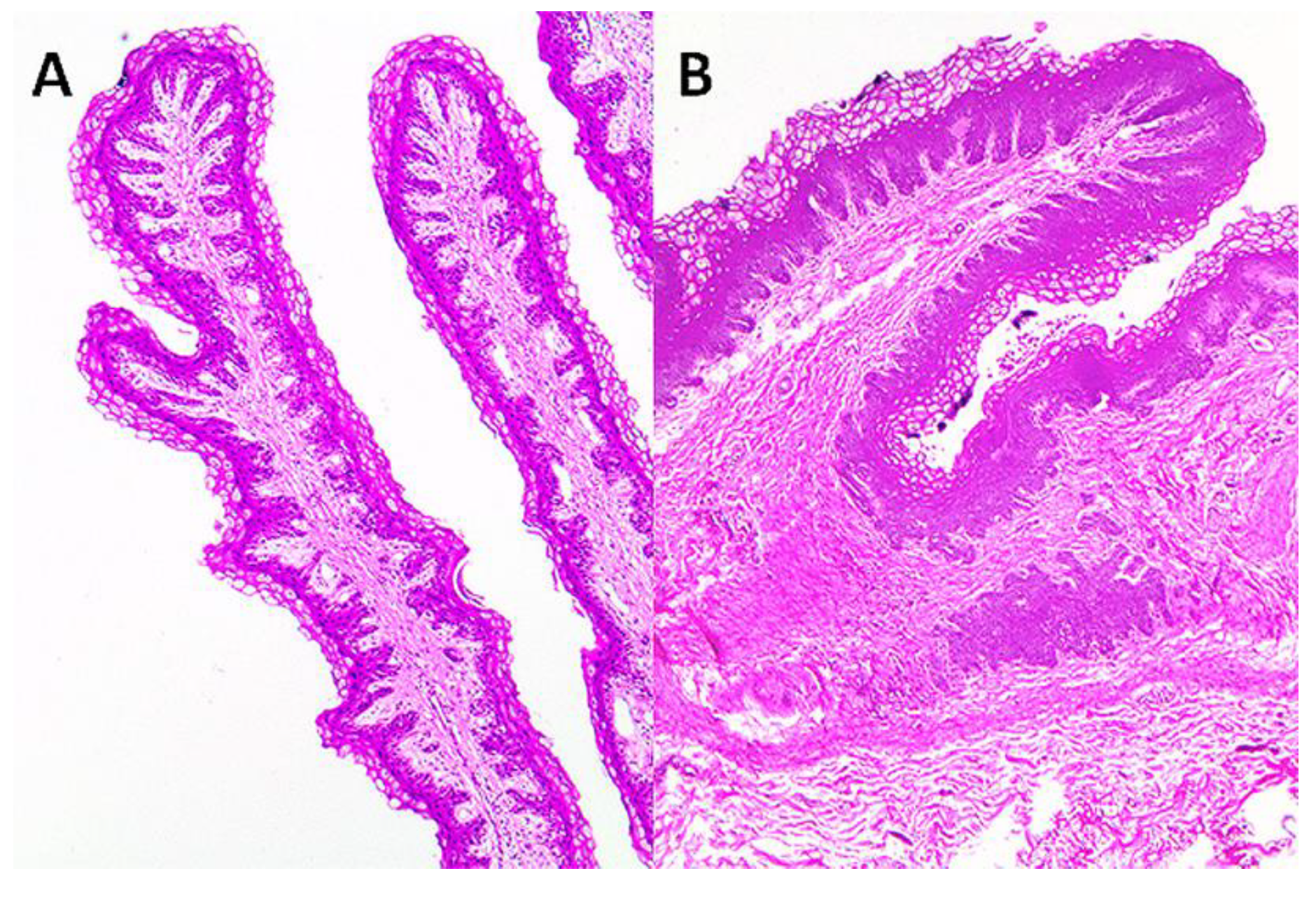
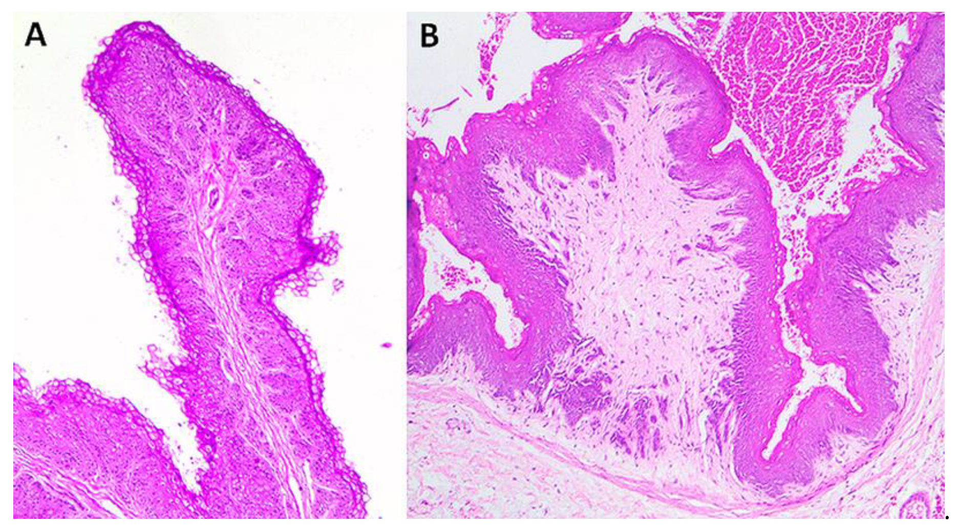
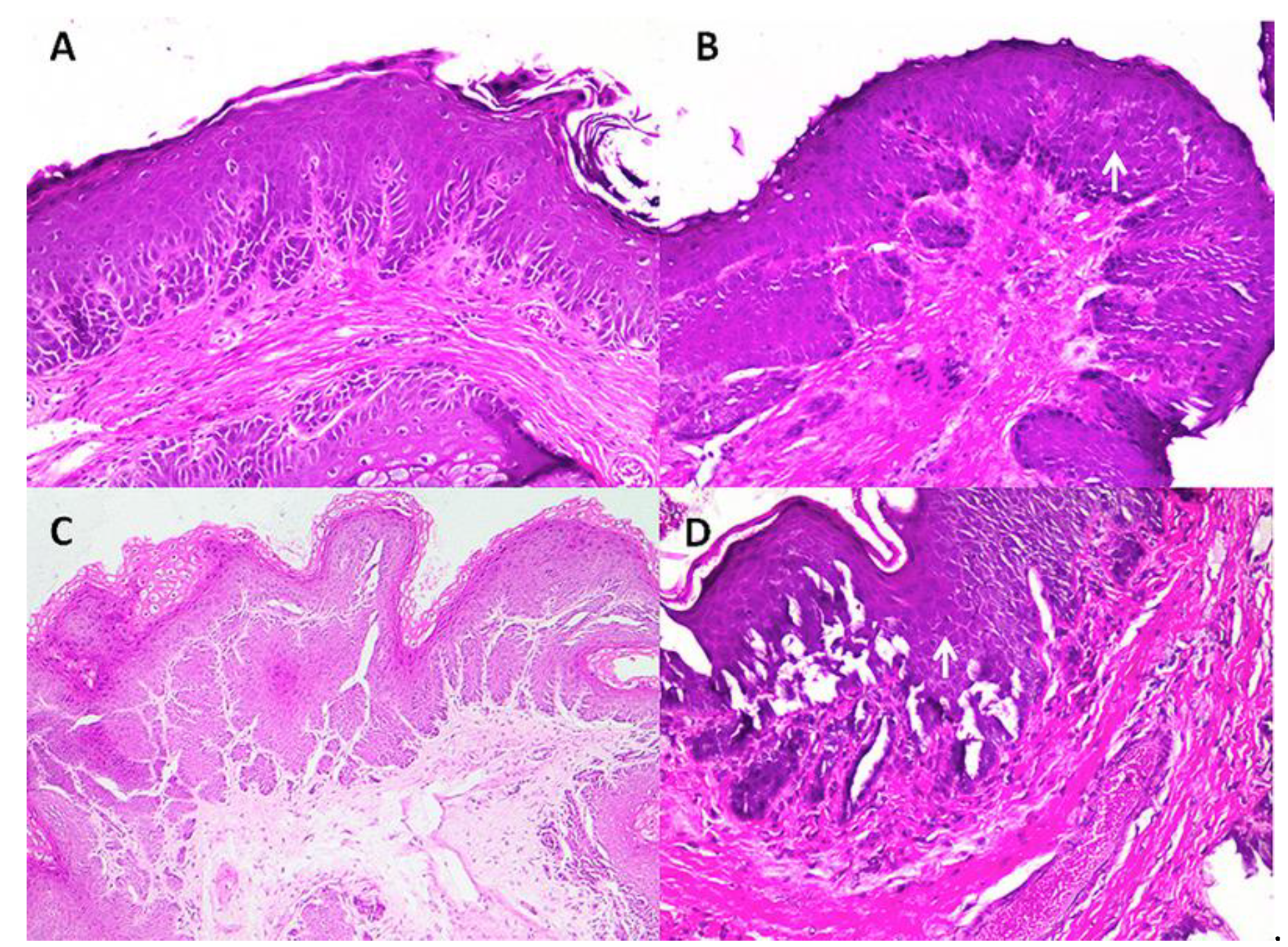
Statistical analyses were completed at a 95% confidence using SPSS 22.0 software (SPSS Inc., Chicago, IL, USA). Because of the sample size and the unknown distribution in the population, a non-parametric Mann–Witney U test was employed to determine the relation of the length of the rumen papillae and the presence/absence (control) of foreign objects. The Spearson correlation coefficient was employed to measure the correlation between the length of the papillae and the foreign body weight.

3. Results

All goats and sheep recovered successfully within the next days after surgery. The animals showed a progressive appetite, and a normal water ingestion and behavior. Ten days later, the skin sutures were removed, and the animals were transported to their farms. Six months after surgery, the farmers were contacted to know the medium-term outcome, and the animals were in good body condition and apparently in complete recovery.
The materials extracted from ruminal cavities were large shapeless masses of tightly compacted indigestible foreign matter and vegetable materials. The masses were composed mainly of synthetic plastics (raffia) and fiber ropes. The mean weight of the foreign objects was 2.750 ± 1.577 kg, which was considered heavy in comparison to the body weights that ranged from 23–38 kg. The heaviest foreign object weighed 5.5 kg.
During the rumenotomies, rumen mucosae appeared macroscopically different from their normal appearance; particularly, the length of the rumen papillae seemed to be shorter. Once measured, statistically significant differences were found (p < 0.05) between the affected animals (1.243 ± 0.621) and the control ones (3.097 ± 0.754). However, when the length of the papillae was compared between species (goats vs. sheep), statistical differences were found in neither the affected group (1.432 ± 0.706 vs. 1.016 ± 0.429) nor the control group (3.382 ± 0.889 vs. 2.812 ± 0.47).
A moderate/high negative correlation between the length of the rumen papillae and the weight of the foreign object was observed in our study (r = −0.665, p < 0.05). The main histological findings are summarized in Table 1.
Reactive changes were particularly characterized by the regular hyperplasia of the squamous epithelium of the ruminal mucosae and mononuclear infiltration in the subepithelial spaces and, less consistently, by the vacuolization of keratinocytes, presence of intraepithelial leukocytes and vascular alterations of the lamina propria (Figure 1, Figure 2 and Figure 3).

4. Discussion

The presence of indigestible foreign objects in the forestomachs of ruminants has been widely described in the literature [10,11,12,13,14,15,16,17,18,19,20,21,22]. The ingestion of a synthetic fiber which formed the nidus of a concretion has been described as a consequence of the consumption of synthetic fiber in an attempt to increase feed intake [35]. Hartnack et al. [36], in a retrospective case series of 95 cattle, performed rumenostomy because of traumatic reticuloperitonitis (n = 31), bloat (9), foreign body (6), choke (5), and other (2). In our case, all animals had foreign bodies by ingestion of non-degradable materials used in agriculture exploitations focused on tomato production. The tomato plants in their vertical growth are attached with different materials, plastic-based materials being one of the most commonly used, at least in the studied area.
Uzal et al. [37] attributed a lesser susceptibility to developing foreign bodies in the rumen of goats and sheep to the selective eating habits when compared to calves. In fact, Sheferaw et al. [14] described that the prevalence of foreign bodies in slaughtered animals was significantly higher in cattle (41.8%) than in sheep (20.6%) and goats (11.9%). If so, the nutritional requirements of the animals would not be covered by conventional foods in the farms, and, consequently, the ingestion of “foreign material” would be higher than desirable. Since all animals were fed in the same way, it is possible that the swallowing behavior was the reason why some animals ingested the foreign bodies and others did not. However, this hypothesis has not been demonstrated and should be studied further in the future.
In a study carried out in Eastern Ethiopia with the free grazing of livestock in highly waste-polluted areas (without plastic waste disposal systems), 43.4% of cattle (144/332), 56.5% of sheep (109/193) and 59.3% of goats (169/285) had foreign bodies, mainly plastic (79.2%), cloths (15.3%) and rope (12.3%) [13]. Therefore, non-degradable agriculture materials could be considered another source of foreign bodies in small ruminants.
Uzal et al. [37] have indicated that the lesser prevalence of foreign bodies in small ruminants could also be explained by the regurgitation phenomena, mainly for those light and small bodies. In fact, regurgitation is recognized to be the main mechanism of bolus losses [38,39,40]. Except for small bodies, the high size and weight of the foreign bodies observed in the current study preclude regurgitation.
The growth and development of the rumen papillae depends mainly on the type of diet ingested. Animals fed with rations containing adequate levels of roughage develop long, slender, regular, white to grey, rumen papillae [37]. The larger length of rumen papillae has been attributed to a higher volatile fatty acids (VFA) content, which influences the development of papillae [41,42,43] in the absence of rumen acidosis by overproduction of VFA [44].
The length of the papillae observed in the affected animals was shorter than for the control ones. We also found that the heavier the foreign body, the shorter the length of the ruminal papillae. This negative correlation was statistically significant. The effect of the foreign bodies on the papillae is unknown; however, one could suppose a pressure effect and continuous mechanical injuries on the ruminal mucosae. Histologically dystrophic changes in the ruminal mucosa were observed, which would have been promoted by the presence of foreign bodies. On the other hand, the foreign bodies would reduce the ruminal capacity with a lesser ingestion of foods and probably digestive impairments. According to Suárez et al. [44], the smaller size of the rumen papillae suggests the scarce presence of VFA in the ruminal fluid, which is directly related to a low carbohydrate ingestion. Nevertheless, chronic ruminal acidosis, usually due to rations high in carbohydrates, is the most common cause of dystrophic changes in the ruminal mucosa. The morphological changes observed in ruminal papillae are clumps, nodules and rosettes [37]. The histological changes observed in our case resemble those described for chronic acidosis, such as acanthosis, hyper- and parakeratosis and hyperpigmentation of the ruminal papillae.
Papillae were shortened, the epithelium increased in thickness and pigmentation, and inflammatory infiltrations with different degrees were present at the mucosal level in both goats and sheep. Epithelial hyperplasia was the most frequent finding in the affected animals. Additionally, the presence of suprabasilar vesicles (9/12) and oedema of the lamina propria (4/12) were more prevalent in goats than in sheep. These changes could be attributed to chronic frictional micro-trauma causing dystrophic epithelial changes, resulting in cellular degeneration and leukocytic infiltration. These lesions can modify the absorption capacity and stimulate inflammation and secondary ruminal infection by the resident microflora. Our results are similar to the histological findings previously described [29,30].
A sustainable animal production could be related to the use of local by-products and local forages because of the high cost and low availability of classical animal feeds in similar geographical areas to that of our study. Further studies related to the composition of tomato by-products are required to completely justify the use of this material as a source of fiber and nutrients for small ruminants.

5. Conclusions

Small ruminants seem susceptible to developing foreign bodies in their forestomachs by ingestion of non-degradable material. The presence of this material produced reactive changes that closely resembled those described in chronic ruminal acidosis. This condition would cause inflammatory and/or degenerative changes in the rumen with severe clinical signs. Therefore, our findings suggest that plastic foreign bodies play an important role in the pathogenesis of rumenitis and epithelial hyperplasia. Oedema of the lamina propria is more prevalent in goats than in sheep. Given its close relationship with agriculture management systems, the use of bio-degradable material is questionable so as to avoid new cases.

Author Contributions

Conceptualization, S.M.M., J.A.C. and M.T.T.-J.; methodology, J.A.C., S.M.M. and F.R.-G.; validation, J.A.C. and M.T.T.-J.; formal analysis, J.A.C.; investigation, S.M.M., M.M., I.M., J.R.J., F.R.-G. and J.A.C.; resources, M.M., S.M.M., F.R.-G. and J.A.C.; writing—original draft preparation, J.A.C., M.T.T.-J., S.M.M. and J.R.J.; writing—review and editing, J.A.C. and M.T.T.-J.; visualization, J.A.C. and F.R.-G.; supervision, J.A.C. and M.T.T.-J.; funding acquisition, M.M. and J.A.C. All authors have read and agreed to the published version of the manuscript.

Funding

This research received no external funding.

Institutional Review Board Statement

An ethical review and approval were waived for this study because the research data were obtained during clinical and surgical procedures during animal patients’ care at the veterinary hospital.

Informed Consent Statement

Not applicable.

Data Availability Statement

Data is contained within the article.

Acknowledgments

The authors thank Jorge Oros for his appreciated help and expertise. His essential contribution on the study was the measurement of the papilla length on the histological specimens and the microphotographs included in the Figures.

Conflicts of Interest

The authors declare no conflict of interest.

References

  1. Abdullah, L. The Development of Integrated Forage Production System for Ruminants in Rainy Tropical Regions-the Case of Research and Extension Activity in Java, Indonesia. Bull. Fac. Agric. Univ. 2006, 58, 125–128. [Google Scholar]
  2. Ajila, C.M.; Brar, S.K.; Verma, M.; Tyagi, R.D.; Godbout, S.; Valéro, J.R. Bio-processing of agro-byproducts to animal feed. Crit. Rev. Biotechnol. 2012, 32, 382–400. [Google Scholar] [CrossRef] [PubMed]
  3. Abubakr, A.; Alimon, A.R.; Yaakub, H.; Abdullah, N.; Ivan, M. Effect of feeding palm oil by-products based diets on muscle fatty acid composition in goats. PLoS ONE 2015, 10, e0119756. [Google Scholar] [CrossRef] [PubMed] [Green Version]
  4. Ahmed, S.T.; Lee, J.-W.; Mun, H.-S.; Yang, C.-J. Effects of supplementation with green tea by-products on growth performance, meat quality, blood metabolites and immune cell proliferation in goats. J. Anim. Physiol. Anim. Nutr. 2015, 99, 1127–1137. [Google Scholar] [CrossRef] [PubMed]
  5. Ben Salem, H.; Priolo, A.; Morand-Fehr, P. Shrubby vegetation and agro-industrial by-products as alternative feed resources for sheep and goats: Effects on digestion, performance and product quality. Anim. Feed Sci. Technol. 2008, 147, 1–2. [Google Scholar] [CrossRef]
  6. Foster, D. Disorders of Rumen Distension and Dysmotility. Vet. Clin. N. Am. Food Anim. Pract. 2017, 33, 499–512. [Google Scholar] [CrossRef]
  7. Abu-Seida, A.M.; Al-Abbadi, O.S. Studies on sharp foreign body syndrome in iraqi buffaloes and its impact on milk production. Asian J. Anim. Sci. 2015, 9, 128–133. [Google Scholar] [CrossRef] [Green Version]
  8. Nongcula, V.V.; Zhou, L.; Nhundu, K.; Jaja, I.F. Association between the prevalence of indigestible foreign objects in the gastrointestinal tract of slaughtered cattle and body condition score. Animals 2017, 7, 80. [Google Scholar] [CrossRef] [Green Version]
  9. Priyanka, M.; Dey, S. Ruminal impaction due to plastic materials-An increasing threat to ruminants and its impact on human health in developing countries. Vet. World 2018, 11, 1307–1315. [Google Scholar] [CrossRef] [Green Version]
  10. Akraiem, A.; Abd Al-Galil, A.S.A. Rumen impaction in cattle due to plastic materials. J. Vet. Med. Res. 2016, 23, 9–14. [Google Scholar]
  11. Bwatota, S.F.; Makungu, M.; Nonga, H.E. Occurrences of Indigestible Foreign Bodies in Cattle Slaughtered at Morogoro Municipal Slaughterhouse, Tanzania. J. Vet. Med. 2018, 2018, 4818203. [Google Scholar] [CrossRef] [PubMed] [Green Version]
  12. Mushonga, B.; Habarugira, G.; Musabyemungu, A.; Udahemuka, J.C.; Jaja, F.I.; Pepe, D. Investigations of foreign bodies in the fore-stomach of cattle at ngoma slaughterhouse, rwanda. J. S. Afr. Vet. Assoc. 2015, 86, 1–6. [Google Scholar] [CrossRef] [PubMed] [Green Version]
  13. Negash, S.; Sibhat, B.; Sheferaw, D. A postmortem study on indigestible foreign bodies in the rumen and reticulum of ruminants, Eastern Ethiopia. Onderstepoort J. Vet. Res. 2015, 82, 1–5. [Google Scholar] [CrossRef] [Green Version]
  14. Sheferaw, D.; Gebru, F.; Asrat, M.; Tesfaye, D.; Debela, E. Ingestion of indigestible foreign materials by free grazing ruminants in Amhara Region, Ethiopia. Trop. Anim. Health Prod. 2014, 46, 247–250. [Google Scholar] [CrossRef]
  15. Dharmaceelan, S.; Kumaresan, A. Kanjanadevi Surgical management of ruminal impaction due to indigestible foreign bodies in cattle. Intas Polivet 2017, 2, 26–27. [Google Scholar]
  16. Dharmaceelan, S.; Kumaresan, A. Devi Kanjana Surgical Management of Ruminal Impaction in a Goat. Intas Polivet 2017, 18, 329–330. [Google Scholar]
  17. Eriksen, M.; Lusher, A.; Nixon, M.; Wernery, U. The plight of camels eating plastic waste. J. Arid Environ. 2021, 185, 104374. [Google Scholar] [CrossRef]
  18. Fromsa, A.; Mohammed, N. Prevalence of indigestible foreign body ingestion in small ruminants slaughtered at Luna Export Abattoir, East Shoa, Ethiopia. J. Anim. Vet. Adv. 2011, 10, 1598–1602. [Google Scholar]
  19. Hayder, A.M.; Bakhiet, A.O.; Mohammed, A.A. Retrospective Study on the Prevalence of Foreign Body in Goats’ Rumen: Omdurman Province, Khartoum State, Sudan (1998–2002). J. Anim. Vet. Adv. 2006, 5, 449–451. [Google Scholar]
  20. Hayder, A.M. Survey Study of Foreign Body in Caprine Rumen in Khartoum State. Ph.D. Thesis, Sudan University of Science and Technology, Khartoum, Sudan, 2004. [Google Scholar]
  21. Igbokwe, I.O.; Kolo, M.Y.; Egwu, G.O. Rumen impaction in sheep with indigestible foreign bodies in the semi-arid region of Nigeria. Small Rumin. Res. 2003, 49, 141–146. [Google Scholar] [CrossRef]
  22. Kh, S.; Sarkar, D.; Gn, R.; Dh, C.; Vishwanth, S. Review of literature with a case report: Severe ruminal impaction caused by different types of foreign bodies in the rumen of a post parturient boer goat. Pharma Innov. J. 2019, 8, 171–175. [Google Scholar]
  23. Beriot, N.; Peek, J.; Zornoza, R.; Geissen, V.; Huerta Lwanga, E. Low density-microplastics detected in sheep faeces and soil: A case study from the intensive vegetable farming in Southeast Spain. Sci. Total Environ. 2021, 755, 142653. [Google Scholar] [CrossRef] [PubMed]
  24. Mahadappa, P.; Krishnaswamy, N.; Karunanidhi, M.; Bhanuprakash, A.G.; Bindhuja, B.V.; Dey, S. Effect of plastic foreign body impaction on rumen function and heavy metal concentrations in various body fluids and tissues of buffaloes. Ecotoxicol. Environ. Saf. 2020, 189, 109972. [Google Scholar] [CrossRef] [PubMed]
  25. Olatunji-Akioye, O.A.; Olawoyin, C.M.; Oyeyemi, M.O. Incidence and consequence of surgical removal of gastric foreign bodies in West African Dwarf goats in Ibadan. Anim. Res. Int. 2019, 16, 3478–3483. [Google Scholar]
  26. Raoofi, A.; Namjoo, A.; Karimi, A.H.; Alizadeh Esfahani, M. A study of clinical signs, hematological changes and pathological findings of experimental ingestion of soft foreign body (plastic rope) in goats. Small Rumin. Res. 2012, 105, 351–354. [Google Scholar] [CrossRef]
  27. Otsyina, H.R.; Mbuthia, P.G.; Nguhiu-Mwangi, J.; Mogoa, E.G.M.; Ogara, W.O. Gross and histopathologic findings in sheep with plastic bags in the rumen. Int. J. Vet. Sci. Med. 2017, 5, 152–158. [Google Scholar] [CrossRef]
  28. Sadan, M.; El-Shafaey, E.S.; Al-Sobayil, F. Diagnosis and treatment of foreign bodies swallowing syndrome in camels (Camelus dromedarius) with special reference to the role of mineral deficiency. J. Vet. Med. Sci. 2020, 82, 1097–1103. [Google Scholar] [CrossRef]
  29. Hailat, N.; Nouh, S.; Al-Darraji, A.; Lafi, S.; Al-Ani, F.; Al-Majali, A. Prevalence and pathology of foreign bodies (plastics) in awassi sheep in jordan. Small Rumin. Res. 1997, 24, 43–48. [Google Scholar] [CrossRef]
  30. Hailat, N.; Al-Darraji, A.; Lafi, S.; Barakat, S.A.; Al-Ani, F.; El-Magrhaby, H.; Al-Qudah, K.; Gharaibeh, S.; Rousan, M.; Al-Smadi, M. Pathology of the rumen in goats caused by plastic foreign bodies with reference to its prevalence in Jordan. Small Rumin. Res. 1998, 30, 77–83. [Google Scholar] [CrossRef]
  31. Canary Islands Institute of Statistics (ISTAC). Available online: http://www.gobiernodecanarias.org/istac/ (accessed on 10 July 2021).
  32. Niehaus, A.J.A.J. Rumenotomy. Vet. Clin. N. Am. Food Anim. Pract. 2008, 24, 341–347. [Google Scholar] [CrossRef]
  33. Martin, S.; López, A.M.; Morales, M.; Morales, I.; Tejedor-Junco, M.T.; Corbera, J.A. Rumenotomy in small ruminants—A review. J. Appl. Anim. Res. 2021, 49, 104–108. [Google Scholar] [CrossRef]
  34. Lesmeister, K.E.; Tozer, P.R.; Heinrichs, A.J. Development and Analysis of a Rumen Tissue Sampling Procedure. J. Dairy Sci. 2004, 87, 1336–1344. [Google Scholar] [CrossRef] [Green Version]
  35. Leask, R.; Bath, G.F. Rumenolith formation in a Bapedi ram. J. S. Afr. Vet. Assoc. 2012, 83, 1–4. [Google Scholar] [CrossRef] [Green Version]
  36. Hartnack, A.K.A.K.; Niehaus, A.J.A.J.; Rousseau, M.; Pentecost, R.L.R.L.; Miesner, M.D.M.D.; Anderson, D.E.D.E. Indications for and factors relating to outcome after rumenotomy or rumenostomy in cattle: 95 cases (1999–2011). J. Am. Vet. Med. Assoc. 2015, 247, 659–664. [Google Scholar] [CrossRef]
  37. Uzal, F.A.; Plattner, B.L.; Hostetter, J.M. Alimentary System. Jubb Kennedy Palmer’s Pathol. Domest. Anim. 2016, 2, 1–257.e2. [Google Scholar]
  38. Riner, J.L.; Byford, R.L.; Stratton, L.G.; Hair, J.A. Influence of density and location on degradation of sustained-release boluses given to cattle. Am. J. Vet. Res. 1982, 43, 2028–2030. [Google Scholar] [PubMed]
  39. Caja, G.; Conill, C.; Nehring, R.; Ribó, O. Development of a ceramic bolus for the permanent electronic identification of sheep, goat and cattle. Comput. Electron. Agric. 1999, 24, 45–63. [Google Scholar] [CrossRef]
  40. Garin, D.; Caja, G.; Conill, C. Performance and effects of small ruminal boluses for the electronic identification of fattening lambs. Livest. Prod. Sci. 2005, 92, 47–58. [Google Scholar] [CrossRef]
  41. Flatt, W.P.; Warner, R.G.; Loosli, J.K. Influence of Purified Materials on the Development of the Ruminant Stomach. J. Dairy Sci. 1958, 41, 1593–1600. [Google Scholar] [CrossRef]
  42. Heinrichs, J. Rumen Development in the Dairy Calf Rudimentary Reticulo-Rumen Changes in Rumen Epithelium. Technology 2005, 17, 179–187. [Google Scholar]
  43. Khan, M.A.; Lee, H.J.; Lee, W.S.; Kim, H.S.; Ki, K.S.; Hur, T.Y.; Suh, G.H.; Kang, S.J.; Choi, Y.J. Structural growth, rumen development, and metabolic and immune responses of Holstein male calves fed milk through step-down and conventional methods. J. Dairy Sci. 2007, 90, 3376–3387. [Google Scholar] [CrossRef] [PubMed] [Green Version]
  44. Suárez, B.J.; Van Reenen, C.G.; Stockhofe, N.; Dijkstra, J.; Gerrits, W.J.J. Effect of roughage source and roughage to concentrate ratio on animal performance and rumen development in veal calves. J. Dairy Sci. 2007, 90, 2390–2403. [Google Scholar] [CrossRef] [Green Version]
Figure 1. Histological comparison between the (A) control and (B) affected ruminal mucosae in sheep. The reduction in the length of the papillae and intense hyperkeratosis, with areas of pseudo-carcinomatous hyperplasia (bottom), were prominent features. H&E, ×40. Microphotography courtesy of Dr. Jorge Orós.
Ruminants 01 00009 g001
Figure 2. (A) Normal and (B) affected goat rumens. The papillae became shorter, and the stratified squamous epithelium showed intense acanthosis. Cellular debris are present in the luminal surface. H&E, ×40. Microphotography courtesy of Dr. Jorge Orós.
Ruminants 01 00009 g002
Figure 3. Details of histological changes in affected animals. (A,B) Hyperplasia with increased number of keratinocytic layers; Exocytosis with the presence of leukocytes within the epithelium (arrows in B and D); (C) Pseudo-carcinomatous hyperplasia and interstitial oedema; (D) Congestion and vacuolar degeneration (spongiosis) of the epithelium, with areas of early detachment. H&E, ×100 (A,B,D), ×40 (C). Microphotography courtesy of Dr. Jorge Orós.
Ruminants 01 00009 g003
Table 1. Histological findings in the bezoar group.
Histological ChangesNumber of Goats/TotalNumber of Ewes/Total
Epithelial changes
Acanthosis9/129/10
Pseudocarcinomatous hyperplasia3/125/10
Hyperkeratosis4/123/10
Spongiosis/suprabasilar vesicles9/121/10
Exocytosis of lymphocytes8/128/10
Hyperpigmentation1/121/10
Subepithelial changes
Mononuclear infiltration7/127/10
Publisher’s Note: MDPI stays neutral with regard to jurisdictional claims in published maps and institutional affiliations.

