Review

The Diagnostic Challenges of Ovine Pulmonary Adenocarcinoma

1
Mountain Research Center (CIMO), School of Agriculture, Polytechnic Institute of Bragança (IPB), Campus de Santa Apolónia, 5300-253 Bragança, Portugal
2
Department of Veterinary Science, University of Trás-os-Montes and Alto Douro Quinta dos Prados, 5001-801 Vila Real, Portugal
3
CITAB—Centre for the Research and Technology of Agro-Environmental and Biological Sciences, University of Trás-os-Montes and Alto Douro, Quinta dos Prados, 5001-801 Vila Real, Portugal
4
INNO Veterinary Laboratory, R. Cândido de Sousa 15, 4710-503 Braga, Portugal
5
Instituto Universitário de Ciências da Saúde, CESPU, Instituto de Investigacão e Formação Avançada em Ciências e Tecnologias da Saúde (IINFACTS), Rua Central de Gandra, 1317, 4585-116 Gandra, Portugal
*
Author to whom correspondence should be addressed.
Ruminants 2021, 1(1), 58-71; https://doi.org/10.3390/ruminants1010005
Submission received: 7 August 2021 / Revised: 10 September 2021 / Accepted: 15 September 2021 / Published: 18 September 2021
(This article belongs to the Special Issue Feature Papers of Ruminants 2021-2022)

Abstract

:
Ovine pulmonary adenocarcinoma (OPA), also known as sheep pulmonary adenomatosis and jaagsiekte, is a contagious pulmonary tumor of sheep, characterized by neoplastic proliferation of type II pneumocyte and club cells. OPA is induced by the oncogenic activity of the envelope glycoprotein (Env) of exogenous jaagsiekte sheep retrovirus (JSRV). This disease is associated with significant economic losses in numerous sheep raising countries. The onset of suggestive clinical signs is often late, making difficult the early diagnosis of the disease and timely implementation of control measures on the affected farms. Further, the lack of diagnostic tests that can be performed routinely by veterinary clinicians to accurately assess infected animals (e.g., serological or others) means that the true prevalence at flock level is not known. Imaging diagnostic methods (e.g., ultrasound, X-ray and computed tomography) can be used to support the clinical diagnosis, even in pre-clinical stages in affected flocks. The diagnosis must be confirmed by PCR of nasal excretions or immunohistochemistry and PCR of tumor lesions. No vaccine for OPA has yet been developed. Thus, in this work, we review the main methods of diagnosis of OPA in order to support the clinician in the identification of the disease, avoid underdiagnosis and allow the implementation of suitable measures to prevent and control its spread.

1. Introduction

Ovine pulmonary adenocarcinoma (OPA), also known as pulmonary adenomatosis and jaagsiekte, is a contagious neoplasia derived from type II pneumocytes and club cells, that occurs in sheep and, exceptionally, in goats and mouflons. The disease is associated with high economic losses not only due to the related mortality but also due to the lack of a vaccine or methods of early detection of the disease [1,2].
One of the first known records of this disease is a letter dated 1825, addressed to the magistrate of the Cape of Good Hope in South Africa, a farmer complained about the loss of many sheep due to a disease called “jaagziekte”, German word translated into africander as jaagsiekte (in Africander, “jaag” means to drive, drive and “siekte” means illness). The origin of the name of the disease reflects the breathing difficulties experienced by the affected animals, particularly evident during herd movements [3].
The infectious nature of the disease was shown by Dungal, Iceland, where the disease took on high proportions and received international veterinary attention, being successfully eradicated by compulsory slaughter [4].
The disease is described around the world in many sheep farming areas, including countries on the European and Asian, African and American continents [1,5,6,7,8]. In Portugal, several sporadic cases have been reported, although the prevalence is not known [9]. It is not reported in Australia and New Zealand, the Falkland Islands and Iceland [10].
OPA is induced by the oncogenic activity of the envelope glycoprotein (Env) of the exogenous jaagsiekte sheep retrovirus (JSRV). JSRV is a single-stranded, positive sense RNA virus in the genus Betaretrovirus, family Retroviridae [11,12]. JSRV genome has ∼7460 nucleotides, including four genes encoding viral proteins: env (encoding surface and transmembrane envelope glycoproteins), gag (encoding the matrix, capsid, and nucleocapsid proteins), pol (encoding reverse transcriptase and integrase enzymes) and pro (encoding aspartic protease) [11,13,14]. The protein env induces oncogenic transformation due its interaction with cell growth pathways such as Hya12-RON, PI3K/Akt, and Ras-MEK-ERK [15].
In the genome of sheep and goats copies of endogenous JSRV, (enJSRV) have been detected, having a similar gene structure to exogenous JSRV (exJSRV), but not associated with the development of tumors [6,12,13]. Despite this high homology, genetic differences are evident in the viral promoter region (U3) that allowed the development of the exJSRV-specific U3PCR. The genetic similarity is also smaller in the intracytoplasmic tail of the transmembrane protein encoded by the env gene [16] the latter being related to the mechanism of oncogenesis induced by Env [17].
In goats, there are few reports of natural infection, mostly unconvincing, possibly because of misdiagnosis [18]. However, in this species, the OPA acquires significant differences and virus replication does not take place within sensitive lung cells [19].
In an immunocytochemical study, antibodies against JSRV capsid were identified in human lung adenocarcinoma, but an association between JSRV and this neoplasm in humans could not be demonstrated [20]. The incubation period of natural infections is prolonged, likely for several months to several years. The incubation period can be shorter in nonendemic herds (6–8 months) and in experimentally infected young lambs [21,22,23,24].
The disease occurs mainly in adult sheep between the ages of 2 and 4, although experimental data show that sheep of all ages are susceptible. The presence of tumours was detected in animals from 2 months to 11 years of age, although cases of natural disease are rarely observed in sheep less than 9 months old [18,22,23]. Animals of either gender are affected [22]. In herds where the disease is endemic, the mortality rate is generally low (1–5%), but can reach higher values (>30%) in newly infected herds [6,22]. Although in endemic regions the disease is pronounced throughout the year, the number of cases tends to be higher in winter [22].
Statistical evidence also suggests that some breeds are more susceptible. In Iceland, animals of the Gottorp breed showed greater susceptibility, with some producers losing about 90% of the sheep, whereas only up to 10% of Adalbol sheep present in those farms presented clinical signs. [25,26,27]. Epidemiological analysis showed that transmission is mainly carried out by aerosols or droplets. However, transmission also takes place vertically, through colostrum and milk. However, transmission also takes place vertically, through colostrum and milk. JSRV has been found in macrophages of infected mother’s milk and has been shown to cross the gut barrier of newborn lambs and spread from mesenteric lymphodes and Peyer’s Patch. The transplacental pathway does not appear to have any epidemiological significance [28,29,30,31].
The use of new methods of diagnosis, has shown that the number of infected animals is far higher than the number of those who will develop lung cancer [32]. It is therefore important to know the clinical and pathological signs of these diseases, so as to understand their effective prevalence. On the other hand, being a viral-induced disease associated with a pulmonary tumor and although no link with human pulmonary tumors has been demonstrated, sheep can provide a natural model for the study of viral tumors in humans [11,20].
In this work, we will briefly look at classical diagnostic methods, based on clinical signs, imaging diagnosis, pathological diagnosis and molecular diagnosis. This work intends to be, more than an exhaustive review, a revisit to the classic diagnostic methods that can be decisive for the early diagnosis and the establishment of timely measures in order to avoid the high economic losses.

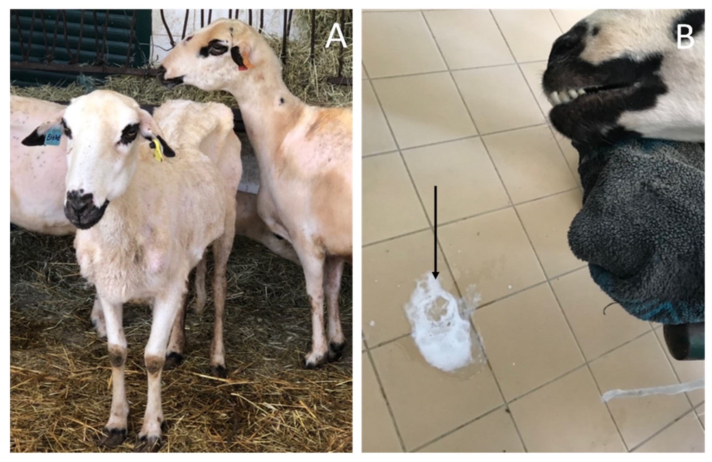
2. Clinical Diagnose

The disease when lesions are well developed, typically after a prolonged incubation period. They include non-specific signs such as progressive wasting (Figure 1A), weight loss and, typically, progressive dyspnea, which was initially detected only after exercise. The gradual replacement of the normal lung with tumor tissue will increase respiratory difficulties. Breathing develops with no apparent clinical signs for extended periods. Clinical signs are observed only becomes deeper and more frequent, associated with abdominal wall motions, orthopneic posture, dilated nostrils and open mouth [3,10,33].
Other signs are due to a build-up of fluid, produced by tumour cells, in the airways. The movement of these fluids, caused by the passage of air through the respiratory tract, produces crackle sounds (Supplementary Material Video S1), easily discernible after auscultation of the thoracic cavity. These fluids can flow into the nostrils of the animal (Figure 1B), especially when the animal drops its head, as happens during feeding. Severe nasal discharge can also be caused by lifting the hind limbs and lowering the head (“wheelbarrow test” performed for at least 30–40 s). The amount of fluid that may be collected from an animal varies from 10 to 60 mL, sometimes up to 400 mL. Occasional excess fluid in the airways can lead to spasmodic cough episodes [8,16,17,18]. This abundant fluid production is especially evident during a pulmonary endoscopy (bronchoscopy) procedure (Figure 2, Supplementary Material Video S2: observe oedema of the glottis, tracheal ulcers and abundant fluid in the trachea).
The usefulness of auscultation is not consensual. Audible pulmonary sounds may be absent especially in the “wheel-barrow negative” cases of OPA [28].
The duration of the disease varies and depends particularly on the age at which clinical signs are detected. In lambs younger than six months, progression is usually acute and death occurs within a few days. In adult sheep, the disease progresses more slowly, with clinical signs extending over several weeks or months, until the animal dies [22,30,31].
OPA can progress without fever, particularly in acute cases, but secondary bacterial complications occur almost always, shortening the duration and changing the course of the disease. The most frequently involved agent is Mannheimia haemolytica, which has a tendency to worsen clinical signs, triggering a febrile reaction in animals [10,34].
The evolution of experimentally acquired disease tends to be faster than in cases of natural disease, especially because the animals, almost always very young, are inoculated with high doses of the etiological agent, which causes death a few days after the appearance of the signs [21,35,36].
Maedi-visna and OPA can coexist in an animal, and the possibility of synergism between the two agents has been speculated. Under such circumstances, respiratory signs tend to be more severe [37,38,39]. Also the association and the possibility of synergistic action between JSRV and the nasal enzootic tumour virus (ENTV) has been pointed out [40,41] and, a enzootic nasal adenocarcinoma was described as associated with JSRV infection once [41].

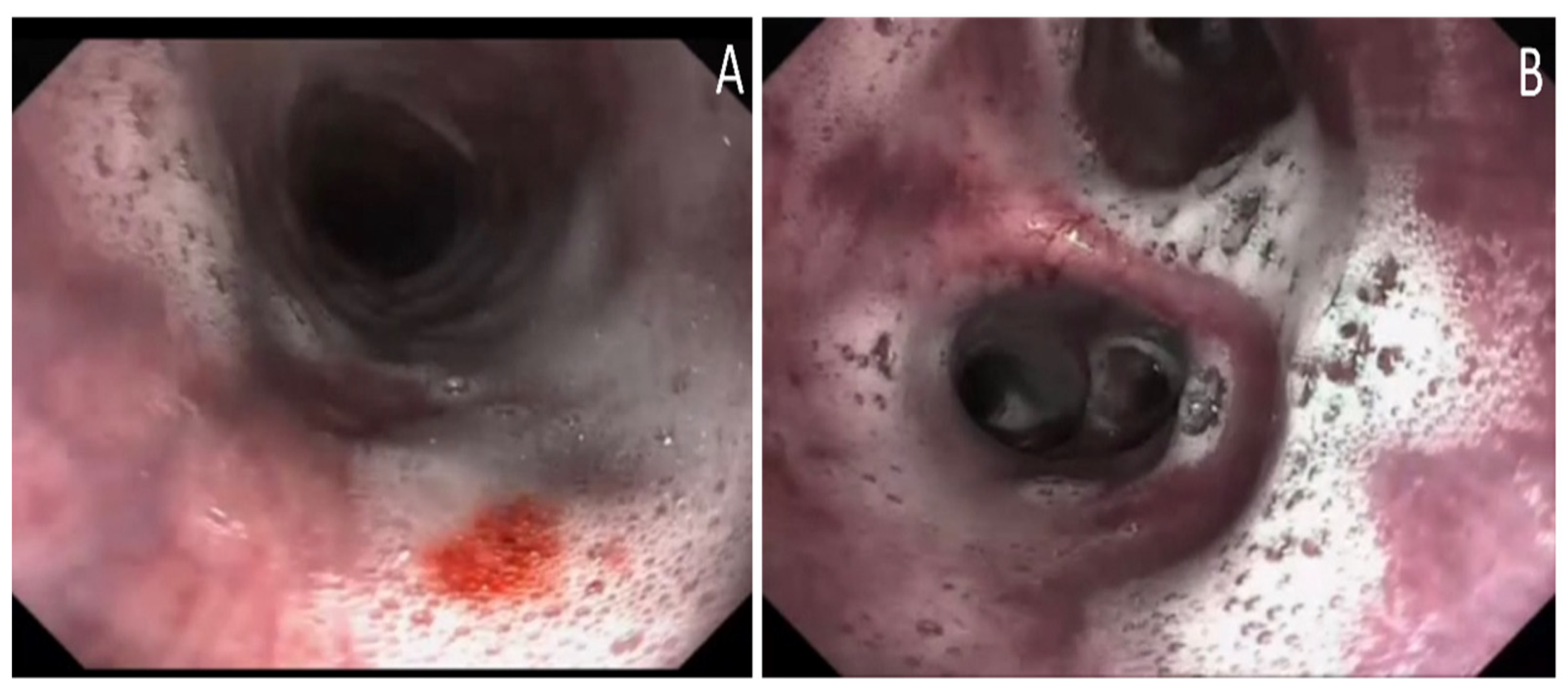
3. Imaging Diagnosis
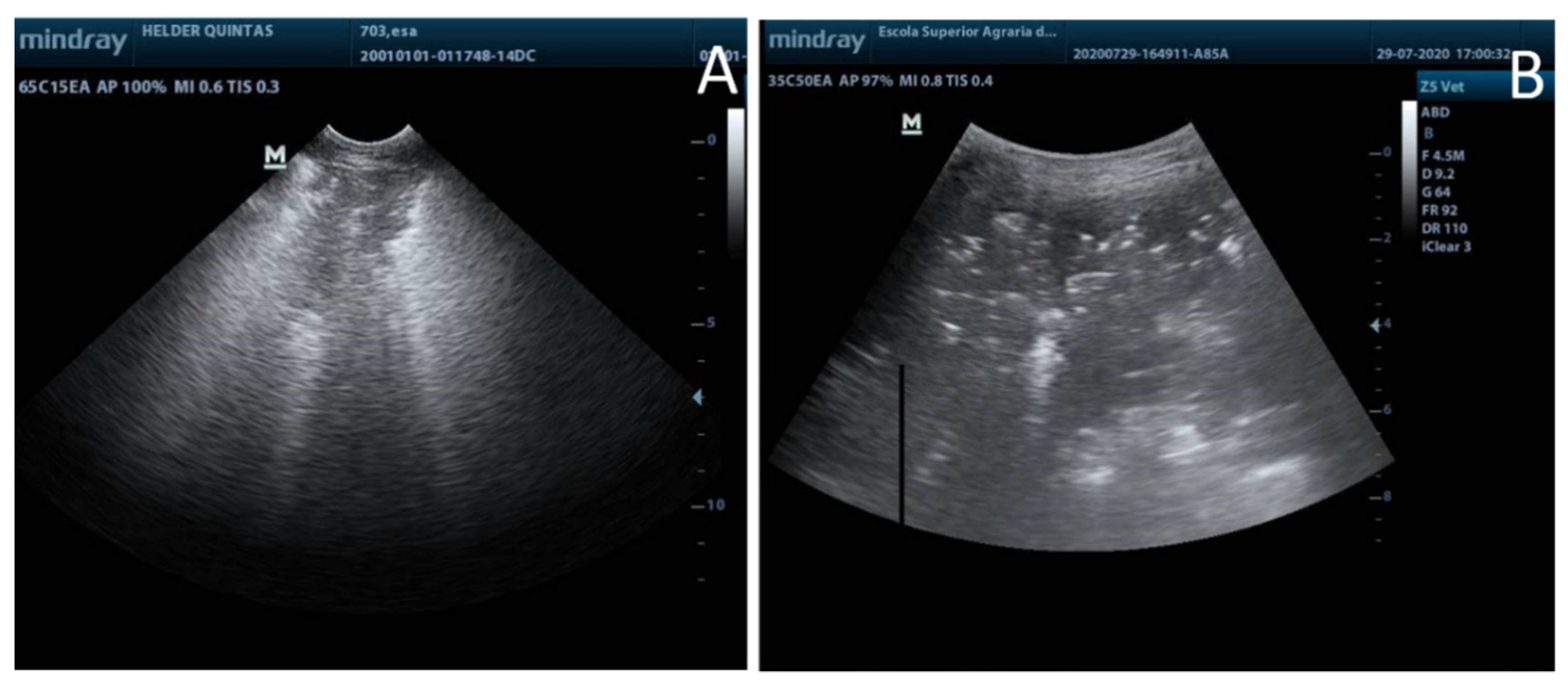
The lack of serological tests for early diagnosis and the low sensitivity and specificity of the clinical diagnosis mean that other diagnostic tools must be taken into consideration. Imaging techniques can be an extra tool for the diagnosis of pulmonary tumour lesions, especially in apparent «negative» cases of OPA. Although not practiced regularly, some of these techniques, especially ultrasound, can be simple, inexpensive and contribute significantly to the diagnosis of OPA under farm conditions [42,43,44].

3.1. Ultrasound

The procedure is well defined [45,46] and can be accurately outlined routinely as an integral part of a disease control plan. Healthy lungs present at ultrasound examination a hyperechoic white line close to the transducer (visceral pleura) and several reverberation artefacts below to aerated parenchyma. Areas consistent with OPA tumour mass can be identified when is observed a loss of this bright white line which is replaced by hypoechoic well-demarcated areas (lung consolidation areas) defined by a broad hyperechoic line [42,47,48] (Figure 3A) Echogenic areas of different sizes corresponding to neoplastic nodules may also be observed [49] (Figure 3B). Transthoracic ultrasonography may identify small tumours (>1 cm in diameter) even before clinical signs are evident [44]. Several authors recommend to perform an ultrasound examination of both sides of the thorax in any investigation of weight loss in sheep with or without respiratory signs [47,50]

3.2. Radiologic Diagnosis

Radiography and, mainly, computed tomography can be important tools to facilitate the understanding of processes at the respiratory level, such as OPA [49,51]. However, the use of ionizing radiation in the diagnosis of lung diseases in sheep is relatively expensive, most often only performed at a veterinary clinic/hospital for research purposes and is restricted by health and safety regulations.

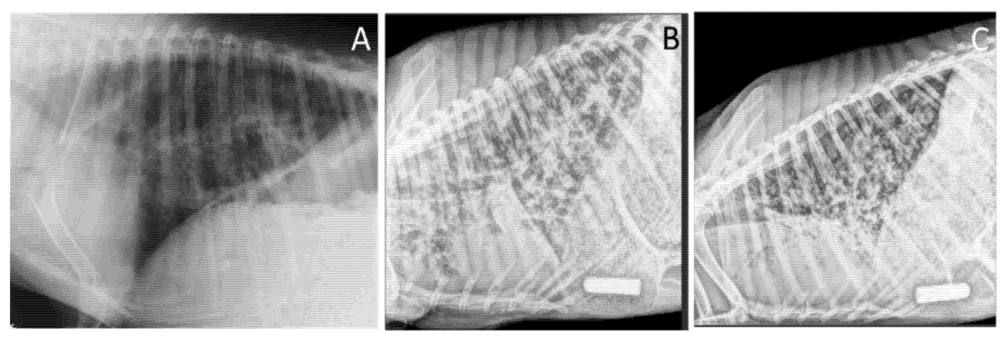
3.2.1. Radiography (X-ray Imaging)

The use of radiography, although it can provide important information about pulmonary lesions, is limited. It is dependent on the availability of portable X-ray equipment and is constrained by the cost and ability to meet health and safety conditions on farms. However, as much as possible, its diagnostic contribution is valuable. The thoracic lateral radiographic projection in a standing position which is limited to the caudo-dorsal lung fields (Figure 4A) which may not detect antero-ventral lesions in the early stages of the disease. Despite causing greater respiratory distress [52] and being more difficult to perform under on-farm conditions a thoracic lateral radiographic projection placing the sheep in lateral decumbency the forelimbs drawn forward allows full visualization of the lung fields (Figure 4B,C). Thoracic radiography can show nodules of different sizes and locations, sometimes a nodular pattern with diffuse nodules. Single small nodules as seen at the onset of the disease or larger lesions when tumor nodules converge. A patchy and hazy area of increased opacity may be present in the ventral lung parenchyma, in classical forms of OPA. An increase in cranioventral opacity may also result from secondary pneumonia at an advanced stage of the disease.

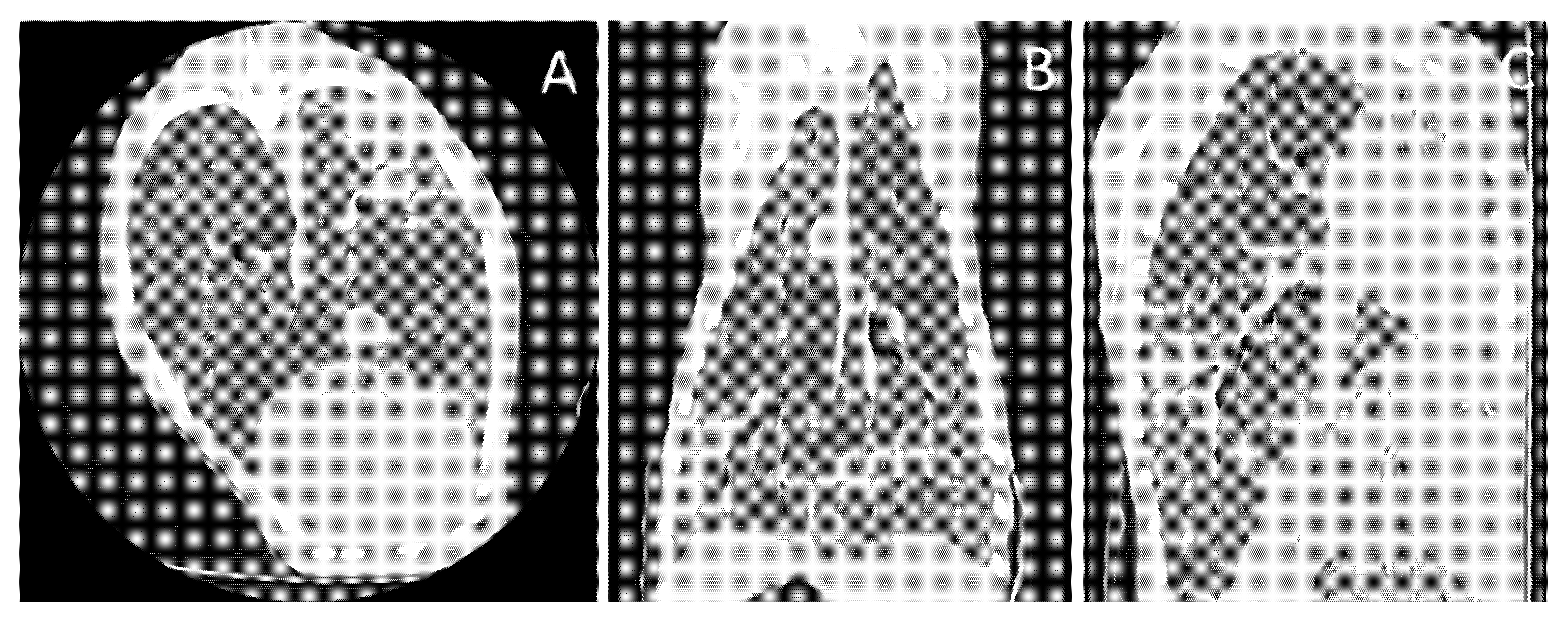
3.2.2. Computed Tomography

Computed tomography (CT) may be a tool to improve in vivo diagnosis and research to understand the pathogenesis of various pulmonary diseases, including OPA [49,53]. CT may enable early diagnosis and accurate detection of tumor nodules, their size and location. CT also enables axial, sagittal, and coronal sections to be obtained (Figure 5) and computerized three-dimensional color reconstruction, consisting of different tumor and respiratory tree densities, which is very helpful in understanding the disease, its pathogenesis, and with serial computed tomography over time, its evolution [51]. Thoracic CT scans generally do not require contrast media to make a precise diagnosis. Non-ionic iodinated contrast agents (usually low osmolality agents) may be useful for opacifying vascular structures. Thus, the need to enhance the tomographic image through intravenous contrast media depends on the option of the clinical team (Figure 5, Supplementary Materials Video S3).

4. Pathological Diagnosis

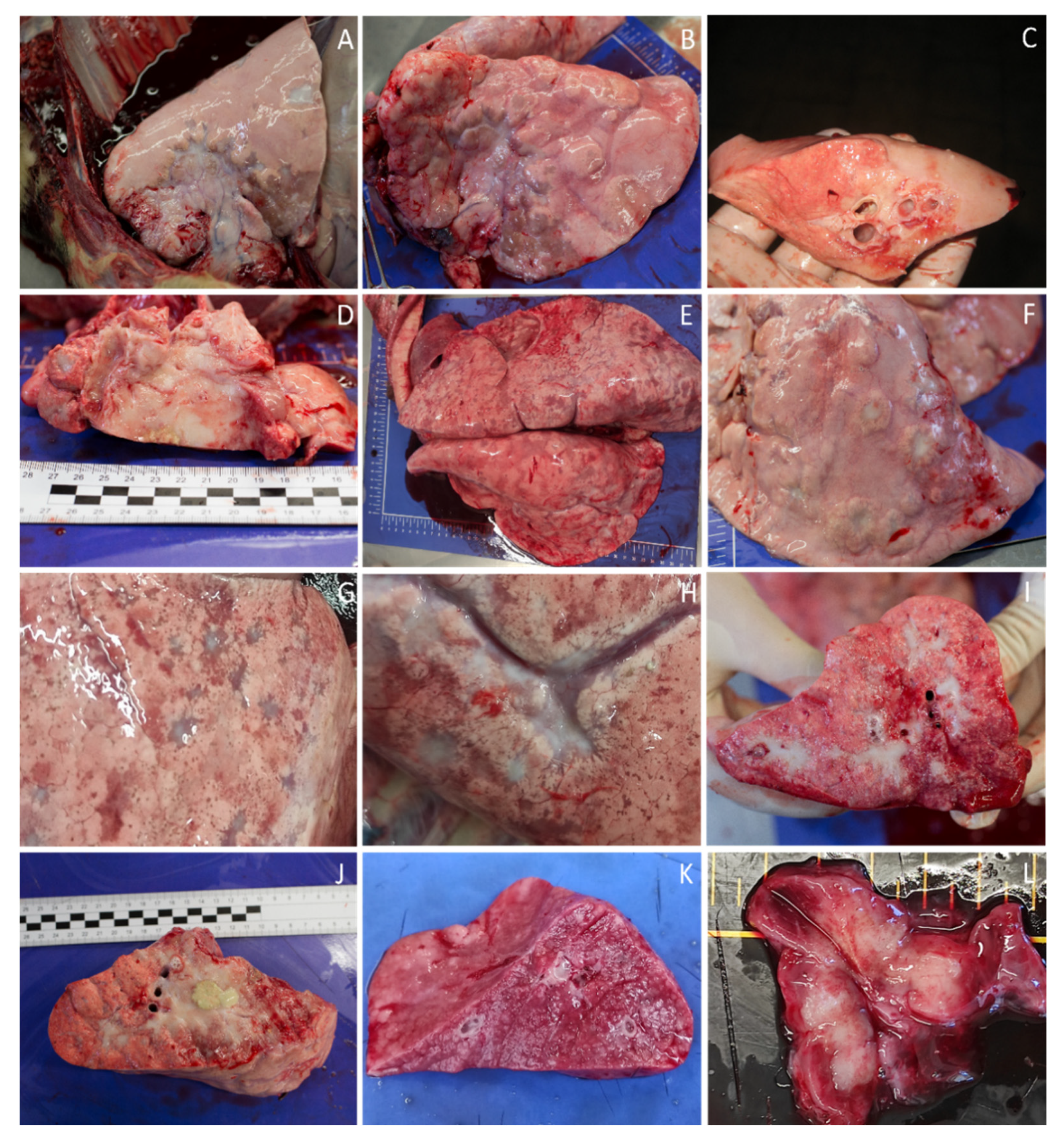
4.1. Macroscopic Diagnosis

OPA is characterized by pulmonary masses lesions ranging in size from small nodules to extensive tumors, sometimes involving half of a lobe. Two types of patterns are now recognised: classic and atypical (Figure 6). The two types may coexist in the same exploration and in the same animal, along with intermediate forms [9,52,53].
In the classic form, which is the most frequent, neoplastic lesions diffusely affect the cranioventral areas of the lung, although any other area may be involved [22,54,55]. The lungs are increased in volume and weight, up to 3 to 4 times, and fail to collapse, during the opening of the chest cavity [22]. Neoplastic masses are diffuse, nodular and slightly grey or pink, which do not extend beyond the surface of the lung. At the cut surface, their perimeter is irregular, small and slightly elevated, with a diffuse glandular appearance and a wet surface. Lesions may develop a diffuse appearance with no visible nodules. The consistency of tumor areas is increased, with a narrow band of pulmonary emphysema surrounding them, and the slight pressure causes clear, cloudy, foamy fluid arising from the bronchioles and bronchi. In advanced cases, the affected areas have a lardaceous, whitish, hard and solid appearance as a result of fibrosis [22,54,55,56].
The tumor pattern can be obscured by lesions of bacterial (namely Mannheimia and Mycoplasma) (Figure 6J,K), viral (Maedi) [37,38,57,58] or parasitic pneumonia [59].
In early cases, when clinical signs are not evident, the lesions may be confined to a very restricted pulmonary area, including in the form of a single tumor nodule, with very small dimensions [54]. The pleura is sometimes affected, with fibrosis and adhesions between the parietal and visceral surfaces, particularly at the anterior lobes [22].
The atypical neoplasm pattern tends to have a larger nodular character. Nodules can be single or multiple, especially in the diaphragmatic lobe. They are round or star-shaped, hard and dry, pearly white, sometimes resembling scar lesions, demarcating very well from the surrounding lung parenchyma [9,54,55,60].
Other organ lesions, associated with metastasis, are described inconsistently by different authors and may be associated with the reduced mean lifespan of a sheep, associated with metastasis. The extrapulmonary locations, in this neoplasm are in the mediastinal lymph nodes, Figure 6 [4,61]. Although very rarely, extrathoracic locations are also described, namely renal, hepatic, splenic and muscular [9,22].

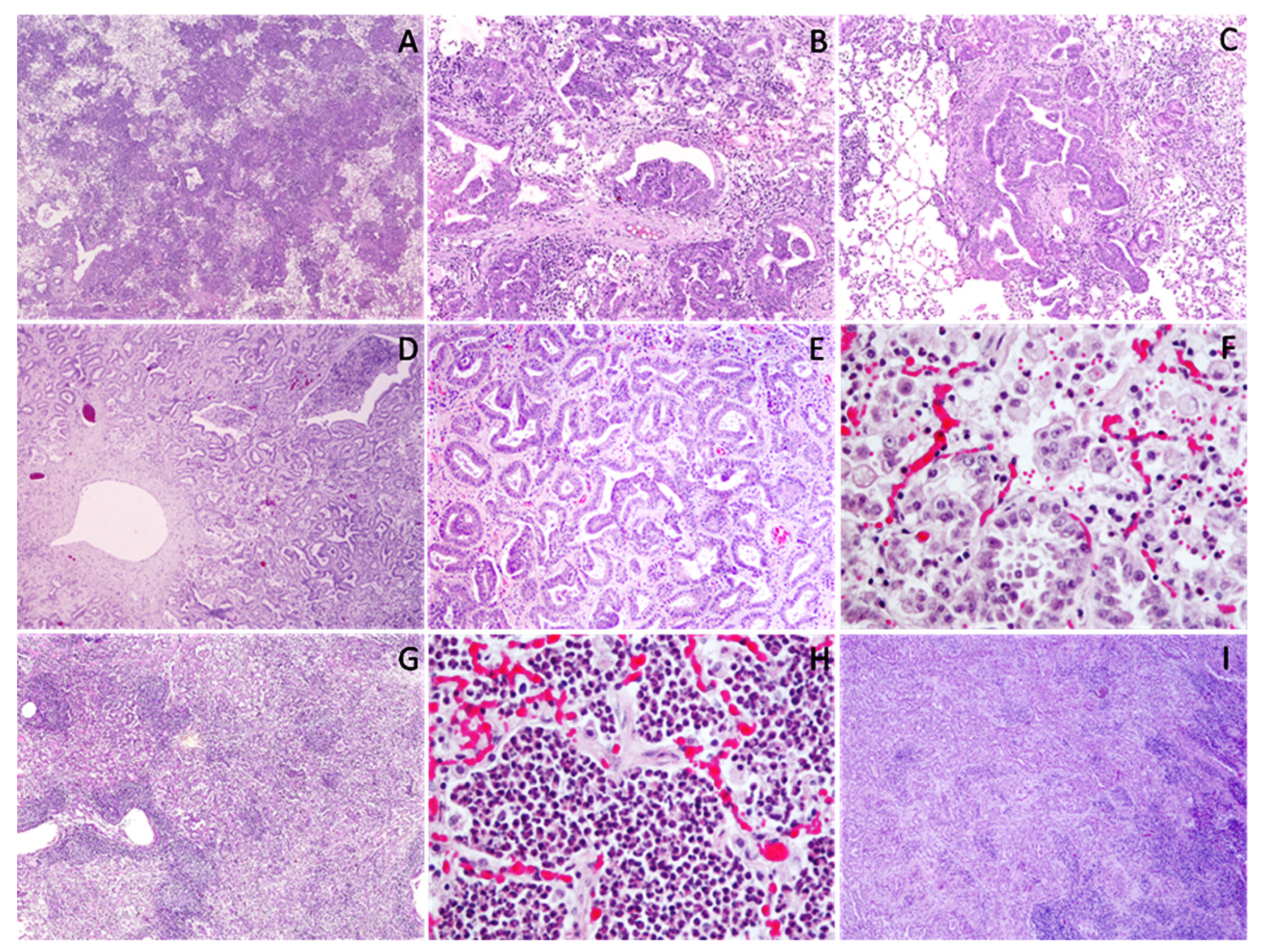
4.2. Microscopic Diagnosis

Microscopically, papillary or acinar proliferations in the alveolar and bronchiolar regions was observed, surrounded by cubic or columnar cells, replacing pneumocytes in alveolar wall. The cells become flatter in more advanced lesions [4,22,58].
The nucleus is generally uniform, located in the basal region in columnar tumors cells and central in the cuboid-shaped cells. The nucleoli tend to have a marginal location and rare mitotic figures were noted [4,54]. The cytoplasm is generally eosinophilic and homogeneous in the cells with a cuboid appearance, while in columnar cells cytoplasm is more pallid and vacuolated [22]. The presence of granules, containing surfactant, is common, especially in the periphery of lesions, but varies in number and size, resemble a fetal lung; [25,54].
Intralveolar macrophages with abundant cytoplasm and high number of lysosomes and phagolysosomes are frequent in the areas surrounding the tumor [22]. The stroma is thin and may be infiltrated by lymphocytes, plasmocytes and connective tissue fibers, accentuating fibrosis, especially in central areas. Sometimes there is an inflammatory reaction, denoted by the presence of neutrophils, both in the alveolar lumen and between tumor cells and the interstitium. This is typically an indication of secondary bacterial infection [9,22,25]. The presence of myxoid tissue masses with a nodular appearance is also reported, associated with neoplastic alveoli. These structures are composed of cells incorporated into a homogeneous basophilic matrix, presumably of mesodermal origin, and it is debatable whether its nature is neoplastic [54,62,63]. Myxoid cells shows positive immunolabeling for vimentin, desmin and alpha smooth-muscle actin but not for multi-cytokeratin [64]. The histopathological appearance of the atypical forms is essentially identical, although the neoplasm presents a more acinous aspect than the papillary one, and the presence of inflammatory cells and connective tissue in the stroma are most noticeable [54,65].
Regional lymph nodes may have a depletion of cortical lymphoid follicles and moderate plasmacytosis in the medullary areas. In addition to these lesions, metastases can also be found in the lymph nodes and in other organs, consisting of neoplastic epithelial cells organized according to the pattern found in the lung [4,6,22]. The frequency of metastases is lower in atypical lesions [54]. Figure 7 illustrates the main microscopic features of OPA.

4.3. Immunohistochemistry Diagnosis

The identification of cells expressing JSRV-related antigens in sheep are of great diagnostic value in this disease. Immunoreaction is detected in alveolar and bronchiolar neoplastic cells in cytoplasm where JSRV actively replicates. Myxoid nodules also show positivity to JSRV proteins and, in early lesions, lymphoreticular cells are immunoreactive too.
In immunohistochemical studies, reagents used included antibodies reacting against JSRV capsid protein (rabbit polyclonal antibodies) and JSRV-Env [66,67,68].

5. Ancillary Tests

The most common methods for identifying disease continue to be clinical signals, pathology, and histology, as mentioned earlier. The presence of JSRV can be detected only when clinical signs or tumors are present, in neoplastic cells or pulmonary fluid by immunoblotting [69], ELISA [70] or polymerase chain reaction (PCR) [71,72,73]. However, there are not methods to identify preclinical OPA because of the absence of measurable JSRV proteins outside of the tumor and circulation of specific JSRV antibodies.

5.1. Serological Diagnostic

Serological diagnostic tests are not available [8]. Despite the genetic differences between JSRV and enJSRV, in infected sheep a systemic humoral response is not detected, probably due to the immunotolerance induced by enJSRV expressed in the placenta and fetus during pregnancy, where they play an important role and lack of circulation of JSRV-specific antibodies [74,75].

5.2. Molecular Diagnosis

The high viral expression in type 2 pneumocytes and club cells is related to the activity of the viral promoter in these cells and accounts for the high viral amount present in bronchial secretions [72,76]. If there is a lung fluid present, is possible to test JSRV nucleic acids or antigens by RT-PCR, immunoblotting or ELISA [11,12]. The development of specific methodologies for the identification of the agent (U3 PCR or U3 hnPCR) demonstrated that the presence of JSRV is not restricted to type II pneumocytes and club cells. JSRV is present in PBLs, lung tissue, lymphoid organs, milk & colostrum in naturally infected sheep in the early stages of the disease before the onset of clinical signs or detectable tumor lesions [29,77,78,79].
Since then, it has been possible to detect the JSRV in asymptomatic animals, and an elevated prevalence has been seen in infected flocks, much higher than the number of animals that further develop the neoplasia [32,73]. The virus can enter into circulation when infected lymphoreticular cells contained in colostrum and milk can pass through the intestinal barrier and arrive to mesenteric lymph nodes [30].
However, the detection of nucleic acids in peripheral blood mononuclear cells (PBMCs), seems to be not very useful and gives many false negative results, due to the low numbers of infected blood mononuclear cells (monocytes, B and T lymphocytes) [6,12]. Some authors refer that this method might be employed as a herd test, but would not be suitable for testing individual animals [80] PCR testing in bronchoalveolar lavage seems to offer better sensitivity than the blood test, but the sampling technique is difficult (sedation required) and usually the samples only represent a small portion of the lung that can have a low or none concentration of infected cells, leading to many false negatives. Another restriction of this method is the limited production of secretions and/or infected cells in early stages [72].

6. Conclusions

The diagnosis of OPA represents a challenge for the veterinary clinician. If OPA is detected in one animal, a diagnostic examination should be carried out in other animals in the same herd. However, there is no precise laboratory technique to identify positive JSRV animals with no obvious clinical signs. It is therefore important to be aware of clinical signs and other diagnostic methods, such as ultrasound, that can be readily performed under flock conditions. Although these have numerous limitations in the early diagnosis of the disease, the use of PCR amplification has allowed a better understanding of the pathogenesis and epidemiology of this process, which would be impossible to achieve only with the use of histopathology. Molecular techniques are also widely used today in diagnosis as a way to establish a causal relationship between JSRV and pulmonary tumor lesions.

Supplementary Materials

The following are available online at www.mdpi.com/article/10.3390/ruminants1010005/s1, Video S1: Dyspnea and audible crackles; Video S2: Bronchoscopy; Video S3: Thoracic CT of OPA.

Author Contributions

Conceptualization, H.Q.; investigation, H.Q. and N.A.; resources, writing—original draft preparation, writing—review and editing: H.Q., I.P., A.G., J.P., F.S. and N.A. All authors have read and agreed to the published version of the manuscript.

Funding

This work was funded by 0687_OVISPID_2_E Interreg España-Portugal (EU) Poctep.

Institutional Review Board Statement

Not applicable.

Informed Consent Statement

Not applicable.

Acknowledgments

The authors thank the Centro Pedagógico Veterinário, Polytechnic Institute of Bragança (IPB) and the Veterinary Hospital of the University of Trás-os-Montes and Alto Douro (HVUTAD) for their support in carrying out diagnostic imaging tests.

Conflicts of Interest

The authors declare no conflict of interest.

References

  1. Shi, W.; Jia, S.; Guan, X.; Yao, X.; Pan, R.; Huang, X.; Ma, Y.; Wei, J.; Xu, Y. A Survey of Jaagsiekte Sheep Retrovirus (JSRV) Infection in Sheep in the Three Northeastern Provinces of China. Arch. Virol. 2021, 166, 831–840. [Google Scholar] [CrossRef] [PubMed]
  2. Sanna, M.P.; Sanna, E.; De Las Heras, M.; Leoni, A.; Nieddu, A.M.; Pirino, S.; Sharp, J.M.; Palmarini, M. Association of Jaagsiekte Sheep Retrovirus with Pulmonary Carcinoma in Sardinian Moufflon (Ovis Musimon). J. Comp. Pathol. 2001, 125, 145–152. [Google Scholar] [CrossRef] [PubMed]
  3. York, D.F.; Querat, G. A History of Ovine Pulmonary Adenocarcinoma (Jaagsiekte) and Experiments Leading to the Deduction of the JSRV Nucleotide Sequence. In Jaagsiekte Sheep Retrovirus and Lung Cancer; Fan, H., Ed.; Current Topics in Microbiology and Immunology; Springer: Berlin/Heidelberg, Germany, 2003; Volume 275, pp. 1–23. ISBN 9783642628979. [Google Scholar]
  4. Verwoerd, D.W.; Tustin, R.C.; Payne, A.L. Jaagsiekte: An infectious pulmonary adenomatosis of sheep. In Comparative Pathobiology of Viral Diseases; Olsen, R.G., Krakowka, S., Blakeslee, J.R., Eds.; CRC Press: Boca Raton, FL, USA, 2021; pp. 53–76. ISBN 9780367252212. [Google Scholar]
  5. Lee, A.M.; Wolfe, A.; Cassidy, J.P.; Messam, L.L.M.; Moriarty, J.P.; O’Neill, R.; Fahy, C.; Connaghan, E.; Cousens, C.; Dagleish, M.P.; et al. First Confirmation by PCR of Jaagsiekte Sheep Retrovirus in Ireland and Prevalence of Ovine Pulmonary Adenocarcinoma in Adult Sheep at Slaughter. Ir. Vet. J. 2017, 70, 33. [Google Scholar] [CrossRef]
  6. Griffiths, D.J.; Martineau, H.M.; Cousens, C. Pathology and Pathogenesis of Ovine Pulmonary Adenocarcinoma. J. Comp. Pathol. 2010, 142, 260–283. [Google Scholar] [CrossRef] [PubMed]
  7. Hashemnia, M.; Chalechale, A.; Malmir, E. Pulmonary Lesions in Slaughtered Sheepin Western Iran: Gross and Histopathological Findings. Vet. Ital. 2019, 55, 47–56. [Google Scholar] [CrossRef]
  8. Mekibib, B.; Mikir, T.; Fekadu, A.; Abebe, R. Prevalence of Pneumonia in Sheep and Goats Slaughtered at Elfora Bishoftu Export Abattoir, Ethiopia: A Pathological Investigation. J. Vet. Med. 2019, 2019, 1–10. [Google Scholar] [CrossRef] [PubMed] [Green Version]
  9. Alegria, N. Adenocarcinoma pulmonar dos ovinos em Portugal: Desenvolvimento de métodos para a detecção do agente etiológico. Ph.D. Thesis, Universidade de Trás-os-Montes e Alto Douro, Vila Real, Portugal, 2006. pp. 97–106, 125–134. [Google Scholar]
  10. De las Heras, M.; Borobia, M.; Ortín, A. Neoplasia-Associated Wasting Diseases with Economic Relevance in the Sheep Industry. Animals 2021, 11, 381. [Google Scholar] [CrossRef] [PubMed]
  11. Gray, M.E.; Meehan, J.; Sullivan, P.; Marland, J.R.K.; Greenhalgh, S.N.; Gregson, R.; Clutton, R.E.; Ward, C.; Cousens, C.; Griffiths, D.J.; et al. Ovine Pulmonary Adenocarcinoma: A Unique Model to Improve Lung Cancer Research. Front. Oncol. 2019, 9, 335. [Google Scholar] [CrossRef]
  12. Zhang, K.; Kong, H.; Liu, Y.; Shang, Y.; Wu, B.; Liu, X. Diagnosis and Phylogenetic Analysis of Ovine Pulmonary Adenocarcinoma in China. Virus Genes 2014, 48, 64–73. [Google Scholar] [CrossRef] [Green Version]
  13. Ortín, A.; De las Heras, M.; Borobia, M.; Ramo, M.A.; Ortega, M.; Ruíz de Arcaute, M. Ovine Pulmonary Adenocarcinoma: A Transmissible Lung Cancer of Sheep, Difficult to Control. Small Rumin. Res. 2019, 176, 37–41. [Google Scholar] [CrossRef]
  14. York, D.F.; Vigne, R.; Verwoerd, D.W.; Querat, G. Nucleotide Sequence of the Jaagsiekte Retrovirus, an Exogenous and Endogenous Type D and B Retrovirus of Sheep and Goats. J. Virol. 1992, 66, 4930–4939. [Google Scholar] [CrossRef] [Green Version]
  15. Wilson, D.W. Tumors of the Respiratory Tract. In Tumors in Domestic Animals; Meuten, D.J., Ed.; John Wiley & Sons, Inc.: Hoboken, NJ, USA, 2016; pp. 467–498. ISBN 9781119181200. [Google Scholar]
  16. Palmarini, M.; Cousens, C.; Dalziel, R.G.; Bai, J.; Stedman, K.; DeMartini, J.C.; Sharp, J.M. The Exogenous Form of Jaagsiekte Retrovirus Is Specifically Associated with a Contagious Lung Cancer of Sheep. J. Virol. 1996, 70, 1618–1623. [Google Scholar] [CrossRef] [Green Version]
  17. Palmarini, M.; Maeda, N.; Murgia, C.; De-Fraja, C.; Hofacre, A.; Fan, H. A Phosphatidylinositol 3-Kinase Docking Site in the Cytoplasmic Tail of the Jaagsiekte Sheep Retrovirus Transmembrane Protein Is Essential for Envelope-Induced Transformation of NIH 3T3 Cells. J. Virol. 2001, 75, 11002–11009. [Google Scholar] [CrossRef] [PubMed] [Green Version]
  18. Rai, S.K.; DeMartini, J.C.; Miller, A.D. Retrovirus Vectors Bearing Jaagsiekte Sheep Retrovirus Env Transduce Human Cells by Using a New Receptor Localized to Chromosome 3p21.3. J. Virol. 2000, 74, 4698–4704. [Google Scholar] [CrossRef]
  19. Caporale, M.; Martineau, H.; De las Heras, M.; Murgia, C.; Huang, R.; Centorame, P.; Di Francesco, G.; Di Gialleonardo, L.; Spencer, T.E.; Griffiths, D.J.; et al. Host Species Barriers to Jaagsiekte Sheep Retrovirus Replication and Carcinogenesis. J. Virol. 2013, 87, 10752–10762. [Google Scholar] [CrossRef] [Green Version]
  20. Yousem, S.A.; Finkelstein, S.D.; Swalsky, P.A.; Bakker, A.; Ohori, N.P. Absence of Jaagsiekte Sheep Retrovirus DNA and RNA in Bronchioloalveolar and Conventional Human Pulmonary Adenocarcinoma by PCR and RT-PCR Analysis. Hum. Pathol. 2001, 32, 1039–1042. [Google Scholar] [CrossRef] [PubMed]
  21. Sharp, J.M.; DeMartini, J.C. Natural History of JSRV in Sheep. In Jaagsiekte Sheep Retrovirus and Lung Cancer; Fan, H., Ed.; Current Topics in Microbiology and Immunology; Springe: Berlin/Heidelberg, Germany, 2003; Volume 275, pp. 55–79. ISBN 9783642628979. [Google Scholar]
  22. De Las Heras, M.; González, L.; Sharp, J.M. Pathology of Ovine Pulmonary Adenocarcinoma. In Jaagsiekte Sheep Retrovirus and Lung Cancer; Fan, H., Ed.; Current Topics in Microbiology and Immunology; Springer: Berlin, Heidelberg, 2003; Volume 275, pp. 25–54. ISBN 9783642628979. [Google Scholar]
  23. Salvatori, D.; González, L.; Dewar, P.; Cousens, C.; de Las Heras, M.; Dalziel, R.G.; Sharp, J.M. Successful Induction of Ovine Pulmonary Adenocarcinoma in Lambs of Different Ages and Detection of Viraemia during the Preclinical Period. J. Gen. Virol. 2004, 85, 3319–3324. [Google Scholar] [CrossRef] [PubMed]
  24. Palmarini, M.; Fan, H. Retrovirus-Induced Ovine Pulmonary Adenocarcinoma, an Animal Model for Lung Cancer. JNCI J. Natl. Cancer Inst. 2001, 93, 1603–1614. [Google Scholar] [CrossRef] [PubMed] [Green Version]
  25. Verwoerd, D.W. Jaagsiekte (ovine pulmonary adenomatosis virus). In Virus Infections of Ruminants; Dinta, Z., Morein, B., Eds.; Elsevier: Amsterdam, The Netherlands, 1990; pp. 453–462. ISBN 9780444873125. [Google Scholar]
  26. Palsson, P.A. Maedi/Visna of sheep in Iceland. Introduction of the disease to Iceland, clinical features, control mesures and eradication. In Slow Viruses in Sheep, Goats and Cattle; Sharp, J.M., Hoff-Jørgensen, R., Eds.; Office for Official Publications of the European Communities [distributor]: Luxembourg, 1985; pp. 3–19. ISBN 9789282557150. [Google Scholar]
  27. Sharp, J.M.; Scott, F.M.M.; Herring, A.J.; Angus, K.W.; Gray, E.W.; Cuthbertson, J.C. Studies of the epidemiology, aetiology and transmission of jaagsiekte in Scotland. In Slow Viruses in Sheep, Goats and Cattle; Sharp, J.M., Hoff-Jørgensen, R., Eds.; Office for Official Publications of the European Communities [distributor]: Luxembourg, 1985; pp. 167–170. ISBN 9789282557150. [Google Scholar]
  28. Cousens, C.; Thonur, L.; Imlach, S.; Crawford, J.; Sales, J.; Griffiths, D.J. Jaagsiekte Sheep Retrovirus Is Present at High Concentration in Lung Fluid Produced by Ovine Pulmonary Adenocarcinoma-Affected Sheep and Can Survive for Several Weeks at Ambient Temperatures. Res. Vet. Sci. 2009, 87, 154–156. [Google Scholar] [CrossRef] [PubMed]
  29. Grego, E.; De Meneghi, D.; Álvarez, V.; Benito, A.A.; Minguijón, E.; Ortín, A.; Mattoni, M.; Moreno, B.; de Villarreal, M.P.; Alberti, A.; et al. Colostrum and Milk Can Transmit Jaagsiekte Retrovirus to Lambs. Vet. Microbiol. 2008, 130, 247–257. [Google Scholar] [CrossRef] [Green Version]
  30. Borobia, M.; De Las Heras, M.; Ramos, J.J.; Ferrer, L.M.; Lacasta, D.; De Martino, A.; Fernández, A.; Loste, A.; Marteles, D.; Ortín, A. Jaagsiekte Sheep Retrovirus Can Reach Peyer’s Patches and Mesenteric Lymph Nodes of Lambs Nursed by Infected Mothers. Vet. Pathol. 2016, 53, 1172–1179. [Google Scholar] [CrossRef]
  31. Borobia, M.; De las Heras, M.; Godino, J.; Ferrer, L.M.; Lacasta, D.; Loste, A.; Ramos, J.J.; Ortín, A. Jaagsiekte Sheep Retrovirus Found in Milk Macrophages but Not in Milk Lymphocytes or Mammary Gland Epithelia of Naturally Infected Sheep. J. VET Diagn. Investig. 2021. [Google Scholar] [CrossRef] [PubMed]
  32. Caporale, M.; Centorame, P.; Giovannini, A.; Sacchini, F.; Di Ventura, M.; de las Heras, M.; Palmarini, M. Infection of Lung Epithelial Cells and Induction of Pulmonary Adenocarcinoma Is Not the Most Common Outcome of Naturally Occurring JSRV Infection during the Commercial Lifespan of Sheep. Virology 2005, 338, 144–153. [Google Scholar] [CrossRef] [PubMed] [Green Version]
  33. Sonawane, G.G.; Tripathi, B.N.; Kumar, R.; Kumar, J. Diagnosis and Prevalence of Ovine Pulmonary Adenocarcinoma in Lung Tissues of Naturally Infected Farm Sheep. Vet. World 2016, 9, 365–370. [Google Scholar] [CrossRef] [Green Version]
  34. Sharp, J.M.; De Las Heras, M. Contagious Respiratory Tumors. In Diseases of Sheep; Blackwell Science: Oxford, UK, 2000; pp. 181–186. [Google Scholar]
  35. Rosadio, R.H.; Lairmore, M.D.; Russell, H.I.; DeMartini, J.C. Retrovirus-Associated Ovine Pulmonary Carcinoma (Sheep Pulmonary Adenomatosis) and Lymphoid Interstitial Pneumonia. I. Lesion Development and Age Susceptibility. Vet. Pathol. 1988, 25, 475–483. [Google Scholar] [CrossRef] [Green Version]
  36. Summers, C.; Neill, W.; Dewar, P.; Gonzalez, L.; van der Molen, R.; Norval, M.; Sharp, J.M. Systemic Immune Responses Following Infection with Jaagsiekte Sheep Retrovirus and in the Terminal Stages of Ovine Pulmonary Adenocarcinoma. J. Gen. Virol. 2002, 83, 1753–1757. [Google Scholar] [CrossRef] [PubMed] [Green Version]
  37. Weikel, J. Double infection with maedi virus and adenomatosis virus in Merino sheep. Tierarztl. Prax. 1991, 19, 596–598. [Google Scholar] [PubMed]
  38. Gonzalez, L.; Juste, R.A.; Cuervo, L.A.; Idigoras, I.; Saez de Ocariz, C. Pathological and Epidemiological Aspects of the Coexistence of Maedi-Visna and Sheep Pulmonary Adenomatosis. Res. Vet. Sci. 1993, 54, 140–146. [Google Scholar] [CrossRef]
  39. Pritchard, G.C.; Done, S.H. Concurrent Maedi-Visna Virus Infection and Pulmonary Adenomatosis in a Commercial Breeding Flock in East Anglia. Vet. Rec. 1990, 127, 197–200. [Google Scholar]
  40. Ortín, A.; Pérez de Villarreal, M.; Minguijón, E.; Cousens, C.; Sharp, J.M.; De las Heras, M. Coexistence of Enzootic Nasal Adenocarcinoma and Jaagsiekte Retrovirus Infection in Sheep. J. Comp. Pathol. 2004, 131, 253–258. [Google Scholar] [CrossRef]
  41. Jahns, H.; Cousens, C. Nasal Adenocarcinoma Associated with Jaagsiekte Sheep Retrovirus Infection in a Sheep. J. VET Diagn. Invest. 2020, 32, 152–155. [Google Scholar] [CrossRef] [PubMed]
  42. Scott, P. Practical Use of Ultrasound Scan in Small Ruminant Medicine and Surgery. Vet. Clin. N. Am. Food Anim. Pract. 2016, 32, 181–205. [Google Scholar] [CrossRef] [PubMed]
  43. Cousens, C.; Graham, M.; Sales, J.; Dagleish, M.P. Evaluation of the Efficacy of Clinical Diagnosis of Ovine Pulmonary Adenocarcinoma. Vet. Rec. 2008, 162, 88–89. [Google Scholar] [CrossRef] [PubMed]
  44. Cousens, C.; Scott, P.R. Assessment of Transthoracic Ultrasound Diagnosis of Ovine Pulmonary Adenocarcinoma in Adult Sheep. Vet. Rec. 2015, 177, 366. [Google Scholar] [CrossRef]
  45. Scott, P.R.; Sargison, N.D. Ultrasonography as an Adjunct to Clinical Examination in Sheep. Small Rumin. Res. 2010, 92, 108–119. [Google Scholar] [CrossRef]
  46. Scott, P.R.; Gessert, M.E. Ultrasonographic Examination of the Ovine Thorax. Vet. J. 1998, 155, 305–310. [Google Scholar] [CrossRef]
  47. Scott, P.R. Use of Ultrasonographic Examination in Sheep Health Management—A General Appraisal. Small Rumin. Res. 2017, 152, 2–9. [Google Scholar] [CrossRef]
  48. Scott, P.R. Thoracic Ultrasonography as an Adjunct to Clinical Examination in Sheep. Small Rumin. Res. 2017, 152, 107–118. [Google Scholar] [CrossRef]
  49. Castells, E.; Lacasta, D.; Climent, M.; Pérez, M.; Sanromán, F.; Jiménez, C.; Ferrer, L.M. Diagnostic Imaging Techniques of the Respiratory Tract of Sheep. Small Rumin. Res. 2019, 180, 112–126. [Google Scholar] [CrossRef]
  50. Crilly, J.P.; Politis, A.P.; Hamer, K. Use of Ultrasonographic Examination in Sheep Veterinary Practice. Small Rumin. Res. 2017, 152, 166–173. [Google Scholar] [CrossRef] [Green Version]
  51. Ferrer, L.M.; Ramos, J.J.; Castells, E.; Ruíz, H.; Climent, M.; Lacasta, D. Use of Computed Tomography and Thermography for the Diagnosis of Respiratory Disorders in Adult Sheep. In Sheep Farming—An Approach to Feed, Growth and Health; Monteiro, A., Ed.; IntechOpen: London, UK, 2021; ISBN 978-1-78985-975-1. [Google Scholar]
  52. Scott, P.R. Sheep Medicine, 2nd ed.; CRC Press—Taylor & Francis Group: Boca Raton, FL, USA, 2015; pp. 149–186. ISBN 9780367575786. [Google Scholar]
  53. Rodríguez-Largo, A.; Castells, E.; de Miguel, R.; Gómez, Á.; Ruiz, H.; Pérez, M.; Reina, R.; Ferrer, L.M.; Luján, L. Detection of Aluminium Hydroxide-Induced Granulomas in Sheep by Computed Tomography: A Feasible Approach for Small Ruminant Lentiviruses Diagnosis and Research. Vet. Med. Sci. 2021. [Google Scholar] [CrossRef] [PubMed]
  54. García-Goti, M.; González, L.; Cousens, C.; Cortabarría, N.; Extramiana, A.; Minguijón, E.; Ortín, A.; De las Heras, M.; Sharp, J. Sheep Pulmonary Adenomatosis: Characterization of Two Pathological Forms Associated with Jaagsiekte Retrovirus. J. Comp. Pathol. 2000, 122, 55–65. [Google Scholar] [CrossRef] [PubMed]
  55. Khodakaram-Tafti, A.; Razavi, Z. Morphopathological Study of Naturally Occurring Ovine Pulmonary Adenocarcinoma in Sheep in Fars Province, Iran. IJVR 2010, 11, 134–138. [Google Scholar] [CrossRef]
  56. Kumar, M.A.; Kumar, R.; Varshney, K.C.; Palanivelu, M.; Sridhar, B.G.; Sivakumar, M. Incidence of Ovine Pulmonary Adenocarcinoma in Southern Parts of India: A Slaughter House Based Study. Indian J. Vet. Pathol. 2014, 38, 149. [Google Scholar] [CrossRef]
  57. Woldemeskel, M.; Tibbo, M. Pulmonary Adenomatosis and Maedi-Visna in Ethiopian Central Highland Sheep: A Microscopic Study. Trop. Anim. Health Prod. 2010, 42, 995–999. [Google Scholar] [CrossRef] [PubMed]
  58. Armezzani, A.; Varela, M.; Spencer, T.; Palmarini, M.; Arnaud, F. “Ménage à Trois”: The Evolutionary Interplay between JSRV, EnJSRVs and Domestic Sheep. Viruses 2014, 6, 4926–4945. [Google Scholar] [CrossRef]
  59. Amara, A.; Jemli, M.H.; Boutouria, M.; Chetoui, C. First case of adenomatosis in a ewe in Tunisia. Rev. Elev. Med. Vet. Pays Trop. 1994, 47, 33–34. [Google Scholar] [CrossRef]
  60. Azizi, S.; Tajbakhsh, E.; Fathi, F. Ovine Pulmonary Adenocarcinoma in Slaughtered Sheep: A Pathological and Polymerase Chain Reaction Study. J. S. Afr. Vet. Assoc. 2014, 85, 5. [Google Scholar] [CrossRef]
  61. Palmarini, M.; Fan, H.; Sharp, J.M. Sheep Pulmonary Adenomatosis: A Unique Model of Retrovirus Associated Lung Cancer. Trends Microbiol. 1997, 5, 478–483. [Google Scholar] [CrossRef]
  62. Cutlip, R.C.; Young, S. Sheep Pulmonary Adenomatosis (Jaagsiekte) in the United States. Am. J. Vet. Res. 1982, 43, 2108–2113. [Google Scholar]
  63. Bouljihad, M.; Drommer, W.; Leipold, H.W. Pathologic and Ultrastructural Findings in Sheep in Morocco with Naturally Occuring Jaagsiekte (Sheep Pulmonary Adenomatosis). Small Rumin. Res. 1996, 19, 275–280. [Google Scholar] [CrossRef]
  64. Toma, C.; Bâlteanu, V.A.; Tripon, S.; Trifa, A.; Rema, A.; Amorim, I.; Pop, R.M.; Popa, R.; Catoi, C.; Taulescu, M. Exogenous Jaagsiekte Sheep Retrovirus Type 2 (ExJSRV2) Related to Ovine Pulmonary Adenocarcinoma (OPA) in Romania: Prevalence, Anatomical Forms, Pathological Description, Immunophenotyping and Virus Identification. BMC Vet. Res. 2020, 16, 296. [Google Scholar] [CrossRef] [PubMed]
  65. Uzal, F.A.; Delhon, G.; Murcia, P.R.; De las Heras, M.; Luján, L.; Fernández Miyakawa, M.E.; Morris, W.E.; Gonzalez, M.J. Ovine Pulmonary Adenomatosis in Patagonia, Argentina. Vet. Res. Commun. 2004, 28, 159–170. [Google Scholar] [CrossRef]
  66. De las Heras, M.; de Martino, A.; Borobia, M.; Ortín, A.; Álvarez, R.; Borderías, L.; Giménez-Más, J.A. Solitary Tumours Associated with Jaagsiekte Retrovirus in Sheep Are Heterogeneous and Contain Cells Expressing Markers Identifying Progenitor Cells in Lung Repair. J. Comp. Pathol. 2014, 150, 138–147. [Google Scholar] [CrossRef] [PubMed]
  67. Wootton, S.K.; Metzger, M.J.; Hudkins, K.L.; Alpers, C.E.; York, D.; DeMartini, J.C.; Miller, A.D. Lung Cancer Induced in Mice by the Envelope Protein of Jaagsiekte Sheep Retrovirus (JSRV) Closely Resembles Lung Cancer in Sheep Infected with JSRV. Retrovirology 2006, 3, 94. [Google Scholar] [CrossRef] [Green Version]
  68. Murgia, C.; Caporale, M.; Ceesay, O.; Di Francesco, G.; Ferri, N.; Varasano, V.; de las Heras, M.; Palmarini, M. Lung Adenocarcinoma Originates from Retrovirus Infection of Proliferating Type 2 Pneumocytes during Pulmonary Post-Natal Development or Tissue Repair. PLoS Pathog. 2011, 7, e1002014. [Google Scholar] [CrossRef] [Green Version]
  69. Sharp, J.M.; Herring, A.J. Sheep Pulmonary Adenomatosis: Demonstration of a Protein Which Crossreacts with the Major Core Proteins of Mason-Pfizer Monkey Virus and Mouse Mammary Tumour Virus. J. Gen. Virol. 1983, 64, 2323–2327. [Google Scholar] [CrossRef]
  70. Palmarini, M.; Dewar, P.; De las Heras, M.; Inglis, N.F.; Dalziel, R.G.; Sharp, J.M. Epithelial Tumour Cells in the Lungs of Sheep with Pulmonary Adenomatosis Are Major Sites of Replication for Jaagsiekte Retrovirus. J. Gen. Virol. 1995, 76, 2731–2737. [Google Scholar] [CrossRef]
  71. Bai, J.; Zhu, R.Y.; Stedman, K.; Cousens, C.; Carlson, J.; Sharp, J.M.; DeMartini, J.C. Unique Long Terminal Repeat U3 Sequences Distinguish Exogenous Jaagsiekte Sheep Retroviruses Associated with Ovine Pulmonary Carcinoma from Endogenous Loci in the Sheep Genome. J. Virol. 1996, 70, 3159–3168. [Google Scholar] [CrossRef] [Green Version]
  72. Voigt, K.; Brügmann, M.; Huber, K.; Dewar, P.; Cousens, C.; Hall, M.; Sharp, J.M.; Ganter, M. PCR Examination of Bronchoalveolar Lavage Samples Is a Useful Tool in Pre-Clinical Diagnosis of Ovine Pulmonary Adenocarcinoma (Jaagsiekte). Res. Vet. Sci. 2007, 83, 419–427. [Google Scholar] [CrossRef]
  73. De las Heras, M.; Ortín, A.; Salvatori, D.; Pérez de Villareal, M.; Cousens, C.; Miguel Ferrer, L.; Miguel Cebrián, L.; García de Jalón, J.A.; Gonzalez, L.; Michael Sharp, J. A PCR Technique for the Detection of Jaagsiekte Sheep Retrovirus in the Blood Suitable for the Screening of Ovine Pulmonary Adenocarcinoma in Field Conditions. Res. Vet. Sci. 2005, 79, 259–264. [Google Scholar] [CrossRef]
  74. Palmarini, M.; Mura, M.; Spencer, T.E. Endogenous Betaretroviruses of Sheep: Teaching New Lessons in Retroviral Interference and Adaptation. J. Gen. Virol. 2004, 85, 1–13. [Google Scholar] [CrossRef]
  75. Spencer, T.E.; Mura, M.; Gray, C.A.; Griebel, P.J.; Palmarini, M. Receptor Usage and Fetal Expression of Ovine Endogenous Betaretroviruses: Implications for Coevolution of Endogenous and Exogenous Retroviruses. J. Virol. 2003, 77, 749–753. [Google Scholar] [CrossRef] [Green Version]
  76. McGee-Estrada, K.; Palmarini, M.; Fan, H. HNF-3β Is a Critical Factor for the Expression of the Jaagsiekte Sheep Retrovirus Long Terminal Repeat in Type II Pneumocytes but Not in Clara Cells. Virology 2002, 292, 87–97. [Google Scholar] [CrossRef] [Green Version]
  77. González, L.; García-Goti, M.; Cousens, C.; Dewar, P.; Cortabarría, N.; Extramiana, A.B.; Ortín, A.; De las Heras, M.; Sharp, J.M. Jaagsiekte Sheep Retrovirus Can Be Detected in the Peripheral Blood during the Pre-Clinical Period of Sheep Pulmonary Adenomatosis. J. Gen. Virol. 2001, 82, 1355–1358. [Google Scholar] [CrossRef] [PubMed] [Green Version]
  78. Palmarini, M.; Holland, M.J.; Cousens, C.; Dalziel, R.G.; Sharp, J.M. Jaagsiekte Retrovirus Establishes a Disseminated Infection of the Lymphoid Tissues of Sheep Affected by Pulmonary Adenomatosis. J. Gen. Virol. 1996, 77, 2991–2998. [Google Scholar] [CrossRef] [PubMed]
  79. Borobia, M.; Ortín, A.; Ferrer, L.M.; Ramos, J.J.; Lacasta, D.; De Las Heras, M. Cells Infected with Jaagsiekte Sheep Retrovirus Are Detected in the Bone Marrow of Asymptomatic Sheep. Can. J. Vet. Res. 2014, 78, 237–240. [Google Scholar] [PubMed]
  80. Lewis, F.I.; Brülisauer, F.; Cousens, C.; McKendrick, I.J.; Gunn, G.J. Diagnostic Accuracy of PCR for Jaagsiekte Sheep Retrovirus Using Field Data from 125 Scottish Sheep Flocks. Vet. J. 2011, 187, 104–108. [Google Scholar] [CrossRef] [PubMed]
Figure 1. Three-year-old sheep with clinical signs of OPA. (A) Progressive weight loss and respiratory distress (video: Supplementary Materials); (B) a large amount of clear frothy fluid flowed freely from both nostrils (arrow) after taking “wheelbarrow test” for 30 s.
Ruminants 01 00005 g001
Figure 2. Presence of tracheal ulcer (A) and of abundant surfactant secretions in the windpipe (B).
Ruminants 01 00005 g002
Figure 3. (A) The characteristic loss of the bright linear echo formed by normal aerated lung tissue replaced by a large 5 cm deep hypoechoic area bordered distally by a hyperechoic line delineating the OPA tumor, in the ventral margins of the right lung lobes at the 7th or 8th intercostal spaces (6.5 MHz sector scanner); (B) Small neoplastic nodules: multiple echogenic areas may be present in animals with OPA lesions (4.5 MHz sector scanner).
Ruminants 01 00005 g003
Figure 4. (A) Lateral radiographic projection in a standing position permitting confirmation, of OPA: tumor mass present in the caudo-dorsal lung field; (B) Lateral radiographic projection in lateral recumbency: small and diffused nodules in a nodular pattern distributed throughout the lung field; (C) image consistent with the regions of neoplastic foci and a secondary pneumonia in in advanced stages of the disease.
Ruminants 01 00005 g004
Figure 5. Thoracic CT images of OPA (A) Axial, (B) Coronal, and (C) Sagittal planes. In addition to other nodules spread throughout the lung parenchyma, a large area of increased radiopacity is seen within the right dorsal lobe consistent with advanced OPA tumor.
Ruminants 01 00005 g005
Figure 6. Macroscopic features of spontaneous OPA: (A,B) Classical form of OP with craneo-ventral tumor; (C,D) Typical OPA pulmonary cut surface, note the transition between normal tissue and tumor tissue: (E,F) The lungs do not collapse and are asymmetrically enlarged with a nodular appearance; (G) Atypical OPA profile with subpleural grey-white myxoid nodules in diaphragmatic lobes; (H) Pleura with irregular, depressed areas and fibrosis; (I) Pulmonary cross-section area of atypical OPA profile; (J) OPA and bacterial infection with abscess and (K) bronchopneumonia; (L) Lymph node metastasis.
Ruminants 01 00005 g006
Figure 7. Microscopical features of spontaneous OPA: (A) Lung neoplasic proliferation (H&E, 40×); (B,C) Tumoral cells shows a papillary pattern (H&E, 100×); (D,E) Cubical to polygonal epithelial cells lining alveoli within a fibrous stroma (H&E, 40×, 100×); (F) Numerous macrophages with abundant cytoplasm in alveoli space (H&E, 400×); (G) Tumoral OPA lesions and Maedi compatible lesions (H&E, 40×); (H) Supurative bronchopneumonia (H&E, 400×); (I) Lymph node metastasis (H&E, 40×).
Ruminants 01 00005 g007
Publisher’s Note: MDPI stays neutral with regard to jurisdictional claims in published maps and institutional affiliations.

