Review

A Concise Review of Prodigious Salinomycin and Its Derivatives Effective in Treatment of Breast Cancer: (2012–2022)

1
Department of Chemistry and Biochemistry, Rowan University, Glassboro, NJ 08028, USA
2
Department of Pharmaceutical Chemistry, R. C. Patel Institute of Pharmaceutical Education and Research, Shirpur 425405, Maharashtra, India
3
Shri Vile Parle Kelavani Mandal, Institute of Pharmacy, Dhule 424001, Maharashtra, India
*
Authors to whom correspondence should be addressed.
Int. J. Transl. Med. 2023, 3(2), 217-245; https://doi.org/10.3390/ijtm3020016
Submission received: 21 February 2023 / Revised: 19 April 2023 / Accepted: 27 April 2023 / Published: 5 May 2023
(This article belongs to the Special Issue Trends of Translational Medicine for Oncology)

Abstract

:
Cancer stem cells (CSCs) are the cells in a primary tumor that have the opportunity to self-renew as well as differentiate into certain cell types, thus forming a mixed tumor. CSCs have been shown to be involved in every aspect of cancer development, including tumor initiation, proliferation, and metastatic activity; they are also involved in chemotherapeutic drug resistance and the recurrence of certain cancers. Based on these capabilities, CSCs have been explored as the next target for the treatment and management of cancer. Salinomycin (SAL), a polyether ionophore antibiotic being used in the poultry industry, was identified as a powerful anti-cancer compound that possesses broad-spectrum activities, especially against CSCs. Here we point out the noteworthy work reported on SAL’s mechanism of action, anticancer activities, toxicity, and clinic applications. In addition, SAL derivatives synthesized by different research groups and their biological activity will also be highlighted.

1. Introduction

Most recently, cancer research has shifted from a complete suspension of cancer proliferation to a mechanistic understanding of the cause that initiates cancer or metastasis, as well as a decrease in chemotherapeutic drug effectiveness. Based on recent results, a specific group of cancerous cells that are covered up within a tumor mass and have the ability to induce tumorigenesis and reconstruct the tumor tissue population to increase their immunity to chemotherapeutic agents has been identified as cancer stem cells (CSCs). Originally established as tumor initiating cells (TICs), these were eventually renamed stem-like cancer cells (CSCs) [1]. According to research, CSCs have plasticity and can flip between stasis and active proliferation [2,3]. CSCs may be governed by dual cells (stromal and immune) in the microenvironment of the tumor (TME) or control the heterogeneous cancerous mass by modulating the architecture of the TME, which plays an important role in CSC plasticity [4,5,6,7,8]. The existence of CSCs was determined to be one of the main causes of relapse and chemotherapeutic insensitivity (Figure 1).
The exact mechanism of origination of CSCs has not yet been fully understood, so the initial step was to differentiate them from the tumor bulk. Although the proposed hypothesis for CSCs origins includes a few major points, firstly, they can be formed from progenitor cells [9], following mutations [10,11] or escaping regulation [9,12]. Secondly, CSCs could develop from mesenchymal stem cells during the healing of damaged tissues or from normal somatic cells after they have been transformed into stem-like cells [11,13,14].
CSCs and tumor bulk can be classified by dual methods (marker expression and functional method) for precise differentiation. These markers are identified as indicators displayed on normal stem cells for differentiation of particular groups, such as prominin-1 (CD-133), CD-44, and nestin. In the functional method, flow cytometry was used to measure the presence of ABCG2 and ABCA3 (adenosine triphosphate-binding cassette subfamily G member 2 and transporter A3) as membrane pumps, generally responsible for multidrug resistance. On the other hand, due to the variety of CSC populations and the expanse of CSC regulation (i.e.), ref. [15] a benchmark in vitro protocol was devised to directly improve the identification of CSCs apart from their oncogenic capabilities in xenograft (PDX) animal models, which are not well established. Nonetheless, worldwide researchers are working hard to learn more about CSCs and find ways to target them. Recent research breakthroughs for breast CSCs have been reviewed, and their multifold interactions (intercellular and intracellular) have been identified [16]. Based on the findings, the development of unique drugs targeting only CSCs is necessary and has shown promising clinical applications in the treatment of cancer by causing programmed cell death (apoptosis) through the inhibition of cell signaling pathways (Wnt and Notch) and by altering the TME or an immunotherapy approach targeting surface markers on the cells [14,17,18]. Weinberg et al. [19] in 2009 were the first to report a monocarboxylic polyether ionophore, salinomycin (SAL), with anti-CSC activity. After the discovery, efforts were focused on the function and mechanism of action of SAL’s activity against CSCs. Ginestier et al. [20,21] utilized a screening technique called genome-wide ribonucleic acid (RNA) interference (RNAi) with the goal of finding key genes that regulate the destiny of breast CSCs. They subsequently designed a series of SAL derivatives and evaluated them to check the inhibitory effects of CSC-fate regulators, where SAL emerged as one of the three most powerful effectors.

1.1. Triple Negative Breast Cancer (TNBC)

In 2020, breast cancer emerged as the leading and most frequent cancer in the United States as well as worldwide, with an estimated 685,00 deaths [22]. Additionally, it has been highlighted that there are two million incidences of breast cancer each year [23]. Current methodologies, such as surgery and chemotherapy, are currently in use for the treatment of breast cancer. Over the past ten years, significant progress has been made in the development of preventive measures and our understanding of breast cancer, leading to more effective and less toxic treatment options [24]. As per the current statistics, there will likely be 287,850 new cases in 2022 [25]. If the cancer was metastatic in nature at the time of diagnosis or developed later, this could result in compromised functioning of some vital organs, such as the lungs and brain, which account for about 90% of breast cancer fatalities [26,27]. The US has failed for almost five decades to alter the rate of metastatic breast cancer at the initial stage, despite the extensive use of mammograms for early detection [28]. There have been recent advances in our knowledge of the mechanisms that cause it to resurface. However, there is no recognized treatment for metastatic breast cancer [29].
TNBC had the worst outcomes of all the breast cancer subtypes studied. The name “TNBC” was actually developed to characterize a subtype of breast cancer that does not overexpress the triple receptors, namely, the human epidermal growth factor 2 receptor (HER2), the progesterone receptor (PR), and the estrogen receptor (ER) [30,31]. TNBC is distinguished by distinct molecular features that include a very active aggressiveness, a considerable ineffective chemotherapeutic response, characteristics of metastatic spread, and a dearth of viable therapeutic alternatives [32]. TNBC research has been very important over the years for researchers and clinicians for several reasons: (1) the outcome for disease-free and overall survival is low; (2) there is no widely accessible, efficient specialized treatment; (3) there is a higher incidence of cases in premenopausal and African American women; and (4) there is a phenotypic commonality between TNBC and breast cancer gene-1 (BRCA1)-linked breast tumors [30].

1.2. Epidemiology

Based on current data from 2020 (Singh et al.) [22], 2,088,849 cases with TNBC characteristics were diagnosed, with approximately 75% of the cases being basal-like [33] and 39% being diagnosed in African women [34]. About 15% of non-African American women in this age bracket have TNBC, which is a substantially lower prevalence. The ER+/HER2+ (+ indicates positive and − indicates negative) subgroup as well as the ER+/HER2 subgroup of breast cancer do not exhibit these racial or menopausal differences [34]. Researchers discovered that the prevalence of TNBC among African American women was more than double that of white women, as was originally presented in 2006 at the San Antonio Breast Cancer Symposium in a study examining racial variations in the incidence of triple-negative invasive breast cancers [30]. In addition, they noted that, compared to white women, just 22% of African American women’s tumors were triple-negative. African American women were nearly three times more likely than white women to have triple-negative tumors after adjusting for age and stage at diagnosis [31]. It is unclear if genes or epigenetic changes could potentially lead women to develop TNBC, given the inequalities between different ethnicities and the prevalence of TNBC, which varies greatly.

1.3. Pathology and Molecular Features

TNBC and basal-like breast cancer can be used interchangeably, although they are not the same thing. These immunological analyses are carried out on formalin-fixed and paraffin-embedded tumor sections, and the term “TNBC” refers to the immunophenotype of breast cancer that is immunologically negative to ER, PR, and HER2 [30]. The molecular phenotype of the tumor as determined by c-DNA microarrays is referred to as basal-like breast cancer [30]. According to reports, out of all TNBC occurrences, almost 75% had a basal-like appearance. The first team to outline the distinct molecular subtypes and profiles of breast tumors was Perou et al. [35], who identified four c-DNA microarray-based subcategories, which included a basal-like breast cancer subtype, and found that the majority of TNBCs were grouped into this subtype. Since then, research has concentrated on gene expression profiling, which has significantly improved oncologists’ comprehension of the molecular diagnosis and given them a framework for adopting the triple negative phenotype to characterize the basal-like subtype.
Polyether ionophores are a diverse and significant class of naturally occurring molecules generated by Streptomyces spp. In recent decades, there has been a rise in interest in compounds of this sort. Thus far, approximately 120 ionophores found naturally have been identified, with ionophores used commercially to prevent coccidiosis and enhance growth in grazing animals. These molecules selectively target rumen bacteria species, and their application increases cattle production efficiency. Polyether ionophores are particularly interesting because of their powerful efficacy against a variety of cancer cells.
The review was constructed with the critical demands of advancement of more potent entities effective against TNBC and reduction in chemotherapeutic medication efficiency in mind. We sought to investigate the increasing use of polyether ionophores and their derivatives by corroborating the previous investigations. The review is organized into parts that discuss the bioactivity and physicochemical properties of targeted polyether ionophore and its varients.

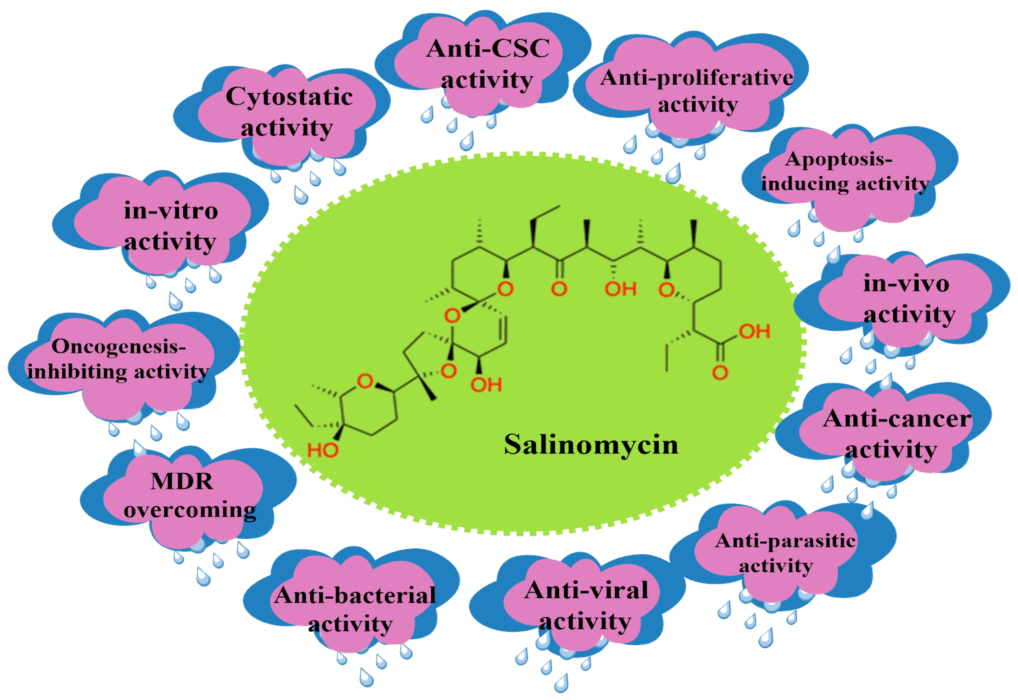
2. Salinomycin (SAL) and Its Bioactivity

A polyether ionophore, SAL, can target CSCs by facilitating the movement of polar alkali metals through hydrophobic membranes [36]. It has also been found that SAL is able to minimize the fraction of cancer cells that have stem cell-like features by inhibiting the Wnt pathway [37]. Haruyasu et al. [38] identified the novel polyether ionophore antibiotic SAL (molecular formula C42H70O11), followed by the Miyazaki group [39]. This molecule is a polyether antibiotic identified from the culture broth of Streptomyces albus (strain no. 80614) by Miyazaki and colleagues [37] at Kaken Chemicals Co., Ltd.’s research division in Tokyo, Japan, during a screening effort for novel antibiotics. SAL was produced by tank fermentation, filtering of culture broth, column chromatography on alumina or silica gel, and crystallization. SAL was isolated as a colorless prism of sodium salt using this method [40]. SAL can also be extracted from a source of chicken feed, as described by Borgstrom et al. [41], in which the granular feed was suspended and extracted in organic solvents, followed by purification through column chromatography. The variety of already reported pharmacological activities of SAL are depicted in Figure 2.

2.1. SAL’s Mechanism of Action against CSCs

Although SAL’s ability to affect CSCs is still unclear, studies have shown that SAL’s biological effects on CSCs depend on the cell type. Based on the literature, the mechanisms of action of SAL on CSCs include:

2.1.1. Apoptosis of CSCs

SAL causes cell death in a different way depending on the cancer being targeted. SAL has been shown to affect the mitochondria, leading to caspase-3 cleaving poly-ADP ribose polymerase (PARP), resulting in apoptosis. SAL has shown the ability to affect prostate cancer (PC-3) cell lines through the production of reactive oxygen species (ROS), leading to programmed cell death. On the other hand, in myeloid leukemia, breast and osteosarcoma cells are also susceptible to apoptosis, while some other cell lines may not [42]. More research is being conducted on the exact reason why some cell lines are affected by SAL, leading to apoptosis, while others do not experience apoptotic cell death.

2.1.2. Interference of ATP-Binding Cassette (ABC) Transporters

It has been shown that SAL has the ability to overcome the ABC transporters in most cancer cell lines, which affect cancer cells drug efflux capabilities, as exhibited in acute myeloid leukemia (AML) cancers [42]. Based on the structure of SAL, it is a K+ ionophore and embeds itself in the cytoplasmic or mitochondrial cell membrane. Due to the fact that ABC transporters expel substrates from the cytosol and SAL is not present in that part of the cell, it is unlikely that SAL will be affected by the transporters, and it has been shown to be a potential blocker. More research is being performed to explore the potential use of SAL as an ABC transporter inhibitor.

2.1.3. Inhibition of Oxidative Phosphorylation and Glycolysis

It has been demonstrated that cancers depend on aerobic glycolysis rather than oxidative phosphorylation; the neoplastic transition of human mesenchymal stem cells goes through this mechanism. It was also determined that glioma cancer cells depend on oxidative phosphorylation, and inhibition could affect these cells. In light of this knowledge, an increase in oxidative phosphorylation in certain stem cells can lead to a malignant transformation in these cells. These cells depend on this activity, and because SAL is known to impede oxidative phosphorylation in the mitochondria, blocking this route could be a useful function of this chemical as a chemotherapeutic agent against tumors [42]. Based on this knowledge, SAL has been shown to have the effect of inhibiting the increase in oxidative phosphorylation and can contribute to the elimination of these cells.
In addition to the effects of oxidative phosphorylation inhibition by SAL, it also has an effect on the glycolysis of different cancer cells. This was seen when SAL in combination with glucose analogs (2-DG, 2-FDG) increased the toxicity of SAL towards cancer cells and showed that cancer cells are dependent on glycolysis for ATP production [43]. In the same study, dichloroacetate (DCA), an inhibitor of pyruvate dehydrogenase kinase, which results in the activation of mitochondrial pyruvate dehydrogenase complex, which aids in converting pyruvate formichloroactate to acetyl-CoA molecules that enter the TCA cycle, was used in combination with SAL, resulting in an increase in cell death, and it can be concluded that the inhibition of oxidative phosphorylation further causes cell death induced by SAL. Glucose starvation-mediated inhibition of salinomycin-induced autophagy amplifies cancer-specific cell death [21,43].

2.1.4. Polyether Ionophore Effects on the Mitochondria

SAL is a potassium ionophore that affects the transmembrane potential and increases potassium efflux from the mitochondria and cytoplasm. It has been documented that there is an increased expression of potassium channels in AML and neuroblastoma cancerous cells, although not observed in their non-tumorigenic counterparts, hinting that potassium channels have a role in these cancers. A decrease in the intracellular concentration due to the efflux of potassium leads to a cytotoxic effect on these cells [42,44]. It has been found that SAL can have an effect on the mitochondria, in which enhanced Na+ inflow leads to the suppression of breast CSCs, as discovered by Mai et al. [21]. Miyazaki et al. [39] were the first to explore the dual action of SAL on mitochondrial ion transport and respiration. From a rat’s liver, they discovered that 0.4 μM of SAL may release preloaded potassium from the mitochondria. The pre-treatment may limit potassium intake because SAL inhibits oxidative phosphorylation in mitochondria without selectivity and reverses cell swelling caused by potassium absorption. Furthermore, SAL inhibited mitochondrial storage of potassium more efficiently than sodium, even though with the addition of potassium uptake stimulants, it inhibited respiration at medium and low concentrations [45]. This research demonstrates that cation transport by SAL might be conditional and influenced by ion gradients.

2.1.5. Induction of Autophagy, ROS, and DNA Damage

For cancer cells, autophagy is the mechanism for protecting and inhibiting cell viability. In one way, it can impede ROS production and protect against apoptotic cell death; in another way, SAL enhances the expression of microtubule-associated proteins, implying autophagy induction. It has been noticed by a number of groups that SAL itself, or in conjunction with sensitizing drugs or radiation, displayed a moderate to strong increase in DNA damage, which caused G2 cell cycle arrest and a decrease in p21 protein levels in cancer cells. Zhao et al. [45] discovered DNA damage caused by SAL and G1 arrest in glioblastoma cells (GBM) through ROS, implying that DNA damage caused by SAL therapy could be a side effect. As a result, it is postulated that a putative autophagic mechanism for SAL in relation to radiation-induced DNA damage and subsequent tumor recurrence could be utilized as a potential avenue for treating different cancers [46,47].

2.1.6. Endoplasmic Reticulum Stress

Li et al. [48] demonstrated that SAL causes autophagic activity; however, it is a double-edged sword in different cell types. Autophagic activity could be a defensive measure for SAL-affected cells. If the factors that inhibit autophagy are not present, the apoptotic rate increases in SAL-treated cells. Strand et al. have worked hard to create different experimental procedures, such as SAL fluorescent conjugate, to reveal SAL’s anti-CSC impact. This group also discovered that SAL causes the release of ER calcium ions and promotes their stress in breast cancer cells [37,47]. This was accomplished by the dispersion of a fluorescent SAL conjugate (SAL-NBD) in the ER and lipid droplets (LDs) following treatment with this conjugate. The disruption of Ca2+ induces ER stress as cellular Ca2+ homeostasis is closely maintained to manage Ca2+ levels in both forms (free and bound) in all the sections of a cell [49]. Protective factors, such as the unfolded protein response (UPR) and the mobilization of pathways to reestablish ER equilibrium, are activated. Strand et al. [46] discovered that SAL’s fluorescent conjugate activated the UPR proteins GPR78 and ATF6. In breast cancer cells, removing CHOP hampered SAL-NBD’s capacity to downregulate β-catenin, and in addition, this compound boosts the enzymatic activity of protein kinase-C (PKC), a Wnt pathway antagonist, by 30%. This shows indirectly the potential calcium ionophore capabilities that SAL possesses, as it could have the potential to regulate calcium levels as well as Ca2+-influenced cellular processes.

2.1.7. Inhibition of the Wnt Signaling Cascade

Lu et al. [50] focused on the Wnt signaling pathway when assessing SAL’s anticancer activity, and it was found in vitro that SAL reduced Wnt1 and β-catenin while having a mild impact on Fizzled class receptor 5 (Fzd5), in addition to the fact that SAL induces an effect of lipoprotein receptor-related protein-6 (LRP6), which is essential for Wnt signaling. One example of this effect can be seen with chronic lymphocytic leukemia (CLL) cells, in which they discovered that these cells are more sensitive to SAL and had a 100-fold greater amount of apoptosis as compared to healthy donors’ peripheral blood mononuclear cells (PBMC) [51], as well as SAL decreasing the expression of Wnt signaling, thus effecting the TCF4E complex [52]. This finding was observed by Qi et al. [53], who found that SAL decreased the amount of protein kinase-A (PKA) by the phosphorylation of β-catenin in NB cells. It was also found that this pathway is vital for preservation, cloning, and aspects of cancer cells, and most importantly, it aids the resistance to radiation and anticancer drugs for many cancers. SAL, however, demonstrated an ability to suppress chronic lymphocytic leukemia cells by lowering the expression of LRP6 and downregulating the Wnt target genes LEF1, cyclin D1, and fibronectin, resulting in cell death.

2.1.8. Sequestration of Iron in the Lysosome

In the cell, iron (Fe2+) is closely controlled in preserving homeostasis, and based on this finding, Rodriguez et al. [54] investigated and discovered dramatic changes in iron in CD-24 low breast cancer cells. Hence, SAL was tested, and at 0.5 μM iron accumulation in the lysosome, a reduction in iron keeper ferritin expression and elevated iron regulatory protein-2 (IRP2) were observed. Based on this finding, a novel mechanism of action of SAL affecting breast CSCs is iron accumulation in the lysosome [1,44,53], and an increased amount of iron in the lysosome produces ROS, which leads to apoptosis [53].

2.1.9. Intracellular Binding Targets

Based on what others have shown about SAL’s ability to suppress CSCs among different cancers, which include breast cancer, neuroblastoma (NB), GBM, medulloblastoma, pancreatic, colon, prostate, melanoma, and lung cancers, particularly in NB cells, CD-34 and CD-133 play a key role in cell proliferation and tumor formation, respectively. It was discovered that NCL is associated with binding to the CD-34 gene promoter region, which initiates the transcription of the respective gene. SAL significantly decreased the cell population of CD-34+ and CD-133+ NB cells through its ability at the mRNA level to prevent the binding of NCL to the CD-34 promoter region, which significantly reduced the CD-34 gene expression and affected the cell proliferation and tumor formation of NB cells [55].
Qi et al. [53,55] just recently found and explored nucleolin (NCL), a protein that has many functions and is necessary for cell proliferation. Based on studies, it has the ability to bind nucleic acids (RNA and DNA) and numerous other proteins. For example, after iron chelator therapy, NCL controls matrix metallopeptidase 9 (MMP9) mRNA translation by binding to its promoter regions [56]. NCL also controls CD-133 and CD-34 expression in hematopoietic stem/progenitor cells (HSPC) [57,58]. Surprisingly, Qi and colleagues discovered by combining the techniques of Drug Affinity Responsive Target Stability (DARTS) and co-immunoprecipitation (co-IP) [59] that for SAL, NCL is a functional cellular binding target. Qi et al. showed that SAL significantly suppresses NB development with an IC50 that is substantially lower than that seen with the majority of presently-utilized NB chemotherapeutic agents. This presents a new mechanism of SAL in the treatment of cancer. Overall, based on these findings regarding SAL’s mechanism of action, it demonstrates how versatile SAL is and how its multiple mechanisms of action can be used to treat cancer.

2.1.10. Differentiation of CSCs and SAL Bioactivity

There are different extracellular biomarkers, such as CD-44, that were found in solid and hematological cancers and aid in the proliferation, self-renewal, and metastasis of cancers. CD-133 is another biomarker that was identified as present in tumors of breast, liver, stomach, and colon cancers. The presence of this marker gives rise to the notion that these caners have a high rate of tumor and spheroid formation. CD-33 has been known to be a prototypical marker for CSCs; for example, it is the most common marker for AML stem cells. It is also expressed in high quantities in CML and hematopoietic stem cells [60].
In addition to the extracellular biomarkers (CD-44 and CD-133), intracellular biomarkers also play a key role in tumorgenesis. These intracellular biomarkers, known as pluripotent transcription factors (e.g., Nanog, Oct4, and Sox2), are present in embryonic stem cells [61]. It has been found that overexpression of these transcription factors, which are present in various carcinomas and breast and prostate cancers, causes tumorgenesis and malignant progression. The ability of these transcription factors to induce tumorgenicity is demonstrated by Oct4’s ability to transform normal mammary epithelial cells into mammosphores, which increases tumorgenicity in vivo. The expression of Sox2 and Nanog in MCF7 cells can aid in cell proliferation and the formation of mammosphores, as knockdown of either transcription factor showed inhibition of cell proliferation and mammosphore formation.
In the TNBC cell line MDA-MB-231, the administration of SAL decreased tumor growth by reducing CD-44 levels rather than inducing apoptosis. Also observed from this study was that a downregulation of Nanog, Oct4, and Sox2 leads to the suppression of mammosphore formation [62].
SOX2 is another transcription factor that is found in embryonic stem cells and CSCs, and an increase in tumorgenesis and chemoresistance was observed when this transcription factor was expressed. This could be a potential target for the treatment of cancers at the CSC level, as seen in Sox2-positive glioblastoma cancer stem cells (GCs) treated with SAL, and it was observed that SAL decreased the expression of SOX2 at the transcriptional and translational levels. This indicated a decrease in SOX2 expression, which resulted in a decline in the tumorgenesis of GBM [63].
SAL has the ability to promote differentiation of cancer cells early on in development, and it can reprogram epithelial growth that undergoes epithelial mesenchymal transition (EMT) and leads to the upregulation and expression of certain genes, such as mammary epithelial differentiation [42,64]. Based on these findings, SAL can eliminate CSCs through multiple mechanisms of action known from the literature (Table 1). Future research is needed to find out if SAL has other relevant mechanisms of action for targeting CSCs.

3. Clinical History

3.1. In Vitro Studies

In a very specific TNBC cell line, MDA-MB-201, SAL has been shown to reduce cell motility even at very dilute concentrations. In this experiment, the experimenters infected the MDA-MB-201 cells with a vector of cytokeratin-18 (CK-18), which is a liver bilayer filament protein, to judge the cell’s parameters. However, in the same cell lines infected with CK-18, there was no such effect on cell motility. Another parameter that was measured was the elucidation distance (ED) of the cells. In the study, SAL reduced the ED significantly, whereas in the control group, no such effect was observed [57]. According to Dewangan et al. [65], SAL decreases the activity of the ABC transporters in leukemia stem cells [65]. The Wnt signaling pathway is present in colorectal cancer cells alongside the growth phases of prostate cancer cells in vitro. Cancer cells in direct contact, which will be mentioned in the studies below, always induce apoptosis or inhibit the development of tumors in certain cancer cell lines.
SAL has been found to inhibit the growth of tumors in both in vitro and in vivo studies. In one case study, SAL was treated for hepatocellular carcinoma (HCC) in several different cell lines [66] and found to reduce proliferating nuclear cell antigen (PNCA) levels. The molecule itself causes cell cycle arrest in different phases of growth. When treated with SAL, the HCC cells in the G0 and G1 growth phases were halted. In the same study, it was determined that SAL also affected BAX and Bcl-2 concentrations, which induce apoptosis. SAL promotes DNA breaks in BC cells and phosphorylated p53 and H2AX levels in Hs578T cells. SAL has been shown to induce DNA damage, which leads to the induction of apoptosis. The anti-proliferative action of SAL is mediated by a variety of mechanisms. SAL also suppresses MDA-MB-231 (Anderson Metastatic Breast) cells’ ability to proliferate at different concentrations and over a period of time, preventing the G1-to-S phase conversion by downregulating genes downstream of the Hedgehog signaling pathway. The β-catenin expression dramatically decreased compared to the control with SAL. Flow cytometry was used to analyze the Ca2+ concentration in HCC cells, and it was discovered that SAL therapy groups had greater Ca2+ concentrations. SAL in the apoptosis process has interacted with necrotic flesh. However, necrotic cell death is characterized by an increase in the size of the endoplasmic reticulum, mitochondria, and cytoplasm, leading to a tear in the plasma membrane. SAL delivers significant ER stress in glioblastoma cells, causing the unfolded protein response and an abnormal autophagic flux that leads to necrosis due to mitochondrial and lysosomal changes.
P53, a tumor suppressor protein, has been shown to be upregulated by SAL. The inverse of protein pH2AX caused cell cycle arrest by decreasing cyclin D1 levels. Researchers who performed a comet assay alongside immune-cytochemical staining of the protein pH2AX confirmed that SAL causes DNA damage in specific cancer cell lines. The Hs578T and MDA-MB231 cell lines mentioned above reduced cyclin D1 levels, leading to apoptosis in those cells. SAL has an overwhelming toxic effect on cancer cells in vitro. Over time, exposure of SAL to Hs578T cancer cells requires fewer concentrations to reduce the toxicity.

3.2. In Vivo Studies

SAL’s effects on mice have significant weight loss differences compared to untreated mice. Their testis, seminal vesicles, and epididymis weights decreased irreversibly. Significant oxidative stress was present in the testis, alongside lower regulation of the enzymes lactate dehydrogenase (LDH), superoxide dismutase (SOD), and catalase (CAT).
Other animals were tested as well for SAL’s toxic effects; horses treated with SAL developed symptoms of ataxia, persistent weakness, and muscular atrophy, while feeding hens SAL-infused chicken feed resulted in more than a third of them dying. In pigs, SAL was fed to the animals at concentrations of 720 and 441 ppm. Pigs who ingested these concentrations of SAL developed symptoms of elevated rectal temperatures and an unwillingness to stand, which led to their deaths. SAL poisoning in humans causes shortness of breath, dizziness, nausea, weakness in the legs, photophobia, and elevated blood pressure [65].

4. SAL’s Toxicological and Pharmacological Properties and Clinical Applications

4.1. In Human Cells

It was observed that SAL’s EC50 values varied against different tumor cells depending on the cancer cell type and had different mechanisms of action, as mentioned above. The toxicity profile of SAL was examined using the ProTox-II tool (Figure 3). Based on the analysis, it was found that SAL was classified under toxicity Class 2, which had a LD50 of 16 mg/kg, and since the LD50 is between 5 mg/kg and 50 mg/kg for Class 2, it is fatal when swallowed at high doses; hence, the ideal dose for SAL should be 5 mg/kg. Some of the toxicity effects that SAL possesses are carcinogenicity, mitochondrial membrane potential heptatoxicity, and cytotoxicity (Figure 3).
Based on the previous work by Qi et al., SAL can effectively eliminate CSC-high NB cells at a concentration of 1–2 μM in under 48 h. In vitro, the EC50 values of SAL’s ability to eliminate CSC-high GBM cells were determined to be 1.25 μM [67]; the IC50 of SAL in effectively killing medulloblastoma cells ranges between 0.1 and 2 μM [68]; and lastly, it was determined that the EC50 of SAL on CSC-high pancreatic cancer cells was observed to be 0.5–2 μM. According to Boehmerle et al. [69], SAL may have a neurotoxic impact on human dorsal root ganglia and Schwann cells (neural cells) at a dose of 10 mΜ. It would cause an influx of sodium and calcium ions, which would further cause calpain and cytochrome c, leading to apoptosis. In these cells, high-dose SAL-induced peripheral neuropathy was dramatically reversed by inhibiting the Na+/Ca2+ exchanger (NCX) [69]. Fuchs et al. [70] investigated the effects of SAL on leukemia cells and discovered that, at effective dosages, SAL promoted the death of human CD4+ T-cell leukemia cells but not normal CD4+ T-cells.
In another study, Scherzed et al. [71] examined, in vitro, the functional impairment of human bone marrow mesenchymal stem cells (hBMSC) by SAL and discovered the cytotoxic effects at concentrations of 30 μM after the first day of treatment. This research was conducted in light of one of the CSC-related hypotheses, which postulated that these cells may be produced from hBMSC. In addition, it was found that there were no negative impacts on the hBMSC’s characteristics, immunophenotype, or potential for multi-differentiation [71]. Overall, although high concentrations or prolonged exposure at low concentrations might cause neural toxicity and differentiation inhibition of mesenchymal stem cells, significant toxicity of normal cells was not often seen at the concentrations used to treat CSCs [72,73,74].
Additionally, the genotoxicity and cytotoxicity of SAL for human non-cancerous cells were explored by examining peripheral blood lymphocytes obtained from human nasal mucosa cells [74]. As a result, nasal mucosa cells and lymphocytes delivered cytotoxic effects at concentrations of 10–20 μM or more, but genotoxicity was absent. At 5 μM, there was a small increase in interleukin 8 (IL-8) release, indicating that SAL has a pro-inflammatory activating potential [75,76]. Furthermore, Szkudlarek-Mikho et al. [77] found that SAL inhibited adipogenesis at concentrations of 10 nM and above. An inhibition of pre-adipocyte differentiation into adipocytes was found, which does not appear to be connected with apoptotic cell death or cell proliferation interference. The discovery indicated SAL’s potential significance as an anti-obesity therapeutic as well as hinted at its toxicity on adipocytes. Later, Scherzed et al. [78] investigated the effects of SAL’s chronic exposure over a 4-week period against the hBMSC cell line at a low dosage (0.1 μM) and discovered a modest dampening effect on the migratory capacity with no change in cytoskeletal organization. Cytotoxic and pro-inflammatory effects of SAL were detected at concentrations 5-fold and 2-fold greater than those used in anticancer therapy, respectively, but cell differentiation inhibition was seen at lower doses. These investigations, summarized in Table 2, however, did not include other human cells; thus, unfavorable effects on noncancerous cells must be evaluated. SAL’s dose-dependent behavior represents the notion of therapeutic index, and the administration methods are critical in evaluating its possible safety risk.

4.2. In Animals

The available research in Table 3 shows that SAL effects are dependent on dose, species, and cell type and that animals do not experience any substantial abnormal effects. Horses, pigs, sheep, hens, bunnies, and cats are among the species that succumb to SAL overdose and combination overconsumption [79,80,81,82,83,84,85,86].
For hens, a 60 mg/kg dose of SAL was found to be safe and had no major detrimental effects, but increasing the dose to double damaged their immune systems [87]. LD50 (lethal dose, 50%) was determined to be 108 and 104 mg/kg, respectively, for broiler chickens and laying hens, although in horses it was determined to be 0.6 µg/kg [88]. Sipman et al. [86] conducted clinical investigations, and the results revealed a distant polyneuropathy in poisoned cats. Miyasaki et al. performed a study with mice and found acute toxicity of SAL with an LD50 of 18 mg/kg intra-peritoneally and 50 mg/kg orally [39], while Boehmerle et al., who also investigated SAL toxicity in mice, showed that a dose of 5 mg/kg was well tolerated by the specimen.
Ojo et al. [89] investigated the effects of SAL, at doses of 1, 3, and 5 mg/kg, on male mice’s fertility for 28 days and found a decline in motility and spermatozoa number. Yet spermatogenesis was found in the testis again 28 days following SAL cessation, indicating that SAL has temporary dose-dependent deleterious effects on the male reproductive organs of mice [89].

4.3. Pharmacokinetics and Pharmacodynamics

Based on the studies and investigations, the lipid solubility of SAL indicates that it is quickly absorbed in the gastro-intestinal tract and disseminated through the serum and tissues [88]. It was determined that adipose tissue had the highest affinity for hepatic and muscle tissues in chickens. In animals, pharmacokinetics and pharmacodynamics were explored. Resham et al. [90] reported that the metabolism and pharmacokinetics of SAL in an in vitro setting showed that SAL is quickly metabolized in the liver, yielding many metabolic products in which this occurs in microsomes. In chickens, it has been found that elimination is moderately fast within a 24-hour period. The cytochrome P450 (CYP) enzymes, specifically CYP3A4, play an important role in the metabolism of SAL. A higher SAL percentage in human plasma (the unbound fraction) was also detected when compared with the plasma of rats and mice, justifying the faster metabolism of SAL in humans [86]. Since the pharmacodynamic and pharmacokinetic aspects have been established in terms of an anti-coccidial agent, further investigation in terms of human cancers must be conducted.

4.4. Clinical Studies

Owing to the potential therapeutic benefits SAL possesses, Naujokat et al. conducted a few clinical trials [42]. SAL was given to a 40-year-old woman with TNBC (specifically, bone and subcutaneous invasive ductal breast cancer) that did not respond to standard chemotherapy. The results showed that the proliferation of the cancerous lesion decreased significantly after 12 iterations of SAL (200 μg/kg) intravenously administered to the respective patient every second day. Another three patients, who had metastatic cancers (breast, ovarian, and head/neck), saw a significant regression in tumor growth and metastasis.
A female patient (82 years old) who had squamous cell carcinoma, in which the cancer metastasized through the pelvic lymphatic vessels, did not respond to the standard chemotherapeutic regiment. The patient was administered 200 μg/kg of SAL as well as Erlotinib, and after 14 interactions of the chemotherapeutic agents, there was a dramatic decrease in tumor size. However, the patient experienced the negative side effects of Erlotinib, and the cancerous lesion re-emerged after a period of three months. Then, 12 cycles of SAL (250 μg/kg) were administered every other day, and the patient was examined for four months, showing a stable disease state with no progression. These studies show the safety and efficacy of the potential clinical use of SAL. Further clinical studies of SAL should be continued to shed light on its potential chemotherapeutic benefits as well as its toxicity in order for it to be used clinically.

5. Physicochemical Analysis of SAL and Its Analogs
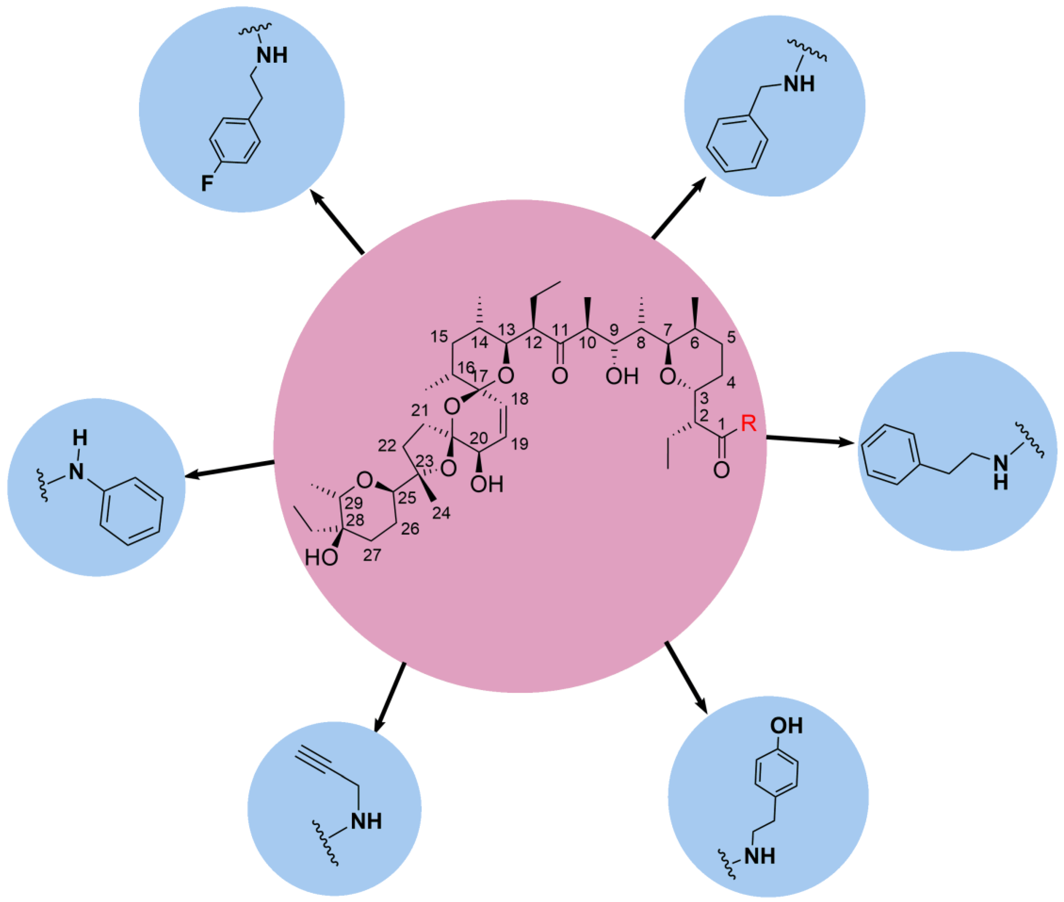
A growing set of bioactivity data for SAL suggests that the drug may also be effective against cells other than CSCs (TNBC) and comparable cell lines. Though the drug’s effects are dosage-dependent and fatal at high doses, in vivo experiments using a variety of groups and species of animals show promising findings, further supporting the drug’s bioactivity. As a result, it was crucial to look into the drug’s structural and physiochemical characteristics in further detail and test its derivatives on different breast cancer cell lines and cancer types.

5.1. C1 Analogs

Based on SAL’s different mechanisms of action and its therapeutic effects on CSCs, chemical modification of SAL has emerged over the years as an interesting research area for improving antitumor effects while reducing possible toxicity, along with the generation of prospective active pharmaceutical ingredients (APIs) with high therapeutic indexes. SAL was determined to be a polyether carboxylic ionophore antibiotic with a molar mass of 751 g/mol. SAL is a polyether ionophore with a pseudocyclic structure (Figure 4) that permits it to form complexes with metal cations and enhance their transport through biological membranes.
It has been demonstrated that chemically changing polyether antibiotics can alter the capacity and selectivity of metal cation interactions as well as the mechanism of cation movement, resulting in novel antibacterial and anticancer compounds [40]. Much of the research that has been conducted and reported has focused on chemically modifying the C-1, C-9, C-11, C-20, and C-28 positions on the molecule. At the C-1 position of the molecule, there is a carboxylic acid (-COOH) and several alcohol residues (-OH), which aid in the ionophoric transport of the SAL molecule.
It has been reported that acylation of the alcohol moieties decreases the K+ transport rate, while esterification of the C-1 carboxylic acid leads to the loss of ionophoric activity of SAL. There has been a lot of work on the derivatization of SAL to increase its antitumor efficacy, either by structural modification or dimer synthesis. The noteworthy derivatives or dimers were achieved by either modifying only the C-1 or C-20 positions or by modifying both the C-1 and C-20 positions [91].
The Antoszczak group briefly described their recent work on the derivatization of SAL and noted the improvements in the efficacy of the derivatives against different types of cancer, which varied. It was found that esterification of SAL at the C1 position sensitizes doxorubicin-resistant LoVo colon cancer cells nearly eight times more while having a comparable impact on vincristine-resistant human promyelocytic leukemia cells (HL-60), thus showing its prospect for cell-dependent applications down the line [87]. Additional research was conducted, and it was determined that three modified amide (Figure 5) and ester (Figure 6) derivatives possessed greater potency than unmodified SAL against ALL cancer cell lines, while the 1,1,1-trifluoro-2-methoxyethane displayed a 2-fold greater efficacy against leukemia cell lines than SAL, despite the fact that it did not have a synergistic significant overlap with a Bcl-2 inhibitor [92].
A series of tertiary amides of SAL were synthesized. A C1 tertiary amide previously reported in the literature was tested against a triple negative breast cancer cell line (MDA-MB-231), which demonstrated an enhanced selectivity index (SI) [89]. Furthermore, Urbaniaka et al. [92] demonstrated that these derivatives had indicated substantial gains in the bioactivity portfolio; they displayed decreased toxicity against noncancerous mammalian cells while preserving activities in the micromolar concentrations against MDR cancer cell lines, noting their potential therapeutic effects.
Similarly, Kuran et al. [32] found that C-1 modified ester derivatives of SAL showed potential therapeutic activity against TNBC cells (MDA-MB-231), which induced ER stress, leading to apoptosis in this cancer cell line. It was determined by biological evaluation after the synthesis of these analogs that the ester derivatives were more potent than SAL itself and its amide counterparts. The compounds of particular interest were the 2,2,2-trifluoroethyl and benzotriazole ester (Figure 6) of SAL due to the fact that, respectively, a rise in the level of p-eIF2α (Ser51) and IRE1α proteins was observed. Additionally, an increased level of DNA damage indicators, such as γH2AX protein and modified guanine (8-oxoG), was also observed.
The results suggest that ER stress and DNA damage response pathways interact to cause cell death in MDA-MB-231 cells, which is triggered by the SAL ester derivatives. These analogs were synthesized by modifying the C1 carboxylic acid. The 2,2,2-trifluoroethyl and benzotriazole esters (Figure 6) exhibited 3-fold and 5-fold greater cytotoxicity towards TNBC cells, respectively. Piperno et al. [40] also devised a series of esters and amide derivatives of SAL in which the anti-proliferative properties of these analogs were explored. The anti-tumor effects of the esters and amide analogs were evaluated in vitro using mammalian leukemia cells, which were susceptible and resilient to Vincristine HL-60, human colon cancer cells to Doxorubicin (LoVo and LoVo/DX), and murine embryonic fibroblasts (BALB/3T3) [40]. Based on the results of the testing of all the derivatives, there was a change in anti-proliferative capability in regards to the ester or amide and the respective cell line. The respective SAL analogues substantially or mildly suppress multi-drug resistance (MDR) effects in cancer cell lines, and the extent of this impact was determined by the chemical properties of the SAL analogues. More specifically, the aniline (Figure 5), 4-fluorophenethyl (Figure 5), dopamine (Figure 5), and 2-(1H-indol-3-yl) ethan-amides (Figure 5) derivatives displayed greater biological efficacy against LoVo/DX. Ester derivatives containing the aliphatic chain (Figure 6), trifluoroethyl ester group (Figure 6), polar di-o-nitrobenzyl (Figure 6), and α-naphthyl methyl (Figure 6) ester substituents show the most effective chemotherapeutic molecules.
Li et al. [93] also modified SAL at the C1 position by developing hydroxamic acid derivatives in the hope of developing potential anticancer agents that focused on improving membrane permeability and antitumor activities. It was found through their studies that most of the hydroxamic derivatives displayed better anti-proliferative effects as compared to SAL itself against human colorectal Adenocarcinoma (HT-29), human gastric Cellosaurus (HGC-27), and TNBC cells. This study discovered that hydroxamic acid analogs have a better capacity to hydrolyze and some have the ability to bind to ions, which is an important feature in a number of metalloproteinase inhibitors, such as the histone deacetylase inhibitors Vorinostat and Panobinostat. These conjugates were evaluated against HT-29 colorectal, HGC-27 gastric, and triple-negative MDA-MB-231 breast cancer cell lines, which had better anti-proliferative efficacy as compared to SAL specifically against MDA-MB-231 cells [93].
The biological evaluation of these molecules specifically showed that brominated products (Figure 7) had greater efficacy, particularly the p-brominated species, which displayed 7-fold greater efficacy in HGC-27 and MDA-MB-231 triple-negative mammalian cancer cell lines. Of the two cell lines, SAL-hydroxamic acid (SAL-HA) analogs demonstrated greater activity, specifically in MDA-MB-231 cells. Wu et al. also synthesized these SAL-HA derivatives, and their anti-proliferative efficacies were evaluated amongst different cancer cell lines. The findings indicate that the analogs improved the efficacy, particularly the C-1 hydroxamic acid conjugates phenyl, phenol, and octanoyl (Figure 7), which demonstrated 2- to 3-fold greater efficacy in colon, gastric, and triple negative breast cancer cell lines (HT-29, HGC-27, and MDA-MB-231, respectively) as compared to SAL.
Because of the improved membrane permeability, the corresponding HA conjugates can have robust biological action and can be easily cleaved to release SAL. The efficiency of such molecules appears to be attributable to the membrane permeability and pace of bond hydrolysis within the cell [93]. The octanoyl analog (Figure 7) has a longer chain length than the acetyl analog (Figure 7), which leads to it being more potent [93]. Among the phenyl substituted molecules, those with electron-withdrawing groups, such as the ortho and para phenyl species, cleaved faster after entering the cells and displayed higher antitumor capabilities. On the other hand, analogs encompassing conjugated double bonds (2E,4E)-hexa-2,4-diene and (E)-prop-1-en-1-ylbenzene (Figure 7) were observed to be more arduous to cleave, hence a decrease in activity for these analogs. Based on the findings of this study, it was determined that the conjugates’ improved membrane permeability and hydrolysis rate resulted in higher activity and function as a precursor for SAL to enter the cell.

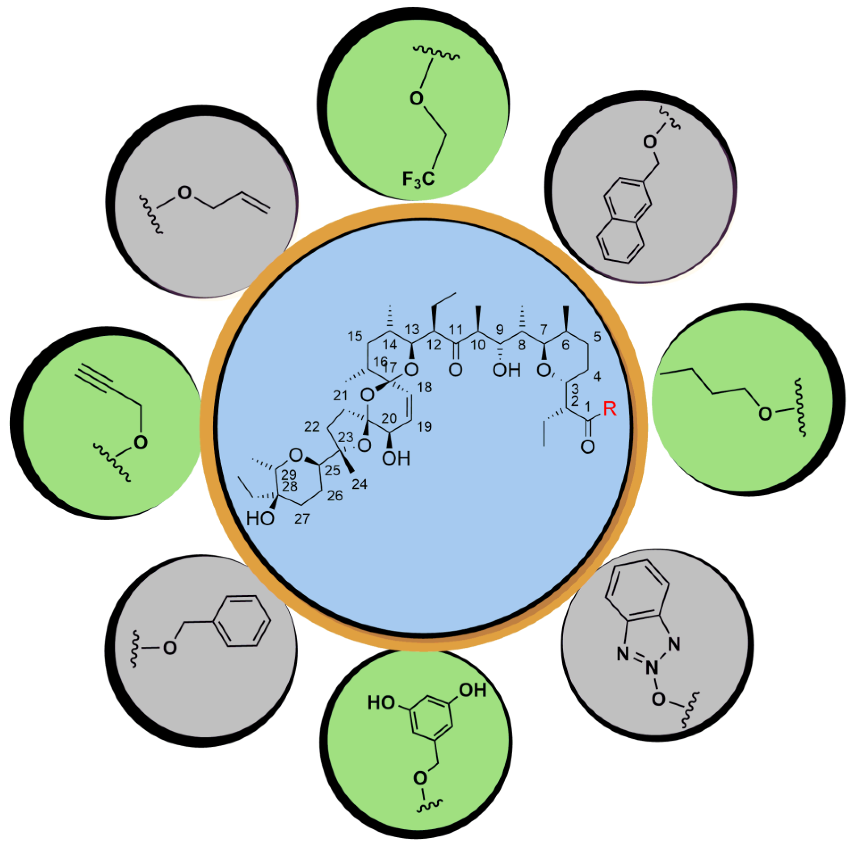
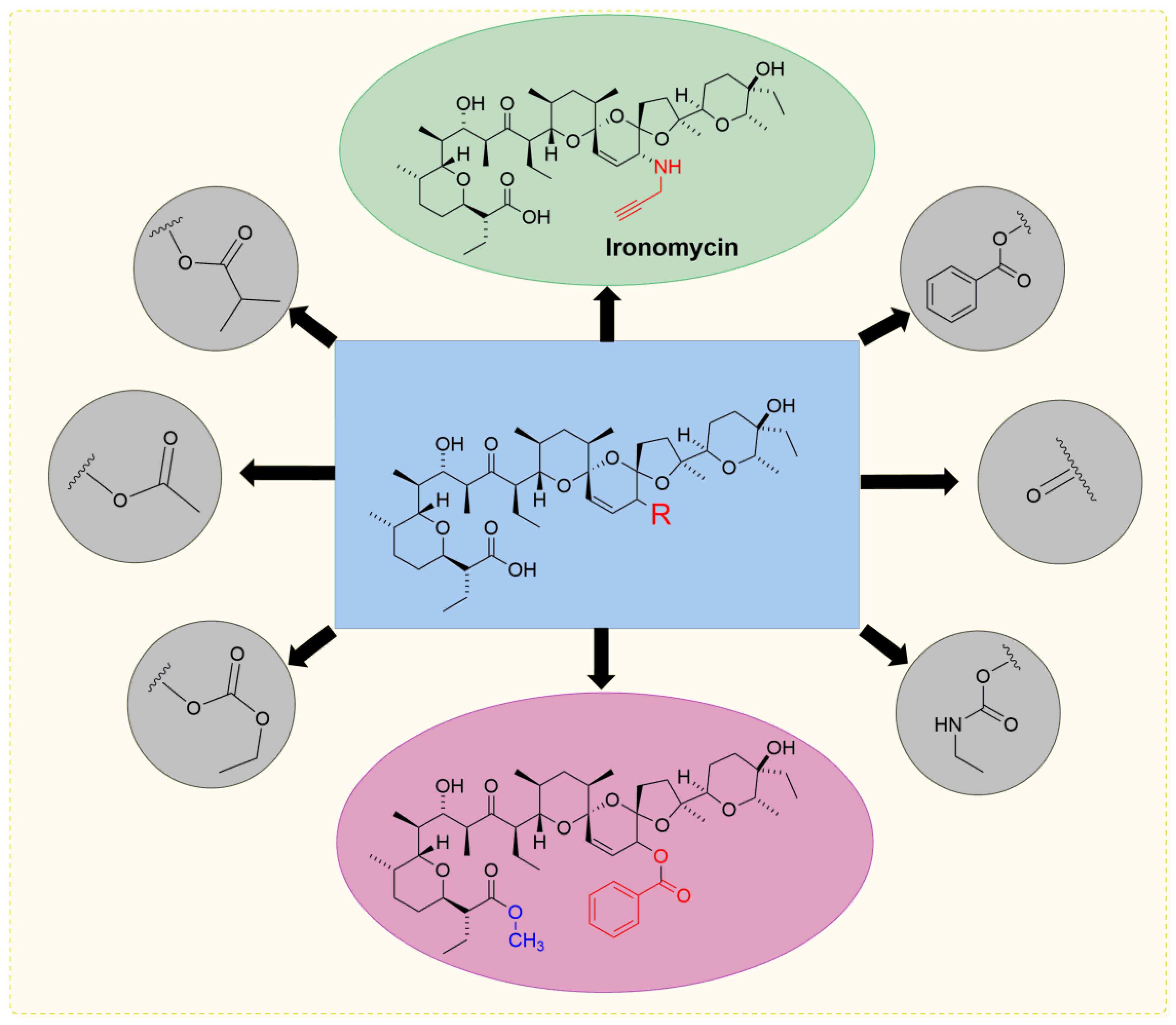
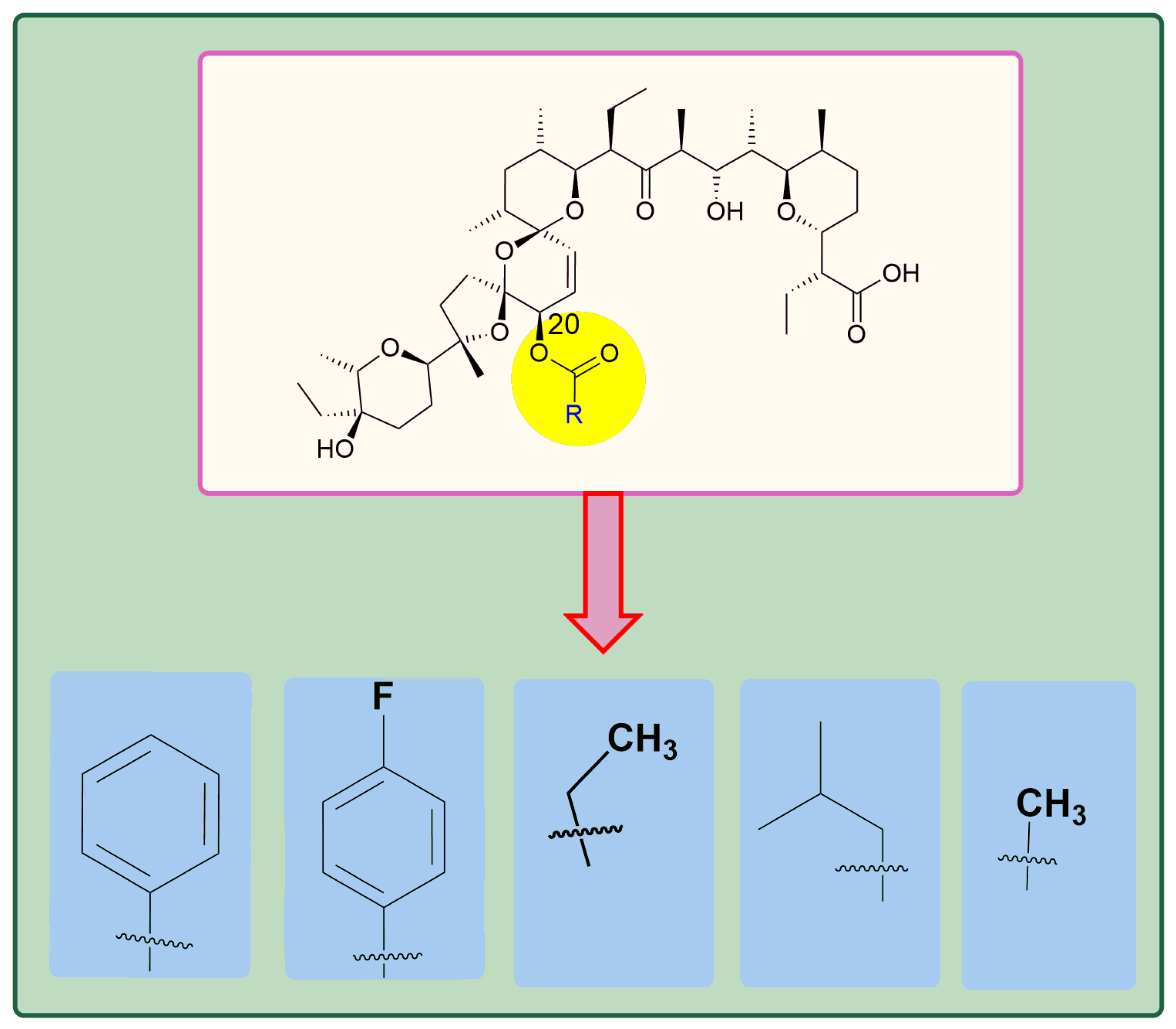
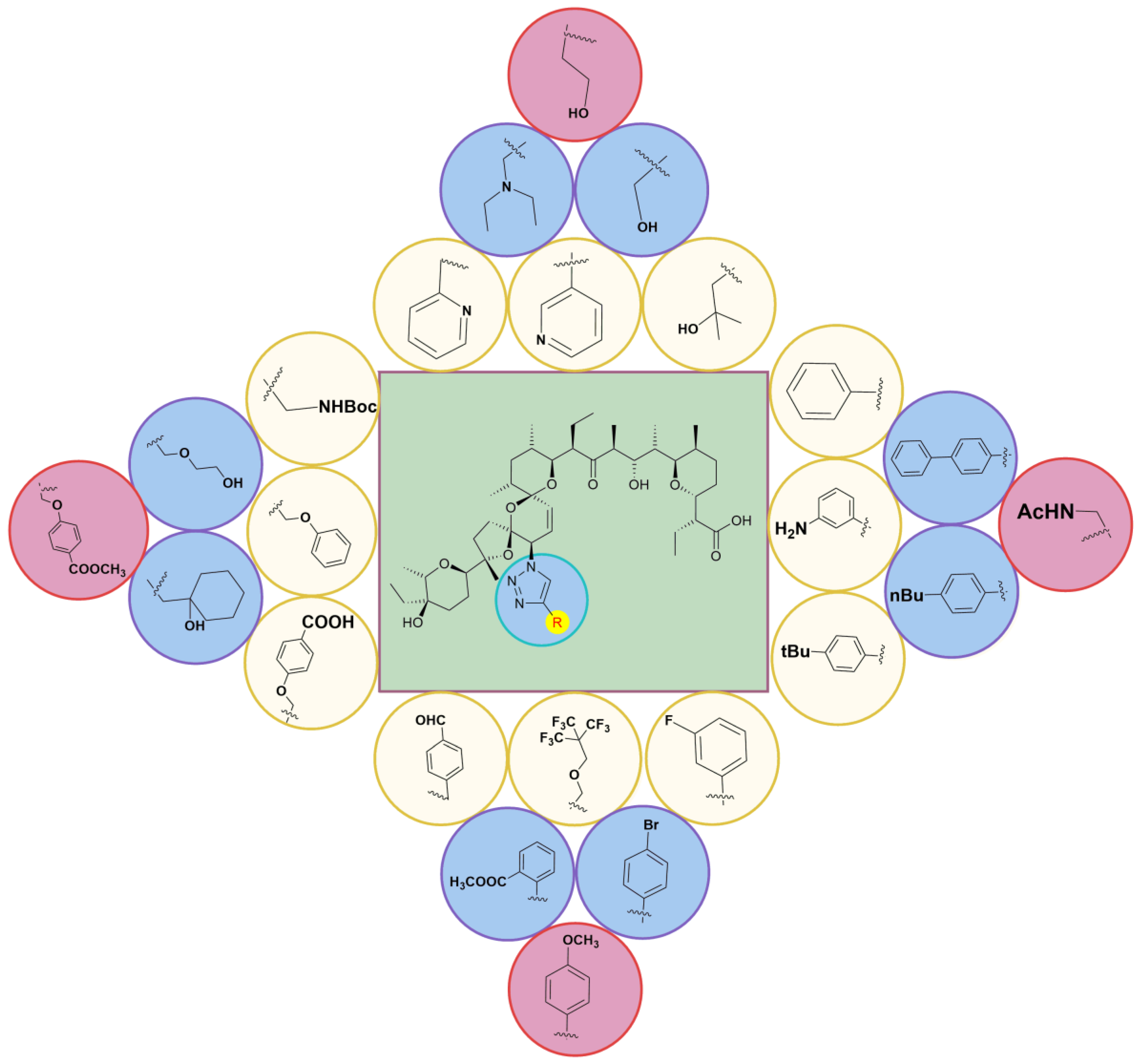
5.2. Chemistry of C20 Analogs

Strand et al. [41] demonstrated that the IC50 against breast cancer cells was reduced by 80% when the acylation of SAL at the C20 hydroxyl group was performed, and the findings emphasize the significance of the C20 hydroxyl group in SAL’s anti-proliferative effects. SAR studies of SAL revealed that C11 and C1 are important ion-coordinating features, and the C20-O-acylated analogs (Figure 8) have significant anti-tumor properties that are both anti-proliferative and CSC-selective [47,94]. The C20-O-analogs impeded breast cancer cell movement, reduced expression of vimentin, a marker of mesenchymal phenotype, and raised the level of E-cadherin, a marker of epithelial phenotype, implying that analogs of SAL and itself cause mesenchymal to epithelial change and may thus prevent malignancy [47].
Since the C1 analogs that were synthesized showed similar or more potent effects with a better SI index against various cell lines when compared to SAL, C20 analogs were synthesized, and their biological activity was evaluated. The C20 ketone analog (Figure 8) exhibited lethal effects equivalent to SAL in an ex vivo study of breast cancer but did not stand out in any in vitro cell-based experiments [95]. Based on the biological activity of the C20 ketone (Figure 8), Rodriguez et al. [54] synthesized a series of C20-amine analogs, of which, when compared to SAL, ironomycin (IRO) exhibited ten times more activity while retaining the selectivity of the CSC high cell population over the CSC low cell population. The amine analog IRO (Figure 8) produced by Mai et al. [21] showed better cytostatic action on breast cancer CSCs and decreased toxicity on non-cancerous breast cells in contrast to SAL, with the alteration occurring at the C20 position [44,53]. Furthermore, IRO demonstrated much higher cytotoxic activity against the ALDH-positive population of breast cancer cells, which is also well known as a cell subgroup with CSC characteristics. SAL and IRO anticancer effects were verified in patient PDX models. Rodriguez’s group created this compound and found it to have a lower IC50 against EMT in CSC-high breast cancer cells in vitro and a stronger specificity over CSC-low cells when compared to SAL, although they did not test for toxicity to normal cells. The findings emphasized IRO’s significant anti-CSC effectiveness with low toxicity to normal cells as well as its potential as a therapeutic agent. Wu et al. [96] stated that the diastereoisomers were synthesized at the C17 and C21 sites of SAL, and their benzoylated derivatives were tested in vitro for anti-proliferative properties on colon cancer, breast cancer, and rat cerebral cortex neuron cells. The sodium salt of 17,21-di-epi-20-O-Bz-SAL (Figure 8) improved the activity by approximately double and had a much higher therapeutic efficacy. The C20-cycloalkyl-modified analogs (Figure 8) with a similar mechanism and a subtle structural variance to IRO demonstrated promising efficacy and specificity against CSC-high breast cancer cells, necessitating further research into toxicity against noncancerous cells and in vivo studies.
Zhang et al. [97] modified SAL and yielded 20-epi-SAL and the resulting SAL-O-acyl derivatives. The first step in this process was the synthesis of the SAL TMSEt-ester, in which the bulky functional group changed SAL’s conformity and the available reactive site, thus leading to a smooth inversion of the hydroxyl configuration [97]. One of the protected products was synthesized, and the respective O-acylated species were introduced to yield the desired 20-O-acyl-SAL derivatives.
Using the MTT assay, the 20-epi-SAL sodium salt and its corresponding C-20-acylated analogs were tested for anti-proliferative efficacy against HT-29 colorectal cancer, HGC-27 gastric cancer, and triple negative MDA-MB-231 breast cancer cells. The results showed that the 20-epi-SAL showed comparable action to SAL, while the 20-epi-O-acylated derivatives displayed greater potency against the cancer cell lines by 2- to 10-fold when compared to SAL. The simple fatty acylated and benzoylated analogs exhibited greater anti-proliferative activity than SAL and its 20-epimer counterpart, except for the p-nitobenzoylated analog. When comparing simple fatty acylated analogs (Figure 9), the benzoylated analogs were 10 times more potent when compared to SAL [97]. It was also observed that the benzoylated analogs (Figure 9) showed greater selectivity when compared to SAL among cancer cells. Based on the results obtained, the respective analogs could be considered for future consideration as therapeutic agents [91].
Shi et al. [98] studied the x-ray crystal structure and modeling of SAL and discovered that the C20 hydroxyl group does not participate in ion chelation, that acylation causes significant obstruction for ion chelation, and that the inversion of the C20 structure may alleviate steric hindrance and improve ion chelation and efficacy [91]. Based on this discovery, another modification site on the SAL molecule was possible for targeted SAL administration without affecting its ion chelation or efficacy [99]. As a result, Shi et al. investigated the inversion of the C20 hydroxyl group by a substitution of an azide group as it allows simple accessibility to numerous SAL derivatives, which they used under mild conditions in Cu-catalyzed azide-alkyne cycloaddition (CuAAC) processes [100]. The Shi group synthesized a series of C20 triazole conjugates with a variety of aromatic species and evaluated them for their biological activity. The triazole-substituted compounds of SAL (Figure 10) were tested for their cytotoxicity on murine breast cancer cells (4T1).
It was determined that the C20-azide analog showed a 27-fold decrease in efficacy when compared to SAL. The phenyl-triazole species showed greater or similar efficacy when compared to SAL. It is worth noting that triazole conjugates with bulky substituents had better biological activity than SAL, while substituents with heteroatoms were less potent due to the strong chelating capabilities of the nitrogen atoms, which led to a hindrance of chelating ions and subsequently decreased the potency of those analogs [98], except for the 3-pyridine conjugate (Figure 10). The hydroxyl-containing conjugates also displayed a similar trend. It is worth noting that triazoles containing ethylene glycol units, which are recognized to chelate ions, showed no appreciable cytotoxicity. Perfluoro-tert-butyl ether was found to be the most powerful of the ether-containing triazoles, being 2-fold more effective than SAL [98].
SAL-triazole conjugates diphenyl, butyl benzene, 3-pyridine, and perfluoro-tert-butyl ether, as well as SAL, were chosen for further testing on human hepatic cells (L02) and cancer cell lines, such as human glioblastoma cells (U87), cervical cancer cells (Hela), and epithelial colorectal adenocarcinoma cells (MCF-7) [98]. According to the findings, triazoles had significantly decreased cytotoxicity against non-cancerous L02 cells and significantly increased cytotoxicity against cancer cells. Tert-butyl benzene, 3-pyridine, and perfluoro-tert-butyl ether (Figure 10) showed a 2-fold increase in cytotoxicity towards U87, and diphenyl displayed a 2.9-fold increase in cytotoxicity towards MCF-7 than that of SAL. In comparison to SAL, perfluoro-tert-butyl ether was 29.5-fold more lethal to colon carcinoma CaCo2 cells [98].
The therapeutic potential of diphenyl, butyl benzene, 3-pyridine, and perfluoro-tert-butyl ether triazoles was investigated, and the selectivity index (SI) was obtained. Except for Hela cells, SAL had a poor SI in selected cancer cells. These triazoles, on the other hand, exhibited a much higher SI than SAL. Triazole diphenyl had the greatest SI of 8.85 for Hela, triazole tert-butyl had the highest SI of 5.89 for colon carcinoma Caco2, and perfluoro-tert-butyl ether is the most promising of these triazoles since it has the highest potency and SI for Hela and Caco2 cells [96]. Based on these findings, the inverse of the C20 position of SAL can relieve steric hindrance, enhance chelation of ions, and increase efficacy.
Additionally, Shi et al. [98] synthesized a library of 20-epi-amino-20-deoxy SAL derivatives with the aim of increasing the drug ability of SAL. The first step in the synthesis was the protection of the C1 carboxylic acid with the TMSEt-ester after the conversion of the C20-hydroxyl to the reactive azide intermediate. The resulting azide was then reduced by Staudinger reduction to yield the desired amine, which then reacted with a variety of acyl chlorides, and the deprotection of the C-1 position resulted in the desired C20-(S)-amide derivatives. Similarly, C20-(S)-N-carbamate derivatives were also synthesized utilizing the same methodology (Figure 11) [100,101].
The molecules from the library, as seen above in Figure 10, were evaluated for their activity against murine breast cancer cells (4T1), HL-60, adenocarcinomic human alveolar basal epithelial cells (A549), human cervical cancer cells (HeLa), human breast cancer cells (MCF-7), human colon adenocarcinoma cells (SW480), and human hepatocarcinoma cells (SMMC-7721). The synthesized amine intermediate showed moderate efficacy and little effectiveness. Because the amine group is a powerful chelating agent for metal ions, the efficacy decrease could be attributed to an ion-chelating-induced conformational shift. Second, acylation of the amine to the respective C20-(S)-N-carbamates (Figure 11) resulted in increased potency and selectivity, thereby alleviating the undesired chelation effect [101]. Of the carbamates synthesized, the benzyl group demonstrated over an 80-fold more potent analog than SAL against SMMC-7721 cells [101], whereas carbamate with a fluorenylmethyl group had the highest potency in A549 cells. Finally, acylation of the amine intermediate produced C20-(S)-N-amides (Figure 11) and amides containing methyl, cyclopropyl, alkene, ethyl ester, phenyl, p-fluorobenzene, m-fluorobenzene, p-cyanobenzene, and p-nitrobenzene, to observe the mixed outcome. The perfluoro-tert-butyl group present on one of the amide analogs of SAL exhibited greater efficacy amongst cell lines, especially the greatest against MCF-7 cells, which was 2-fold higher than SAL [100,101].
SAL’s perfluoro-tert-butyl ether triazole, developed by Shi et al., along with carbamates containing functional groups of Oprn, methoxypentane, methoxy-2-methylpropane, t-butyl, methoxy-methyl benzene, and methoxymethyl)-H-fluorine and amides containing p-fluorobenzene, 1,1,1,3,3,3-hexafluoro-2-methoxy-2-(trifluoromethyl)propane, tert-butyl)benzene, m-fluorobenzene, 2,6-dichlorobenzene, o-methoxybenzene, o-ethoxybenzene, m-methoxybenzene, and methyl(phenyl)sulfane (Figure 11) were evaluated on human bronchial epithelial cells (BEAS-2B). Carbamates and amides showed greater or comparable cytotoxicity when compared to SAL, excluding p-fluorobenzene; when compared to perfluoro-tert-butyl ether triazole against BEAS-2B, the derivatives displayed a 1.2 to 4.0 less effect [100,101]. The selectivity index (SI) was calculated to assess the therapeutic potential of the analogs, and those with great specificity towards a panel of malignant cells, such as analog methoxy-methyl benzene, had the greatest SI of 87.07 towards SMMC-7721, and 2,6-dichlorobenzene demonstrated the largest SI of 48.23 against HL-60. Further exploration of the clinical aspects of these needs to be pursued based on these promising findings.

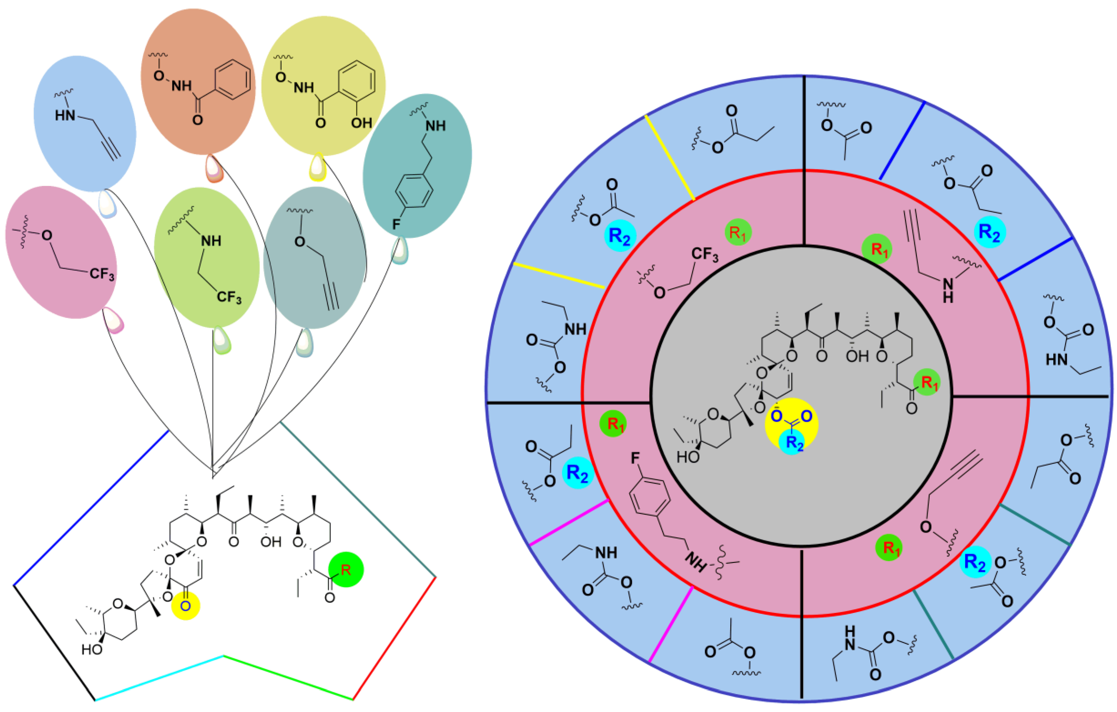
5.3. SAL Double-Modified Analogs

Based on the concept of a promising drug candidate through single modification of SAL, as mentioned, Urbaniak et al. [95] synthesized a series of SAL analogs by modification of the C1 or C20 positions, which led to the development of double-modified SAL analogs (Figure 12).
These molecules were then screened for their improved biological activity against breast cancer stem cells, and it was determined that six single- and two double-modified analogs had a higher potency when compared to SAL against MDA-MB-231 cells. Of these doubly modified SAL analogs, N-methoxybenzamide was determined to have more efficacy when compared to SAL. It was reported that N-methoxybenzamide displayed greater efficacy versus SAL by reducing MDA-MB-231 cell motility and regeneration, as well as generating preferential loss of the CD44+/CD24 low stem-like cells in both monolayer (2D) and organoid (3D) cultures [29]. In addition, anti-proliferative effects against breast cancer stem cells were explored, in which a series of SAL derivatives, singly and doubly modified, were synthesized and found to be more potent when compared to SAL when testing their activity against MDA-MB-231 cells. It was found that the molecules caused DNA to fragment, indicating apoptosis.
N-methoxybenzamide was found to have greater efficacy than SAL, where cell migration and regeneration were inhibited and the CD44+/CD24 stem cells were reduced, including both monolayer and organoid cultures [29,32]. N-methoxybenzamide in the majority of NCI-60 cancer cell lines, including the full panel of breast tumor cell lines, was shown to be more powerful than SAL. N-methoxybenzamide caused apoptosis and increased LC3II, an autophagy-associated marker, which was upregulated in both cell monolayers and organoids [29]. Based on these findings, of the doubly modified analogs, N-methoxybenzamide showed the most therapeutic potential, and further research will need to be pursued in order to determine this analog’s potential as a therapeutic drug in the treatment of metastatic breast cancer.
Modification at the C1 carboxylic acid, either by esterification, amidation, or acylation of SAL at the C20 position, enhances its biological properties. Hence, Antoszczak et al. [37] initially synthesized a series of single-modified SAL analogs, either by esterification or amidation at the C1 position, from previously studied biologically efficacious C1 analogs, in which the respective C1 ester or amide series were further modified at the C20 position by developing an ester, carbonate, and carbamate series (Figure 12), and the doubly-modified SAL analog’s anti-proliferative capabilities when treating different cancer cell lines were explored.
SAL analogs were synthesized by modification of the C1 and C20 positions based on knowledge of the beneficial changes at those positions. The cancer cell line that was mainly evaluated was the primary acute lymphoblastic leukemia (ALL) cells, and the C20 hydroxyl group when acylated increased the efficacy of the amides as compared to the esters, as this was an unexpected finding. It is worth noting that the 20 carbamates showed greater potency as compared to the carbonates and esters. Several doubly modified analogs were discovered to be more effective against the multi-drug-resistant LoVo/DX cell line than the regularly used anticancer agents cisplatin and doxorubicin [37,38]. In addition, three doubly modified compounds were found to have greater potency than SAL against primary ALL cells, and after treatment with SAL or C1-2-(4-fluorophenyl)-N-methylethan-1-amine and C20-(ethylamino)-oxidaneyl)-methanone in an ex vivo model of breast tumor cells, survival was considerably reduced in a time-dependent fashion. Antoszczak et al. demonstrated that doubly-modified SAL analogs show potential lead molecules for the treatment of different cancers [93].

6. Discussion

SAL has been utilized in poultry medicine for many years and was discovered as an anticancer agent by Weinberg et al. in 2009. There has been plenty of data establishing SAL’s effect against different types of CSCs, and the results of the studies have demonstrated promising applications of SAL as a potential chemotherapeutic agent in an in vitro and in vivo setting. Knowing the pre-clinical capabilities, the clinical applications were explored. Although there have been a few investigations (two clinical cases) utilizing SAL itself and in conjunction with other chemotherapeutic agents with the outcomes of advanced cancer regression, SAL has not been used in the treatment of other cancers up to this point. Further investigations of SAL for its clinical applications should be pursued, as this polyether ionophore shows good chemotherapeutic properties.
Knowing the therapeutic benefits SAL possesses, many groups synthesized analogs in order to decrease its toxicity toward normal cells while maintaining or increasing its potency towards cancerous cells. SAL was tested against the CSCs population of breast cancer (Table 4), and it was observed that at 0.5 μM there was iron accumulation in the lysosome, a decrease in ferritin, and an increase in iron regulatory protein 2 (IRP2).
Based on these findings, a novel mechanism of SAL was hypothesized in which it can affect breast CSCs by sequestering iron in the lysosome, where iron homeostasis is tightly regulated intracellularly. An analog of SAL by the modification of the C20 position was named IRO, or coined as ironomycin, which was designed particularly for this mechanism of action. A series of C20-amine analogs, of which one particular analog of SAL, IRO, exhibited a 10 times greater effect against HMLER cells than SAL and was more selective, and this molecule affected CSCs through iron sequestering in the lysosome in the cell, and a significant iron change in breast cancer stem cells was observed. IRO was tested, and an accumulation of iron in lysosomes, which leads to the production of ROS and causes apoptosis to occur, could be due to the alteration at the C20 position. When compared to SAL, this analog demonstrated better potential anticancer activity in breast cancer stem cells and less toxicity in non-cancerous cells. This highlights that IRO has higher anti-CSC efficacy and lower toxicity to normal cells. This analog was found to be about 10-fold more potent and not as detrimental to non-cancerous cells as compared to SAL (Table 5). Owing to the promising results, IRO has demonstrated that further research should be pursued in the development and optimization of this more potent analog.

7. Conclusions

SAL is obtained from Streptomyces albus by isolation from culture broth, column chromatography, and crystallization to yield the polyether ionophore as colorless crystals of the molecules screened. The isolation of Streptomyces albus resulted in the discovery of SAL, which has shown versatility in its biological properties. Over the years, many different research groups worked on the biological activity of SAL, and when tested against breast CSCs, it was found to be one of the most potent of around 16,000 bioactive molecules. The mechanism of action of SAL was explored when interacting with CSCs, and it was determined that this potent molecule affects CSCs through different modes of action. Based on these findings, SAL has been explored for its anticancer properties, and it has shown promising findings in vitro with micromolar concentrations when treating different cancer cell lines, especially TNBC. The in vivo studies have also shown promising results in terms of tumor reduction. This polyether ionophore antibiotic has been introduced in clinical studies that have shown promising results in which SAL significantly decreased tumor metastasis when administered to a middle-aged woman with TNBC and in another clinic study of an elderly female patient who had small cell carcinoma in which SAL by itself was administered and a stable disease state with no progression was observed.
Based on the promising findings of SAL’s biological activity, many groups have been experimenting with the modification of this new potential chemotherapeutic drug at the C1, C17, C20, and C21 positions of the molecule, as mentioned earlier, with the intention of either modifying to achieve increased potency against cancer cells, decreasing toxicity towards non-cancerous cells, or maintaining the same potency but decreasing the toxicity. Many groups were successful in the synthesis of analogs that were more potent than SAL and possessed less cellular toxicity to normal cells.
Based on the initial studies and the encouraging results, more work should be pursued in the development and optimization of this promising, more potent analog of SAL, as it could be a potential chemotherapeutic agent.

Author Contributions

Conceptualization, V.S. and A.N.; methodology, V.S. and A.N.; writing—original draft preparation, V.S., A.N., J.M. and L.V.S.; editing, R.B., S.R. and C.S.; review, A.N. and V.S. All authors have read and agreed to the published version of the manuscript.

Funding

This research received no external funding.

Institutional Review Board Statement

Not applicable.

Informed Consent Statement

Not applicable.

Data Availability Statement

Not applicable.

Acknowledgments

The authors acknowledge the faculty of Rowan University, NJ and Principal, R. C. Patel Institute of Pharmaceutical Education and Research, Shirpur, Maharashtra, India for supporting this work. The work is edicated to our mentor Subash Jonnalagadda, Rowan University, NJ, USA.

Conflicts of Interest

The authors declare no conflict of interest.

References

  1. Qi, D.; Liu, Y.; Li, J.; Huang, J.H.; Hu, X.; Wu, E. Salinomycin as a potent anticancer stem cell agent: State of the art and future directions. Med. Res. Rev. 2022, 42, 1037–1063. [Google Scholar] [CrossRef]
  2. Von Suskil, M.; Sultana, S.N.; Elbezanti, W.O.; Al-Odat, O.S.; Chitren, R.C.; Tiwari, A.K.; Challagundla, K.B.; Srivastava, S.K.; Jonnalagadda, S.C.; Alpdogan-Budak, T.; et al. Bruton’s Tyrosine Kinase Targeting in Multiple Myeloma. Int. J. Mol. Sci. 2021, 22, 5707. [Google Scholar] [CrossRef]
  3. Hermann, P.C.; Sainz, B., Jr. Pancreatic cancer stem cells: A state or an entity. Semin. Cancer Bio. 2018, 53, 223–231. [Google Scholar] [CrossRef] [PubMed]
  4. Chaffer, C.L.; Brueckmann, I.; Scheel, C. Normal and neoplastic nonstem cells can spontaneously convert to a stem-like state. Proc. Nat. Acad. Sci. USA 2011, 108, 7950–7955. [Google Scholar] [CrossRef] [PubMed]
  5. Papaccio, F.; Paino, F.; Regad, T.; Papaccio, G.; Desiderio, V.; Tirino, V. Concise Review: Cancer Cells, Cancer Stem Cells and Mesenchymal Stem Cells: Influence in Cancer Development. Stem. Cells Transl. Med. 2017, 6, 2115–2125. [Google Scholar] [CrossRef]
  6. Nayak, A.; Warrier, N.M.; Kumar, P. Cancer Stem Cells and the Tumor Microenvironment: Targeting the Critical Crosstalk through Nanocarrier Systems. Stem Cell Rev. Rep. 2022, 18, 2209–2233. [Google Scholar] [CrossRef] [PubMed]
  7. Waghray, M.; Yalamanchili, M.; Dziubinski, M.; Zeinali, M.; Erkkinen, M.; Yang, H.; Schradle, K.A.; Urs, S.; Pasca Di Magliano, M.; Welling, T.H.; et al. GM-CSF Mediates Mesenchymal-Epithelial crosstalk in Pancreatic Cancer. Cancer Discov. 2016, 6, 886–899. [Google Scholar] [CrossRef]
  8. Raggi, C.; Mousa, H.S.; Correnti, M.; Sica, A.; Invernizz, P. Cancer stem cells and tumor-associated macrophages: A roadmap for multitargeted strategies. Oncogene 2016, 35, 671–682. [Google Scholar] [CrossRef] [PubMed]
  9. Tan, S.H.; Swathi, Y.; Tan, S.; Goh, J.; Seishima, R.; Murakami, K.; Oshima, M.; Tsuji, T.; Phuah, P.; Tan, L.T.; et al. AQP5 enriches for stem cells and cancer origins in the distal stomach. Nature 2020, 578, 437–443. [Google Scholar] [CrossRef]
  10. Reya, T.; Morrison, S.J.; Clarke, M.F.; Weissman, I.L. Stem cells cancer and cancer stem cells. Nature 2001, 414, 105–111. [Google Scholar] [CrossRef]
  11. Lytle, N.K.; Barber, A.G.; Reya, T. Stem cell fate in Cancer growth, progression, and therapy resistance. Nat. Rev. Cancer 2018, 18, 669–680. [Google Scholar] [CrossRef] [PubMed]
  12. Plaks, V.; Kong, N.; Werb, Z. The cancer stem cell niche: How essential is the niche in regulating stemness of tumor cells? Cell Stem. Cell 2015, 16, 225–238. [Google Scholar] [CrossRef] [PubMed]
  13. Yu, Z.; Pestell, T.G.; Lisanti, M.P.; Pestell, R.G. Cancer stem cells. Int. J. Biochem. Cell Biol. 2012, 44, 2144–2151. [Google Scholar] [CrossRef] [PubMed]
  14. Shibue, T.; Weinberg, R.A. EMT, CSCs, and drug resistance: The mechanistic link and clinical implications. Nat. Rev. Clin. Oncol. 2017, 14, 611–629. [Google Scholar] [CrossRef]
  15. Dosch, J.S.; Ziemke, E.K.; Shettigar, A.; Rehemtulla, A.; Sebolt-Leopold, J.S. Cancer Stem Marker Phenotypes Are Reversible and Functionally Homogeneous in a Preclinical Model of Pancreatic Cancer. Cancer Res. 2015, 75, 4582–4592. [Google Scholar] [CrossRef] [PubMed]
  16. Li, J.; Qi, D.; Hsieh, T.C.; Huang, J.H.; Wu, J.M.; Wu, E. Trailblazing perspectives on targeting breast cancer stem cells. Pharmacol. Ther. 2021, 223, 107800. [Google Scholar] [CrossRef]
  17. Clara, J.A.; Monge, C.; Yang, Y.; Takebe, N. Targetting signaling pathways and the immune microenvironment of cancer stem cells-a clinical update. Nat. Rev. Clin. Oncol. 2020, 17, 204–232. [Google Scholar] [CrossRef]
  18. Takebe, N.; Miele, L.; Harris, P.J.; Jeong, W.; Bando, H.; Kahn, M.; Yang, S.X.; Ivy, S.P. Targetting Notch, Hedgehog, and Wnt pathways in cancer stem cells: Clinical update. Nat. Rev. Clin. Oncol. 2011, 8, 97–106. [Google Scholar] [CrossRef]
  19. Gupta, P.B.; Onder, T.T.; Jiang, G.; Tao, K.; Kuperwsser, K.; Weinberg, R.A.; Lander, E.S. Identification of selective inhibitors of cancer stem cells by high throughput screening. Cell 2009, 138, 645–659. [Google Scholar] [CrossRef]
  20. Arfaoui, A.; Rioualen, C.; Azzoni, V.; Pinna, G.; Finetti, P.; Wicinski, J.; Josselin, E.; Macario, M.; Castellano, R.; Léonard-Stumpf, C.; et al. A genome-wide RNAi screen reveals essential therapeutic targets of breast cancer stem cells. EMBO Mol. Med. 2019, 11, 9930. [Google Scholar] [CrossRef]
  21. Elbezanti, W.O.; Al-Odat, O.S.; Chitren, R.; Singh, J.K.; Srivastava, S.K.; Gowda, K.; Amin, S.; Robertson, G.P.; Nemmara, V.V.; Jonnalagadda, S.C.; et al. Development of novel bruton’s tyrosine kinase inhibitor that exerts anti-cancer activities potentiates response of chemotherapeutic agents in multiple myeloma stem-like cells. Front. Pharmacol. 2022, 13, 1–18. [Google Scholar] [CrossRef]
  22. Singh, S.; Numan, A.; Maddiboyina, B.; Arora, S.; Riadi, Y.; Md, S. The emerging role of immune checkpoint inhibitors in the treatment of triple-negative breast cancer. Drug Discov. Today 2021, 26, 1721–1727. [Google Scholar] [CrossRef]
  23. Ataollahi, M.R.; Sharifi, J.; Paknahad, M.R.; Paknahad, A. Breast cancer and associated factors: A review. J. Med. Life 2015, 8, 6–11. [Google Scholar] [PubMed]
  24. Sharma, G.N.; Dave, R.; Sanadya, J.; Sharma, P.; Sharma, K.K. Various types and management of breast cancer: An overview. J. Adv. Pharm. Technol. Res. 2010, 1, 109–126. [Google Scholar]
  25. Bray, F.; Ferlay, J.; Soerjomataram, I.; Siegel, R.L.; Torre, L.A.; Jemal, A. Global cancer statistics 2018: GLOBOCAN estimates of incidence and mortality worldwide for 36 cancers in 185 countries. CA Cancer J. Clin. 2018, 6, 394–424. [Google Scholar] [CrossRef]
  26. Jin, X.; Mu, P. Targeting breast cancer metastasis. Breast Cancer Basic Clin. Res. 2015, 9, 23–34. [Google Scholar] [CrossRef]
  27. Gupta, G.P.; Massagué, J. Cancer metastasis: Building a framework. Cell 2006, 127, 679–695. [Google Scholar] [CrossRef]
  28. Narod, S.A.; Iqbal, J.; Miller, A.B. Why have breast cancer mortality rates declined? J. Cancer Policy 2015, 5, 8–17. [Google Scholar] [CrossRef]
  29. Urbaniak, A.; Reed, M.R.; Fil, D.; Moorjani, A.; Heflin, S.; Antoszczak, M.; Sulik, M.; Huczynski, A.; Kupsik, M.R.; Eoff, L.; et al. Single and double modified salinomycin analogs target stem-like cells in 2D and 3D breast cancer models. J. Biomed. Pharmacother. 2021, 141, 111815. [Google Scholar] [CrossRef] [PubMed]
  30. Ismail-Khan, R.; Bui, M. A Review of Triple-Negative Breast Cancer. Cancer Control 2010, 17, 173–176. [Google Scholar] [CrossRef]
  31. Kaplan, H.G.; Malmgren, J.A.; Atwood, M.K. Impact of triple negative phenotype on breast cancer prognosis. In Proceedings of the 29th Annual San Antonio Breast Cancer Symposium, San Antonio, TX, USA, 14–17 December 2006. [Google Scholar]
  32. Kuran, D.; Flis, S.; Antoszczak, M.; Piskorek, M.; Huczynski, A. Ester derivatives of Salinomycin efficiently eliminate breast cancer cells via ER-stress-induced apoptosis. Eur. J. Pharmacol. 2021, 893, 173824. [Google Scholar] [CrossRef]
  33. Rakha, E.A.; Reis-Filho, J.S.; Ellis, I.O. Basal-Like Breast Cancer: A Critical Review. J. Clin. Oncol. 2008, 26, 2568–2581. [Google Scholar] [CrossRef]
  34. Carey, L.A.; Perou, C.M.; Livasy, C.A.; Dressler, L.G.; Cowan, D.; Conway, K.; Karaca, G.; Troester, M.A.; Tse, C.K.; Edmiston, S.; et al. Race, breast cancer subtypes, and survival in the Carolina Breast Cancer Study. J. Am. Med. Assoc. 2006, 295, 2492–2502. [Google Scholar] [CrossRef]
  35. Perou, C.M.; Sørlie, T.; Eisen, M.B.; van de Rijn, M.; Jeffery, S.S.; Rees, C.A.; Pollack, J.R.; Ross, D.T.; Johnsen, H.; Akslen, L.A.; et al. Molecular portraits of human breast tumours. Nature 2000, 406, 747–752. [Google Scholar] [CrossRef]
  36. Mai, T.T.; Hamaï, A.; Hienzsch, A.; Cañeque, T.; Müller, S.; Wicinski, J.; Cabaud, O.; Leroy, C.; David, A.; Acevedo, V.; et al. Salinomycin kills cancer stem cells by sequestering iron in lysosomes. Nat. Chem. 2017, 9, 1025–1033. [Google Scholar] [CrossRef] [PubMed]
  37. Antoszczak, M.; Urbaniak, A.; Delgado, M.; Maj, E.; Borgstrom, B.; Wietrzyk, J.; Huczynski, A.; Yua, Y.; Chambers, T.C. Biological activity of doubly modified salinomycin analogs-Evaluation in vitro and ex vivo. Eur. J. Med. Chem. 2018, 156, 510–523. [Google Scholar] [CrossRef]
  38. Haruyasu, K.; Otake, N.; Yonehara, H.; Sato, S.; Saito, Y. The structure of salinomycin, a new member of the polyether antibiotics. Tetrahedron Lett. 1973, 14, 4955–4958. [Google Scholar] [CrossRef]
  39. Miyazaki, Y.; Shibuya, M.; Sugawara, H.; Kawaguchi, O.; Hirose, C. Salinomycin, a new polyether antibiotic. J. Antibio. 1974, 27, 814–821. [Google Scholar] [CrossRef]
  40. Piperno, A.; Marrazzo, A.; Scala, A.; Rescifina, A. Chemistry and Biology of Salinomycin and its Analogues. Ital. J. Chem. Sci. 2016, 19, 177–209. [Google Scholar] [CrossRef]
  41. Borgstrom, B.; Xiaoli, H.; Posta, M.; Hegardt, C.; Ordesson, S.; Strand, D. Synthetic modification of saliomycin: Selective O-acylation and biological evaluation. Chem. Commun. 2013, 49, 9944–9946. [Google Scholar] [CrossRef]
  42. Naujokat, C.; Steinhart, R. Salinomycin as a drug for targeting human cancer stem cells. J. Biomed. Biotech. 2012, 950658. [Google Scholar] [CrossRef]
  43. Jangamreddy, J.R.; Jain, M.V.; Hallbeck, A.L.; Roberg, K.; Lotfi, K.; Łos, M.J. Glucose starvation-mediated inhibition of salinomycin induced autophagy cancer cell specific cell death. Oncotarget 2015, 6, 10134–10145. [Google Scholar] [CrossRef] [PubMed]
  44. Mitani, M.; Yamanishi, T.; Miyazaki, Y.; Otake, N. Salinomycin effects on mitochondrial ion translocation and respiration. Antimicrob. Agents Chemother. 1976, 9, 655–660. [Google Scholar] [CrossRef]
  45. Zhao, S.J.; Wang, X.J.; Wu, Q.J.; Liu, C.; Li, D.A.; Fu, X.T.; Zhang, H.F.; Shao, L.R.; Sun, J.Y.; Sun, B.L.; et al. Induction of G1 Cell Cycle Arrest in Human Glioma Cells by Salinomycin Through Triggering ROS-Mediated DNA Damage In Vitro and In Vivo. Neurochem. Res. 2017, 42, 997–1005. [Google Scholar] [CrossRef]
  46. Huang, X.; Borgstro, B.; Kempengren, S.; Persson, L.; Hegardt, C.; Strand, D.; Oredsson, S. Breast cancer stem cell selectivity of synthetic nanomolar-active salinomycin analogs. BMC Cancer 2016, 16, 145–157. [Google Scholar] [CrossRef] [PubMed]
  47. Huang, X.; Borgström, B.; Stegmayr, J. The Molecular Basis of Inhibition of Stemlike Cancer Cells by salinomycin. ACS Cent. Sci. 2018, 4, 760–767. [Google Scholar] [CrossRef]
  48. Li, B.; Wu, J.; Tang, L.; Lian, X.; Li, Z.; Duan, W.; Qin, T.; Zhao, X.; Hu, Y.; Zhang, C.; et al. Synthesis and anti-tumor activity evaluation of salinomycin C20-O-alkyl/benzyl oxime derivatives. Org. Biomol. Chem. 2022, 20, 870–876. [Google Scholar] [CrossRef] [PubMed]
  49. Krebs, J.; Agellon, L.B.; Michalak, M. Ca2+ homeostasis and endoplasmic reticulum (ER) stress: An integrated view of calcium signaling. Biochem. Biophys. Res. Commun. 2015, 460, 114–121. [Google Scholar] [CrossRef]
  50. Lu, D.; Choi, M.Y.; Yu, J.; Castro, J.E.; Kipps, T.J.; Carson, D.A. Salinomycin inhibits Wnt signaling and selectively induces apoptosis in chronic lymphocytic leukemia cells. Proc. Natl. Acad. Sci. USA 2011, 108, 13253–13257. [Google Scholar] [CrossRef]
  51. Wang, Z.; Zhou, L.; Xiong, Y. Salinomycin exerts anti-colorectal cancer activity by targeting Beta-catenin/T-Cell factor complex. Br. J. Pharmacol. 2019, 176, 3390–3406. [Google Scholar] [CrossRef]
  52. Hamaï, A.; Cañeque, T.; Müller, S. An iron hand over cancer stem cells. Autophagy 2017, 13, 1465–1466. [Google Scholar] [CrossRef] [PubMed]
  53. Wang, F.; Zhou, S.; Qi, D.; Xiang, S.H.; Wong, E.T.; Wang, X.; Fonkem, E.; Hsieh, T.C.; Yang, J.; Kirmani, B.; et al. Nucleolin is a Functional Binding Protein for Salinomycin in Neuroblastoama Stem Cells. J. Am. Chem. Soc. 2019, 141, 3613–3622. [Google Scholar] [CrossRef]
  54. Versini, A.; Colombeau, L.; Hinzsch, A.; Gaillet, C.; Retailleau, P.; Debieu, S.; Muller, S.; Caneque, T.; Rodriguez, R. Salinomycin Derivatives Kill Breast Cancer Stem Cells by Lysosomal Iron Targeting. Chem-A. Eur. J. 2020, 26, 7416–7424. [Google Scholar] [CrossRef] [PubMed]
  55. Fähling, M.; Steege, A.; Perlewitz, A.; Nafz, B.; Mrowka, R.; Persson, P.B.; Thiele, B.J. Role of nucleolin in postranscriptional control of MMP-9 expression. Biochim. Biophys. Acta 2005, 1731, 32–40. [Google Scholar] [CrossRef]
  56. Bhatia, S.; Reister, S.; Mahotka, C.; Meisel, R.; Borkhardt, A.; Grinstein, E. Control of AC133/CD133 and impact on human hematopoietic progenitor cells through nucleolin. Leukemia 2015, 29, 2208–2220. [Google Scholar] [CrossRef] [PubMed]
  57. Grinstein, E.; Du, Y.; Santourlidis, S.; Christ, J.; Uhrberg, M.; Wernet, P. Nucleolin Regulates Gene Expression in CD34-positive Hematopoietic Cells. J. Biol. Chem. 2007, 282, 12439–12449. [Google Scholar] [CrossRef]
  58. Pipeline—Hillstream Biopharma, Inc. Available online: https://hillstreambio.com/pipeline/ (accessed on 12 April 2022).
  59. Walcher, L.; Kistenmacher, A.K.; Suo, H.; Kitte, R.; Dluczek, S.; Strauß, A.; Blaudszun, A.R.; Yevsa, T.; Fricke, S.; Kossatz-Boehlert, U. Cancer Stem Cells-Origins and Biomarkers: Perspectives for Targeted Personalized Therapies. Front. Immunol. 2020, 11, 1–33. [Google Scholar] [CrossRef]
  60. Paredes, F.; Williams, H.C.; San Martin, A. Metabolic adaptation in hypoxia and cancer. Cancer Lett. 2021, 502, 133–142. [Google Scholar] [CrossRef]
  61. An, H.; Kim, J.Y.; Lee, N.; Cho, Y.; Oh, E.; Seo, J.H. Salinomycin possesses anti-tumor activity and inhibits breast cancer stem-like cells via an apoptosis-independent pathway. Biochem. Biophys. Res. Commun. 2015, 466, 696–703. [Google Scholar] [CrossRef]
  62. Tefas, L.R.; Barbalata, C.; Tefas, C.; Tomuta, I. Salinomycin-Based drug delivery system: Overcoming the hurdles in cancer Therapy. Pharmaceutics 2021, 13, 1120. [Google Scholar] [CrossRef]
  63. Magrath, J.W.; Raney, W.R.; Kim, Y. In vitro demonstration of salinomycin as a novel chemotherapeutic agent for the treatment of Sox2-positive glioblastoma cancer stem cells. Oncol. Rep. 2020, 44, 777–785. [Google Scholar] [CrossRef] [PubMed]
  64. Zou, M.; Yin, X.; Zhou, X.; Niu, X.; Wang, Y.; Su, M. Salinomycin-Loaded High-Density Lipoprotein Exerts Promising Anti-Ovarian Cancer Effects by Inhibiting Epithelial-Mesenchymal Transition. Int. J. Nanomed. 2022, 17, 4059–4071. [Google Scholar] [CrossRef] [PubMed]
  65. Dewangan, J.; Srivastava, S.; Srikanta, K.R. Salinomycin Inhibits Breast Cancer Progression via Targeting HIF-1α/VEGF Mediated Tumor Angiogenesis in Vitro and in Vivo. Biochem. Pharmacol. 2019, 164, 326–335. [Google Scholar] [CrossRef] [PubMed]
  66. Wang, F.; He, L.; Dai, W.Q.; Xu, Y.P.; Wu, D.; Lin, C.L.; Wu, S.M.; Cheng, P.; Zhang, Y.; Shen, M.; et al. Salinomycin inhibits proliferation and induces apoptosis of hepatocellular carcinoma in vitro and in vivo. E-Bio Med. (PLoS ONE) 2012, 7, 506–538. [Google Scholar] [CrossRef]
  67. Wang, F.; Zheng, Z.; Guan, J.; Qi, D.; Zhou, S.; Shen, X.; Wang, F.; Wenkert, D.; Kirmani, B.; Solouki, T.; et al. Identification of a panel of genes as a prognostic biomarker for glioblastoma. E-Bio Med. (PLoS ONE) 2018, 37, 68–77. [Google Scholar] [CrossRef]
  68. Zhou, S.; Wang, F.; Zhang, Y.; Austin, J. Salinomycin Suppresses PDGFRβ, MYC, and Notch Signaling in Human Medulloblastoma. Pharmacol. Ther. 2014, 3, 1020. [Google Scholar]
  69. Boehmerle, W.; Endres, M. salinomycin induces calpain and cytochrome-c mediated neuronal cell death. Cell Death Dis. 2011, 2, e168. [Google Scholar] [CrossRef]
  70. Fuchs, D.; Heinold, A.; Opelz, G.; Daniel, V.; Naujokat, C. Salinomycin induces apoptosis and overcomes apoptosis resistance in human cancer cells. J. Biochem. Biophys. Res. Commun. 2009, 390, 743–749. [Google Scholar] [CrossRef]
  71. Scherzed, A.; Hackenberg, S.; Froelich, K.; Rak, K.; Technau, A.; Radeloff, A.; Nöth, U.; Koehler, C.; Hagen, R.; Kleinsasser, N. Effects of salinomycin on human bone marrow-derived mesenchymal stem cells in vitro. Toxicol. Lett. 2013, 218, 207–214. [Google Scholar] [CrossRef]
  72. Shen, H.; Sun, C.C.; Kang, L.; Tan, X.; Shi, P.; Wang, L.; Liu, E.; Gong, J. Low dose salinomycin inhibits breast cancer metastasis by repolarizing tumor hijacked macrophages toward the M1 phenotype. Eur. J. Pharm. Sci. 2021, 157, 105629. [Google Scholar] [CrossRef]
  73. Jiang, J.; Li, H.; Qaed, E. Salinomycin, as an autophagy modulator—A new avenue to anticancer: A review. J. Exp. Clin. Cancer Res. 2018, 37, 26. [Google Scholar] [CrossRef] [PubMed]
  74. Scherzad, A.; Hackenberg, S.; Schramm, C.; Froelich, K.; Ginzkey, C.; Rudolf, H.; Kleinsasser, N. Geno-and cytotoxicity of salinomycin in human nasal mucosa and peripheral blood lymphocytes. Toxicol. Vitro 2015, 29, 813–818. [Google Scholar] [CrossRef] [PubMed]
  75. Koch, A.E.; Polverini, P.J.; Kunkel, S.L.; Harlow, L.A.; DiPietro, L.A.; Elner, V.M.; Elner, S.G.; Strieter, R.M. Interleukin-8 as a macrophage-derived mediator of angiogenesis. J. Sci. 1992, 258, 1798–1801. [Google Scholar] [CrossRef]
  76. Arenberg, D.A.; Kunkel, S.L.; Polverini, P.J.; Glass, M.; Burdick, M.D.; Strieter, R.M. Inhibition of interleukin-8 reduces tu- morigenesis of human non-small cell lung cancer in SCID mice. J. Clin. Investig. 1996, 97, 2792–2802. [Google Scholar] [CrossRef] [PubMed]
  77. Szkudlarek-Mikho, M.; Saunders, R.A.; Yap, S.F.; Ngeow, Y.F.; Chin, K.V. Salinomycin, a polyether ionophoric antibiotic, inhibits adipogenesis. Biochem. Biophys. Res. Commun. 2012, 428, 487–493. [Google Scholar] [CrossRef]
  78. Scherzad, A.; Hackenberg, S.; Froelich, K.; Rak, K.; Hagen, R.; Taeger, J.; Bregenzer, M.; Kleinsasser, N. Chronic exposure of low dose salinomycin inhibits MSC migration capability in vitro. Biomed. Rep. 2016, 4, 325–330. [Google Scholar] [CrossRef]
  79. Potter, L.M.; Blake, J.P.; Blair, M.E.; Bliss, B.A.; Denbow, D.M. Salinomycin toxicity in turkeys. Poult. Sci. 1986, 65, 1955–1959. [Google Scholar] [CrossRef]
  80. Chaudhari, P.J.; Bari, S.B.; Surana, S.J.; Nagar, A. Discovery and Anticancer Activity of Novel 1,3,4-Thiadiazole- and Aziridine-Based Indolin-2-ones via in silico Design Followed by Supramolecular Green Synthesis. ACS Omega 2022, 7, 17270–177294. [Google Scholar] [CrossRef]
  81. Salinomycin toxicity causes deaths of calves on two Scottish dairy farms. Vet. Rec. 2012, 170, 118–121. [CrossRef]
  82. Holliman, A.; Howie, F.; Payne, J.; Scholes, S. Salinomycin toxicity in dairy calves. Vet. Rec. 2011, 169, 561. [Google Scholar] [CrossRef]
  83. Plumlee, K.H.; Johnson, B.; Galey, F.D. Acute salinomycin toxicosis of pigs. J. Clin. Investig. 1995, 7, 419–420. [Google Scholar] [CrossRef] [PubMed]
  84. Ashrafihelan, J.J.; Eisapour, H.; Erfani, A.M.; Kalantary, A.A.; Amoli, J.S.; Mozafari, M. High mortality due to accidental salinomycin intoxication in sheep. Interdiscip. Toxicol. 2014, 7, 173–176. [Google Scholar] [CrossRef] [PubMed]
  85. Rudrapal, M.; Issahaku, A.; Agoni, C.; Nagar, A.; Nedale, A.B.; Lokwani, D.K. In silico screening of phytopolyphenolics for the identification of bioactive compounds as novel protease inhibitors effective against SARS-CoV-2. J. Biomol. Struct. Dyn. 2021, 40, 10437–10453. [Google Scholar] [CrossRef]
  86. Van der Linde-Sipman, J.S.; Van den Ingh, T.S.G.A.M.; Van Nes, J.J.; Verhagen, H.; Kersten, J.G.T.M.; Beynen, A.C.; Plekkringa, R. Salinomycin-induced polyneuropathy in cats: Morphologic and epidemiologic data. Vet. Pathol. 1999, 36, 152–156. [Google Scholar] [CrossRef]
  87. Shalaby, M.A.; el-Sanousi, A.A.; Yehia, M.M.; Naser, A.; Reda, I.M. The effect of salinomycin on the immune response of chicks. Dtsch. Tierarztl. Wochenschr. 1993, 5, 182–185. [Google Scholar]
  88. Shuang, Z.; Wang, F.; Wong, E.T.; Fonkem, E.; Hsieh, T.-C.; Wu, J.M.; Wu, E. Salinomycin: A novel anti-cancer agent with known anti-coccidial activities. Curr. Med. Chem. 2013, 20, 4095–4101. [Google Scholar] [CrossRef]
  89. Ojo, O.O.; Bhadauria, S.; Rath, S.K. Dose-dependent adverse effects of salinomycin on male reproductive organs and fertility in mice. E-Bio Med. (PLoS ONE) 2013, 8, e69086, Erratum in 2019, 14, e0226872. [Google Scholar] [CrossRef] [PubMed]
  90. Resham, K.; Patel, P.N.; Thummuri, D.; Guntuku, L.; Shah, V.; Bambal, R.B.; Naidu, V.G.M. Preclinical drug metabolism and pharmacokinetics of salinomycin, a potential candidate for targeting human cancer stem cells. Chem. Biol. Interact. 2015, 240, 146–152. [Google Scholar] [CrossRef]
  91. Paulus, E.F.; Kurz, M.; Matter, H.; Vertesy, L. Solid State Solution Structure of the Salinomycin-Sodium Complex: Stabilization of Different Conformers for an Ionophore in Different Environments. J. Am. Chem. Soc. 1998, 120, 8209. [Google Scholar] [CrossRef]
  92. Urbaniaka, A.; Delgado, M.; Antoszczak, M.; Huczyński, A.; Chambers, T.C. Salinomycin derivatives exhibit activity against primary acute lymphoblastic leukemia (ALL) cells in vitro. Biomed. Pharmacother. 2018, 99, 384–390. [Google Scholar] [CrossRef]
  93. Li, B.; Wu, J.; Zhang, W.; Zhongwen, L.; Chen, G.; Zhou, Q.; Wu, S. Synthesis and biological activity of salinomycin-hydroxamic acid conjugates. Bioorg. Med. Chem. Lett. 2017, 27, 1624–1626. [Google Scholar] [CrossRef]
  94. Borgstrom, B.; Huang, X.; Hegardt, C.; Oredsson, S.; Strand, D. Structure-activity relationships in salinomycin: Cytotoxicity and phenotype selectivity of semi-synthetic derivatives. Chem.–A Eur. J. 2017, 23, 2077–2083. [Google Scholar] [CrossRef] [PubMed]
  95. Czerwonka, D.; Urbaniak, A.; Sobczak, S.; Pina-Ovidedo, S.; Chambers, T.C.; Antoszczak, M.; Huczynski, A. Synthesis and anticancer activity of tertiary amides of salinomycin and their C20-oxo analogues. Chem. Med. Chem. 2020, 15, 236–246. [Google Scholar] [CrossRef] [PubMed]
  96. Zhang, W.; Wu, J.; Li, B.; Wu, H.; Wang, L.; Hao, J.; Wu, S.; Zhou, Q. Structure-activity & structure-toxicity relationship study of salinomycin diastereoisomers and their benzoylated derivatives. Org. Biomol. Chem. 2016, 14, 2840–2845. [Google Scholar] [CrossRef] [PubMed]
  97. Zhang, W.; Wu, J.; Li, B.; Xia, J.; Wu, H.; Wang, L.; Hao, J.; Zhou, Q.; Wu, S. Synthesis and biological activity evaluation of 20-epi-salinomycin and its 20-O-acyl derivatives. RSC. Adv. 2016, 6, 41885. [Google Scholar] [CrossRef]
  98. Shi, Q.; Li, Y.; Bo, S.; Li, X.; Zhao, P.; Liu, Q.; Yang, Z.; Cong, H.; Deng, H.; Chen, M.; et al. Discovery of a 19F MRI sensitive salinomycin derivative with high cytotoxicity towards cancer cells. Chem. Commun. 2016, 52, 5136. [Google Scholar] [CrossRef]
  99. Zhao, P.; Dong, S.; Bhattacharyya, J.; Chen, M. iTEP Nanoparticle-Delivered Salinomycin Displays an Enhanced Toxicity of Cancer Stem Cells in Orthotopic Breast Tumors. Mol. Pharm. 2014, 11, 2703. [Google Scholar] [CrossRef]
  100. Meldal, M.; Tornøe, C.W. Cu-Catalyzed Azide-Alkyne Cycloaddition. Chem. Rev. 2008, 108, 2952–3015. [Google Scholar] [CrossRef]
  101. Li, Y.; Shi, Q.; Shao, J.; Yuan, Y.; Yang, Z.; Chen, S.; Zhou, X.; Wen, S.; Jiang, Z.X. Synthesis and biological evaluation of 20-epi-amino-20-deoxysalinomycin derivatives. Eur. J. Med. Chem. 2018, 148, 279–290. [Google Scholar] [CrossRef]
  102. Antoszczak, M. A medicinal chemistry perspective on salinomycin as a potent anticancer and anti-CSCs agent. Eur. J. Med. Chem. 2019, 164, 366–377. [Google Scholar] [CrossRef]
Figure 1. Schematic representing a chemotherapeutic drug approach to targeting CSCs as a potential treatment for cancer.
Ijtm 03 00016 g001
Figure 2. SAL’s versatility as a naturally occurring polyether ionophore antibiotic.
Ijtm 03 00016 g002
Figure 3. Predicted toxicity profile of SAL using the ProTox-II tool.
Ijtm 03 00016 g003
Figure 4. The complex and versatile pseudocyclic structure of SAL. The green color indicates the carbon and hydrogen chain, color indicate the methyl group, where as red color indicate the oxygen atom.
Ijtm 03 00016 g004
Figure 5. SAL C1-amide analogs were synthesized by modification of the C1 carboxylic acid group, and three amides showed greater potency against ALL cancer cell lines when compared to SAL.
Ijtm 03 00016 g005
Figure 6. SAL C-1-ester analogs were devised with the modification of the C-1 carboxylic acid group, yielding a series of ester analogs. Of these analogs, the 1,1,1-trifluoro-2-methoxyethane moiety demonstrated 2-fold greater efficacy than SAL against leukemia cell lines (top center structure in the green bubble).
Ijtm 03 00016 g006
Figure 7. SAL‘s C-1-hydroxamic acid conjugates were developed with the notion of increasing membrane permeability with the introduction of the hydroxamic acid functional group. They demonstrated greater effects against colorectal, gastric, and triple negative breast cancers. The analogs to note that had the greatest effect were the brominated species, especially the p-brominated species, which displayed seven times greater potency against gastric and triple negative breast cancers.
Ijtm 03 00016 g007
Figure 8. Promising reported derivatives and conjugates of SAL in which modification at the C20 position with various function groups was performed. Of the derivatives synthesized, ironomycin (green oval) has shown greater efficacy than SAL and displays promising chemotherapeutic potential through a novel mechanism of action.
Ijtm 03 00016 g008
Figure 9. SAL C20-epi-acyl analogs were synthesized, and the 20-epi-O-acylated derivatives showed a 2- to 10-fold increase in activity, particularly the benzoyl and fatty acid acyl analogs, which showed greater potency and selectivity when compared to SAL.
Ijtm 03 00016 g009
Figure 10. SAL C20-triazole conjugates showed promising effects against murine breast cancer cells, in which the bulky groups displayed better activity than the other analogs synthesized.
Ijtm 03 00016 g010
Figure 11. SAL C20-epi-amino-C20-deoxy SAL analogs were synthesized and demonstrated promising lead compounds against various cell lines, such as human cervical cancer cells (HeLa), human breast cancer cells (MCF-7), human colon adenocarcinoma cells (SW480), and human hepatocarcinoma cells (SMMC-7721). The carbamate analogs showed greater potency when compared to SAL, especially the benzyl functional group, against hepatocarcinoma cells.
Ijtm 03 00016 g011
Figure 12. Double-modified SAL analogs were synthesized with the same success as previously synthesized C1 analogs by modification of the C-1 carboxylic acid with the active moiety and modification of the hydroxyl at the C20 position. The N-methoxybenzamide analog showed the most promising biological effects against different cancer cell lines, particularly triple negative breast cancer.
Ijtm 03 00016 g012
Table 1. SAL’s effects on different cancer cell types.
Tumor Cell LineIn Vitro (IC50/EC50)References
Chronic Lymphocytic Leukemia (CCL)100-fold[49]
CD-24 low Breast Cancer0.4 µm[1,43,51]
CSCs-high NB1.2 µm[1]
CSCs-high GBM1.25 µm[60]
Medulloblastoma0.1–2 µm[61]
CSCs-high Pancreatic0.5–2 µm[1]
Table 2. SAL’s effects on different cell types.
Cell TypeIn Vitro (IC50/EC50)Reference
Human Bone Marrow Mesenchymal Stem Cells (hBMSC)30 µm[71]
Human Bone Marrow Mesenchymal Stem Cells (hBMSC)
Chronic Exposure
0.1 μM[78]
Human Nasal Mucosa10–20 µm[74]
Table 3. Different in vivo studies and effects of SAL.
AnimalIn Vivo (LD50)Reference
Rats0.4 µm[21]
Hens60 mg/kg[82]
Broiler Chickens and Laying Hens108 mg/kg and 104 mg/kg[83]
Horses0.6 µg/kg[83]
Mice18 mg/kg (Intraperitoneally)
50 mg/kg (Orally)
[39]
Table 4. Effects of SAL derivatives on different cancer cell lines.
Tumor Cell LineC1-ModificationIn Vitro Activity Compared to SALReference
Doxorubicin-resistant Lovo Colon CancerEsterification8-fold[87]
Vincristine-resistant Human Promyelocytic Leukemia (HL-60)Esterification8-fold[87]
Acute Lymphoblastic Leukemia Cells1,1,1-trifluoro-2-methoxyethane2-fold[88]
Triple Negative Breast Cancer (MDA-MB-231)2,2,2-trifluoroethyl Ester3-fold[32]
Triple Negative Breast Cancer (MDA-MB-231)Benzotriazole Ester5-fold[32]
Triple Negative Breast Cancer (MDA-MB-231)p-brominated Hydroxamic acid7-fold[89]
Human Gastric Carcinoma (HCG-27)p-brominated Hydroxamic acid7-fold[89]
Colon, Gastric and Triple Negative Breast CancersPhenyl, Phenol and Octanoyl hydroxamic acid2-3-fold[102]
Table 5. Effects of SAL derivatives on different cancer cell types.
Cancer TypeC 17, C 20 or C 21- ModificationIn Vitro Activity Compared to SALReferences
CSCs-high Breast CancerC 20-Amine (IRO)10-fold[43,51]
Colon and Breast Cancers17, 21-di-epi-20-O-Bz-SAL Sodium salt2-fold[102]
Triple Negative Breast Cancer (MDA-MB-231)20-epi-O-acylated2-10-fold[95]
Gastric Cancer (HGC-27)
Colorectal Cancer (HT-29)
Triple Negative Breast Cancer (MDA-MB-231)Benzoyl10-fold[93]
Gastric Cancer (HGC-27)
Colorectal Cancer (HT-29)
Murine Breast Cancer (4T1)Perfluoro-tert-butyl Ether Triazole2-fold[96]
Human Glioblastoma (U87)Tert-butyl Benzene
3-pyridine
Perfluoro-tert-butyl Ether
Epithelial Colorectal Adenocarcinoma (MCF-7)Diphenyl Triazole2.9-fold
Perfluoro-tert-butyl Amide2-fold[54,97]
Colon Carcinoma (CaCo2)Perfluoro-tert-butyl Ether Triazole29.5-fold[96]
Disclaimer/Publisher’s Note: The statements, opinions and data contained in all publications are solely those of the individual author(s) and contributor(s) and not of MDPI and/or the editor(s). MDPI and/or the editor(s) disclaim responsibility for any injury to people or property resulting from any ideas, methods, instructions or products referred to in the content.

