Article

Variations in Urinary Renin-Angiotensin-Aldosterone Component Concentrations in Paediatric IgA Vasculitis Nephritis

1
Department of Women’s and Children’s Health, Institute of Life Course and Medical Sciences, University of Liverpool, Liverpool L12 2AP, UK
2
Centre for Proteome Research, Institute of Systems, Molecular and Integrative Biology, University of Liverpool, Liverpool L69 7ZB, UK
3
Department of Paediatric Nephrology, Alder Hey Children’s NHS Foundation Trust Hospital, Liverpool L14 5AB, UK
*
Author to whom correspondence should be addressed.
Int. J. Transl. Med. 2022, 2(4), 597-606; https://doi.org/10.3390/ijtm2040045
Submission received: 4 November 2022 / Revised: 22 November 2022 / Accepted: 28 November 2022 / Published: 2 December 2022

Abstract

:
IgA Vasculitis (IgAV) is the most common form of vasculitis in children, and 1–2% of patients develop chronic kidney disease. In other forms of glomerulonephritis, there is strong evidence to support the role of the renin-angiotensin-aldosterone system (RAAS); however, data are lacking in IgAV nephritis. This study evaluated urinary RAAS components in children with IgA vasculitis, both with nephritis (IgAVN) and without nephritis (IgAVwoN). Urinary concentrations of renin, angiotensinogen and aldosterone were quantified using ELISAs. In total, 40 patients were included: IgAVN n = 9, IgAVwoN n = 17, HC n = 14, with a mean age of 8.3 ± 3.3 years. Urinary renin demonstrated no trend with nephritis. Urinary angiotensinogen was statistically significantly elevated in IgAV (1.18 ± 1.16 ng/mmol) compared to HC (0.28 ± 0.27 ng/mmol, p = 0.0015), and IgAVN (2.00 ± 1.22 ng/mmol) was elevated compared to IgAVwoN (0.74 ± 0.89 ng/mmol, p = 0.0492) and HC (p = 0.0233). Urinary aldosterone levels were significantly elevated in IgAV (1236 ± 1438 pg/mmol) compared to HC (73.90 ± 65.22 pg/mmol, p < 0.0001); this was most increased in IgAVwoN patients (1793 ± 1507 pg/mmol; IgAVN 183.30 ± 111.30 pg/mmol, p = 0.0035, HC p < 0.0001). As expected, the RAAS system is activated in patients with IgAVN and, more surprisingly, even in those without active nephritis. Further studies are needed to fully understand the role of the RAAS system in IgA vasculitis.

1. Introduction

Immunoglobulin A Vasculitis (IgAV), formerly known as Henoch-Schönlein purpura [1], is the most common form of vasculitis in childhood [2,3]. It is diagnosed at a rate of 3–27 per 100,000 children per year [3]. While it can present at any age, 90% of cases are diagnosed before the age of 10 years old, with a peak incidence between 4 and 6 years of age [2]. IgAV typically presents with a palpable purpuric non-blanching rash, predominantly localised to the lower limbs and associated with at least one of the following other symptoms: abdominal pain, joint pain and/or nephritis [4]. In the majority of cases, IgAV is self-limiting and resolves with minimal intervention over a few weeks [2]. However, around 40% of patients will develop renal inflammation, known as IgAV-nephritis (IgAVN), which is the most serious clinical manifestation of IgAV in terms of long-term morbidity and mortality. In total, 1–2% of all patients goes on to develop chronic kidney disease (CKD) stage 5 [2,5]. Early stratification of the patients at high risk of disease progression is essential, and understanding the pathophysiology of IgAVN may allow for earlier intervention [2].
Angiotensinogen (AGT), produced by the liver, is cleaved by renin to form angiotensin 1. This is further shortened to angiotensin II, the main effector of the renin-angiotensin-aldosterone system (RAAS), via the angiotensin converting enzyme 1 (ACE). Angiotensin II acts upon the adrenal zona glomerulosa of the adrenal gland, regulating the secretion of aldosterone. The RAAS pathway is vital for homeostatic control of blood pressure, volume and electrolyte levels through systematic effects on the heart, vasculature and kidneys [6,7]. Angiotensin II drives vasoconstriction on the primary post-glomerular arterioles, impacting upon glomerular hydraulic pressure, protein ultrafiltration and the secretion of aldosterone.
ACE inhibitors (ACEi), angiotensin receptor blockers and aldosterone antagonists have been used for their renoprotective effects by slowing or halting the progression of CKD in a variety of glomerular diseases, including those in the paediatric population [6,8,9]. A recent protein array analysis of urinary proteins, carried out in a small patient cohort with IgAV, suggested RAAS as a pathway of significant interest [10], where angiotensinogen was elevated and renin was unchanged [10,11,12,13]. This study aimed to evaluate urinary RAAS components in children with IgA vasculitis nephritis.

2. Materials and Methods

2.1. Patient Selection and Definitions

The patients were selected using those recruited to the IgA Vasculitis study, a single-centre observational study at Alder Hey Children’s NHS Foundation Trust, Liverpool, UK, between 28 August 2019 and 6 May 2022. Patients of any gender who were aged <18 years old at their first presentation of IgAV were included. The diagnosis was made in line with the EULAR/PRINTO/PReS 2008 criteria [4]. The participant exclusion criteria were as follows: (A) patients with an uncertain diagnosis of IgAV; (B) patients with another concurrent inflammatory or renal condition; and/or (C) patients undergoing dialysis.
Participants were grouped into IgAV and healthy controls (HC), and those with IgAV were further divided according to the presence of nephritis (IgAVN) or without nephritis (IgAVwoN). IgAVN was defined as a urinary albumin to creatinine ratio (UACR) of >30 mg/mmol at the time of urine sampling [4]. The HC participants were age and sex-matched, and consisted of children aged <18 years old with no past medical history of autoimmune or renal disease and those who were not taking any regular medication for other conditions. The HC participants were recruited from patients attending for day-case investigations or day-case surgery. There was no minimum follow-up required for inclusion; however, for the participants included in the IgAV without active nephritis (IgAVwoN) group, a sample was obtained within 4 weeks of diagnosis. All urine samples were collected as a midstream spot urine sample.

2.2. Data Collection

Demographic and clinical data were collected at the time of sampling for baseline characteristics. This included sex, age, ethnicity, blood pressure (BP), height, serum creatinine, serum albumin (where available), UACR, renal histology report and any medication. The renal histology (in cases where a renal biopsy was conducted) was graded according to the International Study of Kidney Disease in Children classification for IgAVN [14]. The participants BP was defined as hypertensive if the systolic BP was above the 95th centile for the child’s age, sex and height for those participants aged <16 years old, or a systolic BP > 140 mmHg for children aged 16 years and older [15]. In cases with missing height data, the value corresponding to the 50th centile for the participants age was used to improvise where height data was not available within 2 months of the BP reading. The age-specific laboratory reference ranges used at Alder Hey Children’s NHS Foundation Trust (Liverpool, UK) were used to define out-of-range blood results. At the last medical review, renal outcome, UACR and medication were recorded. For the IgAVN patients, full remission was defined as a UACR < 30 mg/mmol, partial remission was defined as a UACR that had any improvement from baseline but remained >30 mg/mmol and no response/worsening disease was defined as a UACR that was greater than the baseline result. A UACR of 0 mg/mmol was assumed for patients with a urine dipstick that was negative for any protein.

2.3. Sample Processing

Healthy control urine samples were discarded if a urine dipstick test was positive for leukocytes, nitrites, blood or >+1 for protein. Urine was centrifuged twice at 300× g for 10 min before being frozen for storage at −80 °C. Plasma was isolated from 1 mL heparinised blood and centrifuged twice at 300× g for 10 min, then the plasma was stored at −80 °C. Samples were defrosted on the day of analysis and vortexed for 10 s.

2.4. ELISA Assay

To quantify the renin and aldosterone, commercially available enzyme-linked immunosorbent assays (ELISA) kits were purchased from Bio-Techne (Abingdon, UK) and angiotensinogen ELISA was purchased from Life Technologies Limited (Paisley, UK). All urine samples were run neat, following the manufacturer’s protocols. In the case of renin, the plasma samples were run following ten-fold dilution.

2.4.1. Renin

For the renin assay, 100 µL of assay diluent and 50 µL of sample or standard were added into each well, incubated for 2 h at 36 °C and agitated at 500 rpm. The plate was washed, and 200 µL renin conjugate was added and incubated for 2 h. The wells were washed again, loaded with 200 µL substrate solution and incubated for 30 min. Then, 50 µL stop solution was added and results were measured.

2.4.2. Angiotensinogen

For the angiotensinogen assay, 100 µL of standard or sample was aliquoted into each well and incubated at 36 °C for 2.5 h whilst mixing at 500 rpm. The wells were washed, and 100 µL biotin conjugate was added and incubated for 1 h. After washing, 100 µL streptavidin-HRP solution was loaded and incubated for 45 min. The plate was washed, then 100 µL TMB substrate was added and incubated for 30 min before the addition of stop solution and measurement of the results.

2.4.3. Aldosterone

For this assay, the wells were coated with Aldosterone primary antibody and allowed to incubate for 1 h at 36 °C, while mixing at 500 rpm. The wells were washed and loaded with 50 µL assay or calibrant diluent, then 75 µL of sample/standard was loaded, followed by 50 µL aldosterone conjugate, and incubated for 2 h. Wells were washed, and then 200 µL of substrate solution was added and incubated for 30 min. Finally, 100 µL of stop solution was added before data recording.
All ELISAs were read using a POLARstar Omega plate reader (BMG LABTECH GmbH, Ortenberg, Germany), measuring at 450 nm, with subtracting noise that was measured at 570 nm. Calibration curves were constructed and sample concentrations were determined using MARS Data Analysis Software version 3.32 (BMG LABTECH GmbH).

2.5. Urinary Creatinine Quantification

In order to normalise for varying urine concentrations, all concentrations were divided by the urinary creatinine concentration for each patient. Automated quantification of urinary creatinine was performed using a previously described enzymatic method [16] on an Abbott Architect Ci8200 (Abbott, Chicago, IL, USA).

2.6. Ethical Approval

All experiments were conducted in accordance with the Declaration of Helsinki and the Human Tissue Act. The work was carried out under the IgA Vasculitis study, which was approved by the HRA and Health and Care Research Wales on 21 June 2019 (REC 17/NE/0390, IRAS 236599). Written informed consent was obtained from parents and children before sample collection.

2.7. Statistical Analysis

Statistical analysis was performed using GraphPad Prism version 8.0 (GraphPad Software, San Diego, CA, USA). Due to the relatively small sample size in some of the renal status groups, it was assumed all data were non-normally distributed and evaluated using a Mann–Whitney U test or a Kruskal–Wallis with a Dunn–Bonferroni post hoc test. Pearson’s correlation coefficient (r) was used to assess for correlation between urinary RAAS components and proteinuria (UACR) in the patients with IgAV. A negligible coefficient is deemed to be <0.30, with 0.30–0.50 considered a low correlation, 0.50–0.70 a moderate correlation, 0.70–0.90 a high correlation and 0.90–1.00 a very high correlation [17]. A p-value of <0.05 was considered statistically significant. All data are reported as mean plus/minus standard deviation and the number of samples in each group during analysis. When samples returned a value too low for detection, they were excluded from the statistical analysis.

3. Results

3.1. Paediatric Cohort

In total, 40 children were recruited in this study, which consisted of 26 children with IgAV (IgAVN n = 9, IgAVwoN n = 17, HC n = 14); their baseline characteristics are presented in Table 1. The mean age of the cohort was 8.3 ± 3.3 years old, 58% of the cohort were male and 80% were Caucasian. Children with IgAVwoN were significantly younger (6.7 ± 3.0 years old) than patients in the HC group (10.3 ± 2.7 years old; p = 0.04). At baseline, three patients with IgAVN had hypertension compared to one IgAVwoN. Two patients had elevated serum creatinine levels outside of their age-specific reference range (IgAVN, n = 1; IgAVwoN, n = 1, range 75–85 mg/dL), whilst four had mildly reduced serum albumin levels (IgAVN, n = 3; IgAVwoN, n = 1; range 32–39 g/L). No patients had any evidence of peripheral oedema. At the time of sample collection, no patients were on any immunosuppressive medications or RAAS inhibitors.
Within the IgAVN group, four patients had a renal biopsy demonstrating IgA predominant deposits on immunofluorescence. C3 positivity was present in all four patients: IgG in two and fibrinogen in one.
The mean follow-up was 11.4 ± 17.1 months. At the last review, none of the IgAVwoN patients progressed onto IgAVN during follow up, and amongst IgAVN, five were in partial remission and four in full remission. Of these IgAVN patients, two were on corticosteroids, two on mycophenolate mofetil and two on an ACEi (lisinopril) at the time of the last clinical review.

3.2. Urinary and Plasma Renin Concentration

Urine renin concentrations were below the level of detection in the majority of samples, i.e., 6 HC (43%), 12 IgAVwoN (71%) and 6 IgAVN (67%) of the samples. No trends were discernible between patient groups; however, the sample size where renin was detected was too small for reliable statistical analysis.
Due to the low urinary concentration of renin, plasma renin was analysed in a subset of HC, IgAVwoN and IgAVN participants (selected participants from those where plasma was available). No significant differences were observed in the plasma renin concentrations in the HC (169.60 ± 56.19 pg/mL, n = 9) compared to all IgAV participants (254.90 ± 146.50 pg/mL, n = 14; p = 0.1093). However, the plasma renin concentration was increased between IgAVN (2825 ± 1644 pg/mL, n = 10) and IgAVwoN (1857 ± 540.60 pg/mL, n = 4), although the number of samples available did not allow for reliable statistical analysis to be performed.

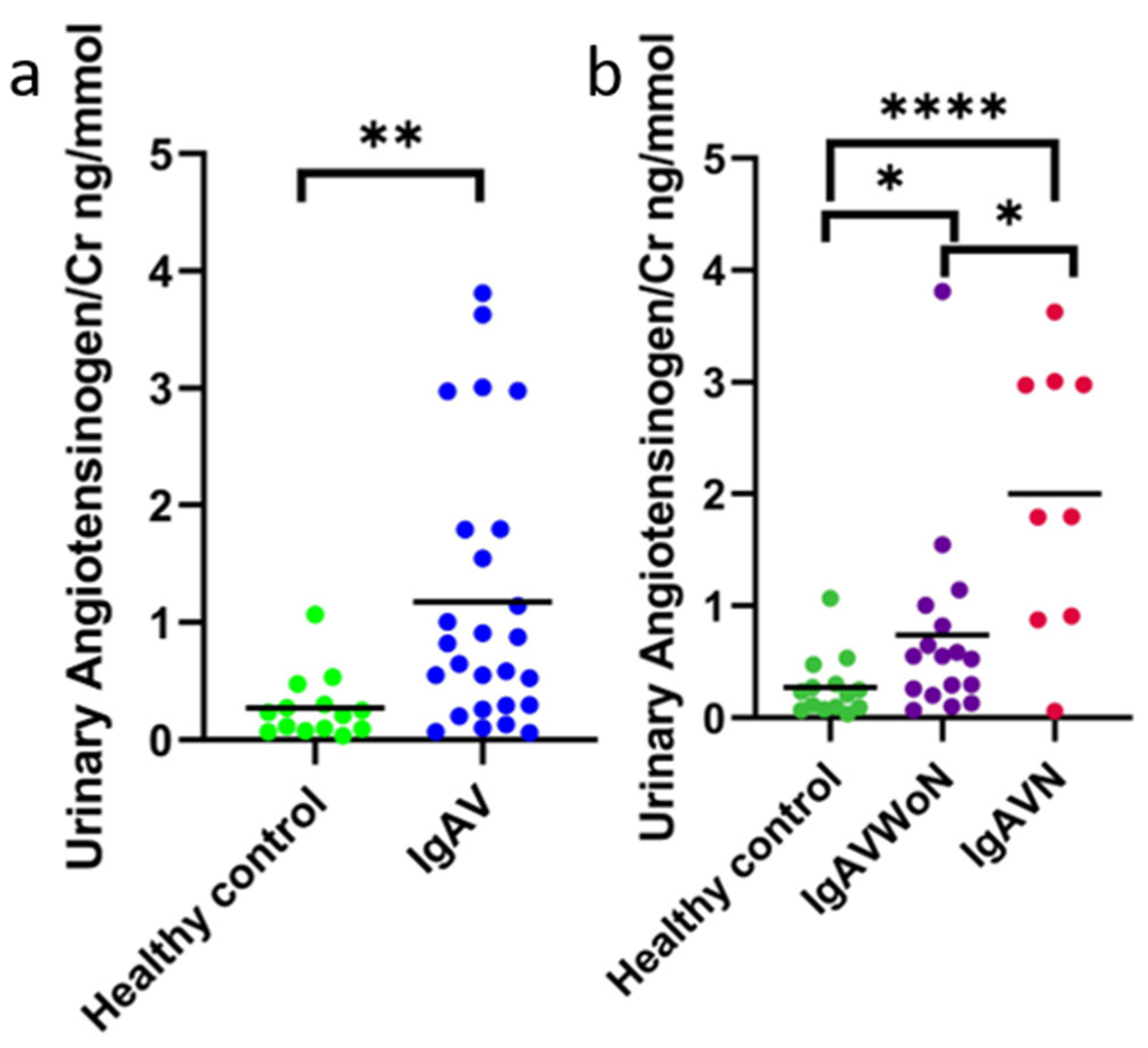
3.3. Urinary Angiotensinogen Concentration

Urinary angiotensinogen (AGT) was detected in all of the samples. The urinary concentrations of AGT/Cr in all participants with IgAV (1.18 ± 1.16 ng/mmol, n = 26) were statistically significantly increased compared to HC (0.28 ± 0.27 ng/mmol, n = 14; p = 0.0015) (Figure 1a). When grouping patients according to the presence of nephritis, the urinary AGT/Cr concentrations were statistically significantly increased by 7.2-fold in the IgAVN group (2.00 ± 1.22 ng/mmol, n = 9) compared to HC (p = 0.0006) (Figure 1b). Similarly, the urinary AGT/Cr concentrations were greater in the IgAVwoN patients (0.74 ± 0.89 ng/mmol, n = 17) when compared to the HC participants (p = 0.0233). There were also statistically significantly increased urinary AGT/Cr concentrations in the patients with IgAVN compared to the IgAVwoN group (2.7-fold, p = 0.0492). An obvious outlier was noted within the IgAVwoN group (urinary AGT/Cr 1.8 ng/mmol), as illustrated.

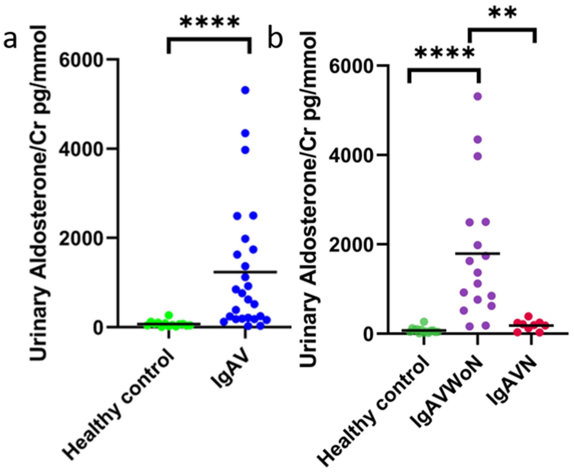
3.4. Urinary Aldosterone Concentration

Urinary aldosterone was detected in all of the samples. The concentration of urinary aldosterone/Cr in all IgAV patients (1236 ± 1438 pg/mmol, n = 26) was statistically significantly increased compared to HC (73.90 ± 65.22 pg/mmol, n = 14; p < 0.0001) (Figure 2a). When patients were divided according to the presence of nephritis, surprisingly, the IgAVwoN had a statistically significantly increased urinary aldosterone/Cr concentration (1793 ± 1507 pg/mmol, n = 17) when compared to the participants with IgAVN (p = 0.0035) and HC (p < 0.0001), as seen in Figure 2b.

3.5. Correlation to Proteinuria

The correlation between urinary angiotensinogen, aldosterone and the extent of proteinuria is shown in Table 2. Angiotensinogen was moderately positively correlated (r = 0.502, p = 0.009) and aldosterone had a low negative correlation with proteinuria values (r = −0.444, p = 0.023).

4. Discussion

In this study, a cohort of 40 paediatric IgAV patients had constituents of the RAAS pathway, renin, angiotensinogen and aldosterone, which was quantified in urine samples. We evaluated their mechanistic role in IgAV associated nephritis. This study found no differences in the urinary renin concentration or plasma; however, urinary angiotensinogen was significantly elevated in patients with IgAV nephritis. Interestingly, aldosterone was significantly elevated in patients with IgAV without nephritis, and this warrants further investigation. This study offers a characterisation of these urinary components of RAAS in a cross-sectional paediatric IgAV cohort, and it offers preliminary insight into guiding therapeutic RAAS inhibition in this disease.
The relationship between urinary angiotensinogen concentrations and nephritis is well established, and it is a well-recognised marker of intrarenal RAAS activity [6,18,19,20]. Alongside hepatic production of AGT, the RAAS pathway is also entrenched within the renal system, with angiotensinogen produced locally via the proximal tubule cells, renin being primarily produced in juxtaglomerular cells and ACE being abundant within the kidney [18,21,22]. In this study, urinary AGT was found at a 4.3-fold greater concentration in IgAVN compared to HC and a 2.9-fold greater concentration in IgAVwoN compared to HC. This indicates that intrarenal RAAS is activated even where renal inflammation is not evident, and the effect becomes more pronounced once nephritis has evolved. These findings imply that many children have activation of RAAS even without overt nephritis, and that elevated urinary AGT may have a key role in the disease [7,18,21,23,24]. It could be that the vascular inflammation activates the RAAS system triggering AGT production, which would potentially explain the lack of renin dysregulation. Previous studies have also observed elevated urinary AGT levels in paediatric IgAVN [10] and IgAV [25], with a proteomic study investigating serum levels of AGT showing that circulating concentrations of AGT were increased in IgAVN [26]. In glomerular histology samples in IgAN, the AGT concentration was strongly correlated with glomerular angiotensin II, TGF-β (a trigger of renal fibrosis) and proteinuria. It has also been demonstrated in patients with IgAN that the M235T polymorphism in AGT exhibits elevated levels of circulating AGT, which is associated with an elevated risk of renal disease progression and adverse cardiac outcomes [27,28,29]. Downstream proteins in the RAAS pathway, such as ACE, which acts on AGT to produce angiotensin I and II, have been found to contain a polymorphism in their encoding genes, affecting the expression and circulating concentrations of these proteins [27]. Previous work has demonstrated that the use of ACEi in IgAN results in a drop in AGT concentration and improved renal outcomes [30]. This suggests that early ACEi may be a possible intervention in patients with IgAV who are at a high risk of evolving nephritis.
The significant elevation in the urinary concentration of aldosterone of IgAVwoN patients is a novel finding and, if these findings are replicable, it suggests that aldosterone may play a role in the early vasculitic process during the acute phase of IgAV [11]. There is an established body of research demonstrating that aldosterone has a direct effect on the vasculature, with aldosterone receptors being expressed in endothelial cells and vascular smooth muscle cells in addition to renal specific, podocytes and mesangial cells [31,32]. Vascular inflammation induced by aldosterone is a result of oxidative stress and inflammatory cytokines [11,31,32]. When aldosterone is infused for 12 h, inflammatory cytokines IL-6 and IL12 are elevated [32]. Activation of the aldosterone or mineralocorticoid receptor triggers the generation of reactive oxygen species, such as superoxide and hydrogen peroxide, triggering the transcription of proinflammatory transcription factors and increased NADPH oxidase activity, which then complements angiotensinogen mediated activation of NADPH oxidase [31]. All of these factors lead to vascular inflammation and leukocyte adhesion [11].
A potential explanation for the elevated levels of aldosterone and AGT in IgAV may be the elevated renal levels of IgA, which has been demonstrated to increase the expression of aldosterone synthase in mesangial cells. As a result, concentrations of TGF-β and AGT become elevated, which may aggravate inflammation [33]. There is also evidence that the galactose-deficient form of IgA (gd-IgA), associated with the early pathogenesis of IgAVN and IgAN, form immune complexes and deposit within the mesangium. This elevated gd-IgA concentration is also associated with elevated oxidative stress and the generation of superoxide, potentially exacerbating the oxidative damage caused by the RAAS pathway or triggering RAAS [34]. This study does have limitations, including the small, heterogenous cohort, and the cross-sectional study design. This may have influenced the findings, and future work would benefit from longitudinal sample collection to determine the precise relationship, as well as the timing of onset and evolution of nephritis, of the RAAS system has in IgAV. Despite these limitations, this study contributes to the literature to demonstrate that there are measurable components of the RAAS system in children with IgAV. This area of study warrants further investigation.

5. Conclusions

This study demonstrates elevated urinary angiotensinogen and aldosterone in patients with IgAV, which supports the suggestion that the RAAS system has a key contributory role to the vascular inflammation associated with this disease. These findings offer preliminary evidence to direct further studies to determine whether the early use of RAAS inhibitors may reduce the risk of developing nephritis in children with IgAV.

Author Contributions

All authors declare that this is an original manuscript and that they meet the criteria for authorship. Conceptualization: L.O. and A.J.C.; investigation: A.J.C., J.M., D.J.H. and S.J.N.; formal analysis: A.J.C. and J.M.; manuscript preparation: A.J.C., J.M. and L.O.; manuscript review and revisions: A.J.C., J.M., D.J.H., S.J.N. and L.O.; funding acquisition: L.O.; supervision: L.O. All authors have read and agreed to the published version of the manuscript.

Funding

This study was financially supported by F.A.I.R. through a project grant (Funding Autoimmune Research Charity, Charity; number: 1176388) and a project grant provided by Kidney Research UK (Paed_RP_008_20190926). The funders had no role in study design, data collection and analysis, decision to publish or preparation of the manuscript. This work was further supported by the UK’s Experimental Arthritis Treatment Centre for Children (supported by Versus Arthritis, Alder Hey Children’s NHS Foundation Trust, the Alder Hey Charity and the University of Liverpool) and partially carried out at the NIHR Alder Hey Clinical Research Facility.

Institutional Review Board Statement

This study was conducted in accordance with NIHR Good Clinical Practice, HTA Codes of Practice, the Declaration of Helsinki, and comparable ethical standards. This study was part of the IgA Vasculitis Study, which was approved by HRA and Health and Care Research Wales (HCRW) on 21 June 2019, REC 17/NE/0390, protocol UoL001347, IRAS 236599.

Informed Consent Statement

Informed consent was obtained from all subjects (or their parents/guardians) involved in the study.

Data Availability Statement

The datasets analysed in this study are available from the corresponding author on reasonable request.

Acknowledgments

The authors would like to acknowledge all children and their families who participated in the IgA vasculitis study. They would also like to acknowledge Catherine McBurney, Silothabo Dliso and Jessica Tiffin for patient recruitment and biofluid collection. We also thank Andrew Hodgkinson and the Biochemistry Department, Alder Hey Children’s NHS Foundation Trust, for performing the urinary creatinine quantification.

Conflicts of Interest

The authors declare no conflict of interest.

Abbreviations

ACEangiotensin converting enzyme
ACEiangiotensin converting enzyme inhibitors
AGTAngiotensinogen
CKDChronic Kidney Disease
CrCreatinine
ELISAEnzyme-linked immunosorbent assays
HCHealthy Control
IgAImmunoglobulin A
IgAVImmunoglobulin A Vasculitis
IgAVNImmunoglobulin A Vasculitis-Nephritis
IgAVwoNImmunoglobulin A Vasculitis without Nephritis
IgANImmunoglobulin A Nephropathy
RAASRenin-angiotensinogen-aldosterone system
UACRUrine albumin creatinine ratio

References

  1. Jennette, J.C. Overview of the 2012 Revised International Chapel Hill Consensus Conference Nomenclature of Vasculitides. Clin. Exp. Nephrol. 2013, 17, 603. [Google Scholar] [CrossRef] [PubMed] [Green Version]
  2. Oni, L.; Sampath, S. Childhood IgA Vasculitis (Henoch Schonlein Purpura)—Advances and Knowledge Gaps. Front. Pediatr. 2019, 7, 257. [Google Scholar] [CrossRef] [PubMed] [Green Version]
  3. Demir, S.; Kaplan, O.; Celebier, M.; Sag, E.; Bilginer, Y.; Lay, I.; Ozen, S. Predictive Biomarkers of IgA Vasculitis with Nephritis by Metabolomic Analysis. Semin. Arthritis Rheum. 2020, 50, 1238–1244. [Google Scholar] [CrossRef] [PubMed]
  4. Ozen, S.; Pistorio, A.; Iusan, S.M.; Bakkaloglu, A.; Herlin, T.; Brik, R.; Buoncompagni, A.; Lazar, C.; Bilge, I.; Uziel, Y.; et al. EULAR/PRINTO/PRES Criteria for Henoch–Schönlein Purpura, Childhood Polyarteritis Nodosa, Childhood Wegener Granulomatosis and Childhood Takayasu Arteritis: Ankara 2008. Part II: Final Classification Criteria. Ann. Rheum. Dis. 2010, 69, 798–806. [Google Scholar] [CrossRef] [PubMed] [Green Version]
  5. Pillebout, E. IgA Vasculitis and IgA Nephropathy: Same Disease? J. Clin. Med. 2021, 10, 2310. [Google Scholar] [CrossRef]
  6. Remuzzi, G.; Perico, N.; Macia, M.; Ruggenenti, P. The Role of Renin-Angiotensin-Aldosterone System in the Progression of Chronic Kidney Disease. Kidney Int. 2005, 68, S57–S65. [Google Scholar] [CrossRef] [Green Version]
  7. Thethi, T.; Kamiyama, M.; Kobori, H. The Link between the Renin-Angiotensin-Aldosterone System and Renal Injury in Obesity and the Metabolic Syndrome. Curr. Hypertens. Rep. 2012, 14, 160–169. [Google Scholar] [CrossRef] [Green Version]
  8. Praga, M.; Gutiérrez, E.; González, E.; Morales, E.; Hernandez, E. Treatment of IgA Nephropathy with Ace Inhibitors: A Randomized and Controlled Trial. J. Am. Soc. Nephrol. 2003, 14, 1578–1583. [Google Scholar] [CrossRef] [Green Version]
  9. Van den Belt, S.M.; Heerspink, H.J.L.; Gracchi, V.; de Zeeuw, D.; Wühl, E.; Schaefer, F.; Anarat, A.; Bakkaloglu, A.; Ozaltin, F.; Peco-Antic, A.; et al. Early Proteinuria Lowering by Angiotensin-Converting Enzyme Inhibition Predicts Renal Survival in Children with CKD. J. Am. Soc. Nephrol. 2018, 29, 2225–2233. [Google Scholar] [CrossRef] [Green Version]
  10. Marro, J.; Chetwynd, A.J.; Wright, R.D.; Dliso, S.; Oni, L. Urinary Protein Array Analysis to Identify Key Inflammatory Markers in Children with IgA Vasculitis Nephritis. Children 2022, 9, 622. [Google Scholar] [CrossRef]
  11. Briet, M.; Schiffrin, E.L. Vascular Actions of Aldosterone. J. Vasc. Res. 2013, 50, 89–99. [Google Scholar] [CrossRef]
  12. Franiek, A.; Sharma, A.; Cockovski, V.; Wishart, D.S.; Zappitelli, M.; Blydt-Hansen, T.D. Urinary Metabolomics to Develop Predictors for Pediatric Acute Kidney Injury. Pediatr. Nephrol. 2022, 37, 2079–2090. [Google Scholar] [CrossRef]
  13. Williams, C.E.C.; Toner, A.; Wright, R.D.; Oni, L. A Systematic Review of Urine Biomarkers in Children with IgA Vasculitis Nephritis. Pediatr. Nephrol. 2021, 36, 3033–3044. [Google Scholar] [CrossRef]
  14. Counahan, R.; Winterborn, M.H.; White, R.H.; Heaton, J.M.; Meadow, S.R.; Bluett, N.H.; Swetschin, H.; Cameron, J.S.; Chantler, C. Prognosis of Henoch-Schönlein Nephritis in Children. Br. Med. J. 1977, 2, 11–14. [Google Scholar] [CrossRef] [Green Version]
  15. Lurbe, E.; Agabiti-Rosei, E.; Cruickshank, J.K.; Dominiczak, A.; Erdine, S.; Hirth, A.; Invitti, C.; Litwin, M.; Mancia, G.; Pall, D.; et al. 2016 European Society OfHypertension Guidelines for Themanagement of High Blood Pressure in Children and Adolescents. J. Hypertens. 2016, 34, 1887–1920. [Google Scholar] [CrossRef] [Green Version]
  16. Asano, K.; Iwasaki, H.; Fujimura, K.; Ikeda, M.; Sugimoto, Y.; Matsubara, A.; Yano, K.; Irisawa, H.; Kono, F.; Kanbe, M.; et al. Automated Microanalysis of Creatinine by Coupled Enzyme Reactions. Hiroshima J. Med. Sci. 1992, 41, 1–5. [Google Scholar]
  17. Mukaka, M.M. A Guide to Appropriate Use of Correlation Coefficient in Medical Research. Malawi Med. J. 2012, 24, 69. [Google Scholar]
  18. Burns, K.D.; Hiremath, S. Urinary Angiotensinogen as a Biomarker of Chronic Kidney Disease: Ready for Prime Time? Nephrol. Dial. Transplant. 2012, 27, 3010–3013. [Google Scholar] [CrossRef]
  19. Struthers, A. Review of Aldosterone- and Angiotensin II-Induced Target Organ Damage and Prevention. Cardiovasc. Res. 2004, 61, 663–670. [Google Scholar] [CrossRef] [Green Version]
  20. Kim, Y.-G.; Song, S.-B.; Lee, S.-H.; Moon, J.-Y.; Jeong, K.-H.; Lee, T.-W.; Ihm, C.-G. Urinary Angiotensinogen as a Predictive Marker in Patients with Immunoglobulin A Nephropathy. Clin. Exp. Nephrol. 2011, 15, 720–726. [Google Scholar] [CrossRef]
  21. Kobori, H.; Navar, L.G. Urinary Angiotensinogen as a Novel Biomarker of Intrarenal Renin-Angiotensin System in Chronic Kidney Disease. Int. Rev. Thromb 2011, 6, 108. [Google Scholar]
  22. Sparks, M.A.; Crowley, S.D.; Gurley, S.B.; Mirotsou, M.; Coffman, T.M. Classical Renin-Angiotensin System in Kidney Physiology. Compr. Physiol. 2014, 4, 1201–1228. [Google Scholar] [CrossRef] [PubMed] [Green Version]
  23. Mills, K.T.; Kobori, H.; Hamm, L.L.; Alper, A.B.; Khan, I.E.; Rahman, M.; Navar, L.G.; Liu, Y.; Browne, G.M.; Batuman, V.; et al. Increased Urinary Excretion of Angiotensinogen Is Associated with Risk of Chronic Kidney Disease. Nephrol. Dial. Transplant. 2012, 27, 3176–3181. [Google Scholar] [CrossRef] [PubMed]
  24. Takamatsu, M.; Urushihara, M.; Kondo, S.; Shimizu, M.; Morioka, T.; Oite, T.; Kobori, H.; Kagami, S. Glomerular Angiotensinogen Protein Is Enhanced in Pediatric IgA Nephropathy. Pediatr. Nephrol. 2008, 23, 1257–1267. [Google Scholar] [CrossRef] [PubMed] [Green Version]
  25. Mao, Y.N.; Liu, W.; Li, Y.G.; Jia, G.C.; Zhang, Z.; Guan, Y.J.; Zhou, X.F.; Liu, Y.F. Urinary Angiotensinogen Levels in Relation to Renal Involvement of Henoch-Schonlein Purpura in Children. Nephrology 2012, 17, 53–57. [Google Scholar] [CrossRef]
  26. He, X.; Yin, W.; Ding, Y.; Cui, S.J.; Luan, J.; Zhao, P.; Yue, X.; Yu, C.; Laing, X.; Zhao, Y.L. Higher Serum Angiotensinogen Is an Indicator of IgA Vasculitis with Nephritis Revealed by Comparative Proteomes Analysis. PLoS ONE 2015, 10, e0130536. [Google Scholar] [CrossRef] [Green Version]
  27. Rudnicki, M.; Mayer, G. Significance of Genetic Polymorphisms of the Renin–Angiotensin–Aldosterone System in Cardiovascular and Renal Disease. Pharmacogenomics 2009, 10, 463–476. [Google Scholar] [CrossRef]
  28. Chen, S.; Zhang, L.; Wang, H.W.; Wang, X.Y.; Li, X.Q.; Zhang, L.L. The M235T Polymorphism in the Angiotensinogen Gene and Heart Failure: A Meta-Analysis. JRAAS—J. Renin-Angiotensin-Aldosterone Syst. 2014, 15, 190–195. [Google Scholar] [CrossRef] [Green Version]
  29. Huang, H.D.; Lin, F.J.; Li, X.J.; Wang, L.R.; Jiang, G.R. Genetic Polymorphisms of the Renin-Angiotensin-Aldosterone System in Chinese Patients with End-Stage Renal Disease Secondary to IgA Nephropathy. Chin. Med. J. 2010, 123, 3238–3242. [Google Scholar] [CrossRef]
  30. Urushihara, M.; Nagai, T.; Kinoshita, Y.; Nishiyama, S.; Suga, K.; Ozaki, N.; Jamba, A.; Kondo, S.; Kobori, H.; Kagami, S. Changes in Urinary Angiotensinogen Posttreatment in Pediatric IgA Nephropathy Patients. Pediatr. Nephrol. 2015, 30, 975–982. [Google Scholar] [CrossRef] [Green Version]
  31. Brown, N.J. Contribution of Aldosterone to Cardiovascular and Renal Inflammation and Fibrosis. Nat. Rev. Nephrol. 2013, 9, 459–469. [Google Scholar] [CrossRef]
  32. Brown, N.J. Aldosterone and Vascular Inflammation. Hypertension 2008, 51, 161–167. [Google Scholar] [CrossRef] [Green Version]
  33. Leung, J.C.K.; Chan, L.Y.Y.; Tang, S.C.W.; Lam, M.F.; Chow, C.W.; Lim, A.I.; Lai, K.N. Oxidative Damages in Tubular Epithelial Cells in IgA Nephropathy: Role of Crosstalk between Angiotensin II and Aldosterone. J. Transl. Med. 2011, 9, 169. [Google Scholar] [CrossRef]
  34. Camilla, R.; Suzuki, H.; Daprà, V.; Loiacono, E.; Peruzzi, L.; Amore, A.; Ghiggeri, G.M.; Mazzucco, G.; Scolari, F.; Gharavi, A.G.; et al. Oxidative Stress and Galactose-Deficient IgA1 as Markers of Progression in IgA Nephropathy. Clin. J. Am. Soc. Nephrol. 2011, 6, 1903–1911. [Google Scholar] [CrossRef]
Figure 1. Urinary creatinine normalised angiotensinogen concentration, separated by disease and renal involvement status. The mean concentration for each patient group is denoted. (a) Urinary angiotensinogen is significantly elevated in IgAV patients compared to HC. (b) When separated by renal involvement, angiotensinogen levels were elevated compared to HC regardless of the presence of renal disease; which was defined as >30 mg/mmol creatinine, with IgAVN having the greatest angiotensinogen concentration. * p < 0.05, ** p < 0.01, **** p < 0.0001.
Ijtm 02 00045 g001
Figure 2. Urinary creatinine normalised aldosterone concentration, separated by disease and renal involvement status. The mean concentration for each patient group is denoted. (a) Urinary aldosterone is significantly elevated in IgAV patients compared to HC. (b) When separated by renal involvement, aldosterone levels in IgAwoN patients were significantly elevated compared to HC and IgAVNs. There was also a slight but insignificant trend for elevated levels in IgAVN compared to HC. ** p < 0.01, **** p < 0.0001.
Ijtm 02 00045 g002
Table 1. Patients characteristics at baseline. a n (%); b mean ± SD. 1 BP readings available for 8 patients of the IgAVwoN group. 2 Serum creatinine was available for 13 patients (IgAVN, n = 8; IgAVwoN, n = 5). 3 Serum albumin was available for 11 patients (IgAV, n = 7; IgAVwoN, n = 4). UACR: urinary albumin to creatinine ratio. ISKDC: International Study for Kidney Disease in Children Classification. Significant p-values are highlighted in bold. + p < 0.05 compared to IgAVwoN. Due to rounding, percentages may not add up to 100.
OverallIgAVNIgAVwoNHCp-Value
n (%)40 (100%)9 (23%)17 (43%)14 (35%)-
Male a23 (28%)6 (67%)10 (59%)7 (50%)0.725
Age, years b8.3 ± 3.38.3 ± 3.56.7 ± 3.010.3 ± 2.7 +0.006
Ethnicity
British
African
Chinese
Any other Asian background
Not stated

32 (80%)
3 (8%)
2 (5%)
1 (3%)
2 (5%)

7 (79%)
0 (0%)
0 (0%)
0 (0%)
2 (22%)

15 (88%)
0 (0%)
2 (12%)
0 (0%)
0 (0%)

10 (71%)
3 (21%)
0 (0%)
1 (7%)
0 (0%)
Sampling
Sample type
Urine and blood a19 (100%)5 (26%)5 (26%)9 (47%)
Urine only a26 (100%)3 (12%)18 (69%)5 (19%)
Blood only a5 (100%)5 (100%)--
Weeks from diagnosis to sampling b18.4 ± 60.049.7 ± 97.81.8 ± 0.9-<0.001
Renal involvement
Hypertension a,14 (23.5%)3 (33.3%)1 (12.5%)-0.312
Serum creatinine mg/dL b,251.5 ± 18.553.3 ± 17.248.6 ± 22.1-0.769
Serum albumin g/L b,339.3 ± 4.337.1 ± 3.143.0 ± 3.7-0.029
UACR mmol/mg b90.7 ± 147.6257.8 ± 141.22.2 ± 5.5-<0.001
Urinary creatinine mmol/L b8.7 ± 7.66.2 ± 4.77.7 ± 7.711.5 ± 8.40.146
Biopsy proven nephritis a 4 (44%)
ISKDC Grade
IIIa a
IIIb a
IV a

-
-
-

1 (25%)
2 (50%)
1 (25%)

-
-
-

-
-
-

-
-
-
Medications
Corticosteroids a 2
Follow-up
Months b11.4 ± 17.123.2 ± 25.05.1 ± 5.2-0.005
UACR at last review b10.9 ± 20.531.6 ± 24.00.0 ± 0.0-<0.001
Outcome a Full remission, 4 (44%)
Partial remission, 5 (56%)
Worsened, 0 (0%)
Table 2. Correlation of urinary angiotensinogen and aldosterone to proteinuria.
Pearson rp-Value
Urinary AGT/Cr 0.5020.009
Urinary aldosterone/Cr −0.4440.023
Publisher’s Note: MDPI stays neutral with regard to jurisdictional claims in published maps and institutional affiliations.

