Article

Effects of Phototherapy on Free Vitamin D Levels in Ten Patients with Atopic Dermatitis

by
Andrea Elmelid
1,2,*,
Amra Osmancevic
1,3,
Martin Gillstedt
1,3 and
Mikael Alsterholm
1,3
1
Department of Dermatology and Venereology, Institute of Clinical Sciences, Sahlgrenska Academy, University of Gothenburg, 413 45 Gothenburg, Sweden
2
Region Dalarna, Department of Dermatology and Venereology, Falu Hospital, 791 82 Falun, Sweden
3
Region Västra Götaland, Department of Dermatology and Venereology, Sahlgrenska University Hospital, 413 45 Gothenburg, Sweden
*
Author to whom correspondence should be addressed.
Int. J. Transl. Med. 2022, 2(4), 586-596; https://doi.org/10.3390/ijtm2040044
Submission received: 17 October 2022 / Revised: 22 November 2022 / Accepted: 28 November 2022 / Published: 1 December 2022

Abstract

:
The role of vitamin D in atopic dermatitis (AD) is controversial. Conflicting data could be due to the use of inadequate markers for assessing vitamin D status. So far, directly measured free 25(OH)D concentrations have not been reported in AD patients. Ten adults with AD were treated with narrow band ultraviolet light B (NB-UVB) for 10–12 weeks. SCORing atopic dermatitis (SCORAD) and the visual analogue scale (VAS) were used to assess disease severity before and after NB-UVB therapy. Total and free 25(OH)D and 1,25(OH)2D serum levels were analyzed before and after treatment. Free 25(OH)D concentrations were measured with a two-step immunosorbent assay (ELISA). The majority of patients had sufficient levels of 25(OH)D before treatment (mean 76.4 nmol/L). Mean free 25(OH)D was 11.9 pmol/L and mean 1,25(OH)2D was 108.9 pmol/L. Median SCORAD decreased from 37.1 to 19.8 and VAS improved significantly after phototherapy. Total and free 25(OH)D increased in all subjects. No correlations between disease severity and vitamin D levels were found. There was no correlation between total and free 25(OH)D levels. Larger studies are needed to test the applicability of the free hormone hypothesis in AD pathogenesis.

1. Introduction

Atopic dermatitis (AD) is the most common chronic inflammatory skin disease worldwide. The prevalence is increasing in industrialized countries and is estimated to be 20% in children and 10% in adults [1]. AD is characterized by chronic or relapsing inflammatory, eczematous, and pruritic lesions that usually debut at an early age. The pathogenesis of AD involves a complex interaction between genetic, immunological, and environmental factors [1].
The role of vitamin D in AD remains controversial. Populations living at latitudes with lower sun exposure and therefore possibly lower vitamin D levels have increased AD prevalence [2]. Large population-based studies in Korea have found a positive association between vitamin D deficiency and AD [3,4]. According to several studies, AD patients seem to have lower 25(OH)D serum levels than healthy controls [5,6,7]. A number of studies found an association between vitamin D deficiency and AD severity, with higher serum levels of 25(OH)D in mild AD compared with moderate or severe AD [6,8,9]. A meta-analysis concluded that vitamin D seems to play a role in AD since serum 25(OH)D levels were lower in AD patients, and disease severity decreased with vitamin D supplementation [5]. Newborns with low vitamin D levels have an increased risk of developing AD later in childhood [10]. Meanwhile, there are studies that could not find an association between 25(OH)D levels and AD [11,12].
The conflicting results of previous studies could possibly be explained by the use of an inadequate biomarker for the assessment of vitamin D status in AD patients.
The free hormone hypothesis states that it is the free fraction of a steroid hormone that passively enters cells and exerts biological action, while the protein-bound part is biologically inactive. Vitamin D status is conventionally assessed with the total serum 25(OH)D level, in contrast to other steroid hormones (such as thyroid and sex hormones) where the free concentrations are measured [13]. Only a small part (~0.03%) of total serum 25(OH)D circulates in its free form, while ~85% is tightly bound to vitamin D binding protein (DBP) and ~15% is loosely bound to albumin. Until recently, a mathematical model for calculating free 25(OH)D has been used. Since the introduction of a commercial two-step ELISA that measures free 25(OH)D, studies have shown that the calculated values highly overestimate the directly measured levels of free 25(OH)D [13]. As far as we are aware, directly measured free 25(OH)D levels in AD patients have not been reported.
Vitamin D refers to a group of lipophilic secosteroids that are derived from cholesterol. When human skin is irradiated by UVB rays (290–315 nm), pre-vitamin D3 is produced from 7-dehydrocholesterol (7-DHC) [14]. To convert vitamin D into its active form, it undergoes two enzymatic hydroxylation steps. The first hydroxylation reaction that forms calcidiol (25(OH)D) occurs primarily in the liver and the second hydroxylation reaction that forms the most biologically active form, calcitriol (1,25(OH)2D), occurs in the kidney by 1-alpha-hydroxylase (CYP27B1) [15]. However, it has been found that 1-alpha-hydroxylation also occurs in a number of extra-renal cells including keratinocytes, immune cells, and several cancer cells [16,17]. 1,25(OH)2D binds to its nuclear receptor, vitamin D receptor (VDR), to activate gene transcription [18].
UV treatment is recommended as adjunctive therapy in AD when topical therapy is insufficient. UVB reduces AD symptoms through several mechanisms; suppression of antigen-presenting Langerhans’ cells, suppression of expression of pro-inflammatory cytokines, stimulation of keratinocyte IL-10 production, upregulation of antimicrobial peptides, apoptosis of infiltrating T cells, reduction of colonization of Staphylococcus aureus and Malassezia species, and UV-induced thickening of stratum corneum resulting in reduced antigen presentation [19,20,21].
Some of the beneficial effects of UV exposure for AD patients are mediated through vitamin D [22]. Vitamin D regulates keratinocyte proliferation, differentiation, and stimulates the production of anti-microbial peptides (AMPs) such as cathelicidins. Decreased production of AMPs due to deficiency in vitamin D could predispose to superinfections with S. aureus, as commonly seen in flare-ups in individuals with AD [23].
The aim of this study was to investigate vitamin D status in patients with AD by measuring total and free 25(OH)D levels and the effects of UVB phototherapy. Our primary hypothesis was that vitamin D levels would increase after phototherapy. Our secondary hypothesis was that disease severity would correlate stronger to free 25(OH)D levels compared to total 25(OH)D levels.

2. Materials and Methods

2.1. Study Design, Setting, and Participants

The study was conducted at the Department of Dermatology at Sahlgrenska University Hospital, Gothenburg, Sweden, between 2013 and 2017.
Ten adult individuals (>18 years) with moderate to severe AD who needed UV therapy were included.
Exclusion criteria were pregnancy/lactation, pregnancy plans, other ongoing systemic or chronic diseases, treatment with oral corticosteroids or potent topical corticosteroids, other immunosuppressive treatments, antibiotic treatment, sun holiday or any use of a solarium at any time four weeks prior to inclusion, planned sun holiday or use of a solarium, use of vitamin D supplements at any time four weeks prior to inclusion, and planned use of vitamin D supplements.
Patients recruited from October to March, when the ultraviolet (UV) index in Gothenburg is <3, were classified as recruited in winter. Patients recruited from April to September, when UV index can reach >3 and vitamin D production in the skin is possible, were classified as recruited in summer.
Data were collected at four time points; before treatment (visit 1), after 4 weeks of treatment (visit 2), when treatment was ended at week 10–12 (visit 3), and 4–6 weeks after end of treatment (visit 4). Skin type according to Fitzpatrick was defined before treatment [24]. Information about lifestyle factors that could affect vitamin D status and inflammation was collected via questionnaires at each visit. Weight, height, blood pressure, and waist circumference were measured. Body mass index (BMI) was calculated at each visit. VAS was collected at each visit. The patients were examined by an experienced dermatologist who performed SCORAD evaluation at visit 1 and 3 (before and after treatment). Biochemical analyses were performed on blood samples collected before and after treatment.
Total serum concentration of 25(OH)D > 75 nmol/L was defined as sufficient, 25(OH)D level of 50 to 75 nmol/L was defined as insufficiency, and 25(OH)D < 50 nmol/L was considered as deficiency according to the Endocrine Society [25].

2.2. Phototherapy

All patients were treated with whole body narrowband ultraviolet light B (NB-UVB 311–312 nm, Corona 4, ESSHÅ) 2–3 times/week during a period of 10–12 weeks according to a standardized protocol adjusted for each patient with a maximum of 30 treatments. The subjects were allowed to use emollients and mild corticosteroids, but not potent topical corticosteroids or calcineurin inhibitors.

2.3. Laboratory Analyses

All analyses were performed at the Department of Clinical Chemistry, Sahlgrenska University Hospital, Gothenburg.
Total 25(OH)D [25(OH)D2 and 25(OH)D3] levels were analyzed with an electrochemiluminescence immunoassay (ECLIA) using the Elecsys Vitamin D Total II assay.
Free 25(OH)D concentrations were measured with a two-step immunosorbent assay (ELISA) using a commercial kit (Future Diagnostics B.V., Wijchen, The Netherlands).
In addition, 1,25(OH)2D was analyzed with an automated chemiluminescence immunoassay (CLIA).

2.4. Calculation of Percentage of Free 25(OH)D

The concentration of free 25(OH)D was converted from pg/mL, the unit in which free 25(OH)D was measured, to nmol/L, using the formula 1 pg/mL = (2.496/1000) nmol/L.
Percentage of free 25(OH)D = 100 × (free 25(OH)D (nmol/L)/total 25(OH)D (nmol/L)).

2.5. Assessment of AD Severity

AD severity was assessed with the SCORing Atopic Dermatitis (SCORAD) index [26]. The SCORAD index is comprised of area estimation and intensity grading by the investigator and rating of pruritus and sleep loss by the patient. SCORAD can also be used as a pure investigator-reported outcome measure, excluding pruritus and sleep loss, sometimes referred to as objective SCORAD. Objective SCORAD ranges from 0 (no disease) to 83 (maximal disease). Objective SCORAD > 40 is considered severe disease [27].
The Visual Analogue Scale (VAS) was used for self-evaluation of AD severity. Patients marked the intensity of their symptoms on a ten-centimeter line (0 means no complaints and 10 worst complaints).
VAS was also used for the patients’ rating of itch (0 means no itch and 10 worst possible itch), in the following text referred to as VAS pruritus.
A questionnaire was used to collect information about diet and lifestyle factors which might affect vitamin D status. Questions regarding medical and family history, atopic disease duration, asthma, allergies, smoking habits, dietary supplements, and sun habits were also included.

2.6. Comparative Statistics Regarding the Effect of Phototherapy on Vitamin D Levels

A sample of psoriasis patients (n = 42) underwent the same NB-UVB treatment (at the same clinic using the same protocol and the same lamp), the result of which (previously described) was used for comparative statistics regarding vitamin D metabolites before and after phototherapy [28].

2.7. Statistical Analyses

Data are presented as mean ± SD or median (inter-quartile range (IQR)) if not otherwise stated. Data were analyzed using R version 3.5.3 (The R Foundation for Statistical Computing, Vienna, Austria). Simple descriptive statistics were applied. Spearman’s correlation test was used to test univariate correlations. Wilcoxon’s rank sum test was used for two sample tests. When testing for changes over time, Spearman’s correlation test stratifying with respect to patient was used. Differences in SCORAD before and after treatment were tested with Wilcoxon’s signed rank test. All tests were two-sided and p < 0.05 was considered statistically significant.

2.8. Ethical Considerations

The study was approved by the Ethics Committee at the University of Gothenburg 22 May 2012 (approval number: 089-12). Declarations of Helsinki protocols were followed. Written informed consent was obtained from all subjects.

3. Results

3.1. Demographics

Ten patients with AD (six women and four men) were included in the study. Eight patients completed the study. One dropout was due to personal reasons and one because of side effects of UVB treatment (erythema). Baseline data were analyzed in all ten included patients. Nine out of ten patients were enrolled in the study during winter.
All subjects were adults with a mean age of 28.6 ± 6.6 years. Mean disease duration was 23 ± 8.4 years.
Both objective SCORAD and total SCORAD indices were calculated and gave similar statistical results. Objective SCORAD is reported in the following.
At visit 1 (before treatment), objective SCORAD ranged from 25.1–40.4, which corresponds to moderate to severe AD [29]. Median objective SCORAD was 37.1 (28.9–38.4).
Fitzpatrick skin type II (3/10), III (6/10), and IV (1/10) were represented. All subjects had normal blood pressure and the majority were of normal weight; mean BMI was 24.7 ± 4.3 kg/m2.
All patients reported at least one atopic comorbidity and heredity for atopic disease. The U.K. Working Party’s Diagnostic Criteria for AD were used to confirm AD diagnosis and determine eligibility [30]. No comorbidity outside the atopic spectrum was reported, with the exception of one patient who stated “possibly rheumatism”. There was no information regarding rheumatological disease in the patient’s record. On-going medications were not known to affect vitamin D status except for corticosteroid inhalation, which was used by one patient.
None of the patients reported current intake of vitamin D, omega-3, or any other dietary supplement. None had been on a sun holiday in the two months prior to inclusion.
Demographic data and confounding factors for vitamin D status in AD patients are presented in Table 1.

3.2. Effect of UVB Treatment

The mean number of treatments (the two dropouts excluded) was 25.6 ± 5.8 and the mean cumulative UVB dose was 24.49 ± 14.69 J/cm2. Furthermore, all patients who completed the study were treated in the wintertime when exposure to ambient UVB is negligible at our latitude (Gothenburg 57° N).
Median objective SCORAD decreased from 37.1 (28.9–38.4) to 19.8 (17.0–22.4) (p = 0.016). Median delta objective SCORAD was −13.8 ((−19.4)–(−6.2)).
VAS improved significantly from visit 1 (median 7.7 (5.9–8.2)) to visit 3 (median 2.8 (2.1–4.1)) (p = 0.017). At visit 4 (four to six weeks after end of phototherapy), VAS had increased again in all responders to a median of 5.0 (4.4–6.4). This change was not statistically significant (Table 2). Phototherapy reduced itch (measured with VAS pruritus before and after treatment (visit 1 and 3)), but this was not statistically significant (p = 0.063).
The dynamics of VAS and objective SCORAD for all patients during follow-up are presented in Figure 1.
Total 25(OH)D and free 25(OH)D levels increased significantly from visit 1 to visit 3. Mean delta 25(OH)D was 79.8 ± 36.7 nmol/L and mean delta free 25(OH)D was 14.6 ± 9.3 pmol/L. Percentage of free 25(OH)D did not change significantly after therapy. In addition, 1,25(OH)2D levels varied with no clear trend (Table 2). BMI and blood pressure remained unaltered during UVB treatment.

3.3. Vitamin D Status and Correlation to Disease Severity at Baseline

At baseline, 70% (7/10) of the patients had sufficient levels of 25(OH)D, 20% (2/10) had insufficient levels, and 10% (1/10) were vitamin D deficient (Table 2). Mean 25(OH)D was 76.4 ± 21.1 nmol/L, mean free 25(OH)D was 11.9 ± 2.9 pmol/L, mean 1,25(OH)2D was 108.9 ± 39.8 pmol/L, and mean percentage of free 25(OH)D was 0.0162 ± 0.0037%.
No correlations were found between disease severity and vitamin D metabolite levels including percentage of free 25(OH)D) at baseline.
We found no statistically significant association between total 25(OH)D and free 25(OH)D concentrations. We found an inverse correlation between total 25(OH)D and percentage of free 25(OH)D (p = 0.044). No associations between BMI and vitamin D metabolites (including percentage of free 25(OH)D) were found.

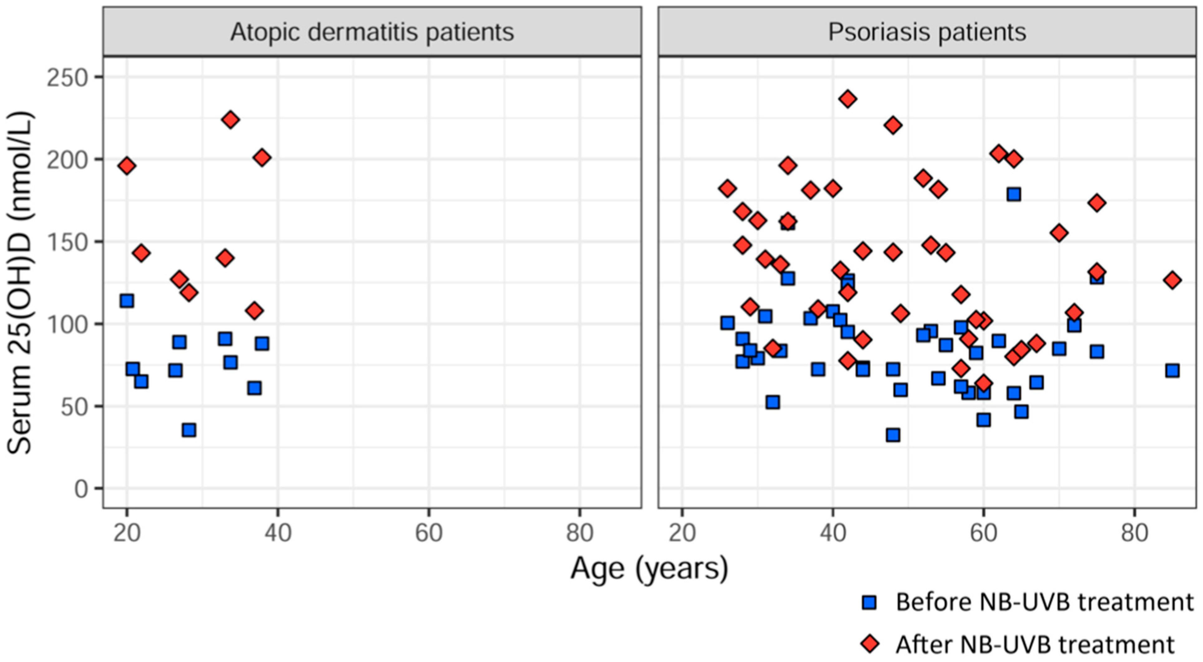
3.4. Comparison of Delta 25(OH)D between AD Patients and Psoriasis Patients after UVB Treatment

AD patients had significantly higher delta 25(OH)D (mean 79.8 ± 36.7 nmol/L) when compared to psoriasis patients (mean 51.1 ± 37.3 nmol/L) (p = 0.033) treated with the same type of UVB therapy (Figure 2) [28].

4. Discussion

This study presents the distribution of directly measured free 25(OH)D levels in AD patients before and after UVB phototherapy. NB-UVB phototherapy significantly raised both total and free levels of 25(OH)D in this cohort.
The results confirm previous findings that NB-UVB is an effective treatment for moderate to severe AD, significantly improving patient- and investigator-reported outcome measures. The effect was short-lived with a raise in VAS score only four to six weeks after the cessation of treatment.
Baseline mean percentage of free 25(OH)D in this cohort of AD patients was 0.016% and was unaltered by UVB phototherapy. The concentrations of free 25(OH)D ranged from 6.49 to 16.7 pmol/L before phototherapy, and from 18.2 to 50.2 pmol/L after phototherapy. In healthy adults, the directly measured free 25(OH)D levels have been reported to be between 0.02% and 0.09% of total 25(OH)D levels and the concentration generally ranges from 1.84 to 29.74 pmol/L [13,31]. Our findings imply that the relationship between free and total 25(OH)D might be altered in individuals with moderate to severe AD, with a lower percentage of free 25(OH)D compared to healthy individuals. Larger studies are needed to investigate this further.
We expected that free 25(OH)D and total 25(OH)D levels at baseline would be positively associated, but this was not confirmed. Although this could be explained by the small sample size, it could also indicate that total 25(OH)D might not be the most accurate measure for assessing vitamin D status in AD patients.
In addition to 25(OH)D, we investigated other vitamin D metabolites (free 25(OH)D and 1,25(OH)2D) but found no correlation with AD severity. The hypothesis that free 25(OH)D correlates better with disease severity than total 25(OH)D could not be confirmed.
This subset of AD patients had higher 25(OH)D levels at baseline (mean 76.4 nmol/L) than previously reported in adult AD patients in a meta-analysis, where the mean 25(OH)D levels ranged from 26.8 nmol/L (Asian patients, n = 70) to 59.5 nmol/L (Caucasian patients, n = 58) [5]. Of note is that different methods for measuring 25(OH)D were used. The fact that our cohort had higher 25(OH)D levels than previously reported was an unexpected finding and the cause is not clear. One possible explanation could be cultural differences in tanning behavior [32].
The patients in this study reported long disease duration (mean 23 years). Presuming a role for vitamin D in AD pathogenesis, it might be more accurate to study vitamin D status in earlier stages of the disease when the disruption of immune responses occurs. It has been shown that pediatric patients with AD have lower 25(OH)D levels compared to controls, while the difference is not significant in adult AD patients [5]. Possibly vitamin D deficiency early in life is a greater risk factor for developing AD than vitamin D deficiency in adults. However, vitamin D supplementation reduces disease severity in both pediatric and adult patients, indicating that vitamin D also plays a role in adult AD patients [5,33].
The assumed link between vitamin D and skin inflammation is further complicated by the finding that topical application of vitamin D3 or the analog MC903 (calcipotriol) in mice can induce an AD-like state in the skin with immunological changes similar to those seen in acute lesions [34]. This paradox could be explained by the fact that vitamin D and its analogues serve as irritants when applied directly on the skin. Furthermore, a topically applied product cannot be compared to the endogenously produced vitamin D which has physiological effects on the skin in an endocrine, paracrine, and autocrine manner. On the other hand, topically applied calcipotriol is an established treatment for psoriasis [35]. This highlights the complex role of vitamin D levels, local and systemical, in cutaneous inflammatory disorders. We have studied the effects of UVB on serum vitamin D levels and clinical outcome measures. It is apparent that there is a need for further studies on the effect of skin tissue levels of vitamin D.
In accordance with previous research on psoriasis patients, we found an inverse correlation between percentage of free 25(OH)D and total 25(OH)D before UVB treatment [36]. This implies a compensating mechanism altering the relationship between free and total 25(OH)D when needed [13,37].
The increase of 25(OH)D was significantly higher in this subset of AD patients (mean change 79.8 nmol/L) compared to NB-UVB treated psoriasis patients (mean change 50.7 nmol/L) described earlier [28]. A previous study on the effects of NB-UVB on 25(OH)D levels in patients with AD, psoriasis, and healthy individuals has shown similar results where AD patients had a higher increase of 25(OH)D compared to psoriasis patients (68.2 nmol/L versus 59.9 nmol/L). NB-UVB-treated healthy subjects had the highest increase of 25(OH)D (90.7 nmol/L), but the difference was not statistically significant between the three groups (p = 0.21) [21]. The differences could not be explained by age or pre-treatment levels of 25(OH)D in the previous study.
The percentage increase of 25(OH)D levels after NB-UVB treatment in this cohort was 49%. This is a lower percentage increase compared to previous reports on AD patients after NB-UVB (110–276% increase) [38]. The large difference in percentage increase of 25(OH)D could be explained by the lower pre-treatment levels of 25(OH)D in the previously studied cohorts compared to the present cohort. The percentage increase of free 25(OH)D was 45% (Table 3). There are no studies to compare this finding with.
Taken together, the partly conflicting findings regarding the role of vitamin D in AD pathogenesis suggest differences in vitamin D metabolism between AD patients, psoriasis patients, and healthy individuals. It can be hypothesized that locally produced vitamin D in the skin does not reach the blood stream to the same extent in those with inflammatory skin disease compared to healthy individuals, possibly because of consumption. The data of this study support the hypothesis that vitamin D plays an important role in skin inflammation and underlines the importance of further investigation of the skin as the main site of vitamin D production. We suggest that the increased vitamin D production in the skin as a result of UV radiation is the primary cause of the improvement in skin lesions, and not the raised serum levels of vitamin D.
The main limitation of this study is the small number of participants. Ten patients were included of which only eight completed the study. The method used for measuring 25(OH)D levels (ECLIA) is not the gold standard method, such as liquid chromatography-tandem mass spectrometry (LC-MS/MS), but it is still a validated method and widely used in assessment of vitamin D status [39]. The psoriasis patients used for comparison after phototherapy were not age and sex matched.

5. Conclusions

NB-UVB increases serum 25(OH)D and free 25(OH)D in AD patients regardless of pre-treatment levels. Phototherapy is an effective treatment in moderate to severe AD, but the effect is short-term. This cohort of AD patients had higher levels of 25(OH)D compared to what has been previously reported in adult AD patients. The increase of serum 25(OH)D levels after NB-UVB treatment was higher in AD patients compared to psoriasis patients. We found no correlation between disease severity and the levels of any of the vitamin D metabolites which were measured.
The question remains whether inadequate assessment of vitamin D status can explain the inconsistency in the literature regarding the impact of vitamin D on AD. Studies with larger number of patients are needed to test the applicability of the free hormone hypothesis in AD pathogenesis. Our findings support previous research that vitamin D plays an important role in skin inflammation. The results imply that vitamin D produced in the skin is locally metabolized in immunological processes resulting in skin healing. Therefore, further research should focus not only on vitamin D in the circulation but also on vitamin D metabolites in the skin.

Author Contributions

Conceptualization, A.E., A.O. and M.A.; Data curation, A.E., A.O., M.G. and M.A.; Formal analysis, A.E., A.O., M.G. and M.A.; Funding acquisition, A.O. and M.A.; Investigation, A.E., A.O. and M.A.; Methodology, A.E., A.O. and M.A.; Project administration, A.E. and A.O.; Resources, A.E., A.O., M.G. and M.A.; Software, A.E., A.O., M.G. and M.A.; Supervision, A.O. and M.A.; Validation, A.E., A.O., M.G. and M.A.; Visualization, A.E., A.O., M.G. and M.A.; Writing—original draft, A.E.; Writing—review and editing, A.O., M.G. and M.A. All authors have read and agreed to the published version of the manuscript.

Funding

This research was funded by The Welander-Finsen Foundation (Reg. no.: application 2238, 2658, 2810, 3302), The Göteborg Medical Society (Grant numbers: 590291 and 884701), The Swedish state under the agreement between the Swedish government and the county councils, the ALF-agreement (ALFGBG-2895) and The Swedish Society of Medicine (grant numbers: SLS-323781 and SLS-404381).

Institutional Review Board Statement

The study was conducted in accordance with the Declaration of Helsinki and approved by the Ethics Committee at the University of Gothenburg 22 May 2012 (approval number: 089-12).

Informed Consent Statement

Informed consent was obtained from all subjects involved in the study.

Data Availability Statement

The data that support the findings of this study are available from the corresponding author upon reasonable request.

Acknowledgments

Maria Siekkeri Vandikas, for support during the preparation and initiation of the study. Christina Halldin and Birgitta Stare Merelaid, research nurses, for clinical assistance during the study.

Conflicts of Interest

The authors declare no conflict of interest.

References

  1. Langan, S.M.; Irvine, A.D.; Weidinger, S. Atopic Dermatitis. Lancet Lond. Engl. 2020, 396, 345–360. [Google Scholar] [CrossRef] [PubMed]
  2. Weiland, S.K.; Hüsing, A.; Strachan, D.P.; Rzehak, P.; Pearce, N. ISAAC Phase One Study Group Climate and the Prevalence of Symptoms of Asthma, Allergic Rhinitis, and Atopic Eczema in Children. Occup. Environ. Med. 2004, 61, 609–615. [Google Scholar] [CrossRef] [PubMed]
  3. Cheng, H.M.; Kim, S.; Park, G.-H.; Chang, S.E.; Bang, S.; Won, C.H.; Lee, M.W.; Choi, J.H.; Moon, K.C. Low Vitamin D Levels Are Associated with Atopic Dermatitis, but Not Allergic Rhinitis, Asthma, or IgE Sensitization, in the Adult Korean Population. J. Allergy Clin. Immunol. 2014, 133, 1048–1055. [Google Scholar] [CrossRef] [PubMed]
  4. Kang, J.W.; Kim, J.H.; Kim, H.J.; Lee, J.-G.; Yoon, J.-H.; Kim, C.-H. Association of Serum 25-Hydroxyvitamin D with Serum IgE Levels in Korean Adults. Auris. Nasus. Larynx 2016, 43, 84–88. [Google Scholar] [CrossRef] [PubMed]
  5. Kim, M.J.; Kim, S.-N.; Lee, Y.W.; Choe, Y.B.; Ahn, K.J. Vitamin D Status and Efficacy of Vitamin D Supplementation in Atopic Dermatitis: A Systematic Review and Meta-Analysis. Nutrients 2016, 8, 789. [Google Scholar] [CrossRef] [PubMed]
  6. El Taieb, M.A.; Fayed, H.M.; Aly, S.S.; Ibrahim, A.K. Assessment of Serum 25-Hydroxyvitamin d Levels in Children with Atopic Dermatitis: Correlation with SCORAD Index. Dermatitis 2013, 24, 296–301. [Google Scholar] [CrossRef] [PubMed]
  7. Yang, A.-R.; Kim, Y.-N.; Lee, B.-H. Dietary Intakes and Lifestyle Patterns of Korean Children and Adolescents with Atopic Dermatitis: Using the Fourth and Fifth Korean National Health and Nutrition Examination Survey (KNHANES IV,V), 2007–2011. Ecol. Food Nutr. 2016, 55, 50–64. [Google Scholar] [CrossRef] [PubMed]
  8. Peroni, D.G.; Piacentini, G.L.; Cametti, E.; Chinellato, I.; Boner, A.L. Correlation between Serum 25-Hydroxyvitamin D Levels and Severity of Atopic Dermatitis in Children. Br. J. Dermatol. 2011, 164, 1078–1082. [Google Scholar] [CrossRef] [PubMed]
  9. Cheon, B.R.; Shin, J.E.; Kim, Y.J.; Shim, J.W.; Kim, D.S.; Jung, H.L.; Park, M.S.; Shim, J.Y. Relationship between Serum 25-Hydroxyvitamin D and Interleukin-31 Levels, and the Severity of Atopic Dermatitis in Children. Korean, J. Pediatr. 2015, 58, 96–101. [Google Scholar] [CrossRef] [PubMed]
  10. Baïz, N.; Dargent-Molina, P.; Wark, J.D.; Souberbielle, J.-C.; Annesi-Maesano, I. EDEN Mother-Child Cohort Study Group Cord Serum 25-Hydroxyvitamin D and Risk of Early Childhood Transient Wheezing and Atopic Dermatitis. J. Allergy Clin. Immunol. 2014, 133, 147–153. [Google Scholar] [CrossRef] [PubMed]
  11. Chiu, Y.E.; Havens, P.L.; Siegel, D.H.; Ali, O.; Wang, T.; Holland, K.E.; Galbraith, S.S.; Lyon, V.B.; Drolet, B.A. Serum 25-Hydroxyvitamin D Concentration Does Not Correlate with Atopic Dermatitis Severity. J. Am. Acad. Dermatol. 2013, 69, 40–46. [Google Scholar] [CrossRef] [PubMed] [Green Version]
  12. Thuesen, B.H.; Heede, N.G.; Tang, L.; Skaaby, T.; Thyssen, J.P.; Friedrich, N.; Linneberg, A. No Association between Vitamin D and Atopy, Asthma, Lung Function or Atopic Dermatitis: A Prospective Study in Adults. Allergy 2015, 70, 1501–1504. [Google Scholar] [CrossRef] [PubMed]
  13. Bikle, D.D.; Schwartz, J. Vitamin D Binding Protein, Total and Free Vitamin D Levels in Different Physiological and Pathophysiological Conditions. Front. Endocrinol. 2019, 10, 317. [Google Scholar] [CrossRef] [PubMed] [Green Version]
  14. Herrmann, M.; Farrell, C.-J.L.; Pusceddu, I.; Fabregat-Cabello, N.; Cavalier, E. Assessment of Vitamin D Status—A Changing Landscape. Clin. Chem. Lab. Med. 2017, 55, 3–26. [Google Scholar] [CrossRef] [Green Version]
  15. Christakos, S.; Dhawan, P.; Verstuyf, A.; Verlinden, L.; Carmeliet, G. Vitamin D: Metabolism, Molecular Mechanism of Action, and Pleiotropic Effects. Physiol. Rev. 2016, 96, 365–408. [Google Scholar] [CrossRef] [PubMed] [Green Version]
  16. Lehmann, B. The Vitamin D3 Pathway in Human Skin and Its Role for Regulation of Biological Processes. Photochem. Photobiol. 2005, 81, 1246–1251. [Google Scholar] [CrossRef]
  17. Zehnder, D.; Bland, R.; Williams, M.C.; McNinch, R.W.; Howie, A.J.; Stewart, P.M.; Hewison, M. Extrarenal Expression of 25-Hydroxyvitamin d(3)-1 Alpha-Hydroxylase. J. Clin. Endocrinol. Metab. 2001, 86, 888–894. [Google Scholar] [CrossRef] [PubMed] [Green Version]
  18. Bikle, D.; Christakos, S. New Aspects of Vitamin D Metabolism and Action—Addressing the Skin as Source and Target. Nat. Rev. Endocrinol. 2020, 16, 234–252. [Google Scholar] [CrossRef] [PubMed]
  19. Wollenberg, A.; Christen-Zäch, S.; Taieb, A.; Paul, C.; Thyssen, J.P.; de Bruin-Weller, M.; Vestergaard, C.; Seneschal, J.; Werfel, T.; Cork, M.J.; et al. ETFAD/EADV Eczema Task Force 2020 Position Paper on Diagnosis and Treatment of Atopic Dermatitis in Adults and Children. J. Eur. Acad. Dermatol. Venereol. 2020, 34, 2717–2744. [Google Scholar] [CrossRef]
  20. Walters, I.B.; Ozawa, M.; Cardinale, I.; Gilleaudeau, P.; Trepicchio, W.L.; Bliss, J.; Krueger, J.G. Narrowband (312-Nm) UV-B Suppresses Interferon Gamma and Interleukin (IL) 12 and Increases IL-4 Transcripts: Differential Regulation of Cytokines at the Single-Cell Level. Arch. Dermatol. 2003, 139, 155–161. [Google Scholar] [CrossRef]
  21. Vähävihu, K.; Ala-Houhala, M.; Peric, M.; Karisola, P.; Kautiainen, H.; Hasan, T.; Snellman, E.; Alenius, H.; Schauber, J.; Reunala, T. Narrowband Ultraviolet B Treatment Improves Vitamin D Balance and Alters Antimicrobial Peptide Expression in Skin Lesions of Psoriasis and Atopic Dermatitis. Br. J. Dermatol. 2010, 163, 321–328. [Google Scholar] [CrossRef]
  22. Camargo, C.A.; Ganmaa, D.; Sidbury, R.; Erdenedelger, K.; Radnaakhand, N.; Khandsuren, B. Randomized Trial of Vitamin D Supplementation for Winter-Related Atopic Dermatitis in Children. J. Allergy Clin. Immunol. 2014, 134, 831–835.e1. [Google Scholar] [CrossRef] [PubMed]
  23. Schauber, J.; Dorschner, R.A.; Coda, A.B.; Büchau, A.S.; Liu, P.T.; Kiken, D.; Helfrich, Y.R.; Kang, S.; Elalieh, H.Z.; Steinmeyer, A.; et al. Injury Enhances TLR2 Function and Antimicrobial Peptide Expression through a Vitamin D-Dependent Mechanism. J. Clin. Invest. 2007, 117, 803–811. [Google Scholar] [CrossRef] [Green Version]
  24. Fitzpatrick, T.B. The Validity and Practicality of Sun-Reactive Skin Types I through VI. Arch. Dermatol. 1988, 124, 869–871. [Google Scholar] [CrossRef] [PubMed]
  25. Holick, M.F.; Binkley, N.C.; Bischoff-Ferrari, H.A.; Gordon, C.M.; Hanley, D.A.; Heaney, R.P.; Murad, M.H.; Weaver, C.M. Endocrine Society Evaluation, Treatment, and Prevention of Vitamin D Deficiency: An Endocrine Society Clinical Practice Guideline. J. Clin. Endocrinol. Metab. 2011, 96, 1911–1930. [Google Scholar] [CrossRef] [PubMed] [Green Version]
  26. Schmitt, J.; Langan, S.; Deckert, S.; Svensson, A.; von Kobyletzki, L.; Thomas, K.; Spuls, P. Harmonising Outcome Measures for Atopic Dermatitis (HOME) Initiative Assessment of Clinical Signs of Atopic Dermatitis: A Systematic Review and Recommendation. J. Allergy Clin. Immunol. 2013, 132, 1337–1347. [Google Scholar] [CrossRef]
  27. Oranje, A.P.; Glazenburg, E.J.; Wolkerstorfer, A.; de Waard-van der Spek, F.B. Practical Issues on Interpretation of Scoring Atopic Dermatitis: The SCORAD Index, Objective SCORAD and the Three-Item Severity Score. Br. J. Dermatol. 2007, 157, 645–648. [Google Scholar] [CrossRef] [PubMed]
  28. Osmancevic, A.; Landin-Wilhelmsen, K.; Larkö, O.; Wennberg, A.-M.; Krogstad, A.L. Vitamin D Production in Psoriasis Patients Increases Less with Narrowband than with Broadband Ultraviolet B Phototherapy. Photodermatol. Photoimmunol. Photomed. 2009, 25, 119–123. [Google Scholar] [CrossRef] [PubMed]
  29. Kunz, B.; Oranje, A.P.; Labrèze, L.; Stalder, J.F.; Ring, J.; Taïeb, A. Clinical Validation and Guidelines for the SCORAD Index: Consensus Report of the European Task Force on Atopic Dermatitis. Dermatol. Basel Switz. 1997, 195, 10–19. [Google Scholar] [CrossRef]
  30. Williams, H.C.; Jburney, P.G.; Hay, R.J.; Archer, C.B.; Shipley, M.J.; Ahunter, J.J.; Bingham, E.A.; Finlay, A.Y.; Pembroke, A.C.; Cgraham-Brown, R.A.; et al. The U.K. Working Party’s Diagnostic Criteria for Atopic Dermatitis: I. Derivation of a Minimum Set of Discriminators for Atopic Dermatitis. Br. J. Dermatol. 1994, 131, 383–396. [Google Scholar] [CrossRef] [PubMed]
  31. Schwartz, J.B.; Gallagher, J.C.; Jorde, R.; Berg, V.; Walsh, J.; Eastell, R.; Evans, A.L.; Bowles, S.; Naylor, K.E.; Jones, K.S.; et al. Determination of Free 25(OH)D Concentrations and Their Relationships to Total 25(OH)D in Multiple Clinical Populations. J. Clin. Endocrinol. Metab. 2018, 103, 3278–3288. [Google Scholar] [CrossRef] [PubMed] [Green Version]
  32. Buller, D.B.; Cokkinides, V.; Hall, H.I.; Hartman, A.M.; Saraiya, M.; Miller, E.; Paddock, L.; Glanz, K. Prevalence of Sunburn, Sun Protection, and Indoor Tanning Behaviors among Americans: Review from National Surveys and Case Studies of 3 States. J. Am. Acad. Dermatol. 2011, 65, S114.e1–S114.e11. [Google Scholar] [CrossRef]
  33. Samochocki, Z.; Bogaczewicz, J.; Jeziorkowska, R.; Sysa-Jędrzejowska, A.; Glińska, O.; Karczmarewicz, E.; McCauliffe, D.P.; Woźniacka, A. Vitamin D Effects in Atopic Dermatitis. J. Am. Acad. Dermatol. 2013, 69, 238–244. [Google Scholar] [CrossRef] [PubMed]
  34. Moosbrugger-Martinz, V.; Schmuth, M.; Dubrac, S. A Mouse Model for Atopic Dermatitis Using Topical Application of Vitamin D3 or of Its Analog MC903. Methods Mol. Biol. Clifton NJ 2017, 1559, 91–106. [Google Scholar] [CrossRef]
  35. McCormack, P.L. Calcipotriol/Betamethasone Dipropionate: A Review of Its Use in the Treatment of Psoriasis Vulgaris of the Trunk, Limbs and Scalp. Drugs 2011, 71, 709–730. [Google Scholar] [CrossRef]
  36. Vandikas, M.S.; Landin-Wilhelmsen, K.; Polesie, S.; Gillstedt, M.; Osmancevic, A. Impact of Etanercept on Vitamin D Status and Vitamin D-Binding Protein in Bio-Naïve Patients with Psoriasis. Acta Derm. Venereol. 2021, 101, adv00604. [Google Scholar] [CrossRef]
  37. Oleröd, G.; Hultén, L.M.; Hammarsten, O.; Klingberg, E. The Variation in Free 25-Hydroxy Vitamin D and Vitamin D-Binding Protein with Season and Vitamin D Status. Endocr. Connect. 2017, 6, 111–120. [Google Scholar] [CrossRef] [PubMed] [Green Version]
  38. Juzeniene, A.; Grigalavicius, M.; Juraleviciute, M.; Grant, W.B. Phototherapy and Vitamin D. Clin. Dermatol. 2016, 34, 548–555. [Google Scholar] [CrossRef] [PubMed]
  39. Batista, M.C.; Menegat, F.D.; Ferreira, C.E.S.; Faulhaber, A.C.L.; Campos, D.A.L.S.; Mangueira, C.L.P. Analytical and Clinical Validation of the New Roche Elecsys Vitamin D Total II Assay. Clin. Chem. Lab. Med. 2018, 56, e298–e301. [Google Scholar] [CrossRef]
Figure 1. Visual analogue scale (VAS) and objective scoring atopic dermatitis (SCORAD) before, during, and after phototherapy with narrow band UVB (NB-UVB) in ten atopic dermatitis (AD) patients. VAS and SCORAD improved from visit 1 to 3 (p = 0.017 and p = 0.016, respectively).
Ijtm 02 00044 g001
Figure 2. 25(OH)D serum levels in atopic dermatitis (AD) patients and psoriasis patients before and after narrow-band UVB phototherapy. The difference between delta 25(OH)D was statistically significant between AD patients and psoriasis patients (p = 0.033).
Ijtm 02 00044 g002
Table 1. Demographic data and confounding factors for vitamin D status in patients with atopic dermatitis (AD) treated with narrow-band UVB phototherapy.
Patient Number12345678910
SexFemaleFemaleMaleMaleFemaleFemaleFemaleMaleMaleFemale
Age (years)33203422273826212837
Duration of AD (years)33151318233615192533
BMI (kg/m2)32253323212221212325
Blood pressure, systolic/diastolic (mmHg)110/70115/70140/85110/70120/70115/80110/60110/60110/80100/80
Current smokingNoNoNoNoNoNoNoYesNoNo
Skin type (Fitzpatrick)IVIIIIIIIIIIIIIIIIIIIIIIII
Month of enrollmentSeptemberNovemberDecemberDecemberJanuaryFebruaryMarchJanuaryFebruaryOctober
Hours per day spent outdoors during summer (winter) 3(3)5(1)8(3)6(1)5(4)8(1)5(1)8(3)7(3)5(1)
Mean number of fish meals/week4.51.52.51.00.52.01.00.00.02.5
Use of medication that could affect vitamin D statusNoNoNoNoNoNoNoNoYesNo
Number of treatments27253026242814122223
Cumulative dose (J/cm2)37.415.6537.5540.632.759.02.73.516.514.5
Table 2. Effects of narrow-band UVB phototherapy on vitamin D levels and disease severity measured with objective SCORing Atopic Dermatitis (SCORAD) in atopic dermatitis (AD) patients. Subjective disease burden measured with visual analogue scale (VAS) and VAS pruritus. Timepoints for assessment were: before treatment (visit 1), during treatment (approximately 4 weeks in) (visit 2), after treatment (when treatment was ended at week 10–12) (visit 3), and 4–6 weeks after end of treatment (visit 4).
Patient Number12345678910
25(OH)D (nmol/L) before treatment (visit 1)90.9114.076.665.088.988.071.772.635.561.0
25(OH)D (nmol/L) after treatment (visit 3)140196224143127201N/AN/A119108
Free 25(OH)D (pmol/L) before treatment
(visit 1)
10.712.510.510.714.716.712.010.56.4914.5
Free 25(OH)D (pmol/L) after treatment
(visit 3)
18.223.734.423.021.250.2N/AN/A18.524.7
Percentage of free 25(OH)D (%) before treatment
(visit 1)
0.01180.01090.01370.01650.01660.01900.01670.01440.01830.0237
Percentage of free 25(OH)D (%) after treatment (visit 3)0.01300.01210.01540.01610.01670.0250N/AN/A0.01550.0229
1,25(OH)2D (pmol/L) before treatment
(visit 1)
15810015814284.013911048.056.094.0
1,25(OH)2D (pmol/L) after treatment (visit 3)145138234168150116N/AN/A11639.0
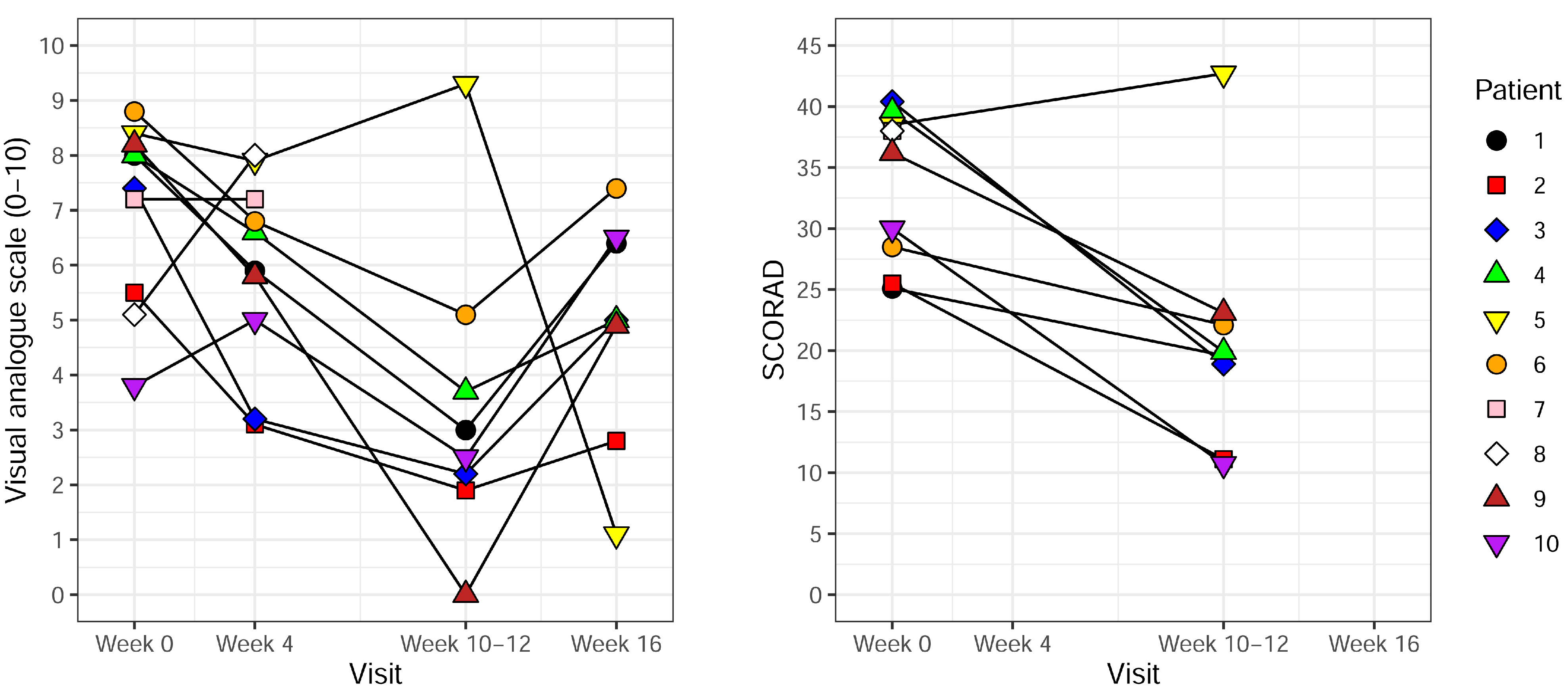
Objective SCORAD before treatment (visit 1)25.125.540.439.638.528.538.038.036.230.0
Objective SCORAD after treatment (visit 3)19.611.118.919.942.722.1N/AN/A23.110.7
VAS before treatment (visit 1)8.05.57.48.08.48.87.25.18.23.8
VAS during treatment (visit 2)5.93.13.26.67.96.87.28.05.85.0
VAS after treatment (visit 3)3.01.92.23.79.35.1N/AN/A0.02.5
VAS 4–6 weeks after end of treatment
(visit 4)
6.42.85.05.01.17.4N/AN/A4.96.5
VAS pruritus before treatment
(visit 1)
8.55.06.36.96.05.06.04.07.07.0
VAS pruritus after treatment (visit 3)3.02.03.02.57.05.0N/AN/A7.02.0
Table 3. Total and free 25(OH)D levels before and after NB-UVB in atopic dermatitis patients. Mean values ± standard deviation (SD) and median values with inter quartile range (IQR) are presented. Percentage increase is based on mean values.
Before UVBAfter UVBIncrease
Total 25(OH)D (nmol/L) mean, median76.4 ± 21.1157.3 ± 43.449%
74.6 (66.7–88.7)141.5 (125.0–197.3)
Free 25(OH)D (pmol/L) mean, median11.9 ± 2.926.7 ± 10.745%
11.4 (10.6–14.0)23.3 (20.5–27.1)
Publisher’s Note: MDPI stays neutral with regard to jurisdictional claims in published maps and institutional affiliations.

