Article

Abundance and Characteristics of Fibrous Microplastics and Microfibers Isolated in Mullus barbatus from the Adriatic Sea—Preliminary Investigation

by
Serena Santonicola
1,2,*,
Michela Volgare
3,
Mariacristina Cocca
2 and
Giampaolo Colavita
1
1
Department of Medicine and Health Sciences “V. Tiberio”, University of Molise, 86100 Campobasso, Italy
2
Institute of Polymers, Composites and Biomaterials, National Research Council of Italy, Via Campi Flegrei 34, 80078 Pozzuoli, Italy
3
Department of Chemical Engineering, Materials and Industrial Production, University of Naples Federico II, P. Tecchio 80, 80125 Naples, Italy
*
Author to whom correspondence should be addressed.
Microplastics 2023, 2(4), 411-421; https://doi.org/10.3390/microplastics2040030
Submission received: 27 November 2023 / Revised: 8 December 2023 / Accepted: 12 December 2023 / Published: 14 December 2023
(This article belongs to the Special Issue Monitoring, Detection and Mitigation of Micro and Nanoplastics)

Abstract

:
Despite the wide distribution of textile microfibers in the marine environment, there is still limited knowledge on microfiber ingestion in fish species intended for human consumption, mostly due to analytical issues. The present study aims to assess the occurrence of microfibers in red mullet (Mullus barbatus) samples collected from the Italian waters of the central Adriatic Sea. M. barbatus is a bottom fish that lives in contact with sediment and therefore was proposed as a sentinel species for the monitoring plastic pollution. A visual approach based on the evaluation of specific microfiber surface morphology was applied for the identification of particles of different origins. The preliminary findings showed the presence of microfibers in 80% of red mullet samples with a mean of 5.95 microfibers/individual. The majority (>80%) of the isolated microfibers were of natural/artificial origin, while the dominant colors were blue and black. The obtained results confirmed that benthic fish species are susceptible to microfiber ingestion and indicate the high availability of these particles in the Adriatic basin. Considering the spectroscopic drawbacks in microfiber analyses and the need to improve the current knowledge on the rate of contamination of fishery products, the visual approach could be a feasible, easy, and accessible method in the study of microfiber pollution, and the assessment of consumer health risks.

1. Introduction

Current studies have revealed the ubiquity of textile microfibers in the environment, including the atmosphere, different water sources, sludge, sediments of rivers, oceans, and topsoil [1,2,3,4]. As reported by Liu et al. [5], microfibers are defined as “particles with a diameter less than 50 µm, and length ranging from 1 µm to 5 mm”. These particles are secondary microplastics that may be released from synthetic garments during laundering [6]. However, personal care products (e.g., wet wipes and face masks), carpeting, and cigarette filters may also contribute to this type of pollution [5]. Other key sources include textile industries, domestic drainages, and wastewater treatment plants [7,8].
Moreover, recently, the demand for synthetic fabrics, linked to the fast fashion trend, has increased the contribution of synthetic textiles to microplastic pollution. In particular, polyester is the predominant fiber (around 60%) in the fast fashion system due to its numerous advantages over other materials [9]. In addition to synthetic textiles, textiles made from natural/artificial fibers, considering in this last case man-made fibers based on cellulose materials (e.g., rayon), are also extensively diffused. Apart from clothing, cellulosic fibers are widely used in furniture, female hygiene products, and diapers, and the fast fragmentation of these fibers is also one of the main reasons for their abundance in the environment [9]. In fact, recently, new emerging debris has been considered, represented by semi-synthetic or natural fibers, which although considered environmentally friendly due to their faster biodegradation, may pose a global hazard because they may be mixed with chemical additives during the textile processing and may also represent a vehicle of environmental pollutants [3].
Once discharged into the environment, microfibers could be ingested by marine species that are part of the food chain [10]. Microfibers may be mistakenly taken up as food by fish and other aquatic animals owing to their tiny size. Larger animals that feed on this seafood, polluted with microfibers, can also be affected [7]. Ingested microfibers can act as carriers of chemical pollutants, heavy metals, and endocrine disruptors, and they may release additives and dyes, used during textile processing, into the tissues of exposed animals, leading to related health problems [10]. In particular, the exposure to polyester microfibers may be associated with oxidative stress, cell damage, and increased mortality in exposed fish [11]. Chronic exposure to microfibers may cause reductions in growth, fecundity, and reproductive output. These health issues in marine biota may also trigger adverse consequences for biodiversity conservation, and food security (referring to the reduction in food availability for the human population) [10].
Literature studies revealed that the ingestion of fibrous microplastics is more prevalent in benthic species than pelagic species [12,13,14,15], probably due to the high occurrence of these particles on the seabed [14,16]. The efforts made to identify a suitable bioindicator to assess the extent of microfiber pollution have pointed out that red mullet (Mullus barbatus), a bottom fish species that lives on the seabed in constant contact with sediment, may be designated as a useful sentinel species [13]. The risk of ingestion is linked to the feeding behavior of M. barbatus, which swallows sediment together with the prey [16]. Previous works have, in fact, documented the exposure to microfibers in specimens of red mullet from the Turkish shore, Adriatic and Tyrrhenian Seas, and the Mediterranean Spanish Coast [12,15,17,18,19,20]. However, the required quality-controlled data are not sufficient in order to make an adequate safety risk assessment, mostly due to analytical issues [21,22]. Although the available literature reported a mixture of natural/artificial and synthetic microfibers in the sea and oceans, these particles have often been excluded from studies on marine biota [23]. The reduced dimensions of these particles and the occurrence of dyes on their surface may hinder their identification using conventional spectroscopic techniques [24]. In this context, intermediate screening steps have been applied to classify synthetic and natural/artificial microfibers [25]. There are key typical features that make the analysis of microfiber surface morphology an essential tool for more a confident identification of microfiber particles of different origins [13,23,25,26,27]. Rodríguez-Romeu et al. [13] characterized different microfiber typologies according to their morphological features (e.g., general and microscopic appearance). Microfibers twisted as ribbons, with broken and frayed ends were identified as cellulosic microfibers through the use of Raman spectroscopy, while microfibers with circular sections and solid edges were identified as polyethylene terephthalate. Stanton et al. [25] proposed visual identification as a useful screening method in order to reduce the number of microfibers for the subsequent spectroscopic identification. The authors proposed a flowchart to characterize textile fibers step by step as natural or extruded (synthetic) based on the unique morphological features of microfiber typologies. Zhu et al. [25] identified typical microfiber surface features for each polymer. For instance, synthetic microfibers, like polyester are tubular-shaped and look smooth, while cellulosic microfibers tend to be bumpy and have spirals, with fat lips at the end of the fiber that concave inward.
Despite the morphological analyses of synthetic textile fibers having been criticized because they are liable to human error [28], this approach was successfully applied to assess the microfiber contamination in bivalves (Mytilus galloprovincialis) [27], commercial fish species (Mullus barbatus and Engraulis encraiscolous) [13,15,29], and environmental samples [24,25], providing a feasible method to evaluate microfiber pollution along the marine ecosystem.
Therefore, considering the urgent need to acquire further knowledge on the occurrence of these particles in fishery products [21,30,31], the aim of the current research was to apply the morphological analyses to evaluate microfiber ingestion in M. barbatus samples from the Adriatic Sea, a preferential region of plastic accumulation in the Mediterranean Sea [3]. Although visual identification cannot conclusively identify the microfiber origin, this approach proves to be useful in quantifying the extent of natural and synthetic microfiber contamination, considering that this type of pollution in marine biota is still less studied compared to other microplastics.

2. Materials and Methods

2.1. Materials

Sodium chloride; hydrogen peroxide solution, 30%; and potassium hydroxide were supplied by Carlo Erba (Val De Reuil, France). During the sample filtration, cellulose nitrate (pore size 8 μm) and acetate (pore size 0.45 μm) filters (Sartorius Stedim Biotech, Gottingen, Germany) were used. The filtrating system was provided by Advantec (Dublin, CA, USA).

2.2. Fish Sampling

M. barbatus samples (Number = 20), intended for human consumption, were purchased from a fisherman after landing in Molise Region, Italy. Fish come from the Central Mediterranean Sub-region, within the sub-area FAO 37.2.1 of the FAO-GFCM (General Fisheries Commission for the Mediterranean), which includes the Adriatic Sea.
The samples were wrapped in aluminum foil and transported to the laboratory where they were stored at −20 °C until further analysis.

2.3. Microfiber Extraction

Each fish was defrosted at room temperature, washed with previously filtered water, measured, and weighted. The gastrointestinal tract (GIT, including stomach and intestine) was removed, weighed, and transferred individually into a glass Erlenmeyer flask. To digest the fish tissues and isolate microfibers, the extraction protocol of Santonicola et al. [29] was applied. This method was previously validated [32]. In detail, to isolate microfibers, the GIT of each individual was digested using a 10% potassium hydroxide (KOH) solution at 45 °C overnight. The use of KOH is common in many studies in order to digest the tissues of fish samples. According to literature studies and authors’ experiences, samples treated with KOH (10% w/v) showed the best results in terms of filtration ability, and the polymers, including natural/artificial cellulosic materials (e.g., rayon), did not show changes when exposed to temperatures lower than 60 °C [33].
After digestion, density-flotation separation was applied by adding 250 mL of a NaCl hypersaline solution to each sample, which was stirred for 10 min and then decanted. The supernatant was collected and filtered under vacuum using cellulose nitrate filters (pore size 8 μm). To completely digest all tissue residues, a 15% H2O2 solution was added to the membranes and allowed to dry in an oven (45 °C, overnight) before observation. The addition of H2O2 directly to the sample tissues after dissection may result in a low recovery rate due to the dense foam that may also hinder the sample filtration and processing. Therefore, according to Avio et al. [17], the H2O2 solution was applied only to remove tissue residues after sample digestion and filtration. The application of this treatment did not affect the particle colors, and no significant effect on the characteristics of the isolated particles was observed [17,34].
For the correction of potential contamination, one blank control without any tissue was carried out for every sample group (5–6 individuals) analyzed on the same day.

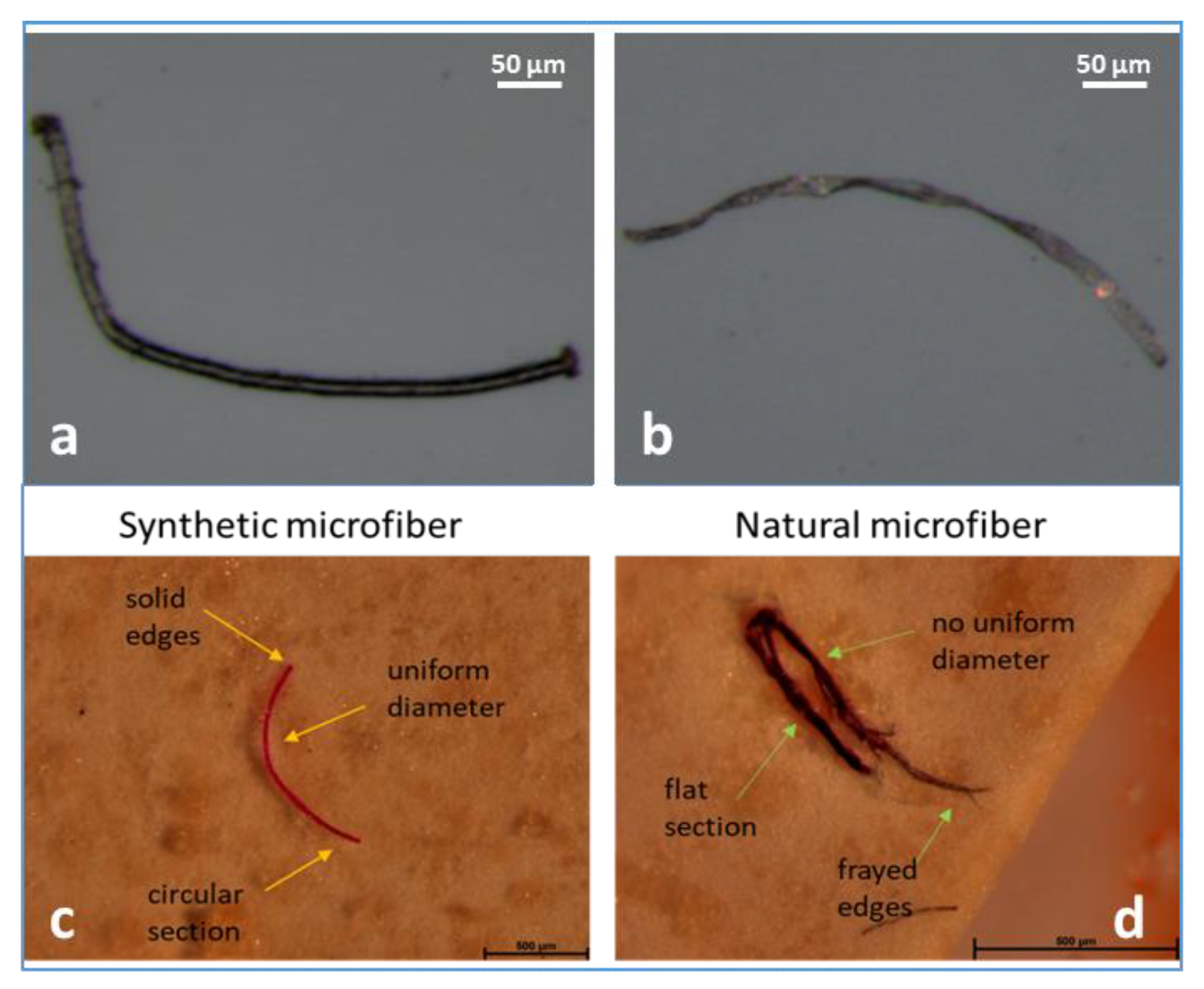
2.4. Microfiber Visual Identification

The filter membranes were observed using a light microscope (M205C; Leica, Wetzlar, Germany) with a magnification of 0.78–16×. For the micrograph acquisition, each filter was considered to have been divided in four sections, which were individually captured with a magnification of 0.78×, then different magnifications were used in order to capture each detail of the filter membranes. Approximately 30 min were spent for the examination of each filter membrane.
Each microfiber isolated on the filter was photographed, and then the micrographs were analyzed by two different operators in order to differentiate synthetic and natural or artificial microfibers according to some morphological characteristics (cross-section, breakages, and the appearance of the fiber body and ends) and the flowchart proposed by Stanton et al. [25]. Prior to sample analysis, the capability of the textile fiber analyst was improved through the observation of synthetic (e.g., polyester) and natural (e.g., cotton) microfibers of known origin [25,27,35] reported in Figure 1. These micrographs were used as references to identify the microfiber morphological features used during the sample analyses. In particular, microfibers from clearly to slightly twisted, with frayed edges and flat sections, were classified as natural/artificial, while microfibers with a smooth surface, solid edges, circular section, and uniform diameter were identified as synthetic [29].
The application of the visual approach was successfully applied by the authors to identify microfibers isolated in bivalves (Mytilus galloprovincialis) [27] and fish (Mullus barbatus and Engraulis encraiscolous) [29] samples from the Tyrrhenian Sea (Western Mediterranean Sea). The previous investigations showed that the spectroscopic analyses confirmed that visual identification corroborated the morphological analyses as a feasible method to assess microfiber contamination in seafood [27,29]. An accurate visual approach has been proven to be useful to differentiate synthetic and cellulosic fiber types [13]. Therefore, taking into account the analytical issues of the use of spectroscopic techniques in microfiber analyses [24], in this preliminary survey, the proposed visual approach was applied also to quantify natural/artificial and synthetic microfibers in red mullet samples collected from the Adriatic Sea (Central Mediterranean Sea).
The microfiber length was measured analyzing optical micrographs using Image J (release 1.43 u, NIH, Bethesda, MD, USA). Then, microfibers were classified based on their color (blue, black, transparent/clear, purple, sky-blue, pink, red, and orange), and counted.

2.5. Contamination Precaution

In order to prevent sample contamination during the analyses, samples were processed in a dedicated previously cleaned laboratory with limited access. Samples were always covered with aluminum foil and exposed only during dissection. Cotton laboratory coats were worn, and all the liquids were filtered through cellulose acetate membranes (pore size 0.45 μm). Moreover, among the applied procedure preventing contamination, all material and working surfaces were cleaned three times with filtered water before use and between samples. During the microscopical observation, cotton lab coats and nitrile gloves were worn. For the correction of potential procedural contamination, microfiber counts within each blank were subtracted from the counts of each associated sample.

2.6. Statistical Analyses
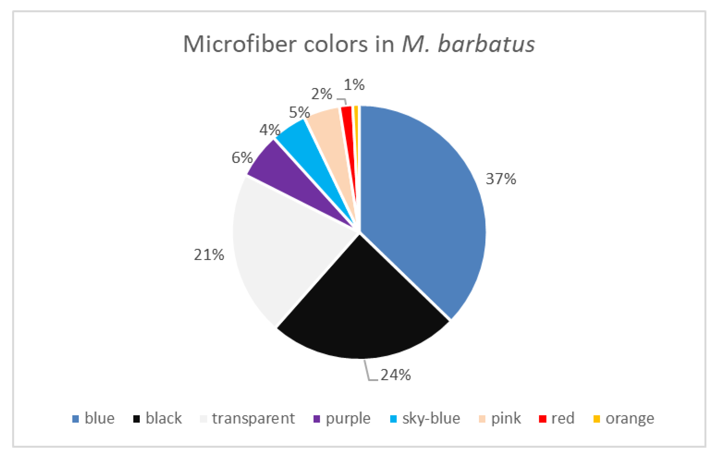
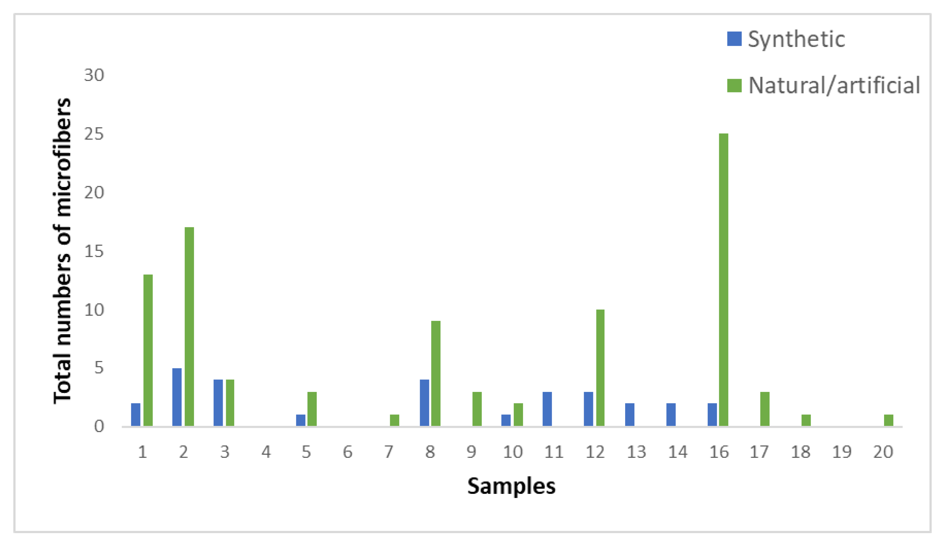
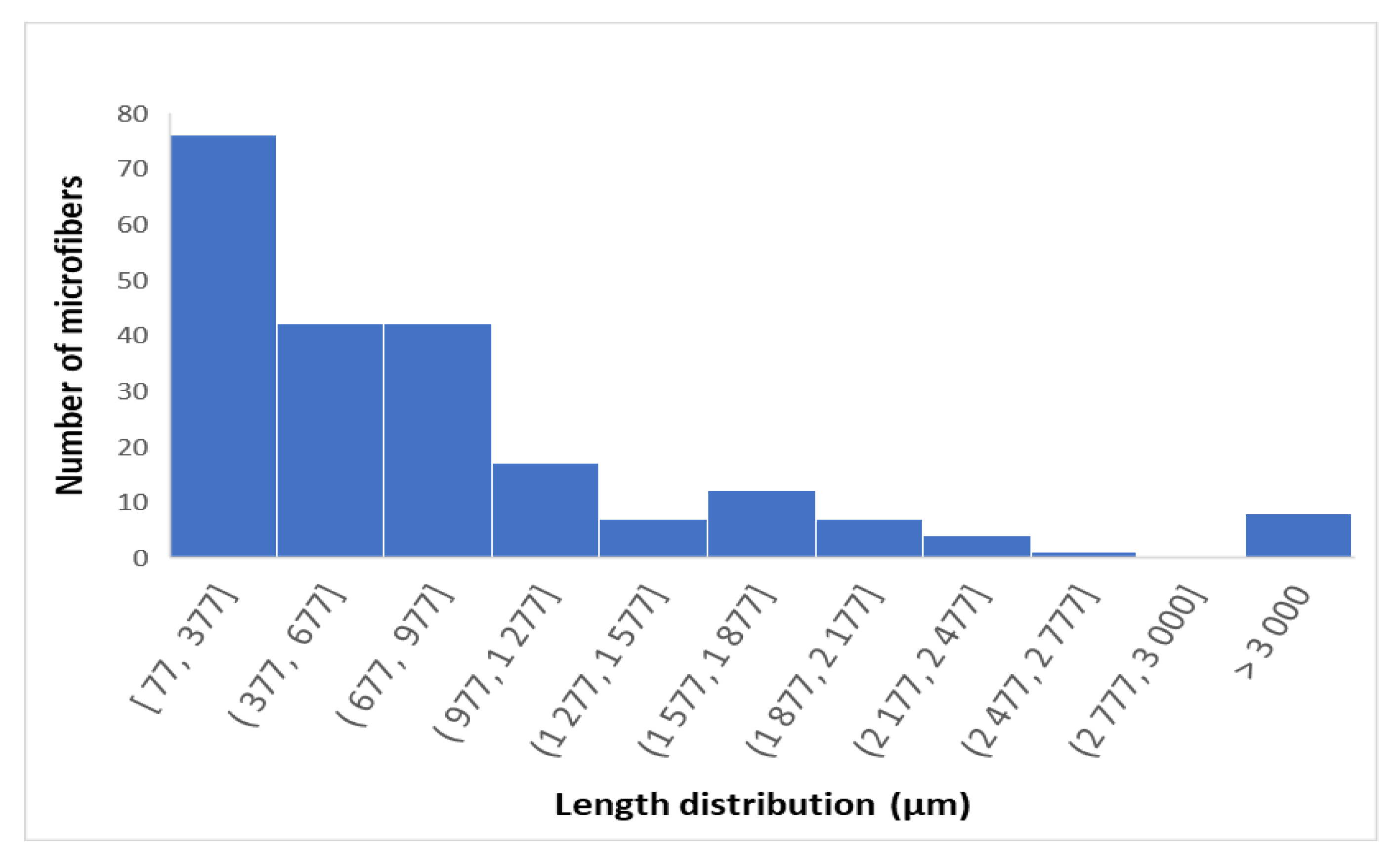
Statistical analysis was carried out by using IBM®SPSS® Statistics software, version 23.0 (IBM, Chicago, IL, USA). To determine the relationship between the data, Pearson correlation test was performed. A 5% significance level was considered for all the statistical tests (p values < 0.05 indicate significant correlation among the data).

3. Results

The mean length and weight (±SD) of red mullet samples were, respectively, 10.89 ± 0.54 cm and 25.27 ± 3.57 g, while the GIT mean weight was 1.15 ± 0.24 g.
The preliminary results showed the occurrence of microfibers in 80% of red mullet samples with a mean of 5.95 microfibers/individual (range: 1–27 microfibers/individual) corresponding to 0.3 microfibers/g of wet weight (w.w.) of each individual and 6.63 microfibers/g w.w.). In the GIT, the number of ingested microfibers did not show a significant relationship with biometric values, or ecological features of the species. However, despite these results, a trend could be observed, since larger red mullet specimens contained more microfibers/g w.w. of each individual (r(20) = 0.139, p = 0.559).
The color distribution of ingested microfibers (both natural and synthetic) revealed that blue (37%), black (24%), and transparent (21%) microfibers were the most common, as reported in Figure 2.
Natural/artificial microfibers, characterized based on the analyses and the observation of fiber morphologies, were the most abundant (Figure 3), representing the 84% of the isolated microfibers.
The average length of synthetic microfibers was determined to be 1015.12 μm, while natural/artificial microfibers showed a mean length of 706.43 μm. Length range distribution was reported in Figure 4.

4. Discussion

Modeling studies on the spatial distribution of marine debris pointed out the Adriatic region as an area of plastic accumulation in the Mediterranean Sea [36]. In particular, the effect of the currents, the inputs of the Po River, the occurrence of commercial seaports together with the intense urbanization of the coasts, may contribute to this phenomenon [12,37]. About 80% of marine plastic litter enters the Adriatic basin through rivers, and from the coastal setting, and the remaining 20% result from shipping and fishing activities [36]. In this context, previous research revealed the abundance of textile microfibers in Adriatic food webs occurring in different fish species with levels of contamination (average numbers: 3–10) and frequency of occurrence (40–70%) higher than those detected for other plastic debris [36]. Microfibers were also the most abundant type (50–80%) of microplastics identified in mussels from the Adriatic Sea [37].
In line with our result, a recent study on M. barbatus exemplars from the central Adriatic Sea has shown the occurrence of microfibers in 80% of red mullet samples with a mean number of 4.37 ± 4.31 microfibers/individual [36]. The available results (Table 1), however, show variable levels of contamination and percentages of ingestion, probably due to the different anthropogenic pressures that could affect the sampling area [12]. In addition to these factors, a limited number of research considered the presence of natural/artificial microfibers in the evaluation of microfiber ingestion [14,36], while in some studies, these particles are completely excluded [38].
The microfiber abundance (97.1%) among the ingested particles was also detected in other bottom fish species, such as the piper gurnard (Trigla lyra) and the blackmouth catshark (Galeus melastomus) from the Southern Tyrrhenian Sea. In particular, in line with the current study, a prevalence of natural microfibers was observed in T. lyra [16]. Considering the documented abundance of cellulosic microfiber in the marine environment [40], the composition of the fibers in the fish gut could reflect the high distribution of these fibers in the surrounding environment [41].
On the other hand, the coloration of the ingested microfibers showed a dominance of blue and black particles, consistent with previous studies [12,18,19,20,39]. The high abundance of these colored microfibers may depend on the ability of fish to distinguish different colors and suggests that fish may mistake microplastics/microfibers with similar colors as food [22,42,43]. Also, transparent/clear (e.g., white and gray) particles may be ingested because they are mistaken for gelatinous prey [10].
Regarding microfibers length, it is also similar to that reported in other investigations on red mullet samples [19,20,39]. The evaluation of the size of ingested particles may be useful in the assessment of microfiber-length-dependent effects, such as DNA damages, that have been observed in seafood after exposure to polyethylene terephthalate microfibers [44]. In addition, microplastics in the size range of 115 to 210 μm were isolated also in the edible tissues of different fish species [45].
In light of this, the issue of microfiber pollution should be addressed considering both the impact on fish health and the hygienic and sanitary implications for the consumers [14]. According to today’s knowledge, mainly bivalves and small pelagic fish species, which are usually eaten whole, may contribute to human microfiber exposure. However, some studies have revealed the occurrence of microfibers also in the gutted meat of some marine organisms [46]. Additional contamination in commercial fish may occur during processing due to airborne fallout from clothing and machinery or from packaging materials [10].
Once ingested, microfiber size may influence the uptake mechanisms and their bioavailability in the human body. Particles with dimensions of a few microns and up to 10 µm may be, respectively, taken up by cells in gut and Peyer’s patch of the ileum, while those as large as 130 µm can enter tissue through paracellular transport [47]. Moreover, microfibers may act as a carrier of potentially dangerous chemicals, such as dyes and plastic additives, or adsorb toxic pollutants from the marine environment, which are transferred through the food chain with negative consequences to human health [10].
However, although the exposure of aquatic organisms and seafood to microfibers has been documented, information on the extent of human exposure has not been deeply explored, and the effect of long-term exposure is not clearly understood. At the moment, a comprehensive assessment of human exposure is not applicable due to the lack of consistent data on the occurrence of microfibers in fish species intended for human consumption. This research gap could be related to the fact that these particles have often been excluded from studies on seafood mostly due to different analytical issues [10].
The application of the visual approach, based on the analyses of unique surface morphological traits of textile fibers, could be an important initial step in the identification of microfibers [13,24,25,26,27]. Through the use of morphological analyses, it is possible to categorize textile fibers as natural/artificial or synthetic only using a stereomicroscopy, allowing for the quantification of microfibers in research where suitable analytical techniques are not available, or reducing the number of fibers for subsequent spectroscopic identification [25]. Moreover, the chemical characterization of microfibers may be hampered by environmental degradation, which may hinder the identification process using conventional spectroscopic techniques. Little knowledge is available on the rates at which different polymers degrade and fragment under varying environmental conditions. In particular, photodegradation, oxidation, and mechanical abrasion may cause physical changes on fiber surfaces, such as the formation of holes and pitting on polyamide and polyester fibers, and the fragmentation of cotton fibers in their structural microfibrils [48,49,50]. However, the degraded microfibers still present the typical morphological features identified during visual identification [27]. The implementation of the morphological analyses could be beneficial also in the evaluation of natural microfiber pollution, which remains understudied despite different studies, consistent with the current survey, showing their abundance in the marine environment [29,33,51,52]. Natural fibers, such as cotton, silk, and wool, are biodegradable, but this phenomenon may be influenced by different processing procedures such as dying, coating, and other fabric treatments to reduce microfiber loss and improve their durability [53]. Both natural and synthetic microfibers cannot in fact be biodegraded within 200 days in natural marine environments [54]. Therefore, a comprehensive evaluation of microfiber contamination in seafood should also include the analyses of natural/artificial microfibers given that it is still unknown whether the toxicological effects and the impacts of these particles may differ from those of synthetic microfibers [10].

5. Conclusions

Recently, synthetic microfibers have been stated as the most common anthropogenic microparticle in the marine environment. The evaluation of the exposure in M. barbatus samples from the Adriatic Sea showed the presence of both natural/artificial and synthetic microfibers. It can be reasonably supposed that microfibers represent an important sediment pollutant that may affect fish species that live and feed on seabed. In this context, there is an urgent need to acquire further information on the rate of contamination of fish species intended for human consumption. Considering the analytical issue of the use of spectroscopic techniques that may hinder microfiber analyses, the visual approach could be a fast, accessible, and easy method in the evaluation of microfiber pollution. The obtained findings showed that natural microfibers fibers constitute a significant proportion of environmental textile fibers, and therefore, considering the current difficulties in spectroscopic analyses in the study of these particles, visual identification may help to improve the knowledge on the extent of natural/artificial microfiber exposure in marine biota. Although morphological analyses cannot conclusively identify the origin of the textile fibers, it may successfully place the abundance of synthetic and natural/artificial microfibers within the study of microplastic pollution. This information could contribute to the application of mitigation strategies, as well as to the assessment of human health risks through the consumption of contaminated seafood.

Author Contributions

Conceptualization, G.C. and M.C.; investigation, S.S. and M.V.; data curation, S.S. and M.V.; writing—original draft preparation, S.S.; writing—review and editing, S.S.; supervision, G.C. and M.C. All authors have read and agreed to the published version of the manuscript.

Funding

This research received no external funding.

Data Availability Statement

The data presented in this study are available on request from the corresponding author.

Conflicts of Interest

The authors declare no conflict of interest.

References

  1. Yu, X.; Ladewig, S.; Bao, S.; Toline, C.A.; Whitmire, S.; Chow, A.T. Occurrence and distribution of microplastics at selected coastal sites along the southeastern United States. Sci. Total Environ. 2018, 613, 298–305. [Google Scholar] [CrossRef] [PubMed]
  2. Campanale, C.; Stock, F.; Massarelli, C.; Kochleus, C.; Bagnuolo, G.; Reifferscheid, G.; Uricchio, V.F. Microplastics and their possible sources: The example of Ofanto river in southeast Italy. Environ. Pollut. 2020, 258, 113284. [Google Scholar] [CrossRef] [PubMed]
  3. Santini, S.; De Beni, E.; Martellini, T.; Sarti, C.; Randazzo, D.; Ciraolo, R.; Cincinelli, A. Occurrence of Natural and Synthetic Micro-Fibers in the Mediterranean Sea: A Review. Toxics 2022, 10, 391. [Google Scholar] [CrossRef] [PubMed]
  4. Welsh, B.; Aherne, J.; Paterson, A.M.; Yao, H.; McConnell, C. Atmospheric deposition of anthropogenic particles and microplastics in south-central Ontario, Canada. Sci. Total Environ. 2022, 835, 155426. [Google Scholar] [CrossRef] [PubMed]
  5. Liu, J.; Yang, Y.; Ding, J.; Zhu, B.; Gao, W. Microfibers: A preliminary discussion on their definition and sources. Environ. Sci. Pollut. Res. 2019, 26, 29497–29501. [Google Scholar] [CrossRef] [PubMed]
  6. De Falco, F.; Di Pace, E.; Cocca, M.; Avella, M. The contribution of washing processes of synthetic clothes to microplastic pollution. Sci. Rep. 2019, 9, 6633. [Google Scholar] [CrossRef] [PubMed]
  7. Mishra, S.; Charan Rath, C.; Das, A.P. Marine microfiber pollution: A review on present status and future challenges. Mar. Pollut. Bull. 2019, 140, 188–197. [Google Scholar] [CrossRef] [PubMed]
  8. Carney Almroth, B.; Cartine, J.; Jönander, C.; Karlsson, M.; Langlois, J.; Lindström, M.; Lundin, J.; Melander, N.; Pesqueda, A.; Rahmqvist, I.; et al. Assessing the effects of textile leachates in fish using multiple testing methods: From gene expression to behavior. Ecotoxicol. Environ. Saf. 2021, 207, 111523. [Google Scholar] [CrossRef]
  9. Balasaraswathi, S.R.; Rathinamoorthy, R. Synthetic textile and microplastic pollution: An analysis on environmental and health impact. In Sustainable Approaches in Textiles and Fashion: Circular Economy and Microplastic Pollution; Springer: Singapore, 2022; pp. 1–20. [Google Scholar]
  10. Santonicola, S.; Volgare, M.; Cocca, M.; Dorigato, G.; Giaccone, V.; Colavita, G. Impact of Fibrous Microplastic Pollution on Commercial Seafood and Consumer Health: A Review. Animals 2023, 13, 1736. [Google Scholar] [CrossRef]
  11. Kim, J.A.; Kim, M.J.; Song, J.A.; Choi, C.Y. Effects of microfiber exposure on medaka (Oryzias latipes): Oxidative stress, cell damage, and mortality. Comp. Biochem. Physiol. Part C Toxicol. Pharmacol. 2023, 265, 109535. [Google Scholar] [CrossRef]
  12. Giani, D.; Baini, M.; Galli, M.; Casini, S.; Fossi, M.C. Microplastics occurrence in edible fish species (Mullus barbatus and Merluccius merluccius) collected in three different geographical sub areas of the Mediterranean Sea. Mar. Pollut. Bull. 2019, 140, 129–137. [Google Scholar] [CrossRef] [PubMed]
  13. Rodríguez-Romeu, O.; Constenla, M.; Carrassón, M.; Campoy-Quiles, M.; Soler-Membrives, A. Are anthropogenic fibres a real problem for red mullets (Mullus barbatus) from the NW Mediterranean? Sci. Total Environ. 2020, 733, 139336. [Google Scholar] [CrossRef] [PubMed]
  14. Keerthika, K.; Padmavathy, P.; Rani, V.; Jeyashakila, R.; Aanand, S.; Kutty, R.; Subash, P. Microplastics accumulation in pelagic and benthic species along the Thoothukudi coast, South Tamil Nadu, India. Mar. Pollut. Bull. 2023, 189, 114735. [Google Scholar] [CrossRef] [PubMed]
  15. Santonicola, S.; Volgare, M.; Di Pace, E.; Mercogliano, R.; Cocca, M.; Raimo, G.; Colavita, G. Research and characterization of fibrous microplastics and natural microfibers in pelagic and benthic fish species of commercial interest. Ital. J. Food Saf. 2023, 12, 11032. [Google Scholar] [CrossRef] [PubMed]
  16. Capillo, G.; Savoca, S.; Panarello, G.; Mancuso, M.; Branca, C.; Romano, V.; Spanò, N. Quali-quantitative analysis of plastics and synthetic microfibers found in demersal species from Southern Tyrrhenian Sea (Central Mediterranean). Mar. Pollut. Bull. 2020, 150, 110596. [Google Scholar] [CrossRef] [PubMed]
  17. Avio, C.G.; Gorbi, S.; Regoli, F. Experimental development of a new protocol for extraction and characterization of microplastics in fish tissues: First observations in commercial species from Adriatic Sea. Mar. Environ. Res. 2015, 111, 18–26. [Google Scholar] [CrossRef] [PubMed]
  18. Bellas, J.; Martínez-Armental, J.; Martínez-Cámara, A.; Besada, V.; Martínez-Gómez, C. Ingestion of microplastics by demersal fish from the Spanish Atlantic and Mediterranean coasts. Mar. Pollut. Bull. 2016, 109, 55–60. [Google Scholar] [CrossRef] [PubMed]
  19. Güven, O.; Gökdağ, K.; Jovanović, B.; Kıdeyş, A.E. Microplastic litter composition of the Turkish territorial waters of the Mediterranean Sea, and its occurrence in the gastrointestinal tract of fish. Environ. Pollut. 2017, 223, 286–294. [Google Scholar] [CrossRef]
  20. Digka, N.; Tsangaris, C.; Torre, M.; Anastasopoulou, A.; Zeri, C. Microplastics in mussels and fish from the Northern Ionian Sea. Mar. Pollut. Bull. 2018, 135, 30–40. [Google Scholar] [CrossRef]
  21. Garrido Gamarro, E.; Ryder, J.; Elvevoll, E.O.; Olsen, R.L. Microplastics in fish and shellfish—A threat to seafood safety? J. Aquat. Food Prod. Technol. 2020, 29, 417–425. [Google Scholar] [CrossRef]
  22. Atamanalp, M.; Köktürk, M.; Uçar, A.; Duyar, H.A.; Özdemir, S.; Parlak, V.; Alak, G. Microplastics in tissues (brain, gill, muscle and gastrointestinal) of Mullus barbatus and Alosa immaculata. Arch. Environ. Contam. Toxicol. 2021, 81, 460–469. [Google Scholar] [CrossRef]
  23. Athey, S.N.; Erdle, L.M. Are we underestimating anthropogenic microfiber pollution? A critical review of occurrence, methods, and reporting. Environ. Toxicol. Chem. 2022, 41, 822–837. [Google Scholar] [CrossRef] [PubMed]
  24. Zhu, X.; Nguyen, B.; You, J.B.; Karakolis, E.; Sinton, D.; Rochman, C. Identification of microfibers in the environment using multiple lines of evidence. Environ. Sci. Technol. 2019, 53, 11877–11887. [Google Scholar] [CrossRef] [PubMed]
  25. Stanton, T.; Johnson, M.; Nathanail, P.; MacNaughtan, W.; Gomes, R.L. Freshwater and airborne textile fibre populations are dominated by ‘natural’, not microplastic, fibres. Sci. Total Environ. 2019, 666, 377–389. [Google Scholar] [CrossRef] [PubMed]
  26. Athey, S.N.; Adams, J.K.; Erdle, L.M.; Jantunen, L.M.; Helm, P.A.; Finkelstein, S.A.; Diamond, M.L. The widespread environmental footprint of indigo denim microfibers from blue jeans. Environ. Sci. Technol. Lett. 2020, 7, 840–847. [Google Scholar] [CrossRef]
  27. Volgare, M.; Santonicola, S.; Cocca, M.; Avolio, R.; Castaldo, R.; Errico, M.E.; Gentile, G.; Raimo, G.; Gasperi, M.; Colavita, G. A versatile approach to evaluate the occurrence of microfibers in mussels Mytilus galloprovincialis. Sci. Rep. 2022, 12, 21827. [Google Scholar] [CrossRef] [PubMed]
  28. Remy, F.; Collard, F.; Gilbert, B.; Compère, P.; Eppe, G.; Lepoint, G. When microplastic is not plastic: The ingestion of artificial cellulose fibers by macrofauna living in seagrass macrophytodetritus. Environ. Sci. Technol. 2015, 49, 11158–11166. [Google Scholar] [CrossRef] [PubMed]
  29. Santonicola, S.; Volgare, M.; Di Pace, E.; Cocca, M.; Mercogliano, R.; Colavita, G. Occurrence of potential plastic microfibers in mussels and anchovies sold for human consumption: Preliminary results. Ital. J. Food Saf. 2021, 10, 9962. [Google Scholar] [CrossRef]
  30. EFSA German Federal Institute for Risk Assessment (BfR). Risk assessment and toxicological research on micro-and nanoplastics after oral exposure via food products. EFSA J. 2020, 18, e181102. [Google Scholar]
  31. Alberghini, L.; Truant, A.; Santonicola, S.; Colavita, G.; Giaccone, V. Microplastics in Fish and Fishery Products and Risks for Human Health: A Review. Int. J. Environ. Res. Public Health 2023, 20, 789. [Google Scholar] [CrossRef]
  32. Mercogliano, R.; Santonicola, S.; Raimo, G.; Gasperi, M.; Colavita, G. Extraction and identification of microplastics from mussels: Method development and preliminary results. Ital. J. Food Saf. 2021, 10, 9264. [Google Scholar] [CrossRef] [PubMed]
  33. Thiele, C.J.; Hudson, M.D.; Russell, A.E. Evaluation of existing methods to extract microplastics from bivalve tissue: Adapted KOH digestion protocol improves filtration at single-digit pore size. Mar. Pollut. Bull. 2019, 142, 384–393. [Google Scholar] [CrossRef] [PubMed]
  34. Ge, A.; Zhao, S.; Sun, C.; Yuan, Z.; Liu, L.; Chen, L.; Li, F. Comparison of three digestion methods for microplastic extraction from aquaculture feeds. Sci. Total Environ. 2023, 912, 168919. [Google Scholar] [CrossRef] [PubMed]
  35. Pittura, L.; Nardi, A.; Cocca, M.; De Falco, F.; d’Errico, G.; Mazzoli, C.; Regoli, F. Cellular disturbance and thermal stress response in mussels exposed to synthetic and natural microfibers. Front. Mar. Sci. 2022, 9, 981365. [Google Scholar] [CrossRef]
  36. Avio, C.G.; Pittura, L.; d’Errico, G.; Abel, S.; Amorello, S.; Marino, G.; Regoli, F. Distribution and characterization of microplastic particles and textile microfibers in Adriatic food webs: General insights for biomonitoring strategies. Environ. Pollut. 2020, 258, 113766. [Google Scholar] [CrossRef] [PubMed]
  37. Pizzurro, F.; Recchi, S.; Nerone, E.; Salini, R.; Barile, N.B. Accumulation Evaluation of Potential Microplastic Particles in Mytilus galloprovincialis from the Goro Sacca (Adriatic Sea, Italy). Microplastics 2022, 1, 303–318. [Google Scholar] [CrossRef]
  38. Valente, T.; Pelamatti, T.; Avio, C.G.; Camedda, A.; Costantini, M.L.; de Lucia, G.A.; Matiddi, M. One is not enough: Monitoring microplastic ingestion by fish needs a multispecies approach. Mar. Pollut. Bull. 2022, 184, 114133. [Google Scholar] [CrossRef] [PubMed]
  39. Cocci, P.; Gabrielli, S.; Pastore, G.; Minicucci, M.; Mosconi, G.; Palermo, F.A. Microplastics accumulation in gastrointestinal tracts of Mullus barbatus and Merluccius merluccius is associated with increased cytokine production and signaling. Chemosphere 2022, 307, 135813. [Google Scholar] [CrossRef]
  40. Suaria, G.; Achtypi, A.; Perold, V.; Lee, J.R.; Pierucci, A.; Bornman, T.G.; Ryan, P.G. Microfibers in oceanic surface waters: A global characterization. Sci. Adv. 2020, 6, eaay8493. [Google Scholar] [CrossRef]
  41. Ergas, M.; Figueroa, D.; Paschke, K.; Urbina, M.A.; Navarro, J.M.; Vargas-Chacoff, L. Cellulosic and microplastic fibers in the Antarctic fish Harpagifer antarcticus and Sub-Antarctic Harpagifer bispinis. Mar. Pollut. Bull. 2023, 194, 115380. [Google Scholar] [CrossRef]
  42. Xiong, X.; Tu, Y.; Chen, X.; Jiang, X.; Shi, H.; Wu, C.; Elser, J.J. Ingestion and egestion of polyethylene microplastics by goldfish (Carassius auratus): Influence of color and morphological features. Heliyon 2019, 5, e03063. [Google Scholar] [CrossRef] [PubMed]
  43. Barboza, L.G.A.; Lopes, C.; Oliveira, P.; Bessa, F.; Otero, V.; Henriques, B.; Guilhermino, L. Microplastics in wild fish from North East Atlantic Ocean and its potential for causing neurotoxic effects, lipid oxidative damage, and human health risks associated with ingestion exposure. Sci. Total Environ. 2020, 717, 134625. [Google Scholar] [CrossRef] [PubMed]
  44. Choi, J.S.; Kim, K.; Park, K.; Park, J.W. Long-term exposure of the Mediterranean mussels, Mytilus galloprovincialis to polyethylene terephthalate microfibers: Implication for reproductive and neurotoxic effects. Chemosphere 2022, 299, 134317. [Google Scholar] [CrossRef] [PubMed]
  45. Daniel, D.B.; Ashraf, P.M.; Thomas, S.N. Microplastics in the edible and inedible tissues of pelagic fishes sold for human consumption in Kerala, India. Environ. Pollut. 2020, 266, 115365. [Google Scholar] [CrossRef] [PubMed]
  46. Barboza LG, A.; Vethaak, A.D.; Lavorante, B.R.; Lundebye, A.K.; Guilhermino, L. Marine microplastic debris: An emerging issue for food security, food safety and human health. Mar. Pollut. Bull. 2018, 133, 336–348. [Google Scholar] [CrossRef] [PubMed]
  47. Cox, K.D.; Covernton, G.A.; Davies, H.L.; Dower, J.F.; Juanes, F.; Dudas, S.E. Human consumption of microplastics. Environ. Sci. Technol. 2019, 53, 7068–7074. [Google Scholar] [CrossRef] [PubMed]
  48. Booth, A.M.; Kubowicz, S.; Beegle-Krause, C.; Skancke, J.; Nordam, T.; Landsem, E.; Jahren, S. Microplastic in global and Norwegian marine environments: Distributions, degradation mechanisms and transport. Miljødirektoratet 2017, M-918, 1–147. [Google Scholar]
  49. Sørensen, L.; Groven, A.S.; Hovsbakken, I.A.; Del Puerto, O.; Krause, D.F.; Sarno, A.; Booth, A.M. UV degradation of natural and synthetic microfibers causes fragmentation and release of polymer degradation products and chemical additives. Sci. Total Environ. 2021, 755, 143170. [Google Scholar] [CrossRef]
  50. Herrera, L.K.; Justo, A.; Duran, A.; de Haro, M.J.; Franquelo, M.L.; Perez Rodríguez, J.L. Identification of cellulose fibres belonging to Spanish cultural heritage using synchrotron high resolution X-ray diffraction. Appl. Phys. A 2010, 99, 391–398. [Google Scholar] [CrossRef]
  51. Athey, S.N.; Carney Almroth, B.; Granek, E.F.; Hurst, P.; Tissot, A.G.; Weis, J.S. Unraveling Physical and Chemical Effects of Textile Microfibers. Water 2022, 14, 3797. [Google Scholar] [CrossRef]
  52. Li, Y.; Lu, Q.; Xing, Y.; Liu, K.; Ling, W.; Yang, J.; Zhao, D. Review of research on migration, distribution, biological effects, and analytical methods of microfibers in the environment. Sci. Total Environ. 2022, 855, 158922. [Google Scholar] [CrossRef]
  53. Liu, J.; Liang, J.; Ding, J.; Zhang, G.; Zeng, X.; Yang, Q.; Gao, W. Microfiber pollution: An ongoing major environmental issue related to the sustainable development of textile and clothing industry. Environ. Dev. Sustain. 2021, 23, 11240–11256. [Google Scholar] [CrossRef]
  54. Zambrano, M.C.; Pawlak, J.J.; Daystar, J.; Ankeny, M.; Cheng, J.J.; Venditti, R.A. Microfibers generated from the laundering of cotton, rayon and polyester based fabrics and their aquatic biodegradation. Mar. Pollut. Bull. 2019, 142, 394–407. [Google Scholar] [CrossRef]
Figure 1. Description of the distinctive features of synthetic and natural microfibers of known origin, (a) polyamide microfiber and (b) cotton microfiber, and of isolated microfibers (c,d).
Microplastics 02 00030 g001
Figure 2. Percentage distribution of microfibers colors in M. barbatus samples.
Microplastics 02 00030 g002
Figure 3. Abundance of natural/artificial and synthetic microfibers in M. barbatus samples.
Microplastics 02 00030 g003
Figure 4. Microfiber length distribution in M. barbatus samples.
Microplastics 02 00030 g004
Table 1. Microplastics (MPs) and microfibers (MFs) detected in M. barbatus samples.
Number of Red Mullet SamplesSampling AreaFrequency of IngestionNumber of Particles/IndividualReferences
128Spanish Mediterranean coast18.8%1.9 ± 1.2 MPs (71% *)[18]
207Turkey Mediterranean coast66%2.12 MPs (70% *)[19]
25Northern Ionian Sea32%1.5 ± 0.3 MPs (17.7% *)[20]
132Mediterranean Sea19.7%1.08 MPs (44% *)[12]
20Adriatic Sea80%4.37 ± 4.31 MFs[36]
16Adriatic Sea100%6.9 ± 2.7 MFs[39]
15Tyrrhenian Sea60%9.2 MFs[15]
* Microfiber percentage among the isolated microplastics.
Disclaimer/Publisher’s Note: The statements, opinions and data contained in all publications are solely those of the individual author(s) and contributor(s) and not of MDPI and/or the editor(s). MDPI and/or the editor(s) disclaim responsibility for any injury to people or property resulting from any ideas, methods, instructions or products referred to in the content.

