
Comparison of Reduced PCR Volume PowerPlex Fusion 6C Kit Validations on Manual and Automated Systems
 , and
, and
Abstract
:1. Introduction
2. Materials and Methods
2.1. Samples
2.2. DNA Extraction
2.3. PCR Amplification and Settings
2.4. Capillary Electrophoresis
3. Validation
3.1. Repeatability and Reproducibility
3.2. Sensitivity
3.3. Mixture Samples
3.4. Stability
3.5. Degraded Casework Samples
4. Results
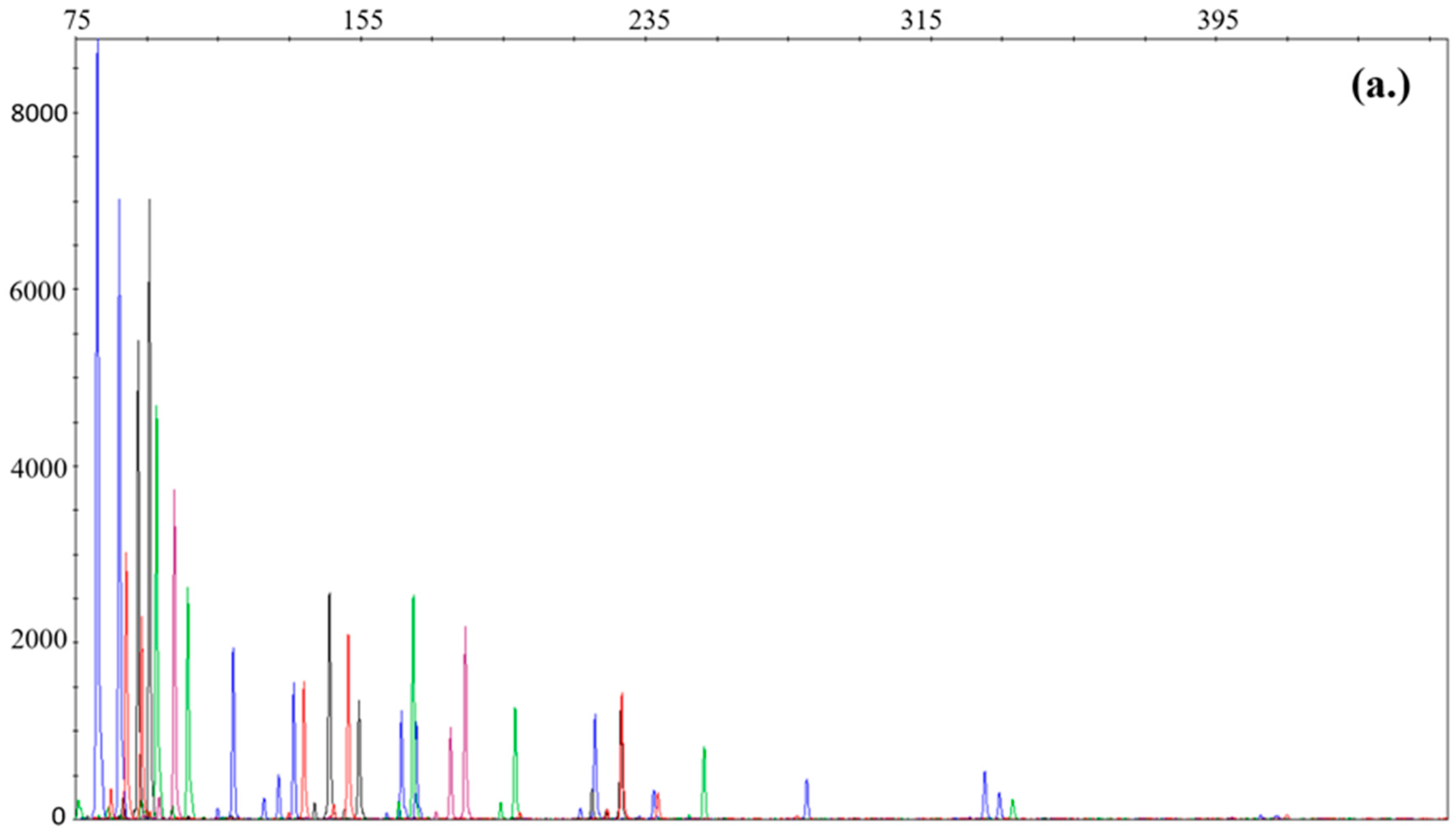
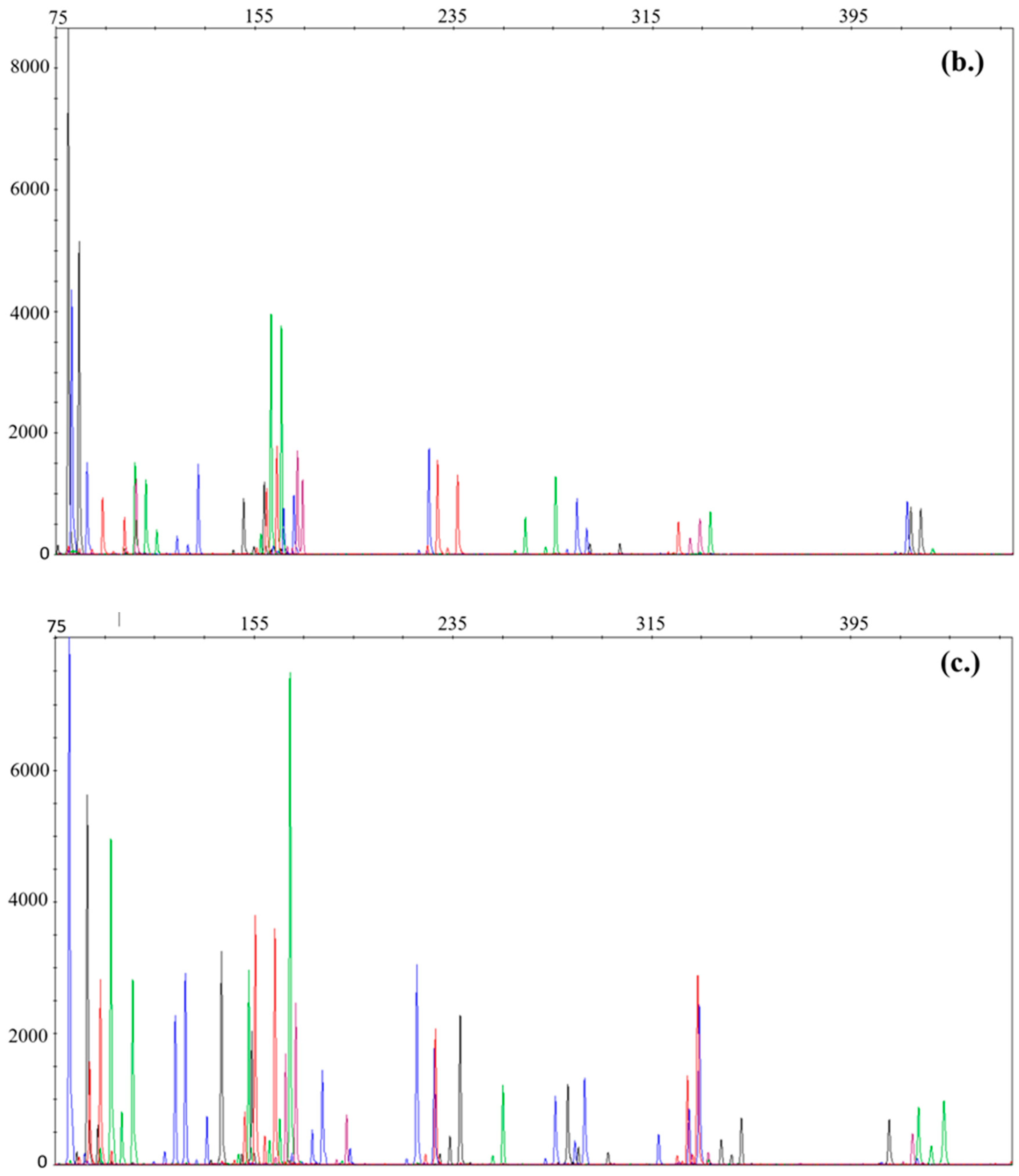
4.1. Sensitivity
4.2. Mixture Analysis
4.3. Stability Assessment
4.4. Degraded Casework Samples
5. Discussion
Author Contributions
Funding
Institutional Review Board Statement
Informed Consent Statement
Data Availability Statement
Acknowledgments
Conflicts of Interest
References
- Haddrill, P.R. Developments in forensic DNA analysis. Emerg. Top Life Sci. 2021, 5, 381–393. [Google Scholar] [CrossRef] [PubMed]
- Gill, P.; Haned, H.; Bleka, O.; Hansson, O.; Dørum, G.; Egeland, T. Genotyping and interpretation of STR-DNA: Low-template, mixtures and database matches—Twenty years of research and development. Forensic Sci. Int. Genet. 2015, 18, 100–117. [Google Scholar] [CrossRef] [PubMed]
- Roewer, L. DNA fingerprinting in forensics: Past, present, future. Investig. Genet 2013, 4, 22. [Google Scholar] [CrossRef] [PubMed]
- Alvarez-Cubero, M.J.; Saiz, M.; Martínez-García, B.; Sayalero, S.M.; Entrala, C.; Lorente, J.A.; Martinez-Gonzalez, L.J. Next generation sequencing: An application in forensic sciences? Ann. Hum. Biol. 2017, 44, 581–592. [Google Scholar] [CrossRef] [PubMed]
- Aalbers, S.E.; Hipp, M.J.; Kennedy, R.S.; Weir, B.S. Analyzing population structure for forensic STR markers in next generation sequencing data. Genetics 2020, 49, 102364. [Google Scholar] [CrossRef] [PubMed]
- Kraemer, M.; Prochnow, A.; Bussmann, M.; Scherer, M.; Peist, R.; Steffen, C. Developmental validation of QIAGEN Investigator® 24plex QS Kit and Investigator® 24plex GO! Kit: Two 6-dye multiplex assays for the extended CODIS core loci. Forensic Sci. Int. Genet. 2017, 29, 9–20. [Google Scholar] [CrossRef] [PubMed]
- Williams, G.; Foley, M.M.; Knight, K.L. Applied Biosystems’ GlobalFiler™ PCR Amplification Kit. In Forensic DNA Analysis: Methods and Protocols; Springer: New York, NY, USA, 2023; Volume 2685, pp. 241–252. [Google Scholar] [CrossRef]
- Ensenberger, M.G.; Lenz, K.A.; Matthies, L.K.; Hadinoto, G.M.; Schienman, J.E.; Przech, A.J.; Morganti, M.W.; Renstrom, D.T.; Baker, V.M.; Gawrys, K.M.; et al. Developmental validation of the PowerPlex(®) Fusion 6C System. Forensic Sci. Int. Genet. 2016, 21, 134–144. [Google Scholar] [CrossRef] [PubMed]
- Burch, S.; Sulzer, A.; Voegeli, P.; Morf, N.V.; Gysi, M.; Kratzer, A. The Applied Biosystems™ NGM Detect™ PCR Amplification Kit—As promising as promised? Forensic Sci. Int. Genet. Suppl. Ser. 2017, 6, 504–506. [Google Scholar] [CrossRef]
- Almohammed, E.; Hadi, S. Internal validation of GlobalFilerTM kit using reduced reaction volume. Forensic Sci. Int. Genet. Suppl. Ser. 2019, 7, 878–883. [Google Scholar] [CrossRef]
- Perry, J.; Munshi, T.; Haizel, T.; Iyavoo, S. Validation of reduced volume VeriFiler™ Express PCR Amplification Kit for buccal swab samples extracted using Prep-n-Go™ Buffer. J. Forensic Sci. 2022, 67, 1971–1978. [Google Scholar] [CrossRef] [PubMed]
- Steffen, C.R.; Doble, M.D.; Gettings, K.B.; Vallone, P.M. Corrigendum to ‘U.S. Population Data for 29 Autosomal STR Loci’. Forensic Sci. Int. Genet. 2017, 31, 36–40. [Google Scholar] [CrossRef] [PubMed]
- McCaughan, C.; Lenz, K.A. DNA Amplification Using Promega’s PowerPlex® Fusion Systems (5C and 6C). In Forensic DNA Analysis: Methods and Protocols; Springer: New York, NY, USA, 2023; Volume 2685, pp. 207–226. [Google Scholar] [CrossRef]
- Available online: https://www.swgdam.org/_files/ugd/4344b0_3f94c9a6286048c3924c58e2c230e74e.pdf (accessed on 20 January 2024).
- Available online: https://enfsi.eu/wp-content/uploads/2016/09/minimum_validation_guidelines_in_dna_profiling_-_v2010_0.pdf (accessed on 20 January 2024).
- Tegally, H.; San, J.E.; Giandhari, J.; de Oliveira, T. Unlocking the efficiency of genomics laboratories with robotic liquid-handling. BMC Genom. 2020, 21, 729. [Google Scholar] [CrossRef] [PubMed]
- Pan, J.Y.; Chang-Yen, D.; Blanchard, D.P.; Lam, W.; Searle, P.A. An Automated Tube Labeler for High-Throughput Purification Laboratories. SLAS Technol. 2021, 26, 113–116. [Google Scholar] [CrossRef] [PubMed]
- Enoch, E.; Councill, A.W.; Axtell, N.B.; Truong, T.; Liang, Y.; Aposhian, A.L.; Webber, K.G.I.; Zhu, Y.; Cong, Y.; Carson, R.H.; et al. Adapting a Low-Cost and Open-Source Commercial Pipetting Robot for Nanoliter Liquid Handling. SLAS Technol. 2021, 26, 311–319. [Google Scholar] [CrossRef]
- Ng, N.; Gately, R.; Ooi, L. Automated liquid handling for microplate assays: A simplified user interface for the Hamilton Microlab STAR. J. Appl. Bioanal. 2021, 7, 11–18. [Google Scholar] [CrossRef]
- Kong, F.; Yuan, L.; Zheng, Y.F.; Chen, W. Automatic Liquid Handling for Life Science: A Critical Review of the Current State of the Art. J. Lab. Autom 2012, 17, 169–185. [Google Scholar] [CrossRef] [PubMed]
- Available online: https://www.qiagen.com/us/resources/resourcedetail?id=46064856-1b88-4b27-a825-d3f616e06c08&lang=en (accessed on 20 January 2024).
- Promega Corporation. PowerPlex® Fusion System for Use on the Applied Biosystems® Genetic Analyzers; Instructions for Use of Products DC2402 and DC2408. Technical Manual TMD039, Revised in July 2020; Promega Corporation: Madison, WI, USA, 2020. [Google Scholar]
- Promega Corporation. PowerPlex® 6C Matrix Standard, Instructions for Use of Product DG4900; Technical Manual TMD046, Revised in October 2015; Promega Corporation: Madison, WI, USA, 2015. [Google Scholar]
- Vernarecci, S.; Ottaviani, E.; Agostino, A.; Mei, E.; Calandro, L.; Montagna, P. Quantifiler® Trio Kit and forensic samples management: A matter of degradation. Forensic Sci. Int. Genet. 2015, 16, 77–85. [Google Scholar] [CrossRef] [PubMed]


| Input DNA (ng) | Manual (M) Automated (A) | Blue Dye | Green Dye | Black Dye | Red Dye | Purple Dye | |||||
|---|---|---|---|---|---|---|---|---|---|---|---|
| 1 | M (mean ± SD. RFU) | 13,796.3 | 2639.2 | 9732.1 | 1829.2 | 12,012.2 | 2275.3 | 12,571.4 | 3246.8 | 7838 | 820.6 |
| M (DO%) | 0 | 0 | 0 | 0 | 0 | ||||||
| 0.5 | M (RFU) | 8221.8 | 2382.1 | 6062.4 | 1511.3 | 5900.6 | 1288.9 | 6291.2 | 1534.4 | 4385.1 | 803.9 |
| M (DO%) | 0 | 0 | 0 | 0 | 0 | ||||||
| A (mean ± SD. RFU) | 6099.3 | 3001.1 | 4420.9 | 2606.7 | 5262.7 | 2191.6 | 5897 | 2566 | 3112.1 | 2236.2 | |
| A (DO%) | 0 | 0 | 0 | 0 | 0 | ||||||
| 0.15 | M (mean ± SD. RFU) | 2583.5 | 1224.9 | 1959.7 | 1279.6 | 1592 | 605.7 | 1813.5 | 712.3 | 1162.7 | 964.3 |
| M (DO%) | 0 | 0 | 0 | 0 | 0 | ||||||
| A (mean ± SD. RFU) | 1808.2 | 1131.8 | 1070.2 | 755.4 | 1546.2 | 867.9 | 1687.6 | 848 | 530.1 | 386.7 | |
| A (DO%) | 0 | 0 | 0 | 0 | 10 | ||||||
| 0.075 | M (mean ± SD. RFU) | 1119.2 | 652.7 | 993.7 | 682.7 | 761.6 | 384 | 729.3 | 307.3 | 583.2 | 381.7 |
| M (DO%) | 0 | 4 | 5 | 8 | 10 | ||||||
| 0.0375 | M (mean ± SD. RFU) | 478.6 | 322.6 | 425.1 | 355.9 | 407.2 | 293.1 | 365.1 | 275.2 | 264.8 | 173.2 |
| M (DO%) | 13 | 4 | 5 | 6 | 10 | ||||||
| 0.015 | M (mean ± SD. RFU) | 127 | 46.6 | 109.6 | 53.2 | 111.6 | 63 | 112.1 | 42.9 | 58.5 | 0.7 |
| M (DO%) | 68 | 70 | 60 | 66 | 80 | ||||||
| 0.0075 | M (mean ± SD. RFU) | 114.6 | 83.1 | 103.9 | 41.2 | 98.1 | 34.9 | 136.2 | 43.7 | 79.7 | 13.1 |
| M (DO%) | 82 | 76 | 73 | 90 | 70 | ||||||
| Input DNA (ng) | Manual (M) Automated (A) | Blue Dye | Green Dye | Black Dye | Red Dye | Purple Dye | |||||
|---|---|---|---|---|---|---|---|---|---|---|---|
| 0.15 | M (mean ± SD. RFU) | 7721.6 | 2223.5 | 5917.6 | 1952.5 | 6628.5 | 2038.6 | 6480.7 | 1874.9 | 4217.7 | 1344.9 |
| M (DO%) | 0 | 0 | 0 | 0 | 0 | ||||||
| A (mean ± SD. RFU) | 9138.8 | 3302.2 | 6454.7 | 3035.6 | 7498.6 | 2395.3 | 7648.6 | 2486.3 | 5300.6 | 1945.3 | |
| A(DO%) | 0 | 0 | 0 | 0 | 0 | ||||||
| 0.075 | M (mean ± SD. RFU) | 4846.1 | 2255 | 3672.9 | 1476.5 | 3652.8 | 1179.3 | 4067.2 | 1652.2 | 2474.6 | 1056.4 |
| M (DO%) | 2 | 0 | 0 | 0 | 0 | ||||||
| A (mean ± SD. RFU) | 4293.3 | 1990.4 | 2936.3 | 1612.7 | 3555.8 | 1511.2 | 3799 | 1847.7 | 2082.6 | 642.3 | |
| A (DO%) | 0 | 0 | 2 | 0 | 0 | ||||||
| 0.0375 | M (mean ± SD. RFU) | 2291.9 | 1108.4 | 1637.2 | 747.2 | 1723.7 | 979.9 | 1734.8 | 1033.1 | 853.6 | 673.1 |
| M (DO%) | 2 | 2 | 0 | 0 | 0 | ||||||
| A (mean ± SD. RFU) | 1520.7 | 842.1 | 1025.9 | 598.8 | 1255.6 | 671.2 | 1423.8 | 805.5 | 749.4 | 384.7 | |
| A (DO%) | 7 | 4 | 7 | 4 | 0 | ||||||
| 0.015 | M (mean ± SD. RFU) | 720.3 | 502.1 | 480.2 | 298.3 | 653.4 | 373.7 | 671.6 | 358.7 | 388.3 | 219.4 |
| M (DO%) | 15 | 14 | 18 | 18 | 10 | ||||||
| A (mean ± SD. RFU) | 431 | 542.2 | 474.3 | 360.8 | 508.1 | 377 | 650.5 | 515.1 | 368 | 218.3 | |
| A(DO%) | 43 | 40 | 35 | 46 | 40 | ||||||
| 0.0075 | M (mean ± SD. RFU) | 385.5 | 375.7 | 284.9 | 216.8 | 348.8 | 241 | 396.4 | 328.8 | 325 | 165.4 |
| M (DO%) | 38 | 48 | 43 | 36 | 40 | ||||||
| M (mean ± SD. RFU) | 294.1 | 176.2 | 273.5 | 186.5 | 326.7 | 291 | 403.7 | 225.2 | 329 | NA | |
| M(DO%) | 53 | 72 | 55 | 38 | 90 | ||||||
| Manually Prepared PCR | Automated PCR | |||
|---|---|---|---|---|
| Mix (Female:Male) | Allele Dropout (No.) | Allele Dropout in % | Allele Dropout (No.) | Allele Dropout in % |
| 1:1 | 0 | 0.0% | 0 | 0.0% |
| 1:4 | 0 | 0.0% | 2 | 0.89% |
| 1:7 | 1 | 0.44% | 7 | 3.11% |
| 1:9 | 5 | 2.22% | 18 | 8.00% |
| 1:19 | 32 | 14.22% | 52 | 23.11% |
| DI | DNA Concentration (ng/µL) | Average DNA Concentration (ng/µL) | Samples (No.) | DNA Profile Suitable for Comparison | Average Locus Dropout (%) | ||
|---|---|---|---|---|---|---|---|
| Manual | Hamilton Biorobot | Total | |||||
| LOW (3–4) | 0.002–0.1465 | 0.0222 | 92 | 60.98% | 50% | 59.78% | 52.22% |
| MEDIUM (4–7) | 0.0031–0.1423 | 0.0175 | 80 | 46.67% | 40% | 46.25% | 64.24% |
| HIGH (>7) | 0.0021–0.1418 | 0.0151 | 29 | 26.92% | NA | 24.14% | 83.65% |
Disclaimer/Publisher’s Note: The statements, opinions and data contained in all publications are solely those of the individual author(s) and contributor(s) and not of MDPI and/or the editor(s). MDPI and/or the editor(s) disclaim responsibility for any injury to people or property resulting from any ideas, methods, instructions or products referred to in the content. |
© 2024 by the authors. Licensee MDPI, Basel, Switzerland. This article is an open access article distributed under the terms and conditions of the Creative Commons Attribution (CC BY) license (https://creativecommons.org/licenses/by/4.0/).
Share and Cite
Lőrincz, E.É.; Mátrai, N.; Rádóczy, K.A.; Cseppentő, T.; Magonyi, N.M.; Heinrich, A. Comparison of Reduced PCR Volume PowerPlex Fusion 6C Kit Validations on Manual and Automated Systems. DNA 2024, 4, 52-63. https://doi.org/10.3390/dna4010003
Lőrincz EÉ, Mátrai N, Rádóczy KA, Cseppentő T, Magonyi NM, Heinrich A. Comparison of Reduced PCR Volume PowerPlex Fusion 6C Kit Validations on Manual and Automated Systems. DNA. 2024; 4(1):52-63. https://doi.org/10.3390/dna4010003
Chicago/Turabian StyleLőrincz, Eszter É., Norbert Mátrai, Katalin A. Rádóczy, Tamás Cseppentő, Nóra M. Magonyi, and Attila Heinrich. 2024. "Comparison of Reduced PCR Volume PowerPlex Fusion 6C Kit Validations on Manual and Automated Systems" DNA 4, no. 1: 52-63. https://doi.org/10.3390/dna4010003