Entry

Saprophytic Filamentous Fungi against Helminths Affecting Captive Wild Animals

by
Rami Salmo
1,
Cándido Viña
1,
João Lozano
2,3,
Antonio M. Palomero
1,
José Ángel Hernández
1,
Rodrigo Bonilla
4,
Rita Sánchez-Andrade
1,
Adolfo Paz-Silva
1,
Luis M. Madeira de Carvalho
2,3,
María Sol Arias
1 and
Cristiana Cazapal-Monteiro
1,*
1
Control of Parasites Group (COPAR, GI-2120), Department of Animal Pathology, Faculty of Veterinary, University of Santiago de Compostela, 27002 Lugo, Spain
2
CIISA—Centre for Interdisciplinary Research in Animal Health, Faculty of Veterinary Medicine, University of Lisbon, Avenida da Universidade Técnica, 1300-477 Lisbon, Portugal
3
Associate Laboratory for Animal and Veterinary Sciences (AL4AnimalS), 1300-477 Lisbon, Portugal
4
CARVAL Pharmaceuticals, Bogotá 110211, Colombia
*
Author to whom correspondence should be addressed.
Encyclopedia 2024, 4(1), 91-100; https://doi.org/10.3390/encyclopedia4010008
Submission received: 2 November 2023 / Revised: 20 December 2023 / Accepted: 2 January 2024 / Published: 5 January 2024
(This article belongs to the Collection Encyclopedia of Fungi)

Definition

:
In recent decades, important modifications have been introduced in zoos in order to guarantee the welfare of captive wild animals. Thus, many of these species are housed in enclosures with access to vegetation, where they can enjoy habitats close to those in their natural surroundings, interact with the environment, etc. These habitats present beneficial conditions for some species of parasites to survive and spread. This is a very similar problem to that affecting livestock, and the same solution, based on deworming, is currently being applied. However, the free-living stages of certain parasites that develop in the soil are responsible for high rates of ground contamination throughout the year, so that animals become infected soon after successful deworming, resulting in chemical parasiticides being frequently administered. Preventive measures are seldom considered, which worsens the situation. This entry summarizes the usefulness of the dissemination of certain saprophytic filamentous fungi with proven antagonism against some of the parasites.

1. Introduction

Wild animals bred in captivity in zoos are often found permanently in the same plot of land, a circumstance that favors high concentrations of parasites in the environment [1]. This situation can be worsened by problems of stress, feeding, hygienic conditions in the plots, and confined spaces, which increase the possibility of parasitic infections [2]. Accordingly, control is primarily focused on deworming the animals, and it has been stated that individuals that receive these treatments regularly do not exhibit clinical signs of infection [3], and this could be interpreted to mean that the infection has fully disappeared. Nevertheless, the periodic administration of anthelmintics does not prevent challenge infections among captive species; it is suggested that a lack of quarantining newly entered animals as well as free-ranging stray animals could be possible sources of infection [4]. Other issues that need attention are the frequent use of parasiticide drugs in combination with inappropriate dosage and administration methods, which may lead to the development of resistant strains of some species of gastrointestinal nematodes [5]. Reports have noted the existence of trichostrongylids resistant to both levamisole and fenbendazole in captive Armenian red sheep, in wapitis to levamisole, and in giraffe to macrocyclic lactones and imidazothiazoles [6,7]. As can be easily understood, this problem is identical to the problems reported for decades in farm animals, which remain unsolved to date. More attention should be paid to prevention with the aim of limiting the risk of infection and, thus, decreasing the need for deworming.

1.1. Main Parasites Affecting Captive-Bred Animals
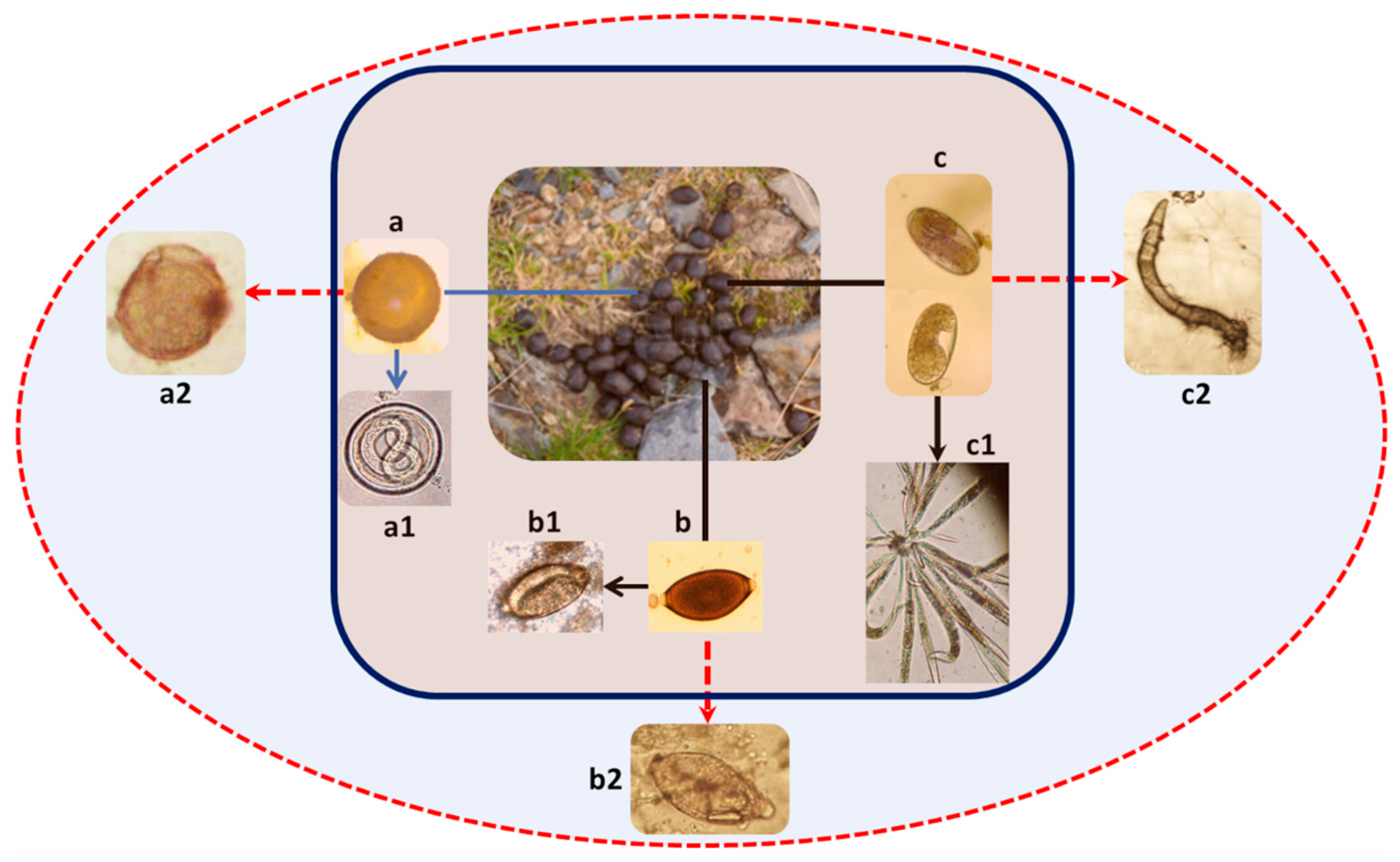
Different parasites involving protozoa, helminths, or ectoparasites have been described among wild captive animals, but helminths are the most frequently detected. These are organisms with a direct life cycle, with part of their development occurring in the environment for days to weeks, and they are frequently detected in the feces of animals kept in captivity [8]. This happens because the animals are separated, housed in parks that normally prevent the entry of other animals and in more or less stable conditions in terms of the condition of their plots. All of this hinders the development of parasites with an indirect life cycle, in which the collaboration of intermediate hosts is necessary [9]. The most frequent groups or families of helminths are ascarids, strongylids, and trichurids.

1.1.1. Ascarids

Ascarids, also known as roundworms, belong to the class Nematoda. The adult stages usually occur inside the small intestine, where females shed unembryonated eggs, which reach the environment through the feces [10,11]. After several weeks under optimal temperature and humidity conditions, one larva 2 (L2) develops inside [12], and the egg become infective. Infection in wild captive species occurs through the accidental ingestion of an infective egg while grazing [13,14], and the species more commonly described in carnivores and omnivores are Toxocara spp., Toxascaris leonina, and Baylisascaris spp. [3,15,16]. In herbivores (cow, buffalo, bison, eland), infection with Toxocara vitulorum, a roundworm without zoonotic characteristics (they cannot parasitize humans), is described with some frequency, while in monogastric herbivores, Parascaris equorum is the most commonly diagnosed ascarid species [15].

1.1.2. Strongylids

Strongylids also belong to the class Nematoda, and usually cause mixed infections involving more than one species. These infections cause a chronic process with low mortality, but, although they do not cause serious health changes, they do cause alterations in growth, poor coat, gastrointestinal disorders, etc. Most grazing animal species are affected depending on the conditions in which they are kept (overcrowding, hygiene, etc.). These parasites are frequently found in temperate areas (9–27 °C) and high relative humidity (≥70%) conditions [17]. The most frequently identified species belong to the genera Ostertagia (cattle), Teladorsagia (sheep, goats, and deer) and Trichostrongylus, although there are others such as Haemonchus, Cooperia, Bunostomum, Nematodirus, and Oesophagostomum. Infected animals excrete the eggs into the environment when defecating, which emerge already embryonated, and in a few days, the larva 1 (L1) forms inside, hatches, and the leaves the egg, transforming into an L2 that feeds on organic matter present in the feces. Depending on the temperature and humidity conditions, L2 larvae molt into L3 larvae (the infective stage), which leave the feces and move towards the apical portion of plant species, especially grasses, and are ingested by herbivores while pasturing.

1.1.3. Trichurids

These nematodes (also called whipworms) constitute a genus of gastrointestinal parasites belonging to the roundworm family Trichuridae, characterized by a direct life cycle involving unembryonated eggs that are passed in the feces of infected animals [18]. The eggs develop outside until a first-stage larva, or L1, is formed inside and turns into the infective stage. Infection occurs when embryonated eggs are ingested, and the L1 hatches in the gut and moves to the large intestine, where the adult stage is attained and the larva feeds on blood.
Infection with Trichuris spp. is commonly detected among captive wild animals [18,19], enhanced by eggs shed in the feces. The feces provide a sticky cover that ensures they can remain not only in soil, but on different structures that exist in animal refuges such as hangers, perches, or swings. Accordingly, the prevention of infection is very difficult.

1.1.4. Bronchopulmonary Nematodes

Bronchopulmonary nematodes of the families Protostrongylidae and Dictyocaulidae, also known as lungworms, are parasites characterized by the location of adults in the lower respiratory tract of domestic and wild mammals [20]. Most infections are asymptomatic, but eventually they can cause respiratory problems such as bronchopneumonia and parasitic bronchitis that could evolve into severe disease. Adults lay their eggs in the lung tissues, where the first-stage larvae hatch and climb up to the throat to be swallowed and finally expelled with the feces. Soil contamination occurs through the spread of L1 larvae in the feces of parasitized animals. There are some differences in the way animals become infected, as Dictyocaulidae has a direct life cycle with a free-living stage, while in Protostrongylidae, a terrestrial mollusk acts as an intermediate host [21]. Animals become infected when grazing through the ingestion of the L3 free-living stage for Dictyocaulidae or the intermediate host carrying the L3 for Protostrongylidae.
Adults of Dictyocaulus spp. produce embryonated eggs in the airways that hatch briefly after oviposition, while the eggs of lungworms are unembryonated. When they both hatch, the first-stage larvae reach the trachea and are passed in the feces. Once in the soil, Dictyocaulus spp. molt to L2 and L3, the infective stage, while the larvae of pulmonary nematodes need to penetrate mollusks to reach the infective L3 stage. After release in the abomasum, the L3 of Dictyocaulus spp. must penetrate the intestine and migrate via lymph nodes to reach the lungs as L4s. The third-stage larvae of Protostrongylidae leave the intermediate host from the stomach and penetrate the intestine, reaching the lungs, and form nodes with L4s within the lungs after thoracic duct–heart–pulmonary artery migration [22].

1.2. Clinical Importance and Detection

Even though parasites are one of the leading causes of death in captive wild animals, especially on those with a heavy parasitic burden, the great majority of these infections are subclinical and so clinical signs are hardly noticeable by veterinarians and zookeepers [9]. As ascarids, strongylids, and trichurids are mainly localized in the gastrointestinal tract, if a clinical syndrome is observed, the clinical signs will be mostly digestive or due to the parasite’s hematophagy. Hence, it is possible to observe anemia, edema, lethargy, malnutrition, and diarrhea. In severe infections, there will also be extreme weight loss and worsening of body condition, and death can also occur [7]. Unless there is a heavy infection, the clinical signs of Trichuris are rare and consist of minor cecitis and diarrhea with mucus and blood.
Therefore, it is crucial to have adequate tools for the diagnosis of parasitic infections. Routine diagnostics consist of the visualization of parasite eggs in feces, confirming the existence of adult worms inside the host. There are also serological probes such as ELISA, although a positive result is not enough to confirm the parasitism, only previous exposure [23].

1.3. Control of Parasites in Captive Animals

Wild animals seem to have a natural resistance to parasitic infections, but when kept in captivity, their sensitivity to parasites appears to be reduced, possibly due to the disruption of their ecosystem [24].
The control of parasites in captive-bred animals is carried out by administering broad-spectrum antiparasitic drugs, normally twice a year, against the stages inside the hosts. For the control of nematodes, benzimidazoles like albendazole or fenbendazole and macrocyclic lactones such as ivermectin are the most utilized families, while praziquantel is used for the control of cestodes. The use of these drugs in wild species involves a series of additional difficulties:
-
In a large number of cases, deworming is carried out without a prior coprological examination.
-
The antiparasitic drugs used for livestock are often not specific for the target species, so they are underdosed to avoid intoxication problems.
-
Animal handling problems can cause most treatments to be administered orally due to the impossibility of giving them parenterally. This oral administration does not ensure an adequate ingestion of antiparasitic drugs, so there will be animals that ingest a higher dose than recommended and others in which the dosage is practically nil.
-
One of the main limiting factors regarding the implementation of preventive measures against parasites is the place where these animals are located, which is usually limited and permanent. The effectiveness of any antiparasitic treatment is reduced as a consequence of numerous reinfections when they are continuously located in the same plot.
-
The residues produced by the metabolism of pesticides can be toxic to some species of microorganisms that are very useful for soil enrichment, such as some coprophagous beetles [25,26].
For the purpose of limiting the risk of infection from certain parasites, regular monitoring for sanitation and cleaning is necessary to minimize the risk, and the frequent cleaning of animal refuges and facilities, as well as the removal of feces to avoid infectious stages from being reached, are also important [4]. It is noteworthy to state that preventing environmental contamination by the eggs and larvae of parasites represents one of the key steps to stopping the transmission of parasites to wildlife.
Despite proper deworming and good hygiene practices, animals living in captivity will still suffer from diseases caused by parasites. This occurs due to the presence of the animals in the same place for prolonged periods of time, favoring their exposure to infective forms, so that reinfection occurs [15,27].
With all of the above in mind, together with the high resistance presented by the eggs of some helminths and even infective larvae under certain conditions, it appears necessary to adopt an alternative approach for the control of these soil-transmitted parasites [16,23,28,29].

2. Application of Helmintophagous Fungi against Parasites Affecting Wild Captive Animals

The use of natural antagonists against pathogens is known as biological control. Some fungi are antagonists of different parasites, thus providing an ecological method of control that is harmless to people, animals, and plants. This strategy is not intended to eliminate all forms of parasites, as is the case with anthelmintic drugs, but rather to seek a balance between animals and parasites that does not cause a decline in their health or productivity [30].
Depending on their activity, parasiticidal fungi can be divided into three main groups: nematode-trapping, predatory, and endoparasitic fungi.
-
Nematode-trapping fungi: These are characterized by producing an extensive system of hyphae or mycelium where, at certain intervals of development, they present specialized structures such as hyphal nets, knobs, branches, or rings to trap and sustain live nematodes (larval stages); therefore, they are ideal for the control of parasites such as strongyles [31]. Examples of this group are Duddingtonia flagrans, Arthrobotrys spp., and Monacrosporium thaumasium [32,33,34,35], which can develop different types of traps in which the mobile parasitic stages become caught, absorbed, and finally destroyed.
-
Predatory fungi or egg parasites: The hyphae of these fungi adhere to and penetrate the egg through small pores in the vitelline layer, causing permeability alterations. These fungi continue their development until they colonize the contents of the egg (embryo or larva). Good examples of this type of fungus are Pochonia chlamydosporia and Mucor circinelloides [36,37,38]. The parasiticide activity of these fungi is classified into three types, depending on how the egg morphology and viability are affected. Predatory fungi exhibit parasiticide activity type 1 when the hyphae surround the eggshell without penetrating it and where no embryo alterations are observed, but parasite development is stopped; activity type 2 occurs when the eggshell and embryo show morphological alterations without hyphae penetration; and, finally, type 3 activity takes place when the hyphae penetrate the eggshell and destroy the embryo so that the eggs become completely unviable, being unable to infect animals. If no damage or alterations are observable in the eggs, they remain viable, and this is considered activity type 0 [38].
-
Endoparasitic fungi: These fungi can infect nematodes through their spores. The fungi do not develop outside the host’s body and are not able to penetrate unless they are ingested, which makes their culture and maintenance in laboratory conditions very difficult and is the main reason for them not being used in current investigations.
It is important to emphasize that depending on the target (the infective stage of the parasitic species), a different fungal mechanism of action must be employed. In ascarids or trichurids, the infective phase is immobile, and, therefore, predatory fungi are useful. On the other hand, strongyles have a mobile infective phase and can be controlled by means of nematode-trapping fungi (Figure 1).

2.1. Biological Control of Helminths Affecting Captive Animals: Analysis in Feces

For the purpose of establishing the usefulness of biological control to prevent the development of parasites in feces until their infective stages, some tests consisting of adding chlamydospores of D. flagrans, M. circinelloides, Verticillium, and Paecilomyces were conducted. Some of these studies took place in a zoological park located in Galicia (northwest Spain) where these filamentous fungi were isolated and tested over several gastrointestinal nematodes from carnivores, equids, ruminants, and monogastric herbivores kept in captivity. Chlamydospores of Mucor circinelloides, Paecilomyces, and Verticillium were sprayed into racoon (Procyon lotor) feces positive for B. procyonis, and reductions of 53–69%, 45–62%, and 52–67%, respectively, were obtained, indicating type 3 activity for all three fungi [38]. Similar results were recorded when M. circinelloides was tested in T. leonina and Parascaris equorum eggs, with 58% and 61–67% of reduction, respectively [39,40]. Ovicidal activity type 3 was identified for all fungal species in each experiment. Moreover, the addition of spores of M. circinelloides or Trichoderma atrobrunneum to the feces of captive dromedaries (Camelus dromedarius) resulted in a 50% reduction in the viability of eggs from Trichuris sp. [18].
For evaluating the larvicidal activity of the nematode-trapping fungi Duddingtonia flagrans over larvae 3 of strongylids from equids, chlamydospores were added to the feedstuff twice a week, reaching a reduction varying between 84 and 89% [41]. The results showed that the viability of the eggs or larvae of gastrointestinal nematodes was reduced by ≥45%. It should be taken into account that, so far, few trials have been conducted on the efficacy of fungi on parasites affecting animals, and most of the tests have been carried out in vitro in Petri dishes. Table 1 shows a resume of all of these studies.
The data from Table 1 demonstrate that the presence of parasiticide fungi in the feces of animals passing eggs provides a very helpful and eco-friendly measure to limit the possibility of parasites attaining infectivity, decreasing the risk of further challenge infections. In this way, an interesting question arises regarding ways to ensure that the fungal stages (mycelium, spores) can reach the feces, interact with different phases of the parasites, and develop their antagonistic roles. There are several possibilities, and the most effective are those involving oral administration to the animals.
There are few trials concerning the antagonism of D. flagrans against the larvae of M. capillaris prior their penetration on the snail, and the results seem to indicate little or no effect [42,43].

2.2. Administration of Helmintophagous Fungi to Captive Animals

Due to the resistance of chlamydospores to high temperatures and pH conditions, these organisms can survive the passage across the digestive tract, and, once excreted, germinate in the feces and form traps, making them ideal candidates for oral administration [44]. In recent decades, different formulations have been tried for the administration of fungi to animals. One of them consisted of mixing the spores with the feed manually, before giving it to the animals. The fungi solution, which could have one or two fungi species, must be mixed with the feed concentrate immediately before the administration to avoid any fungal development that could alter their organoleptic characteristics, making it unappetizing for the animals. Another option was to incorporate these chlamydospores into a supplement feed with grains and sweet feed for improving its consumption by the animals. This strategy was used successfully with giraffes, antelopes, and gerenuks in EEUU [40], but also in Spain with European donkeys, African asses, and zebras [41], achieving excellent percentages of larval reduction. Other options used to provide the spores orally was by means of a powdered product resulting from mashing the fungi, and its utilization was able to reduce the presence of third-stage larvae from gastrointestinal nematodes in reticulated giraffe, scimitar-horned oryx, and roan antelope by mixing it with their feed [45].
To further facilitate the task of giving spores to the animals, some experiments were carried out with nutritional pellets to which the chlamydospores were added during the mixing phase of the manufacturing process. Thus, the ability of M. circinelloides and Duddingtonia flagrans to reduce the infective phases of Trichostrongylus, Nematodirus, Chabertia, and Haemonchus in the soil was demonstrated, reducing the probability of reinfection in all species that were studied, since it was not necessary to deworm them again throughout the 24 months that the study lasted. All of these experiments carried out with domestic or wild living animals required individual deworming before starting the fungal administration, since nematophagous fungi have no effect on the parasites harbored within the animal and only destroy those in the free-living parasitic stages. It is important to underline the absence of side effects when fungal chlamydospores were given to horses, cattle, and sheep, as well as captive wild animals [45,46,47]. Table 2 summarizes all of the studies mentioned above.
These investigations corroborated the findings of those previously performed on livestock species showing that the oral administration of fungal spores is possible by means of different formulated foods. Finally, the existence of collections of wild animals in public places such as parks, gardens of historic buildings, etc., should also be taken into account, as they are at risk of infection from different parasites. Recent investigations have demonstrated that very promising results are possible by supplementing peacocks with chlamydospores of a strain of the saprophytic filamentous fungus M. circinelloides isolated in Lisbon (Portugal) [34].

3. Conclusions and Prospects

According to data collected in recent decades, saprophytic filamentous fungi are able to destroy or interfere with the development of certain parasites in feces and/or soil, which has become of great importance in contributing to the control of different parasites affecting wild animals maintained in zoological parks where the administration of chemical parasiticide compounds often utilized in livestock appears to be the only solution. Nevertheless, despite many effective formulations being commercially available, wild animal species are soon infected again, possibly due to the fact that the animals are always kept in the same location, which increases their exposure to the infective stages of different parasites developing in the soil. This emphasizes the need for preventive measures to avoid large numbers of parasites evolving into their respective infective stages in the environment (enclosure, box) where the animals are sheltered, thus increasing their risk of infection and the need for additional deworming.
A program for the control of parasites affecting wild captive animals that integrates the analysis of feces prior to deworming through the administration of adequate anthelmintics (benzimidazoles or macrocyclic lactones), together with the periodical administration of chlamydospores from soil saprophytic fungi, appears a novel and helpful contribution to the sustainable management of digestive infections from parasites in zoological parks. It seems very plausible that a sustainable and eco-friendly strategy could be developed not only among zoological gardens, but also involving animals maintained in public areas. This suggested strategy constitutes a safe procedure.

Author Contributions

Conceptualization, methodology, data curation, resources, writing—original draft preparation, supervision, funding acquisition, and project administration: R.S.-A., A.P.-S., M.S.A. and C.C.-M.; formal analysis, software, writing, and visualization: R.B. and J.Á.H.; investigation, validation, and conceptualization: R.S., C.V., A.M.P. and J.L.; resources, A.P.-S.; writing—review and editing: R.S.-A., A.P.-S., M.S.A., C.C.-M. and L.M.M.d.C. All authors have read and agreed to the published version of the manuscript.

Funding

This research was funded by the Ministry for Science and Innovation (MCINN, Spain) and the European Regional Development Fund (ERDF) (Project PID2020-120208RB-I00), AGACAL (Consellería de Cultura, Educación e Universidades, Xunta de Galicia, Spain) (Projects FEADER 2022/017B and ED431B 2021/07). The APC was funded by the University of Santiago de Compostela (USC, Spain).

Conflicts of Interest

R.B. is research director for Carval pharmaceuticals, and the authors declare no conflict of interest. The funders had no role in the design of the study; in the collection, analyses, or interpretation of data; in the writing of the manuscript; or in the decision to publish the results.

References

  1. Wolfe, B.A. Bovidae (except sheep and goats) and Antilocapridae. In Fowler’s Zoo and Wild Animal Medicine, 1st ed.; Miller, R.E., Fowler, M.E., Eds.; Elsevier: St. Louis, MO, USA, 2015; Volume 8, pp. 626–645. ISBN 978-1-4557-7397-8. [Google Scholar] [CrossRef]
  2. Palomero, A.M.; Cazapal-Monteiro, C.F.; Valderrábano, E.; Paz-Silva, A.; Sánchez-Andrade, R.; Arias, M.S. Soil fungi enable the control of gastrointestinal nematodes in wild bovidae captive in a zoological park: A 4-year trial. Parasitology 2020, 147, 791–798. [Google Scholar] [CrossRef] [PubMed]
  3. Schieber, M.-C.; Štrkolcová, G. Prevalence of endoparasites in carnivores in a zoo and a wolves park in Germany. Folia Vet. 2019, 63, 54–59. [Google Scholar] [CrossRef]
  4. Nath, T.C.; Eom, K.S.; Choe, S.; Hm, S.; Islam, S.; Ndosi, B.A.; Kang, Y.; Bia, M.M.; Kim, S.; Eamudomkarn, C.; et al. Insight into One Health approach: Endoparasite infections in captive wildlife in Bangladesh. Pathogens 2021, 10, 250. [Google Scholar] [CrossRef] [PubMed]
  5. Shalaby, H.A. Anthelmintics resistance; how to overcome it? Iran. J. Parasitol. 2013, 8, 18–32. [Google Scholar]
  6. Young, K.E.; Jensen, J.M.; Craig, T.M. Evaluation of an anthelmintic activity in captive wild ruminants by fecal egg reduction tests and a larval development assay. J. Zoo Wildl. Med. 2000, 31, 348–352. [Google Scholar] [CrossRef]
  7. Garretson, P.D.; Hammond, E.E.; Craig, T.M.; Holman, P.J. Anthelmintic resistant Haemonchus contortus in a giraffe (Giraffa camelopardalis) in Florida. J. Zoo Wildl. Med. 2009, 40, 131–139. [Google Scholar] [CrossRef]
  8. Sengar, A.; Shrivastav, A.B.; Singh, K.P.; Rokde, A. Noninvasive assessment of gastrointestinal parasites infection in free-ranging wild herbivores and adjoining livestock of Panna Tiger Reserve, Madhya Pradesh, India. Vet. World 2017, 10, 748–751. [Google Scholar] [CrossRef]
  9. Atanaskova, E.; Kochevski, Z.; Stefanovska, J.; Nikolovski, G. Endoparasites in wild animals at the zoological garden in Skopje, Macedonia. JoTT Note 2011, 3, 1955–1958. [Google Scholar] [CrossRef]
  10. Mateus, T.L.; Castro, A.; Ribeiro, J.N.; Vieira-Pinto, M. Multiple zoonotic parasites identified in dog feces collected in Ponte de Lima, Portugal—A potential threat to human health. Int. J. Environ. Res. Public Health 2014, 11, 9050–9067. [Google Scholar] [CrossRef]
  11. Hoopes, J.; Hill, J.E.; Polley, L.; Fernando, C.; Wagner, B.; Schurer, J.; Jenkins, E. Enteric parasites of free-roaming, owned, and rural cats in prairie regions of Canada. Can. Vet. J. 2015, 56, 495–501. [Google Scholar]
  12. Côtes, E. Clinic and Veterinary Advisor. Dogs and Cats, 3rd ed.; Elsevier: Toronto, ON, Canada, 2014. [Google Scholar]
  13. Vandemark, L.M.; Jia, T.-W.; Zhou, X.-N. Social science implications for control of helminth infections in southeast Asia. Adv. Parasitol. 2010, 73, 137–170. [Google Scholar] [CrossRef] [PubMed]
  14. Elsheikha, H.M. Major cestodes infections. In Essentials of Veterinary Parasitology, 1st ed.; Elsheikha, H.M., Khan, N.A., Eds.; Caister Academic Press: Poole, UK, 2011; pp. 71–79. ISBN 978-1-904455-80-6. [Google Scholar]
  15. Maesano, G.; Capasso, M.; Ianniello, D.; Cringoli, G.; Rinaldi, L. Parasitic infections detected by FLOTAC in zoo mammals from Warsaw, Poland. Acta Parasitol. 2014, 59, 343–353. [Google Scholar] [CrossRef] [PubMed]
  16. Gałęcki, R.; Sokół, R.; Koziatek, S. Parasites of wild animals as a potential source of hazard to humans. Ann. Parasitol. 2015, 61, 105–108. [Google Scholar] [PubMed]
  17. Voinot, M.; Bonilla, R.; Sousa, S.; Sanchís, J.; Canhão-Dias, M.; Romero Delgado, J.; Lozano, J.; Sánchez-Andrade, R.; Sol Arias, M.; Madeira de Carvalho, L. Control of Strongyles in First-Season Grazing Ewe Lambs by Integrating Deworming and Thrice-Weekly Administration of Parasiticidal Fungal Spores. Pathogens 2021, 10, 1338. [Google Scholar] [CrossRef] [PubMed]
  18. Hernández, J.A.; Cazapal-Monteiro, C.F.; Arroyo, F.L.; Silva, M.I.; Palomero, A.M.; Paz-Silva, A.; Sanchez-Andrade, R.; Arias, M.S. Biological control of soil transmitted helminths (STHs) in a zoological park by using saprophytic fungi. Biol. Control 2018, 122, 24–30. [Google Scholar] [CrossRef]
  19. Barbosa, A.D.S.; Pinheiro, J.L.; Dos Santos, C.R.; de Lima, C.S.C.C.; Dib, L.V.; Echarte, G.V.; Augusto, A.M.; Bastos, A.C.M.P.; Antunes Uchôa, C.M.; Bastos, O.M.P.; et al. Gastrointestinal Parasites in Captive Animals at the Rio de Janeiro Zoo. Acta Parasitol. 2020, 6, 237–249. [Google Scholar] [CrossRef] [PubMed]
  20. Saidi, A.; Mimouni, R.; Hamadi, F.; Oubrou, W. Coprological survey of protostrongylid infections in antelopes from Souss-Massa National Park (Morocco). Helminthologia 2020, 57, 306–313. [Google Scholar] [CrossRef]
  21. Panayotova-Pencheva, M.S. Species composition and morphology of protostrongylids (Nematoda: Protostrongylidae) in ruminants from Bulgaria. Parasitol. Res. 2011, 109, 1015–1020. [Google Scholar] [CrossRef]
  22. Panuska, C. Lungworms of ruminants. Vet. Clin. Food Anim. 2006, 22, 583–593. [Google Scholar] [CrossRef]
  23. Overgaauw, P.A.M.; van Knapen, F. Veterinary and public health aspects of Toxocara spp. Vet. Parasitol. 2013, 193, 398–403. [Google Scholar] [CrossRef]
  24. Goossens, E.; Dorny, P.; Boomker, J.; Vercammen, F.; Vercruysse, J. A 12-month survey of the gastro-intestinal helminths of antelopes, gazelles and giraffids kept at two zoos in Belgium. Vet. Parasitol. 2005, 127, 303–312. [Google Scholar] [CrossRef]
  25. Martínez, I.M.; Lumaret, J.-P.; Ortiz, R.Z.; Kadiri, N. The effects of sublethal and lethal doses of ivermectin on the reproductive physiology and larval development of the dung beetle Euoniticellus intermedius (Coleoptera: Scarabaeidae). Can. Entomol. 2017, 149, 461–472. [Google Scholar] [CrossRef]
  26. Verdú, J.R.; Cortez, V.; Ortiz, A.J.; Lumaret, J.-P.; Lobo, J.M.; Sánchez-Piñeiro, F. Biomagnification and body distribution of ivermectin in dung beetles. Sci. Rep. 2020, 10, 9073. [Google Scholar] [CrossRef] [PubMed]
  27. Fagiolini, M.; Lia, R.P.; Laricchiuta, P.; Cavicchio, P.; Mannella, R.; Cafarchia, C.; Otranto, D.; Finotello, R.; Perrucci, S. Gastrointestinal parasites in mammals of two Italian zoological gardens. J. Zoo Wildl. Med. 2010, 41, 662–670. [Google Scholar] [CrossRef] [PubMed]
  28. Gavin, P.J.; Kazacos, K.R.; Shulman, S.T. Baylisascariasis. Clin. Microbiol. Rev. 2005, 18, 703–718. [Google Scholar] [CrossRef] [PubMed]
  29. Gawor, J.; Borecka, A. Quantifying the risk of zoonotic geohelminth infections for rural household inhabitants in Central Poland. Ann. Agric. Environ. Med. 2017, 24, 44–48. [Google Scholar] [CrossRef]
  30. Grønvold, J.; Henriksen, S.A.; Larsen, M.; Nansen, P.; Wolstrup, J. Biological control aspects of biological control—With special reference to arthropods, protozoans and helminths of domesticated animals. Vet. Parasitol. 1996, 64, 47–64. [Google Scholar] [CrossRef]
  31. Sagüés, M.F.; Purslow, P.; Fernández, S.; Fusé, L.; Iglesias, L.; Saumell, C. Nematophagous fungi used for the biological control of gastrointestinal nematodes in livestock and administration routes. Rev. Iberoam. Micol. 2011, 28, 143–147. [Google Scholar] [CrossRef]
  32. Maciel, A.S.; Freitas, L.G.; Figueiredo, L.D.; Campos, A.K.; Mello, I.N. Antagonistic activity of the fungus Pochonia clamydosporia on mature and immature Toxocara canis eggs. Parasitology 2012, 139, 1074–1085. [Google Scholar] [CrossRef]
  33. Arias, M.S.; Cazapal-Monteiro, C.F.; Suárez, J.; Miguélez, S.; Francisco, I.; Arroyo, F.L.; Suárez, J.L.; Paz-Silva, A.; Sánchez-Andrade, R.; Mendoza de Gives, P. Mixed production of filamentous fungal spores for preventing soil-transmitted helminth zoonoses: A preliminary analysis. Biomed. Res. Int. 2013, 2013, 567876. [Google Scholar] [CrossRef]
  34. Lozano, J.; Almeida, C.; Oliveira, M.; Paz-Silva, A.; Madeira de Carvalho, L. Biocontrol of avian gastrointestinal parasites using predatory fungi: Current status, challenges and opportunities. Parasitologia 2022, 2, 37–44. [Google Scholar] [CrossRef]
  35. Pérez-Anzúrez, G.; Olmedo-Juárez, A.; von-Son de Fernex, E.; Alonso-Díaz, M.A.; Delgado-Nuñez, E.J.; López-Arellano, M.E.; González-Cortázar, M.; Zamilpa, A.; Ocampo-Gutierrez, A.Y.; Paz-Silva, A.; et al. Arthrobotrys musiformis (Orbiliales) kills Haemonchus contortus infective larvae (Trichostrongylidae) through its predatory activity and its fungal culture infiltrates. Pathogens 2023, 11, 1068. [Google Scholar] [CrossRef] [PubMed]
  36. Lýsek, H.; Fassatiová, O.; Cuervo, N.P.; Lorenzo, N.H. Ovicidal fungi in soils of Cuba. Folia Parasitol. 1982, 29, 265–270. [Google Scholar] [PubMed]
  37. Carvalho, R.O.; Araújo, J.V.; Braga, F.R.; Araujo, J.M.; Alves, C.D. Ovicidal activity of Pochonia chlamydosporia and Paecilomyces lilacinus on Txocara canis eggs. Vet. Parasitol. 2010, 169, 123–127. [Google Scholar] [CrossRef] [PubMed]
  38. Cazapal-Monteiro, C.F.; Hernández, J.A.; Arroyo, F.L.; Miguélez, S.; Romasanta, Á.; Paz-Silva, A.; Sánchez-Andrade, R.; Arias, M.S. Analysis of the effect of soil saprophytic fungi on the eggs of Baylisascaris procyonis. Parasitol. Res. 2015, 114, 2443–2450. [Google Scholar] [CrossRef] [PubMed]
  39. Hernández, J.Á.; Cazapal-Monteiro, C.F.; Sanchís, J.; Sánchez-Andrade, R.; Paz-Silva, A.; Arias, M.S. Potential usefulness of filamentous fungi to prevent zoonotic soil-transmitted helminths. Vector Borne Zoonotic Dis. 2018, 18, 690–696. [Google Scholar] [CrossRef]
  40. Arroyo, F.L.; Arias, M.S.; Cazapal-Monteiro, C.F.; Hernández, J.A.; Suárez, J.; Miguélez, S.; Romasanta, A.; Sánchez-Andrade, R.; Paz-Silva, A. The capability of the fungus Mucor circinelloides to maintain parasiticidal activity after the industrial feed pelleting enhances the possibilities of biological control to livestock parasites. Biol. Control 2016, 92, 38–44. [Google Scholar] [CrossRef]
  41. Arias, M.S.; Arroyo, F.L.; Cazapal-Monteiro, C.F.; Hernández, J.A.; Suárez, J.; Francisco, I.; López-Arellano, M.E.; Sánchez-Andrade, R.; Mendoza de Gives, P.; Paz-Silva, A. Formulating Duddingtonia flagrans in nutritional pellets for the sustainable control of equine strongyles. J. Sci. Technol. Environ. 2015, 5, 3000249. [Google Scholar]
  42. Paraud, C.; Chartier, C. Biological control of infective larvae of a gastro-intestinal nematode (Teladorsagia circumcincta) and a small lungworm (Muellerius capillaris) by Duddingtonia flagrans in goat faeces. Parasitol. Res. 2003, 89, 102–106. [Google Scholar] [CrossRef]
  43. Paraud, C.; Cabaret, J.; Pors, I.; Chartier, C. Impact of the nematophagous fungus Duddingtonia flagrans on Muellerius capillaris larvae in goat faeces. Vet. Parasitol. 2005, 131, 71–78. [Google Scholar] [CrossRef]
  44. Canhão-Dias, M.; Paz-Silva, A.; Madeira de Carvalho, L.M. The efficacy of predatory fungi on the control of gastrointestinal parasites in domestic and wild animals-A systematic review. Vet. Parasitol. 2020, 283, 109173. [Google Scholar] [CrossRef] [PubMed]
  45. Terry, J.A. The Use of Duddingtonia flagrans for Gastrointestinal Parasitic Nematode Control in Feces of Exotic Artiodactylids at Disney’s Animal Kingdom. Master’s Thesis, Louisiana State University (LSU), Baton Rouge, LA, USA, 2013. [Google Scholar]
  46. Arias, M.; Cazapal-Monteiro, C.; Valderrábano, E.; Miguélez, S.; Rois, J.L.; López-Arellano, M.E.; Madeira de Carvalho, L.; Mendoza de Givez, P.; Sánchez-Andrade, R.; Paz-Silva, A. A preliminary study of biological control of strongyles affecting equids in a zoological park. J. Equine Vet. Sci. 2013, 33, 115–1120. [Google Scholar] [CrossRef]
  47. Palomero, A.M.; Hernández, J.A.; Cazapal-Monteiro, C.F.; Arroyo, F.B.; Silva, M.I.; Paz-Silva, A.; Sánchez-Andrade, R.; Arias, M.S. Implementation of biological control to the integrated control of strongyle infection among wild captive equids in a zoological park. Biomed. Res. Int. 2018, 2018, 4267683. [Google Scholar] [CrossRef] [PubMed]
  48. Young, K.R. The Effect of the Nematode Trapping Fungus Duddingtonia flagrans against Gastronintestinal Nematodes of Exotic Ruminant Hoofstock at Disney’s ® Animal Kingdom Lodge. Master’s Thesis, Louisiana State University (LSU), Baton Rouge, LA, USA, 2018. [Google Scholar]
Figure 1. Eggs of ascarids (a), trichurids (b), and strongylids (c) are frequently observed in the feces of wild captive animals. Despite the animals being successfully dewormed, the eggs evolve to the infective phases (a1,b1,c1) in the soil/feces. The saprophytic fungus Mucor circinelloides is able to interfere with the development of the eggs of ascarids (a2) or trichurids (b2), avoiding the infective stage. Duddingtonia flagrans is a nematode-trapping fungus capable of catching the third-stage larvae of strongylids (c2) and taking their nutrients. In both cases, the parasites become nonviable and noninfective.
Encyclopedia 04 00008 g001
Table 1. Effect of soil saprophytic fungi on parasites in feces of captive animals.
HelminthFungal Species% Viability ReductionReference
Baylisascaris procyonisMucor circinelloides53–69%[38]
Paecilomyces45–62%
Verticillium53–69%
Toxascaris leoninaMucor circinelloides58%[39]
Equine strongyle larvaeDuddingtonia flagrans84–89%[41]
Parascaris equorumMucor circinelloides61–67%[40]
Trichuris sp.Mucor circinelloides11–50%[18]
Trichoderma atrobrunnenum13–50%
Table 2. Edible formulations for the administration of fungal spores to captive animals in zoos.
Fungal SpeciesFormulationAnimal SpeciesReference
Duddingtonia
flagrans
Pellets top-dressed with a solution of chlamydosporesGiraffe
Antelope
Gerenuk
[45]
European donkey
African ass
Zebra
[46]
Powdered
chlamydospores
Reticulated giraffe
Scimitar-horned oryx
Roan antelope
[48]
M. circinelloides
+ D. flagrans
Nutritional pellets manufactured with fungal sporesBlackbuck
Gazelle
Mouflon
Bison
Marshbuck
Kob
[47]
Disclaimer/Publisher’s Note: The statements, opinions and data contained in all publications are solely those of the individual author(s) and contributor(s) and not of MDPI and/or the editor(s). MDPI and/or the editor(s) disclaim responsibility for any injury to people or property resulting from any ideas, methods, instructions or products referred to in the content.

