Entry

Interaction of Prokineticin Receptors with Accessory Proteins

by
Roberta Lattanzi
1,* and
Rossella Miele
2,*
1
Department of Physiology and Pharmacology “Vittorio Erspamer”, Sapienza University of Rome, I-00185 Rome, Italy
2
Department of Biochemical Sciences “A. Rossi Fanelli”, Sapienza University of Rome, I-00185 Rome, Italy
*
Authors to whom correspondence should be addressed.
Encyclopedia 2023, 3(4), 1498-1508; https://doi.org/10.3390/encyclopedia3040107
Submission received: 12 September 2023 / Revised: 31 October 2023 / Accepted: 28 November 2023 / Published: 30 November 2023
(This article belongs to the Section Medicine & Pharmacology)

Definition

:
G protein-coupled receptors (GPCRs) are transmembrane proteins that mediate the intracellular pathway of signals not only through heterotrimeric GTP-binding proteins (G proteins) but also through their associations with a variety of additional partner proteins. Prokineticin receptors 1 (PKR1) and 2 (PKR2) are new members of the GPCRs whose ligands are the novel chemokines prokineticin 1 (PK1) and prokineticin 2 (PK2). The multiplicity of G proteins coupled to PKRs, the ability of PKR2 to heterodimerize, the interaction of PKR2 with accessory proteins, and the existence of alternative splice isoforms of PKR2/PK2 explain the complexity of the system in the signal transduction pathway and, consequently, in the modulation of various physiological and pathological functions. Knowledge of these mechanisms provides the basis for the development of targeted drugs with therapeutic efficacy in PK-dependent diseases.

Graphical Abstract

1. Introduction

G protein-coupled receptors (GPCRs) are a family of transmembrane proteins that play a fundamental role in various physiological and pathophysiological processes in humans. For this reason, they are pharmacological targets for the development of new drugs with therapeutic efficacy in many conditions such as chronic pain, inflammation, neurodegenerative diseases, diabetes, stress, and osteoporosis. A substantial number of pharmaceutical drugs have GPCRs as a target, further highlighting their importance in population health [1].
Prokineticin receptors 1 (PKR1) and 2 (PKR2) belong to the family of A-GPCRs and are particularly similar to neuropeptide Y receptors (NPY). They show high sequence identity and greater sequence variability in their N-terminal region [2,3,4]. Prokineticin receptors are widely distributed in all districts: PKR1 is predominantly expressed in peripheral organs and tissues and in the peripheral nervous system (PNS), whereas PKR2 is mainly expressed in the central nervous system (CNS) [5]. In the rat hippocampus, a PKR2 splice variant has been identified that lacks the second exon and results in a protein that does not consist of the seven transmembrane domains typical of GPCRs, but of only four transmembrane elements, and is therefore called TM 4–7 [6].
Prokineticin receptors bind two ligands capable of triggering contraction of the guinea pig ileum and are therefore termed prokineticin 1 (PK1) and prokineticin 2 (PK2) [7]. The prokineticin system is found throughout the evolutionary scale. Orthologs of PK2 were first identified in amphibians [8] and reptiles [9] and then in fish and mammals [7].
PK1 is also known as EG-VEGF (endocrine gland-derived vascular endothelial growth factor) because it can induce proliferation, migration, and fenestration in the endothelial cells of steroid-synthesising glands [10,11]. PK2 is also called mammalian Bv8 (mBv8) [7,12] because it is the orthologue of the Bv8 protein isolated from the skin secretion of the frog Bombina variegata [8].
In humans and mice, the Pk2 gene, consisting of four exons [13], generates three distinct transcripts encoding PK2, PK2L, and PK2C through alternative splicing. PK2L, a long form of PK2 due to the presence of 21 additive basic amino acids, is inactive but rapidly cleaved to PK2β, a more PKR1-selective ligand [14,15]. The PK2C isoform is expressed in the mouse’s central nervous system, particularly in the hippocampus and spinal cord, and is encoded by exon 1 and exon 4 [16]. All proteins are also called AVITGA proteins [17] because they share a common and conserved N-terminal sequence consisting of alanine, valine, isoleucine, tryptophan, glycine, and alanine. The prokineticins are small GPCR ligands with ten cysteine residues forming five disulfide bridges. They act as chemotactic and immunomodulatory factors and are classified as chemokines [18].
Prokineticins bind to PKRs and the ligand/receptor complex undergoes conformational changes that induce activation of intracellular effectors (G-proteins or Arrestins) and cellular responses through various signal transduction pathways. PKRs activate various intracellular signaling pathways in different cell types by coupling to Gq, Gi, and Gs [5,12,19].
The specific functional properties of GPCRs such as ligand binding, activation, and trafficking can be modulated by their dimerisation [20]. PKR2, like several GPCRs, is capable of forming homodimers. By generating PKR2 mutants, it has been shown that dimerization occurs through interactions between transmembrane domains (TMs), particularly TM5, via a domain swapping mechanism. It has been shown that the co-expression of binding-deficient and signal-deficient forms of PKR2 can restore receptor functionality [5,12].
Evidence suggests that the interaction between prokineticins and prokineticin receptors follows the accepted model of chemokine/chemokine receptor interaction. This is a two-sided model involving a first interaction (site 1) that is critical for specificity and consists of the recognition of the N-terminus of the chemokine by the extracellular loop 2 (ECL2) of the receptor. The second interaction (site 2), which is responsible for the complex conformational changes that trigger transduction of the intracellular signaling pathway, results from the chemokine N-terminus entering the orthosteric transmembrane (TM)-binding site pocket of the GPCR [21].
Site 1 of prokineticin receptors was characterized, demonstrating the essential role of the extracellular surface of PKRs, particularly extracellular loop 2 (ECL2), in the binding sites for endogenous prokineticin ligands. Analysis of the PKR2 Q210R mutation, in which the glutamine at position 210 is replaced by arginine, demonstrated that the ECL2 glutamine residue is essential for PK2 binding in patients with Kallmann syndrome (KS) [22].
Using amber codon suppression technology, it has been demonstrated that Tryptophan at position 212 of PKR2 also plays a critical role in PKS ligand binding [23].
Computational analysis identified the PKRs orthosteric TM–binding site 2 and showed that, in humans, this site is almost identical in both PKR1 and PKR2. The only difference concerns the valine residue (V) 207 in PKR1, which is substituted by phenylalanine (F) 198 in PKR2. This allosteric transmembrane site also binds small non-peptide agonists and synthetic small non-peptide ligands that act as PKR antagonists [24,25,26] (Figure 1).
Endogenous prokineticin ligands bind to PKRs via two “hot spots” consisting of the region comprising the amino acid sequence AVITG and the conserved hydrophobic amino acid residue tryptophan at position 24 (Trp24). This is exposed on the surface of prokineticins and is critical for their PKR affinity and activity. The substitution of tryptophan 24 (Trp 24) with Alanine (Ala) produces a Bv8 analog called (Ala24-Bv8) that, when injected at high doses in both mice and rats, similarly to Bv8, acts as an agonist, inducing deep and long-lasting thermal hyperalgesia and tactile allodynia. Conversely, when administered at low, ineffective doses, it behaves like PKR-receptor antagonists and reverts Bv8-induced hyperalgesia and allodynia [12,27].
PKR2 is known to bind not only endogenous ligands but also the pathogenic parasite Trypanosoma cruzi, facilitating its invasion and infiltration into mammalian host cells. PKR2 specifically recognizes Tc85, a Trypanosoma glycoprotein that belongs to the trans-sialidase family [28]. The LamG domain of Tc85 activates PKR2 and induces the activation of ERK, NFAT, and STAT3 in CHO mammalian cells and in mouse spinal ganglion explants [28].
Prokineticins and prokineticin receptors are expressed in a variety of organs, including the central and peripheral nervous systems, heart, ovaries, testes, placenta, adrenal cortex, peripheral blood cells, and bone marrow. They are involved in a number of physiological functions such as hormone secretion and reproduction, angiogenesis, neurogenesis, the regulation of circadian rhythm, and the modulation of food intake and drinking. They are involved in pathological conditions such as pain, cancer, obesity, neurodegenerative diseases such as Alzheimer’s and Parkinson’s disease, and genetic disorders such as Kallmann syndrome [5,12,29,30,31,32,33].
The different biological effects induced by the prokinetic system in various tissues is due to the multiplicity of G-proteins coupled to PKRs together with the alternative splicing isoforms of PK2 and PKR2 and the dimerization of PKRs.
For a long time, it was believed that the signal mediated by GPCRs depended exclusively on G proteins, which in turn were able to activate or inhibit downstream effectors, and that the variability of the response was due to the ability to couple different G proteins. Although the central role of G proteins is still recognized, it is now clear that GPCRs rely on associations with a variety of additional protein partners to complete their cycle. Accessory proteins that interact with GPCRs modulate cell signaling and receptor expression and/or pharmacological profiles.
In this review, we analyze how accessory proteins can control the exact synthesis of the PKRs, stabilize them on the cell membrane, modulate the ligand binding and the intracellular signal transduction pathway, and induce receptor desensitization and endocytosis.
Understanding the role of these PKR accessory proteins is important because they may be a target for drug development with therapeutic efficacy in PK-dependent diseases.

2. Proteins Mediating the Endoplasmic Reticulum Quality Control System

The quality control system of the endoplasmic reticulum (ER) ensures that only correctly folded proteins are transported from this compartment, while incorrectly folded proteins are selectively degraded. At the ER, chaperones, such as BiP (also known as GRP78), bind misfolded proteins to prevent aggregation and promote refolding. When refolding fails, the unfolded proteins aggregate and cause ER stress. This leads to activation of the unfolded protein response (UPR), which reduces ER stress by transporting unfolded proteins out of the ER by a dual mechanism to degrade them. The most commonly used degradation system, called ERAD (ER-associated degradation), targets proteins for ubiquitination and then directs them to the proteasome. The alternative system is lysosomal protein degradation. When the UPR mechanism fails or the ER stress is too high, an additional quality control system (post-ER pathway) acts to reduce the amount of misfolded protein in the ER. This post-ER pathway consists of transient proteins being transported to the Golgi via the COPI pathway and then transported back to the ER for degradation [34,35]. If PKR2 does not achieve proper folding, it is degraded via the ERAD pathway at the ER. This is confirmed by evidence of an interaction with the E3 ubiquitin ligase gp78 and the ERAD-associated E3 ubiquitin protein ligase, HRD1 proteins [36]. These interactions are more pronounced in PKR2 mutations of Kallman syndrome that do not allow proper folding [37]. Under certain conditions, PKR2 may also be subject to the post-ER pathway interacting with RER1, which acts as a cargo receptor and a bridge between PKR2 and the COPI machinery [36] (Figure 2).

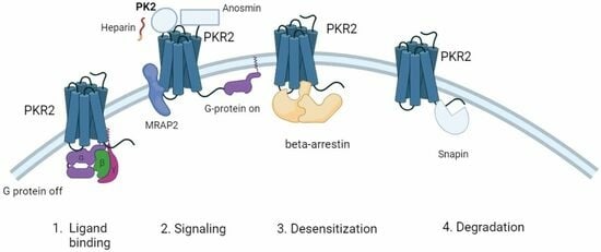
3. Proteins That Stabilize PKRs at the Cell Surface

Prokineticin 2, which binds to PKR1 and PKR2, functions as a novel adipokine that controls food intake [38,39] and plays a critical role in regulating thermoregulation and energy homeostasis by binding to PKR2. PK2 has an anorexic effect that acts at both a central and peripheral level. Centrally, PK2 activates arcuate nucleus neurons (ARC) to release melanocyte-stimulating hormones [38] and, peripherally, it impairs preadipocyte proliferation and their differentiation in adipocytes reducing the adipose tissue growth [40,41]. The balance between PK2 and PK2β regulates food intake in mice. Unlike PK2, the intraperitoneal injection of PK2β did not reduce food intake in mice [42].
The prokineticin and MRAP2 systems interact in the regulation of food intake. The melanocortin receptor accessory proteins (MRAP), containing MRAP1 and MRAP2, are able bind to GPCRs and modulate their trafficking and signal transduction pathway [43]. MRAP2 physically interacts with PKRs and, in this way, participates in the modulation of PKR activation that follows PK2 binding [44].
MRAP2 is a single transmembrane protein found on the cell surface and reticulum membrane. It can dimerize with a dual topology, i.e., parallel and anti-parallel orientations, making it a unique protein with this property in the eukaryote proteome [45]. MRAP2 is mainly expressed in the stomach and endocrine glands, hypothalamus, and adipocytes. It binds and modulates several GPCRs involved in the control of energy homeostasis, such as melanocortin-4 receptor (MC4R), orexin receptor 1, and ghrelin receptor [43]. The involvement of MRAP2 in the control of food intake is confirmed by the fact that mice in which MRAP2 is reduced develop severe early-onset obesity [46].
In vitro experiments showed that stimulation with PK2 induces a significant increase of MRAP2 expression in hypothalamic explants and in adipocytes, demonstrating an involvement of the prokineticin system in the regulation of MRAP in both central and peripheral tissues involved in the control of food intake.
In addition, MRAP2 has been shown to bind PKRs and regulate their plasmatic membrane localization and signal transduction [44].
Direct mutagenesis experiments have shown that glycosylation at position 27 is essential for PKR2 transport at the plasma membrane and that this posttranslational modification is not evident in the presence of MRAP2 [47]. MRAP2 binds the glycosylation sites in the N-terminal region of PKR2, reducing glycosylation of the receptor and thus its proper localization in the plasma membrane [48]. The C-terminal domain of MRAP2 is the most important domain for regulating GPCRs but little is known about its structure, although there is evidence that it contains a disordered secondary structure.
The hypothesis formulated to explain the ability of MRAP2 to interact with multiple protein partners is that the C-terminal region of MRAP2 adopts a more fixed structure only after binding to different GPCRs [45]. Biochemical dissection of the C-terminal MRAP2 domain allowed us to distinguish two distinct parts involved in modulating PKR2 function. One part, encompassing the region from amino acid position 76 to 141, is important for both transport and signal transduction, while the other part, encompassing the region from amino acid position 141 to 205, is important only for signal transduction [49]. In addition, the MRAP2-specific region critical for interaction with PKR2 and for the formation of MRAP2 dimers was identified and characterized; this region extends from amino acid position 78 to 131 [48]. Arginine, at position 125, was shown to play a critical role in protein conformation, dimer formation, PKR2 binding, and metabolic function. The fundamental role of this residue in MRAP2 function is confirmed by evidence that the substitution of the amino acid residue by histidine (R125H) or cysteine (R125C) is found in human patients with extreme obesity [50].

4. Proteins That Modulate Ligand Binding of PKRs

Anosmin-1 has been shown to modulate the binding of PK2 to PKR2 [51,52].
Anosmin-1, encoded by the KAL1 gene, is a glycoprotein of the extracellular matrix, a cell adhesion protein that shows a high degree of sequence identity in different species. Biochemical analysis of anosmin-1 identified six domains that the protein requires to bind its various partners and exert its biological effects: a cysteine-rich region (CR domain), an acid-like domain (WAP), four consecutive fibronectin-type domains III (FNIII), and a C-terminal region characterized by a high number of basic amino acid residues and prolines. The WAP domain is present in protease inhibitors and plays a role in neuronal genesis and migration. FNIII domains are contained in proteins such as tyrosine kinases and phosphatases, which are involved in cell adhesion, neuronal migration, and axon guidance. It also contains five potential heparan sulphate binding sites and six N-glycosylation sites. Anosmin-1 protein is present from embryonic development to adulthood and is highly expressed in the central nervous system as well as in the inner ear, kidney, testes, skin, and vascular endothelial cells [53].
Anosmin-1, PK2, and PKR2 are involved in the pathogenesis of Kallmann syndrome (KS). It is a genetic disease that manifests with hypogonadism and anosmia and is characterized by the involvement of numerous genes that determine several models of genetic inheritance. The pathology is caused by the disruption of the neuronal migration pathway shared by gonadotropin-releasing hormone (GnRH) neurons and olfactory neurons during embryonic development.
Currently, more than twenty pathogenic genes are associated with KS, six of which are relatively common: KAL1 (or ANOS1, the gene responsible for the X-linked form of KS, which encodes anosmin), FGFR1, CHD7, FGF8, PKR2, and PK2, which are involved in the autosomal transmission of KS [54,55].
In mice, PKR2 is widely distributed in the central nervous system, especially from the nuclei of the olfactory bulb to the brainstem [56]. Mutations in the Prok2 or Prokr2 genes lead to impaired development of the olfactory bulb and impaired migration of gonadotropin-releasing hormone (GnRH) neurons, resulting in reproductive organ atrophy and infertility [44,45].
These findings made PK2/PKR2 signaling deficits in mice a relevant preclinical model for KS, a combination of hypogonadotropic hypogonadism (HH) and a diminished (hyposmia) or absent (anosmia) sense of smell [57,58].
Anosmin directly interacts with the N-tail and extracellular loops 2 (ECL2) and 3 (ECL3) of PKR2 and plays a modulatory role in PKR2 function [51,52].
The binding of truncated anosmin to PKR2 enhances PK2-induced activation of the ERK1/2 (extracellular signal-regulated kinase 1/2) pathway [52].

5. Protein That Mediates Receptor Desensitization

Arrestin are a family of four homologous proteins consisting of two visual and two non-visual members (β-Arrestin 1 and β-Arrestin 2) that are ubiquitously expressed in most vertebrates and bind and regulate the activity of several hundred GPCRs. Once bound to a GPCR, arrestins exert at least three functions: (i) desensitization; they prevent further coupling of the receptor to G proteins, (ii) activation of transduction signals; they act as scaffolds for a variety of proteins involved in signaling pathways, such as mitogen-activated protein kinase cascades, Src family tyrosine kinases, and E3 ubiquitin ligases, (iii) internalization of the receptor; they facilitate the compartmentalization of the receptor in endosomes [59].
After ligand binding, an active GPCR is phosphorylated by one or more specialized GPCR kinases (GRKs), and the phosphorylated GPCR is then able to bind arrestins. Arrestins are elongated molecules consisting of two domains, commonly referred to as N- and C-domains. Remarkably, all four vertebrate arrestins have very similar structures in their basal conformation and bind to activated GPCRs in two different ways [59]. The first type of interaction involves the interaction of arrestin with the phosphorylated C-terminal region of GPCRs [60]. In the other, the finger loop region of arrestin is inserted into the cytoplasmic cavity formed by the transmembrane core of the GPCR, resulting in a high-affinity β-arrestin/GPCR complex [60]. The binding of arrestin to the activated form of phosphorylated receptors sterically prevents the coupling of G-proteins, resulting in the desensitization of the receptors [59].
The activation of PKR2 induces the recruitment of β-arrestin, as demonstrated in the bioluminescence resonance energy transfer (BRET) assay by fusing PKR2 C-terminally with yellow fluorescent protein (YFP) and by fusing β-arrestin with Rluciferase (Rluc). The BRET signal obtained was lower than that measured for other receptors such as PAR1 or V2R, suggesting that the interaction of PKR2 with arrestin is less efficient [61].
The interaction between PKR2 and β-arrestin is specific as the BRET ratio increases hyperbolically with the increase in BRET acceptor concentration [62]. Using BRET, it was possible to assess the binding of PKRs with the two isoforms of β-arrestin. These were made fluorescent and then transfected into HEK293 cells and into fibroblasts of knock-out mice for both isoforms to study binding to PKRs in the absence of endogenous β-arrestins. The results obtained show that both receptors, PKR1 and PKR2, are able to form complexes with β-arrestin 2 but not with β-arrestin 1 [61], although there is conflicting information in the literature about this PKRs/arrestin interaction. Conflicting results were obtained in another study using the BRET method in HEK293 cells. In this case, PKR2 was found to interact with the two β-arrestin isoforms with the same affinity but with a lower binding strength than other GPCRs [62]. Another study using the same cell line showed that PKR1 and PKR2 are unable to recruit β-arrestin 1 and β-arrestin 2 to the plasma membrane [63]. The ability of PKR2 to bind β-arrestin was also investigated in nine Kallman PKR2 mutants. No mutations significantly affected targeting to the cell surface for arrestin recruitment; however, only substituting arginine at position 80 in cysteine resulted in a complete absence of binding to arrestin [62].

6. Protein-Mediating Endocytosis of Receptors

Cells are protected from prolonged or repeated agonistic stimulation of GPCRs by the endocytosis of receptors from the cell membrane [64]. Clathrin-mediated endocytosis, using clathrin-coated cavities, is the predominant pathway of GPCR endocytosis in mammalian cells. Once internalized, the receptors can be recycled at the membrane or degraded at the lysosomal level.
In many cases, arrestin binds ligand-activated GPCRs by interacting with adaptor protein-2 (AP-2) on clathrin-coated pits. The binding of arrestins to GPCRs triggers a conformational change to reach the active conformation. In this process, the two arrestin domains are twisted in relation to each other and the C-terminus of arrestin is released, making the arrestin-binding site, the RXR-binding motif, accessible to adaptor protein 2 (AP-2). In addition to the role of AP-2 in arrestin-mediated endocytosis, sometimes a specific subunit of AP-2, m2-adaptin, can bind directly to the intracellular loops or to the carboxy tail of GPCRs, facilitating clathrin-mediated endocytosis in a manner independent of arrestins. GRKs, which are serine/threonine kinases that play a fundamental role in the binding of arrestin to GPCRs, are also involved in facilitating the binding of AP2 to the GPCR in the absence of arrestin. There are several pathways of internalization that depend on GRK phosphorylation but are independent of arrestin. This is the case for PKR2 endocytosis after PK2 binding, which has been shown to depend on GRK2 and clathrin but is independent of β-arrestin [64]. PKR2 trafficking is also regulated by interaction with snapin. Snapin, a binding protein of SNAP-25, stabilizes the SNARE complex formation, performing important facilitatory functions in synaptic transmission and hormone secretion. Indeed, snapin knock-down mice show impaired pre-synaptic vesicle fusion and release and hormone secretion. Snapin has been reported to promote lysosome biogenesis and the transport of endocytosed material toward lysosomes in non-neuronal cells, thus promoting degradation processes [65]. Moreover, the ability of snapin to interact with multiple partners, including the ryanodine receptor, aquaporins, TRPV1 and TRPC6 receptors, and the α1-subunit of the Cav1.3-Ca2+ channel, suggests several potential functions [65,66].
Recently, snapin was also shown to interact with GPCR, particularly the α1A-adrenoceptor [67] and PKR2 [68]. Yeast two-hybrid screening, GST pull-down, and co-immunoprecipitation studies revealed the interaction of snapin with PKR2 C-terminal (from amino acids 333 to 384) through two similar motifs characterized by two aromatic amino acids followed by a basic amino acid. The interaction between snapin and PKR2 does not affect PKR2 activation but induces its degradation upon ligand binding, suggesting a role for snapin in PKR2 transport [68].

7. Glycosaminoglycans and Their Interaction with Proteins

Glycosaminoglycans (GAGs) are linear complex carbohydrates with a molecular weight of 10–100 kDa that regulate many biological functions through interaction with various accessory proteins. Glycosaminoglycans are divided into nonsulfated (hyaluronic acid, HA) and sulfated (chondroitin sulphate CS, dermatan sulphate DS, keratan sulphate KS, heparin and heparan sulphate HS) and consist of disaccharide repeating units composed of uronic acid (D-glucoronic acid or L-iduronic acid) and amino sugars (D-galactosamine or D-glucosamine).
With the exception of HA, the simplest of the nonsulfated GAGs, all GAG chains in mammals are covalently bound to a core protein to form a proteoglycan, which is then incorporated into the plasma membrane.
The sulfated GAGs heparin and heparan sulphate, called glucosaminoglycans because they contain glucosamine, bind a variety of nuclear proteins to form HSPGs, (Heparan Sulfate Proteoglycans) which are the major components of the ECM (Extracellular Matrix) in mammals [69].
Proteoglycans include growth factors, antithrombin, cell adhesion molecules, cytokines, and chemokines. PK1, also known as EG-VEFG, has a consensus sequence specific for heparin binding (motif XBBXBX, where B stands for a basic amino acid) that allows it to bind with high affinity to heparin-Sepharose. EG-VEGF is a basic molecule with a pI of 8.6 that binds to heparin and can therefore be sequestered in the extracellular compartment in vivo [70,71]. This ability may be important for regulating its bioavailability and activity as has already been demonstrated for other angiogenic factors such as bFGF and VEGF. EG-VEGF is structurally unrelated to VEGF, but in certain cellular and tissue contexts, the functional activity of this molecule is indistinguishable from that of VEGF. In addition, PK2 and PK2β bind to heparin. PK2β binds with a higher affinity than PK2 because PK2L (the precursor of PK2β) has an additional region of 21 amino acids compared with PK2, that contains a heparin-binding protein consensus sequence. Heterologous yeast expression experiments have shown that heparin modulates PK2β activity on PKR1 [15] (Table 1).

8. Conclusions

Several proteins, like accessory proteins, have been identified as regulators of GPCRs capable of modulating receptor transport, signal transduction, and ligand specificity [72].
Protein–protein interactions (PPIs), defined as physical connections between proteins and their partners, play a central role in life processes. Interactions are a series of finely tuned recognition events that take place on protein surfaces and may happen with a variable strength and specificity of binding.
Studies have shown that various pathologies such as neurodegenerative, infectious, and oncological diseases are also due to the presence of aberrant and dysfunctional protein–protein interactions. Therefore, PPIs play a central role among the new molecular targets for the development of new pharmacological treatments.
The development of compounds capable of modulating protein–protein interactions is an important goal of modern drug discovery. The identification of the peptide motifs present in the PPI interface allows the synthesis and development of small PPI inhibitory peptides. These are particularly effective for interacting with proteins that are not completely folded in their isolated state but achieve conformational stability when they form complexes with other partners. The data suggest that these types of drugs show a lower risk of developing resistance, representing a major challenge in areas such as oncology and infectious diseases.
The modulation of PPIs is an extremely promising strategy for new drug development. Some molecules have already been tested in clinical trials and others have been brought to market, opening up new perspectives for pharmaceutical research [73].

Author Contributions

R.L. and R.M.: conceptualization, figures, design, writing and reviewing of the review. All authors have read and agreed to the published version of the manuscript.

Funding

This research received no external funding.

Institutional Review Board Statement

Not applicable.

Informed Consent Statement

Not applicable.

Conflicts of Interest

The authors declare no conflict of interest.

References

  1. Rosenbaum, D.M.F.; Rasmussen, S.G.F.; Kobilka, B.K. The structure and function of G-protein-coupled receptors. Nature 2009, 459, 356–363. [Google Scholar] [CrossRef] [PubMed]
  2. Soga, T.; Matsumoto, S.; Oda, T.; Saito, T.; Hiyama, H.; Takasaki, J.; Kamohara, M.; Ohishi, T.; Matsushime, H.; Furuichi, K. Molecular cloning and characterization of prokineticin receptors. Biochim. Biophys. Acta 2002, 1579, 173–179. [Google Scholar] [CrossRef]
  3. Lin, D.C.; Bullock, C.M.; Ehlert, F.J.; Chen, J.-L.; Tian, H.; Zhou, Q.Y. Identification and Molecular Characterization of Two Closely Related G Protein-coupled Receptors Activated by Prokineticins/Endocrine Gland Vascular Endothelial Growth Factor. J. Biol. Chem. 2002, 277, 19276–19280. [Google Scholar] [CrossRef] [PubMed]
  4. Masuda, Y.; Takatsu, Y.; Terao, Y.; Kumano, S.; Ishibashi, Y.; Suenaga, M.; Abe, M.; Fukusumi, S.; Watanabe, T.; Shintani, Y.; et al. Isolation and identification of EG-VEGF/prokineticins as cognate ligands for two orphan G-protein-coupled receptors. Biochem. Biophys. Res. Commun. 2002, 293, 396–402. [Google Scholar] [CrossRef] [PubMed]
  5. Negri, L.; Ferrara, N. The Prokineticins: Neuromodulators and Mediators of Inflammation and Myeloid Cell-Dependent. Angiogenesis. Physiol. Rev. 2018, 98, 1055–1082. [Google Scholar] [CrossRef] [PubMed]
  6. Lattanzi, R.; Maftei, D.; Fullone, M.R.; Miele, R. Identification and characterization of Prokineticin receptor 2 splicing TM4-7 variant and its modulation in an animal model of Alzheimer's disease. Neuropeptides 2019, 73, 49–56. [Google Scholar] [CrossRef] [PubMed]
  7. Li, M.; Bullock, C.M.; Knauer, D.J.; Ehlert, F.J.; Zhou, Q.Y. Identification of two prokineticin cDNAs: Recombinant proteins potently contract gastrointestinal smooth muscle. Mol. Pharmacol. 2001, 59, 692–698. [Google Scholar] [CrossRef]
  8. Mollay, C.; Wechselberger, C.; Mignogna, G.; Negri, L.; Melchiorri, P.; Barra, D.; Kreil, G. Bv8, a small protein from frog skin and its homologue from snake venom induce hyperalgesia in rats. Eur. J. Pharmacol. 1999, 374, 189–196. [Google Scholar] [CrossRef]
  9. Schweitz, H.; Pacaud, P.; Diochot, S.; Moinier, D.; Lazdunski, M. MIT (1), a black mamba toxin with a new and highly potent activity on intestinal contraction. FEBS Lett. 1999, 461, 183–188. [Google Scholar] [CrossRef]
  10. Ferrara, N.; LeCouter, J.; Lin, R.; Peale, F. EG-VEGF and Bv8: A novel family of tissue-restricted angiogenic factors. Biochim. Biophys. Acta 2004, 1654, 69–78. [Google Scholar] [CrossRef]
  11. LeCouter, J.; Kowalski, J.; Foster, J.; Hass, P.; Zhang, Z.; Dillard-Telm, L.; Frantz, G.; Rangell, L.; DeGuzman, L.; Keller, G.A.; et al. Identification of an angiogenic mitogen selective for endocrine gland endothelium. Nature 2001, 412, 877–884. [Google Scholar] [CrossRef]
  12. Lattanzi, R.; Miele, R. Prokineticin-Receptor Network: Mechanisms of Regulation. Life 2022, 12, 172. [Google Scholar] [CrossRef] [PubMed]
  13. Jilek, A.; Engel, E.; Beier, D.; Lepperdinger, G. Murine Bv8 gene maps near a synteny breakpoint of mouse chromosome 6 and human 3p21. Gene 2000, 256, 189–195. [Google Scholar] [CrossRef] [PubMed]
  14. Chen, J.; Kuei, C.; Sutton, S.; Wilson, S.; Yu, J.; Kamme, F.; Mazur, C.; Lovenberg, T.; Liu, C. Identification and pharmacological characterization of prokineticin 2 beta as a selective ligand for prokineticin receptor 1. Mol. Pharmacol. 2005, 67, 2070–2076. [Google Scholar] [CrossRef] [PubMed]
  15. Lattanzi, R.; Maftei, D.; Negri, L.; Fusco, I.; Miele, R. PK2β ligand, a splice variant of prokineticin 2, is able to modulate and drive signaling through PKR1 receptor. Neuropeptides 2018, 71, 32–42. [Google Scholar] [CrossRef] [PubMed]
  16. Lattanzi, R.; Maftei, D.; Vincenzi, M.; Fullone, M.R.; Miele, R. Identification and Characterization of a New Splicing Variant of Prokineticin 2. Life 2022, 12, 48. [Google Scholar] [CrossRef] [PubMed]
  17. Kaser, A.; Winklmayr, M.; Lepperdinger, G.; Kreil, G. The AVIT protein family. Secreted cysteine-rich vertebrate proteins with diverse functions. EMBO Rep. 2003, 4, 469–473. [Google Scholar] [CrossRef]
  18. Monnier, J.; Samson, M. Cytokine properties of prokineticins. FEBS J. 2008, 275, 4014–4021. [Google Scholar] [CrossRef]
  19. Lin, R.; LeCouter, J.; Kowalski, J.; Ferrara, N. Characterization of endocrine gland-derived vascular endothelial growth factor signaling in adrenal cortex capillary endothelial cells. J. Biol. Chem. 2002, 277, 8724–8729. [Google Scholar] [CrossRef]
  20. Gurevich, V.V.; Gurevich, E.V. How and why do GPCRs dimerize? Trends Pharmacol. Sci. 2008, 29, 234–240. [Google Scholar] [CrossRef]
  21. Kufareva, I.; Salanga, C.L.; Handel, T.M. Chemokine and chemokine receptor structure and interactions: Implications for therapeutic strategies. Immunol. Cell. Biol. 2015, 93, 372–383. [Google Scholar] [CrossRef]
  22. Monnier, C.; Dodé, C.; Fabre, L.; Teixeira, L.; Labesse, G.; Pin, J.P.; Hardelin, J.P.; Rondard, P. PROKR2 missense mutations associated with Kallmann syndrome impair receptor signalling activity. Hum. Mol. Genet. 2009, 18, 75–81. [Google Scholar] [CrossRef]
  23. Fullone, M.R.; Lattanzi, R.; Maftei, D.; Bonaccorsi, M.C.; Miele, R. Analysis of role of aromatic residues in extracellular loop 2 of Prokineticin receptor 2 in ligand binding probed with genetically encoded photo-crosslinkers. BBA Biomembr. 2021, 1863, 183549. [Google Scholar] [CrossRef]
  24. Levit, A.; Yarnitzky, T.; Wiener, A.; Meidan, R.; Niv, M.Y. Modeling of human prokineticin receptors: Interactions with novel small-molecule binders and potential off-target drugs. PLoS ONE 2011, 6, e2799. [Google Scholar] [CrossRef]
  25. Lattanzi, R.; Miele, R. Non-Peptide Agonists and Antagonists of the Prokineticin Receptors. Curr. Issues Mol. Biol. 2022, 44, 6323–6332. [Google Scholar] [CrossRef]
  26. Gasser, A.; Brogi, S.; Urayama, K.; Nishi, T.; Kurose, H.; Tafi, A.; Ribeiro, N.; Désaubry, L.; Nebigil, C.G. Discovery and cardioprotective effects of the first non-Peptide agonists of the G protein-coupled prokineticin receptor-1. PLoS ONE 2015, 10, e0121027. [Google Scholar] [CrossRef] [PubMed]
  27. Bullock, C.M.; Li, J.D.; Zhou, Q.Y. Structural Determinants Required for the Bioactivities of Prokineticins and Identification of Prokineticin Receptor Antagonists. Mol. Pharmacol. 2004, 65, 582–588. [Google Scholar] [CrossRef]
  28. Khusal, K.G.; Tonelli, R.R.; Mattos, E.C.; Soares, C.O.; Genova, D.B.M.; Juliano, M.A.; Urias, U.; Alves, M.J.M. Prokineticin receptor identified by phage display is an entry receptor for Trypanosoma cruzi into mammalian cells. Parasitol Res. 2015, 114, 155–165. [Google Scholar] [CrossRef]
  29. Lattanzi, R.; Miele, R. Versatile Role of Prokineticins and Prokineticin Receptors in Neuroinflammation. Biomedicines 2021, 9, 1648. [Google Scholar] [CrossRef]
  30. Lattanzi, R.; Severini, C.; Miele, R. Prokineticin 2 in cancer-related inflammation. Cancer Lett. 2022, 546, 215838. [Google Scholar] [CrossRef] [PubMed]
  31. Sarfati, J.; Dode, C.; Young, J. Kallmann syndrome caused bymutations in the PROK2 and PROKR2 genes: Pathophysiology and genotype-phenotype correlations. Front. Horm. Res. 2010, 39, 121–132. [Google Scholar]
  32. Shojaei, F.; Wu, X.; Zhong, C.; Yu, L.; Liang, X.H.; Yao, J.; Blanchard, D.; Bais, C.; Peale, F.V.; van Bruggen, N.; et al. Bv8 regulates myeloid-cell-dependent tumour angiogenesis. Nature 2007, 450, 825–831. [Google Scholar] [CrossRef]
  33. Traboulsi, W.; Brouillet, S.; Sergent, F.; Boufettal, H.; Samouh, N.; Aboussaouira, T.; Hoffmann, P.; Feige, J.J.; Benharouga, M.; Alfaidy, N. Prokineticins in central and peripheral control of human reproduction. Horm. Mol. Biol. Clin. Investig. 2015, 24, 73–81. [Google Scholar] [CrossRef] [PubMed]
  34. Hetz, C.; Zhang, K.; Kaufman, R.J. Mechanisms, regulation and functions of the unfolded protein response. Nat. Rev. Mol. Cell Biol. 2020, 21, 421–438. [Google Scholar] [CrossRef] [PubMed]
  35. Araki, K.; Nagata, K. Protein folding and quality control in the ER. Cold Spring Harb. Perspect. Biol. 2011, 3, a007526. [Google Scholar] [CrossRef] [PubMed]
  36. Song, Y.B.; Park, S.Y.; Park, K.; Kaiser, U.B. Trafficking-defective mutant PROKR2 cycles between endoplasmic reticulum and Golgi to attenuate endoplasmic reticulum stress. Proc. Natl. Acad. Sci. USA 2022, 119, e2102248119. [Google Scholar] [CrossRef] [PubMed]
  37. Chen, D.N.; Ma, Y.T.; Liu, H.; Zhou, Q.Y.; Li, J.D. Functional rescue of Kallmann syndrome-associated prokineticin receptor 2 (PKR2) mutants deficient in trafficking. J. Biol. Chem. 2014, 289, 15518–15526. [Google Scholar] [CrossRef] [PubMed]
  38. Gardiner, J.V.; Bataveljic, A.; Patel, N.A.; Bewick, G.A.; Roy, D.; Campbell, D.; Greenwood, H.C.; Murphy, K.G.; Hameed, S.; Jethwa, P.H.; et al. Prokineticin 2 is a hypothalamic neuropeptide that potently inhibits food intake. Diabetes 2010, 59, 397–406. [Google Scholar] [CrossRef]
  39. Beale, K.; Gardiner, J.V.; Bewick, G.A.; Hostomska, K.; Patel, N.A.; Hussain, S.S.; Jayasena, C.N.; Ebling, F.J.; Jethwa, P.H.; Prosser, H.M.; et al. Peripheral administration of prokineticin 2 potently reduces food intake and body weight in mice via the brainstem. Br. J. Pharmacol. 2013, 168, 403–410. [Google Scholar] [CrossRef]
  40. Szatkowski, C.; Vallet, J.; Dormishian, M.; Messaddeq, N.; Valet, P.; Boulberdaa, M.; Metzger, D.; Chambon, P.; Nebigil, C.G. Prokineticin receptor 1 as a novel suppressor of preadipocyte proliferation and differentiation to control obesity. PLoS ONE 2013, 8, e81175. [Google Scholar] [CrossRef]
  41. Qureshi, R.; Kindo, M.; Arora, H.; Boulberdaa, M.; Steenman, M.; Nebigil, C.G. Prokineticin receptor-1-dependent paracrine and autocrine pathways control cardiac tcf21 fibroblast progenitor cell transformation into adipocytes and vascular cells. Sci. Rep. 2017, 7, 12804. [Google Scholar] [CrossRef] [PubMed]
  42. Maftei, D.; Lattanzi, R.; Vincenzi, M.; Squillace, S.; Fullone, M.R.; Miele, R. The balance of concentration between Prokineticin 2β and Prokineticin 2 modulates the food intake by STAT3 signaling. BBA Adv. 2021, 1, 100028. [Google Scholar] [CrossRef] [PubMed]
  43. Berruien, N.N.; Smith, C.L. Emerging roles of melanocortin receptor accessory proteins (MRAP and MRAP2) in physiology and pathophysiology. Gene 2020, 757, 144949. [Google Scholar] [CrossRef] [PubMed]
  44. Chaly, A.L.; Srisai, D.; Gardner, E.E.; Sebag, J.A. The Melanocortin Receptor Accessory Protein 2 promotes food intake through inhibition of the Prokineticin Receptor-1. eLife 2016, 5, e12397. [Google Scholar] [CrossRef] [PubMed]
  45. Hinkle, P.M.; Sebag, J.A. Structure and function of the melanocortin2 receptor accessory protein (MRAP). Mol. Cell. Endocrinol. 2009, 300, 25–31. [Google Scholar] [CrossRef] [PubMed]
  46. Asai, M.; Ramachandrappa, S.; Joachim, M.; Shen, Y.; Zhang, R.; Nuthalapati, N.; Ramanathan, V.; Strochlic, D.E.; Ferket, P.; Linhart, K.; et al. Loss of Function of the Melanocortin 2 Receptor Accessory Protein 2 Is Associated with Mammalian Obesity. Science 2013, 341, 275–278. [Google Scholar] [CrossRef] [PubMed]
  47. Verdinez, J.A.; Sebag, J.A. Role of N-Linked Glycosylation in PKR2 Trafficking and Signaling. Front. Neurosci. 2021, 15, 730417. [Google Scholar] [CrossRef]
  48. Fullone, M.R.; Maftei, D.; Vincenzi, M.; Lattanzi, R.; Miele, R. Identification of Regions Involved in the Physical Interaction between Melanocortin Receptor Accessory Protein 2 and Prokineticin Receptor 2. Biomolecules 2022, 12, 474. [Google Scholar] [CrossRef]
  49. Rouault, A.A.J.; Lee, A.A.; Sebag, J.A. Regions of MRAP2 required for the inhibition of orexin and prokineticin receptor signaling. Biochim. Biophys. Acta Mol. Cell Res. 2017, 1864, 2322–2329. [Google Scholar] [CrossRef]
  50. Fullone, M.R.; Maftei, D.; Vincenzi, M.; Lattanzi, R.; Miele, R. Arginine 125 Is an Essential Residue for the Function of MRAP2. Int. J. Mol. Sci. 2022, 23, 9853. [Google Scholar] [CrossRef]
  51. Murcia-Belmonte, V.; Astillero-López, V.; Esteban, P.F. Anosmin 1 Interacts with the Prokineticin Receptor 2 In Vitro Indicating a Molecular Link Between Both Proteins in the Pathogenesis of Kallmann Syndrome. Protein Pept. Lett. 2016, 23, 650–655. [Google Scholar] [CrossRef] [PubMed]
  52. Murcia-Belmonte, V.; Tercero-Díaz, M.; Barrasa-Martín, D.; Lopez de la Vieja, S.; Munoz-Lopez, M.; Esteban, P.F. Anosmin 1 N-terminal domains modulate prokineticin receptor 2 activation by prokineticin 2. Cell. Signal. 2022, 98, 110417. [Google Scholar] [CrossRef] [PubMed]
  53. Choy, C.; Kim, S.-H. Biological actions and interactions of anosmin 1. Front. Horm. Res. 2010, 39, 78–93. [Google Scholar] [PubMed]
  54. Kemal Topaloglu, A.; Damla Kotan, L. Genetics of Hypogonadotropic Hypogonadism. Endocr. Dev. 2016, 29, 36–49. [Google Scholar]
  55. Hardelin, J.P.; Dodé, C. The complex genetics of Kallmann syndrome: KAL1, FGFR1, FGF8, PROKR2, PROK2; et al. Sex Dev. 2008, 2, 181–193. [Google Scholar] [CrossRef]
  56. Cheng, M.; Leslie, F.M.; Zhou, Q.-Y. Expression of prokineticins and their receptors in the adult mouse brain. J. Comp. Neurol. 2006, 498, 796–809. [Google Scholar] [CrossRef]
  57. Matsumoto, S.-I.; Yamazaki, C.; Masumoto, K.H.; Nagano, M.; Naito, M.; Soga, T.; Hiyama, H.; Matsumoto, M.; Takasaki, J.; Kamohara, M.; et al. Abnormal development of the olfactory bulb and reproductive system in mice lacking prokineticin receptor PKR2. Proc. Natl. Acad. Sci. USA 2006, 103, 4140–4145. [Google Scholar] [CrossRef]
  58. Ng, K.L.; Li, J.-D.; Cheng, M.Y.; Leslie, F.M.; Lee, A.G.; Zhou, Q.-Y. Dependence of olfactory bulb neurogenesis on prokineticin 2 signaling. Science 2005, 308, 1923–1927. [Google Scholar] [CrossRef]
  59. Lefkowitz, R.J.; Shenoy, S.K. Transduction of receptor signals by b-arrestins. Science 2005, 308, 512–517. [Google Scholar] [CrossRef]
  60. Zhou, X.E.; He, Y.; de Waal, P.W.; Gao, X.; Kang, Y.; Van Eps, N.; Yin, Y.; Pal, K.; Goswami, D.; White, T.A.; et al. Identification of phosphorylation codes for arrestin recruitment by G protein-coupled receptors. Cell 2017, 170, 457–469. [Google Scholar] [CrossRef]
  61. Casella, I.; Ambrosio, C. Prokineticin receptors interact unselectively with several G protein subtypes but bind selectively to -arrestin 2. Cell. Signal. 2021, 83, 110000. [Google Scholar] [CrossRef]
  62. Sbai, Q.; Monnier, C.; Dodé, C.; Pin, J.-P.; Hardelin, J.-P.; Rondard, P. Biased signaling through G-protein-coupled PROKR2 receptors harboring missense mutations. FASEB J. 2014, 28, 3734–3744. [Google Scholar] [CrossRef]
  63. Yin, W.; Liu, H.; Peng, Z.; Chen, D.; Li, J.; Li, J.-D. Mechanisms that underlie the internalization and extracellular signal regulated kinase 1/2 activation by PKR2 receptor. Cell. Signal. 2014, 26, 1118–1124. [Google Scholar] [CrossRef]
  64. Pathak, C.; Vaidya, F.U.; Waghela, B.N.; Jaiswara, P.K.; Gupta, V.K.; Kumar, A.; Rajendran, B.K.; Ranjan, K. Insights of Endocytosis Signaling in Health and Disease. Int. J. Mol. Sci. 2023, 24, 2971. [Google Scholar] [CrossRef]
  65. Ilardi, J.M.; Mochida, S.; Sheng, Z.H. Snapin: A SNARE-associated protein implicated in synaptic transmission. Nat. Neurosci. 1999, 2, 119–124. [Google Scholar] [CrossRef]
  66. Han, J.; Pluhackova, K.; Böckmann, R.A. The Multifaceted Role of SNARE Proteins in Membrane Fusion. Front. Physiol. 2017, 8, 5. [Google Scholar] [CrossRef]
  67. Fumiko Suzuki, F.; Shigeru Morishima, S.; Takashi Tanaka, T.; Ikunobu Muramatsu, I. Snapin, a New Regulator of Receptor Signaling, Augments 1a-Adrenoceptor-operatedCalcium Influx through TRPC6. J. Biol. Chem. 2007, 282, 29563–29573. [Google Scholar] [CrossRef]
  68. Song, J.; Li, J.; Liu, H.; Liu, W.; Feng, Y.; Zhou, X.T.; Li, Y.D. Snapin interacts with G-protein coupled receptor PKR2. Biochem. Biophys. Res. Commun. 2016, 469, 501–516. [Google Scholar] [CrossRef]
  69. Gandhi, N.S.; Mancera, R.L. The Structure of Glycosaminoglycans and their Interactions with Proteins. Chem. Biol. Drug Des. 2008, 72, 455–482. [Google Scholar] [CrossRef]
  70. LeCouter, J.; Ferrara, N. The role of EG-VEGF in the regulation of angiogenesis in endocrine glands. Cold Spring Harb. Symp. Quant. Biol. 2002, 67, 217–221. [Google Scholar] [CrossRef]
  71. LeCouter, J.; Ferrara, N. EG-VEGF and the concept of tissue-specific angiogenic growth factors. Cell Dev. Biol. 2002, 13, 3–8. [Google Scholar] [CrossRef] [PubMed]
  72. Kotliar, I.B.; Lorenzen, E.; Schwenk, J.M.; Hay, D.L.; Sakmar, T.P. Elucidating the Interactome of G Protein-Coupled Receptors and Receptor Activity-Modifying Proteins. Pharmacol. Rev. 2023, 75, 1–34. [Google Scholar] [CrossRef] [PubMed]
  73. Haiying Lu, H.; Qiaodan Zhou, Q.; He, J.; Jiang, Z.; Peng, C.; Tong, R.; Shi, J. Recent advances in the development of protein–protein interactions modulators: Mechanisms and clinical trials. Signal Transduct. Target. Ther. 2020, 5, 213. [Google Scholar]
Figure 1. Schematic representation of PKR2 topology. Created in BioRender.com.
Encyclopedia 03 00107 g001
Figure 2. Illustration of the synthesis and localization of prokineticin receptors. Created in BioRender.com.
Encyclopedia 03 00107 g002
Table 1. Accessory proteins that modulate PKRs.
Interacting ProteinFunctionsPKR2 Site of InteractionReferences
BiPER quality control systemND[34]
gp78ER quality control systemND[34]
RER1ER quality control systemND[34]
MRAP2Down regulation of PKR s trafficking and signal transduction pathwaysN-terminal region[42,45,46,47,48]
Anosmin-1Modulation of PK2 binding on PKR2N-terminal regionExtracellular loop 2Extracellular loop 3[49,50]
beta-arrestin 2DesensibilizationND[59,60]
Snapin Degradation C-terminal region[66]
Disclaimer/Publisher’s Note: The statements, opinions and data contained in all publications are solely those of the individual author(s) and contributor(s) and not of MDPI and/or the editor(s). MDPI and/or the editor(s) disclaim responsibility for any injury to people or property resulting from any ideas, methods, instructions or products referred to in the content.

