Review

Trace Elements and Their Management in Dialysis Patients—Pathophysiology and Clinical Manifestations

Department of Nephrology, Tokushima University Graduate School of Biomedical Sciences, 3-18-15 Kuramoto-cho, Tokushima 770-8503, Japan
Kidney Dial. 2023, 3(3), 274-296; https://doi.org/10.3390/kidneydial3030025
Submission received: 18 May 2023 / Revised: 25 June 2023 / Accepted: 16 August 2023 / Published: 21 August 2023

Abstract

:
Recently, as the number of elderly dialysis patients has been increasing, complications associated with low nutritional status such as infectious disease have had a strong influence on the prognosis of dialysis patients. Nutritional disorders are caused by the inadequate intake of the three major nutrients—proteins, fats, and carbohydrates—as well as vitamin and mineral deficiencies. Minerals are composed of various elements, including small-amount elements and trace elements, which are present in the human body in very small quantities lower than that of iron. In dialysis and predialysis patients, zinc, manganese, and selenium are the three major elements that are significantly depleted as compared to normal subjects; these deficiencies are sometimes symptomatic. Zinc deficiency is manifest as anemia, taste abnormality, and delayed wound healing, while selenium deficiency is associated with impaired cardiac function and immunocompromised condition. Zinc has multiple functions, since various enzymes, including DNA polymerase and RNA polymerase, need zinc as a cofactor, while selenium is a component of selenoproteins, including glutathione peroxidase and thioredoxin reductases, which are major antioxidative stress enzymes. These elements can only be supplemented exogenously and contribute to the sustainable QOL of dialysis patients. On the other hand, as regards other trace elements, including copper, chromium, manganese, lead, arsenic, etc., the association of their deficiency or intoxication with various involvements of dialysis patients were investigated, although all investigations were performed in cross-sectional studies or observational studies. Therefore, the supplementation of these elements is inconclusive, given the scarcity of other intervention studies. More conclusive studies are endorsed for the establishment of proper supplementation strategies.

1. Introduction

In recent years, the concept of nutritional management for dialysis patients has been changing, and the number of elderly dialysis patients has been increasing. This is due to the fact that the complications associated with low nutritional status, such as sarcopenia and frailty, have a strong influence on the prognosis of dialysis patients, and concerns about nutritional disorders caused by excessive dietary restriction have become apparent. Nutritional disorders are caused by the inadequate intake of the three major nutrients—proteins, fats, and carbohydrates—as well as vitamin and mineral deficiencies, which cannot be ignored. In this article, we focus on trace elements among minerals, especially zinc and selenium, which have attracted considerable attention in recent years.

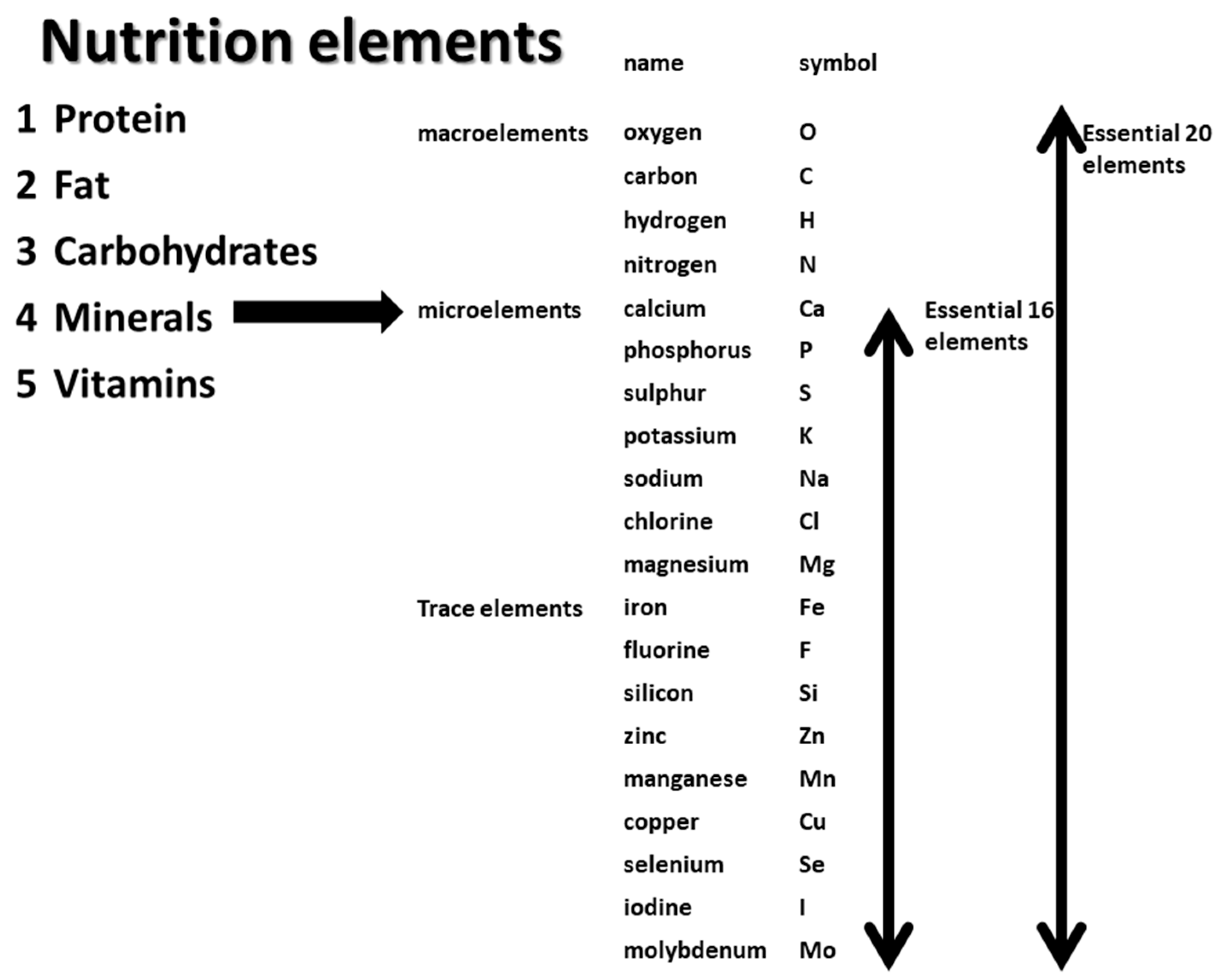
2. What Are Trace Elements?

There are currently 118 known elements on earth, and all substances and objects are composed of these elements. The human body is also composed of various elements. In terms of its composition, 60% of the human body is water (H2O), with the largest content of oxygen, which makes up 90% of the body. Next is C, which makes up organic matter, and H is the third largest by weight, although it is present in large quantities as a molecule because of its small atomic weight of 1. Next is N, which makes up the amino group of amino acids. O, C, H, and N are called macroelements (Figure 1). The largest elements with smaller amounts are Ca and P, which are constituents of bone, and together, they account for 99% of the human body. In contrast, the term mineral is often confused with element. Mineral comes from “mine” and “mineral mine” and is used as a synonym for metal in ordinary daily life but it is used with a different meaning as a term related to nutrition. In other words, proteins, lipids, and carbohydrates are the three major nutrients, and the remaining two become minerals and vitamins, meaning an inorganic substance, which is a general term for elements other than O, C, H, and N, as pointed out earlier. Among them, Ca, P, S, K, Na, Cl, and Mg are contained in relatively large amounts in living organisms and are called small-amount elements among minerals and measured in clinical practice in blood collection tests. In contrast, minerals with lower contents, i.e., Fe and below, are trace elements: Fe, F, Si, Zn, Mn, Cu, Se, I, and Mo. These small-amount elements are called trace elements, and together, they are referred to as the 16 essential elements of minerals. From a nutritional point of view, minerals are not necessarily metals, such as Fe, Zn, and Cu, i.e., substances that are hard at room temperature and have luster. In addition to these metallic elements, F, Se, and I are minerals, although they are nonmetallic elements.
Trace elements or minerals are important for four reasons. First, they are structural materials that form the bones and teeth; Ca, P, and Mg are known to make up bones, and F makes up teeth. Secondly, they are activators of enzymes in the body; Zn, Mn, Cu, Mg, and Ca are examples of this, and I makes up part of the thyroid hormones. I is part of the thyroid hormones, and some proteins (selenoproteins and heme proteins) are part of functional proteins. Thirdly, Na, K, and Cl are ions in body fluids, both intracellularly and extracellularly, that maintain the function of various organs. Finally, minerals do not contain calories, and they can only be obtained from food because they cannot be synthesized in the body.

3. Deficiency of Trace Elements

The importance of these minerals and trace elements is due to the fact that minerals and trace elements are decreasing in the Earth’s soils; data presented at the Earth Summit in Rio de Janeiro in 1992 indicate that the amount of minerals in the world’s soils has decreased over the past 100 years. This is probably due to the abuse of chemical fertilizers. As a result, trace elements in soils and crops have been depleted, with the end result that mineral intakes have decreased and requirements are not being met. Apart from dietary intake, deficiency and excess of trace elements are known to occur in renal failure and dialysis. The blood concentrations of trace elements in renal failure, hemodialysis, and peritoneal dialysis patients are higher than those in normal subjects for many trace metals, including Cr, Mo, and Si, and nonessential elements, such as cobalt, Ni, vanadium, strontium, and Cd. This is because these elements are mainly excreted by the kidneys. In contrast, there are some elements that become deficient, the most common of which are zinc and selenium (Table 1).

4. Zinc Deficiency in Dialysis Patients

4.1. Zinc and Zinc Deficiency

Zn is at the center of the activity of various enzymes and acts as an essential factor in their activity. Zinc is taken up by insulin-secreting beta cells in the pancreas to regulate insulin secretion. It is also a component of retinol-binding protein, which is a protein that transports vitamin A in the blood. It is an activator of enzymes involved in DNA synthesis and RNA synthesis and is therefore involved in cell division. Therefore, it is involved in wound healing. In recent years, zinc has been attracting attention because of its importance for the maintenance of taste bud cells on the tongue, which detect taste. It is also involved in the synthesis of the male sex hormone, testosterone. Zinc is involved in a wide variety of enzymatic reactions in the body, and Al-p, which is also measured in blood tests, is well-known. RNA polymerase and DNA polymerase are particularly important, as they are involved in DNA replication and transcription and are deeply involved in cell division and cell regeneration. The resulting symptoms of zinc deficiency are manifold. The skin is an important tissue for wound healing, tissue regeneration, and metabolism. Vitamin A is also important, and its deficiency causes dermatitis and stomatitis. Hair root cells also actively divide, and hair loss is observed. The intestinal epithelium is also actively regenerates and divides, and a zinc deficiency causes a loss of appetite. Laboratory findings include low blood zinc levels and low levels of alkaline phosphatase, the enzyme primarily responsible for its activity. Zinc deficiency anemia is also observed. Last year, the Japanese Society of Clinical Nutrition developed diagnostic criteria for zinc deficiency (Figure 2). A suspected case is defined as one in which one or more of the clinical symptoms and low alkaline phosphatase levels are observed, other diseases are ruled out, blood levels are less than 60 μg/dL, and symptoms improve with supplementation [1].

4.2. Zinc Deficiency in Dialysis Patients

Zinc deficiency occurs in a variety of pathological conditions, among which dialysis patients are the most common. The causes of zinc deficiency in dialysis patients are (1) elimination by dialysis, (2) dietary restriction, (3) hypoproteinemia, (4) decreased absorption in the small intestine, (5) increased consumption due to increased oxidative stress, and (6) adsorption by adsorbents and ion exchange resins. It is known that cation exchange resins used for the correction of hyperpotassemia adsorb mineral ions other than K. A 2009 study on the effect of zinc supplementation demonstrated that the serum zinc levels of zinc-supplemented individuals and non-zinc-supplemented individuals were 63.29 ± 9.92 mg/dL and 68.07 ± 12.57 mg/dL, respectively, demonstrating that supplementation improved anemia [2].
Several symptoms associated with anemia in dialysis patients have been documented regarding zinc deficiency. In one study, patients on HD with low serum zinc levels (<65 μg/dL) were randomly assigned to two groups: a polaprezinc group (who received daily polaprezinc containing 34 mg/day of zinc) (n = 35) and a control group (no supplementation) (n = 35) for 12 months. In the polaprezinc group, erythropoiesis-stimulating agent dosage and erythropoiesis resistance (ERI) were significantly decreased at 10 months and 9 months, respectively, as compared with the baseline value. Multiple stepwise regression analysis revealed that the change in the serum zinc level was an independent predictor of lowered ERI. In conclusion, zinc supplementation reduces ERI in patients undergoing HD and may be a novel therapeutic strategy for patients with renal anemia and low serum zinc levels [3]. It has also been reported that Zinc sulfate ameliorates pruritus in patients on maintenance hemodialysis [4]. In the study, a double-blind, randomized, placebo-controlled trial was conducted on 40 adults with end-stage renal disease (ESRD) who were on maintenance hemodialysis. Patients were randomized to receive either zinc sulfate (440 mg/day) or placebo for two consecutive months. The authors reported that zinc sulfate was more effective than the placebo for the relief of pruritus. The main mechanism of this effect is considered to be an inhibitory effect exerted by zinc on histamine release from mast cells. Two other RCTs showed that zinc supplementation was more effective at reducing itching than placebo or hydroxyzine [5,6]. Zinc deficiencies and high serum histamine levels have also been observed in itching patients with ESRD. The sense of taste is also affected by zinc, which plays an important role in the proliferation and maintenance of sensory neuron cells in taste buds in the tongue. The role in the sense of taste has long been recognized among HD patients in non-Asian countries. Two studies examined the effects of Zn replacement on hypogeusia, which refers to diminished sensitivity to detect a specific taste quality or class of compounds [7,8]. In a recent study, the authors divided patients on hemodialysis into two groups based on serum zinc concentration. Salt taste acuity and preference were determined by a sensory test using varying concentrations of NaCl solution, and dietary sodium intake was estimated using 3-day dietary recall surveys. They found that the mean salt recognition threshold and salt taste preference were significantly higher in the zinc-deficient group than in the non-zinc-deficient group. They also reported that there was a significant positive correlation between salt taste preference and dietary sodium intake in the zinc-deficient group. In addition, interdialytic weight gain was significantly higher in the zinc-deficient group than in the non-zinc-deficient group [9]. Finally, Zinc plays an important role in regulating every phase of the wound-healing process, ranging from membrane repair, oxidative stress, coagulation, inflammation, immune defense, tissue re-epithelialization, and angiogenesis to fibrosis/scar formation. Several clinical studies have stressed the importance of zinc in skin ulcer treatment. Although not in the case of dialysis patients, a randomized, double-blind, placebo-controlled trial was conducted regarding the treatment of diabetic foot ulcers [10]. The authors demonstrated that 220 mg zinc sulfate supplementation for 12 weeks had beneficial effects on parameters of ulcer size and metabolic profiles among diabetic foot ulcer patients. A more recent study investigated the relationship between zinc deficiency and clinical outcome in patients with critical limb ischemia [11]. However, in a retrospective observational study of a de novo infrainguinal bypass grafting operation, the authors found that patients in the Zn deficiency group were more likely to have undergone hemodialysis and that graft patency, limb salvage, amputation-free survival, and complete wound-healing rates were significantly lower in the Zn-deficient group. Finally, another report showed that zinc deficiency is related to mortality in dialysis patients [12]. Zinc plays an important role in immune systems and the treatment and prevention of infectious disease. A clinical study reported that in long-term dialysis patients, the serum level of zinc was an independent predictor of future hospitalization due to infectious diseases and of overall mortality. The authors enrolled 111 patients on maintenance dialysis and measured serum levels of selenium, copper, and zinc. Patients were followed for 2 years or until death or withdrawal. Multivariate Cox regression analysis indicated that zinc deficiency (HR, 0.979; 95% CI, 0.966–0.992; p = 0.002) were more likely to be hospitalized for infectious diseases. Multivariate Cox regression also indicated low serum levels of zinc independently predict overall mortality. The survival effects of Zn supplementation are considered to be due to its anti-inflammatory and antioxidative effects. Recently, two meta-analyses including intervention studies conducted in both Western and non-Western countries revealed that Zn replacement results in the reduction in serum CRP levels, as well as MDA concentration, a serum oxidative stress marker [13,14]. More recently, it was demonstrated that Zn sulfate supplementation has favorable effects on CRP, fasting blood glucose, and renal function in Zn-deficient diabetic hemodialysis patients [15].

4.3. Treatment of Zinc Deficiency

A list of these trials is presented in Table 2 [3,15,16,17,18,19,20,21,22,23,24,25,26,27,28]. Zinc preparations such as zinc acetate (Novelzine®) and polaprezinc (Promax®) have been used for zinc supplementation. In Japan, a Zn acetate hydrate tablet containing 50 mg of Zn (Nobelpharma Co., Ltd., Tokyo, Japan) is administered orally after each meal (three times daily) (150 mg/day) for the treatment Zinc deficiency. However, in light of the aging population in recent years, the problem is overwhelmingly one of reduced zinc intake due to dietary restrictions and reduced dietary intake. Therefore, dietary supplementation is the best option. Foods rich in zinc include oysters, pork, and beef. Although these foods contain protein, the dietary intake of the elderly is low, so restricting protein intake because of renal failure is likely to result in a deficiency. Another important issue is that the supplementation dose does not always have effects in clinical practice. Therefore, Zn concentration should be evaluated according to serum protein or albumin concentration, since most Zn is bound to albumin or α2-microglobulin in the blood. In addition, Zn is present in the intracellular space of the whole body. Additional evaluation methods need to be explored.

5. Selenium and Selenium Deficiency

5.1. Selenium and Selenium Deficiency

Selenium (Se) is also an important trace element for the maintenance of life. Its functions are described as follows: (1) it promotes the immune system, especially to maintain cellular immunity; (2) it inhibits cancer; (3) it is a constituent of glutathione peroxidase, a representative antioxidant enzyme, and has antioxidant effects; and (4) it is involved in the synthesis and degradation of thyroid hormones. Selenium is a mineral and a trace element but not a metal element. Selenium is a homologous element of sulfur; therefore, selenoamino acids exist in place of amino acids containing sulfur, including selenomethionine and selenocysteine. t-RNA that carries these amino acids, and selenoamino acids are incorporated into proteins, i.e., selenoproteins. In humans, 25 types of selenoproteins have been found (Table 3), major of which are the glutathione peroxidase family, thioredoxin reductase, and iodothyronine deiodinase.
The American Society for Parenteral and Enteral Nutrition has defined symptoms of selenium deficiency as cardiomyopathy, myalgia, myositis, hemolysis, cellular immune disorders, nail and hair abnormalities, large cell changes in red blood cells, and anemia. Nail and skin abnormalities are also observed due to the involvement of selenium in DNA synthesis. In 2017, diagnostic criteria for selenium deficiency were developed (Figure 3) [29]. The presence of any one clinical symptom can be recognized by changes such as whitening, deformity, dermatitis, and alopecia in the nails and skin; cardiomyopathy and abnormalities of the conduction system in myocardial disorders; muscle weakness and myalgia in the lower extremities in muscular disorders; and macrocytic anemia in blood data. Laboratory findings may include abnormal thyroid hormones, abnormal muscle enzymes, and liver abnormalities. Electrocardiogram changes may also be present. If one or more of these symptoms and laboratory findings is present, other diseases can be ruled out, the serum selenium level is low, and the patient is considered suspicious and should be considered for supplementation. If the symptoms improve with supplementation, the case is confirmed. The standard serum concentration is 10 µg/dL or 100 pg/mL in adults.

5.2. Selenium Deficiency in Dialysis Patients

Dialysis patients are known to be a risk population for selenium deficiency. Possible causes include decreased selenium intake due to decreased dietary protein intake, decreased selenium-binding proteins, increased selenium requirements, increased urinary excretion, loss from dialyzer membranes, and altered distribution of selenium in the body. There are few case reports of selenium deficiency up to supplementation in dialysis patients. Myocardial symptoms were the main symptoms, with concentrations of less than 2.5 μg/dL and 8 μg/dL, both of which recovered after 3–4 months of supplementation [30,31]. In contrast, a very detailed cohort study of patients on dialysis as a whole was reported in Iwate Prefecture in Japan in 2011 [32]. Blood levels were measured in a multicenter cohort of dialysis patients, and the overall mean was significantly lower in dialysis patients than in healthy controls, with a median value of 10.3 μg/dL. When the cohort was divided into four groups based on serum selenium levels, BMI and serum albumin levels were significantly correlated. The group with the lowest serum selenium concentration had a significantly lower survival rate than the other groups in all-cause and infectious disease mortality [33]. Recently, studies on selenium and life expectancy have been reported in other countries. A study conducted in Alberta, Canada, reported the results of routine measurements of 25 trace elements [34]. Data on 25 trace elements were collected from 1278 multicenter hemodialysis patients. This cohort was followed-up for 2 years to determine which trace elements were associated with death, cardiovascular accidents, systemic infections, and hospitalizations. In 2 years, there were 260 (20%) deaths, 285 (24%) cardiovascular accidents, 117 (10%) systemic infections, and 928 (77%) hospitalizations. When investigating the relationship between 25 trace elements, low selenium levels were significantly associated with death and total hospitalization. Whereas high levels of copper and cadmium were associated with death, low levels of zinc and magnesium and high levels of lead, arsenic, and mercury were not associated with death or hospitalization, contrary to our expectations. In dialysis retrospective observational cohort study conducted in Spain that included 85 patients with ESRD on three modalities of dialysis, selenium was considered to be closely related to death, with a plasma selenium test performed 5–6 years before the study. Patients with low selenium showed an increased risk of all-cause mortality (hazard ratio, 2.952) compared with patients with normal or high selenium [35]. Although these data were observational and other important factors may contribute to the mortality of dialysis patients, these reports from three independent cohorts imply that selenium has some roles in the prognosis of dialysis patients.
In relation to mortality, special attention should be paid in terms of the causal relationship between infection and selenium status. As previously stated, in a hemodialysis cohort study conducted in Iwate prefecture in Japan, complication with infectious disease affected the selenium status, and supplementation of this element was found to help to ameliorate the condition in infectious disease. More recently, the relationship between COVID-19 infection and selenium deficiency was investigated [36]. Chinese cohort surveillance revealed an association between the reported cure rates for COVID-19 and selenium status in one city. Antiviral effects of selenium have been reported previously, and multiple cellular and viral mechanisms involving selenium and selenoproteins may influence viral pathogenicity, including virally encoded selenium-dependent glutathione peroxidases. Furthermore, a meta-analysis consisting of a total of 13 RCTs comparing selenium and placebo for patients with sepsis were reported [37]. The analyses could not detect the association of selenium treatment with a decreased mortality at different time courses. Selenium supplementation did not show a favorable effect on the incidence of renal failure, secondary infection, or duration of mechanical ventilation. However, the study found that selenium therapy was a benefit for sepsis patients, with reduced duration of vasopressor therapy, time in the intensive care unit and hospital, and incidence of ventilator-associated pneumonia.
Cardiovascular complications are also important causes of death in dialysis patients. The relationship between selenium and cardiovascular diseases has been explored since the famous selenium-deficient disease in which Kashan disease was presented with symptoms of dilated cardiomyopathy. A randomized control trial was performed in France in 1989 in which the effects of oral treatment with 500 μg selenium for 3 months and 200 μg for the next 2 months were tested in terms of cardiac function. As compared with the placebo control, the IVS (interventricular septum) in an echocardiogram decreased, although the cardiac function did not change [38]. In another study, a correlation between decreased serum selenium levels and coronary flow reserve was examined as an indicator of endothelial dysfunction and atherosclerosis in HD patients. Serum selenium levels and coronary flow reserve values were significantly lower in hemodialysis patients compared with controls. There was a significant positive correlation between coronary flow reserve and serum levels of selenium. A linear regression analysis showed that serum levels of selenium were independently and positively correlated with coronary flow reserve [39].
The association between selenium and anemia has been shown in hemodialysis patients. A cross-sectional study was performed, and serum selenium levels were determined in 173 hemodialysis patients. The association of serum selenium with the responsiveness to erythropoiesis-stimulating agents, as defined by the ESA resistance index, was analyzed. First, the study showed that 50% of the subjects had lower selenium levels than the population-based reference values. The authors also found that serum selenium levels were significantly and inversely correlated with the erythropoiesis resistance index (ERI) but not transferrin saturation (TSAT) or ferritin levels. Moreover, an independent association between selenium levels and ESA hyporesponsiveness was detected in multiple regression analyses. When patients were divided according to selenium levels and iron status, both low serum selenium (<10.5 μg/dL) and iron deficiency significantly affected the response to ESA. Finally, the association of low serum selenium with ESA hyporesponsiveness persisted after adjustment of confounding variables [40].
Finally, the association between blood trace element levels and sleep quality in patients on maintenance hemodialysis was reported. This cross-sectional and single-center study performed in 2019 examined sleep quality in 121 enrolled HD patients with the use of the Pittsburgh Sleep Quality Index, which revealed an association between low blood selenium levels and the occurrence of severe sleep disturbances. [41].

5.3. Treatment of Selenium Deficiency in Dialysis Patients

Selenium supplementation in dialysis patients has been attempted for a long time. A lists of these trials is presented in Table 4 [38,42,43,44,45,46,47,48,49,50,51]. The treatment protocol has not been well-determined and varies, including intravenous treatment with 400 mg of sodium selenite. In Japan, sodium selenite solution containing of 100 μg of selenium (Fujimoto Co., Ltd., Osaka, Japan) is administered by daily infusion for the treatment selenium deficiency. In almost all studies, serum selenium concentration and glutathione peroxidase (GPx) activity in serum or red blood cells increased, and several biological markers were improved, including oxidative stress. In a more recent studies, selenium tablets or capsules are frequently used in trials. In 2013 one randomized, double-blind, placebo-controlled trial was conducted using selenium (200 µg) or a placebo capsule daily for 12 weeks [47]. The authors observed that the nutritional condition index, subjective global assessment (SGA) score, the systemic inflammation and nutrition condition score, and the malnutrition inflammation score (MIS) decreased significantly in the selenium group compared to the placebo group. Moreover, serum levels of malondialdehyde (MDA), an oxidative stress marker, decreased significantly in the selenium group compared with increasing levels in the placebo group. Selenium supplementation also hindered an increase in IL-6 levels compared with the placebo group. It can be concluded that selenium may be an effective complementary supplement for reducing the severity of malnutrition in HD patients by alleviating oxidative stress and inflammation.

5.4. Several Controversies about Selenium Supplementation

5.4.1. Optimal Serum Concentration of Selenium

A therapeutic threshold should be established with respect to the effectiveness of serum selenium concentrations. A U-shaped phenomenon is observed for the optimal selenium concentration. Thus, selenium should not be supplemented at overdosage levels. A previous study, selenium therapy had no effects on subjects with normal selenium concentration, and only half of the subjects exhibited an increase in GPx activity [52]. On the other hand, anticancer effects were not evident in patients with very low concentrations of selenium.

5.4.2. The Tissue Distribution of Selenium

As selenium functions are incorporated in the selenoprotein enzyme, the actual physiological effects may not be evaluated by the serum concentration. To overcome this problem, the activity of GPX in blood cells can be utilized as an alternative for the assessment of systemic selenium condition. In this regard, the normal range of serum concentration should be carefully considered.

5.4.3. Gene Polymorphism of Selenoprotein

Some SNPs or mutation on selenoproteins have been reported so far, which reduce the therapeutic effects of supplementation treatment. In these patients, supplementation is of little clinical relevance.

5.4.4. The Unique Bioavailability of Selenium

The unique characteristics of selenium bioavailability make its assessment complicated. Selenium is incorporated in selenoproteins and mobilized by a specific genetic code. In this regard, the adequate evaluation of selenium function is complex.
As selenium deficiency has some significance in the QOL and mortality of patients in end-stage renal disease, an appropriate method for assessment of selenium condition including measurement of serum concentration and cellular activity of GPx should be developed in the future. Considering its role of survival effects, supplementation should be limited to severely compromised patients, including sepsis, SIRS, severe heart failure, or end-stage kidney disease patients. Moreover, the supplementation regimen should be carefully determined because overdose treatment has either no effects or adverse effects. Selenium is an essential and vital element that contributes to mortality, and its supplementation can open a novel path to nutrition support therapy. As selenium plays a pivotal role in the cardiovascular system and defense against infectious disease, clinical trials targeted at the patients with acute heart failure, septic shock, or acute kidney injury are optimal to confirm the efficacy of selenium intervention.

6. Copper

Plasma Copper (Cu) levels can be of clinical relevance in relation to Zn deficiency. In HD patients, as described above, the plasma Zn concentration decreases, and improper Zn supplementation can lead to Cu deficiency, since Zn antagonizes the uptake of divalent cations, including iron (Fe) and Cu, in erythrocyte precursors. Cu is required for Fe transfer from cells to blood, ensuring dietary Fe absorption and systemic Fe distribution [53]. Cu/Zn superoxide dismutase (Cu/Zn-SOD), an antioxidant enzyme, is decreased in Cu-deficient subjects, which may accelerate oxidation reactions and shorten the life span of erythrocytes. In these mechanisms, Cu deficiency may lead to refractory anemia in HD patients, and its correction can improve erythropoietin non-responsive anemia in HD patients [54].
On the other hand, elevated levels of serum Cu have been reported to trigger oxidative stress and activate inflammation [55]. The disruption of Cu and Zn homeostasis increases the risk of adverse outcomes. Significant negative associations between the Cu/Zn ratio and peripheral T-lymphocyte subsets (CD3 and CD4) and B-lymphocytes CD19 exist in CAPD patients, suggesting that variations in the Cu/Zn ratio can indicate oxidative stress and inflammation status in CAPD patients [56]. Zn supplementation significantly increases plasma Zn concentration, decreases Cu concentration and the Cu/Zn ratio, and decreases C-reactive protein and proinflammatory cytokine concentrations in HD patients [25].

7. Chromium

Chromium (Cr) levels in dialysis patients are regarded to be of clinical importance. One cohort study revealed higher concentrations of Cr than normal control in patients with ESKD [34]. In particular, in PD patients, Cr toxicity has been of clinical interest, and the consideration and measurement of dialysate fluid of PD may be necessary in the future [57]. The role of Cr can be either beneficial or harmful, depending on the ionic form. It is well established that Cr (VI) ions can easily enter cells and are associated with oxidative stress due to the reduction to Cr (III) in vivo [58]. In contrast, the more commonly found Cr (III) ion is much less dangerous or may actually be beneficial [58]. Serum Cr concentration is negatively associated with malnutrition in HD patients [59]. A prospective observational study is required to determine its toxicity to PD or HD patients.

8. Manganese

Serum manganese (Mn) levels are reported to be low in hemodialysis patients [60]. The main mechanisms for this deficiency include that Mn absorption is possibly decreased in the iron deficiency state [61], providing an additional rationale for Mn deficiency in HD patients, who are often iron deficient. Another mechanism deduced that dietary sources of Mn include meat, fish, nuts, and dried fruit [62], and the intake of these materials is often restricted in hemodialysis patients. However, a recent study reported that in Canada, there was no evidence that low Mn concentrations exist in hemodialysis patients [63]. Similar results were reported in a Spanish cohort [64]. Several previous reports have revealed the clinical consequences of Mn deficiency in hemodialysis patients. A low blood Mn level was independently associated with lower hemoglobin levels and anemia in patients undergoing hemodialysis [65]. This association was also observed in data and sample information of 110 hemodialysis patients downloaded from the UC San Diego Metabolomics Workbench public repository. Patients with scarce response to erythropoiesis-stimulating agents (ESAs) were shown to be characterized by reduced Mn-to-nickel and Mn-to-antimony (Sb) ratios, which showed that Mn plays a role in the mechanisms underlying the human response to ESAs [66]. Low intake of Mn has also been reported to be related to malnutrition and systemic inflammation [67]. However, data on Mn deficiency in hemodialysis patients are still lacking, without any Mn supplementtation study, and whether its deficiency contributes to ill health in hemodialysis patients remains unknown, with no strong argument in favor of Mn supplementation.

9. Lead

Lead (Pb) is a heavy metal that is widespread and easy to extract and work with. Because of these characteristics, it has been used for thousands of years, and Pb toxicity has been extensively documented. Pb toxicity was previously reported in hemodialysis patients. An 18-month cross-sectional and prospective study included 927 patients on maintenance hemodialysis and revealed that a high blood Pb level is associated with increased risk for all-cause, cardiovascular-cause, and infection-cause 18-month mortality [68]. Similarly, blood Pb concentrations were associated with residual renal function and hyperparathyroidism and were related to an increased hazard ratio for all-cause 18-month mortality in peritoneal dialysis patients [69]. More recent cohort studies conducted in Canada and Spain also reported that excessive Pb concentrations were common [63,64], but the improvement in environmental condition reduced the lead intoxication in dialysis patients, and a higher concentration of Pb was not associated with higher risk of clinical outcomes such as mortality and hospitalization [34]. In general subjects, Pb accumulation affects hematopoiesis and bone formation, as well as the nervous and cardiovascular systems. Pb toxicity may present nonspecific symptoms, including colicky abdominal pain, nausea, constipation, arthralgias and myalgias, headaches and inability to concentrate, and peripheral neuropathy, especially affecting the wrist and finger extensors [70,71]. Similarly, in hemodialysis patients, several studies have delineated the clinical significance of Pb toxicity. Blood Pb levels were positively associated with carpal tunnel syndrome in patients on maintenance hemodialysis [72]. A nationwide analysis linking drinking water supply records to patient data showed that in hemodialysis patients, even low levels of Pb that were commonly encountered in community water systems throughout the United States were associated with lower hemoglobin levels and higher ESA use [73]. These data endorsed the further investigation of Pb toxicity among dialysis patients. Pb tends to be accumulated in bone, and Pb toxicity is associated with bone remodeling. Secondary hyperparathyroidism in dialysis patients can result in increased release of Pb from bone stores. Under this machanism, blood Pb concentration was shown to be effectively suppressed by calcitriol therapy [74].

10. Arsenic

Arsenic (As) is a metalloid element that is naturally present in the Earth. In human beings, As accumulates in multiple tissues, including the peripheral nervous system, skin, gastrointestinal system, bone marrow, and kidneys, and is predominantly cleared from urines [75]. Chronic As poisoning predominantly affects the skin and nervous system. Symmetrical polyneuropathy and cognitive changes are common neurologic sequelae [76,77]. Underweight or malnourished humans, especially those lacking selenium, might also be at increased risk [76], suggesting that hemodialysis patients may be at higher-than-average risk of toxicity. Several previous studies have reported increased serum concentrations of As in dialysis patients [63,64]. However, scace data of clinical significance have been reported on increased As concentration in dialysis patients. One recent study reported that high serum As was associated with cardiovascular risk factors in patients undergoing continuous ambulatory peritoneal dialysis [78]. However, there is an argument that As and its compounds occur in both organic and inorganic forms. Inorganic As compounds are highly toxic, while organic As from food is considered nontoxic. Because only total As concentrations are measured in plasma samples of the hemodialysis patients, it is difficult to determine whether the excessive plasma As concentrations could have adverse health effects on these patients.

11. Other Trace Elements

Previous reports have measured the serum or tissue concentrations of other trace elements than were described above. In a Canadian cohort, the concentrations of several trace elements in hemodialysis patients were measured and evaluated based on the 5th and 95th percentile plasma concentrations from healthy reference populations. In terms of other trace elements, excessive plasma concentrations of cobalt, vanadium, cadmium, barium, antimony, nickel, and molybdenum were common, as were low platinum, tungsten, and beryllium concentrations [63]. Using this cohort, the clinical significances were analyzed by prospective study for 2 years of followup. Higher concentrations of mercury were not independently associated with higher risk of clinical outcomes including mortality or hospitalization. Cadmium levels in the highest decile were associated with higher risk of death [34]. In a Spanish cohort, it was reported that hemodialysis patients showed significantly higher concentrations of nickel as compared with normal controls [64]. Prior to these recent surveillance studies, a systemic review and metanalysis of 128 eligible studies was reported, and levels of cadmium and vanadium were higher [60]. Tissue concentrations of these trace elemtns were also measured to evaluate trace element deficiency or accumulation. In scalp hair of hemodialysis patients, the concentrations of beryllium, molybdenum, iodine, vanadium, and cobalt were significantly higher than those in healthy subjects, while mercury, germanium, and bromine levels were significantly lower than those in the former group. No significant differences were observed for lithium, aluminum, cadmium, boron, or nickel [79]. Significant bone accumulation of aluminium and vanadium occurred in the hemodialyzed azotemic individuals [80].
The abnormalities of several trace elements were investigated in a specific function. High cadmium levels might play a role in coronary artery calcification development in hemodialysis patients [81]. The relationship between serum nickel and homocysteine concentration was reported in hemodialysis patients, and nickel might also be involved in the regulation of the methionine-folate cycle in humans [82]. Finally, a multicenter study indicated that patients from particular dialysis centers are at an increased risk for strontium accumulation [83], and an association between osteomalacia and increased bone strontium concentrations in dialysis patients was reported [84]. However, all these clinical data were obtained in a cross-sectional or observational study, and a cause-and-effect relationship has not been demonstrated. Moreover, the methods of measurement were not the same between past and the recent studies, and the soil, drinking water, and dialysate water contents of trace elements varied among areas or countries. Confirmation of these clinical data awaits further investigation.

12. Conclusions

The major causes of death in dialysis patients are heart failure and infections, which may be closely related to poor nutrition. Although not specific to any one nutrient, trace element deficiency or toxicity is also a factor that cannot be ignored. For example, it has been reported that selenium deficiency is associated with infection and cardiac dysfunction and that its deficiency may affect the life expectancy of hemodialysis patients. However, in most trace elements other than Zn or Se, only association or observation studies have been reported, and there has been no consensus with respect to supplementation of these elements. Additional investigations are necessary to confirm the efficacy of intervention in the future.

Funding

This research received no external funding.

Institutional Review Board Statement

Ethical review and approval were waived for this study because this article is a review.

Informed Consent Statement

Patient consent was waived because this article is review.

Data Availability Statement

No new data were created or analyzed in this study. Data sharing is not applicable to this article.

Conflicts of Interest

The author declares no conflict of interest.

References

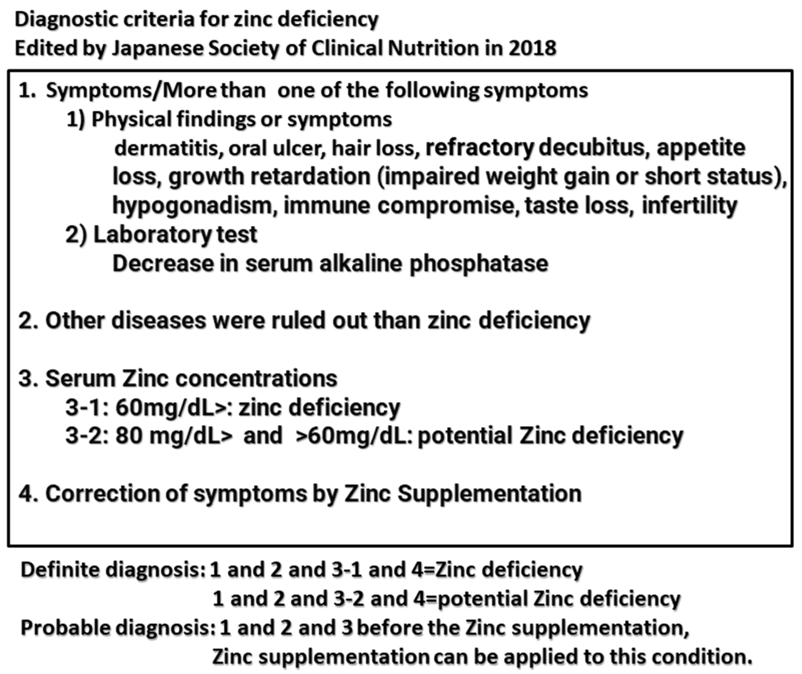
  1. Kodama, H.; Itakura, H.; Omori, H.; Sasaki, M.; Sando, K.; Kamura, T.; Fuse, Y.; Hosoi, T.; Yoshida, H. Clinical guidelines for zinc deficiency. J. Jpn. Soc. Clin. Nutr. 2018, 40, 120–167. [Google Scholar]
  2. Fukushima, T.; Horike, H.; Fujiki, S.; Kitada, S.; Sasaki, T.; Kashihara, N. Zinc deficiency anemia and effects of zinc therapy in maintenance hemodialysis patients. Ther. Apher. Dial. 2009, 13, 213–219. [Google Scholar] [CrossRef]
  3. Kobayashi, H.; Abe, M.; Okada, K.; Tei, R.; Maruyama, N.; Kikuchi, F.; Higuchi, T.; Soma, M. Oral zinc supplementation reduces the erythropoietin responsiveness index in patients on hemodialysis. Nutrients 2015, 7, 3783–3795. [Google Scholar] [CrossRef]
  4. Najafabadi, M.M.; Faghihi, G.; Emami, A.; Monghad, M.; Moeenzadeh, F.; Sharif, N.; Jazi, A.H.D. Zinc sulfate for relief of pruritus in patients on maintenance hemodialysis. Ther. Apher. Dial. 2012, 16, 142–145. [Google Scholar] [CrossRef] [PubMed]
  5. Mapar, M.A.; Pazyar, N.; Siahpoosh, A.; Latifi, S.M.; SS, B.M.; Khazanee, A. Comparison of the efficacy and safety of zinc sulfate vs.placebo in the treatment of pruritus of hemodialytic patients: A pilot randomized, triple-blind study. G Ital. Dermatol. Venereol. 2015, 150, 351–355. [Google Scholar]
  6. Amerian, M.; Nezakati, E.; Ebrahimi, H.; Zolfaghari, P.; Yarmohammadi, M.; Sohrabi, M.B. Comparative effects of zinc sulfate and hydroxyzine in decreasing pruritus among hemodialysis patients: A cross-over clinical trial. J. Maz. Univ. Med. Sci. 2019, 29, 81–90. [Google Scholar]
  7. Atkin-Thor, E.; Goddard, B.W.; O’Nion, J.; Stephen, R.L.; Kolff, W.J. Hypogeusia and zinc depletion in chronic dialysis patients. Am. J. Clin. Nutr. 1978, 31, 1948–1951. [Google Scholar] [CrossRef] [PubMed]
  8. Sprenger, K.B.; Bundschu, D.; Lewis, K.; Spohn, B.; Schmitz, J.; Franz, H.E. Improvement of uremic neuropathy and hypogeusia by dialysate zinc supplementation by dialysate zinc supplementation: A double-blind study. Kidney Int. Suppl. 1983, 16, S315–S318. [Google Scholar]
  9. Kim, S.M.; Kim, M.; Lee, E.K.; Kim, S.B.; Chang, J.W.; Kim, H.W. The effect of zinc deficiency on salt taste acuity, preference, and dietary sodium intake in hemodialysis patients. Hemodial. Int. 2016, 20, 441–446. [Google Scholar] [CrossRef]
  10. Momen-Heravi, M.; Barahimi, E.; Razzaghi, R.; Bahmani, F.; Gilasi, H.R.; Asemi, Z. The effects of zinc supplementation on wound healing and metabolic status in patients with diabetic foot ulcer: A randomized, double-blind, placebo-controlled trial. Wound Repair Regen. 2017, 25, 512–520. [Google Scholar] [CrossRef]
  11. Koyama, A.; Tsuruoka, T.; Fujii, T.; Sugimoto, M.; Banno, H.; Komori, K. Zinc Deficiency and Clinical Outcome After Infrainguinal Bypass Grafting for Critical Limb Ischemia. Circ. Rep. 2020, 2, 167–173. [Google Scholar] [CrossRef] [PubMed]
  12. Yang, C.-Y.; Wu, M.-L.; Chou, Y.-Y.; Li, S.-Y.; Deng, J.-F.; Yang, W.-C.; Ng, Y.-Y. Essential trace element status and clinical outcomes in long-term dialysis patients: A two-year prospective observational cohort study. Clin. Nutr. 2012, 31, 630–636. [Google Scholar] [CrossRef] [PubMed]
  13. Wang, L.-J.; Wang, M.-Q.; Hu, R.; Yang, Y.; Huang, Y.-S.; Xian, S.-X.; Lu, L. Effect of Zinc Supplementation on Maintenance Hemodialysis Patients: A Systematic Review and Meta-Analysis of 15 Randomized Controlled Trials. BioMed. Res. Int. 2017, 2017, 1024769. [Google Scholar] [CrossRef] [PubMed]
  14. Mousavi, S.M.; Djafarian, K.; Mojtahed, A.; Varkaneh, H.K.; Shab-Bidar, S. The effect of zinc supplementation on plasma C-reactive protein concentrations: A systematic review and meta-analysis of randomized controlled trials. Eur. J. Pharmacol. 2018, 834, 10–16. [Google Scholar] [CrossRef] [PubMed]
  15. Hosseini, R.; Montazerifar, F.; Shahraki, E.; Karajibani, M.; Mokhtari, A.M.; Dashipour, A.R.; Ferns, G.A.; Jalali, M. The Effects of Zinc Sulfate Supplementation on Serum Copeptin, C-Reactive Protein and Metabolic Markers in Zinc-Deficient Diabetic Patients on Hemodialysis: A Randomized, Double-Blind, Placebo-Controlled Trial. Biol. Trace Elem. Res. 2022, 200, 76–83. [Google Scholar] [CrossRef] [PubMed]
  16. Brodersen, H.-P.; Holtkamp, W.; Larbig, D.; Beckers, B.; Thiery, J.; Lautenschläger, J.; Probst, H.-J.; Ropertz, S.; Yavari, A. Zinc supplementation and hepatitis B vaccination in chronic haemodialysis patients: A multicentre study. Nephrol. Dial. Transpl. 1995, 10, 1780. [Google Scholar]
  17. Jern, N.A.; Vanbeber, A.D.; Gorman, M.A.; Weber, C.G.; Liepa, G.U.; Cochran, C.C. The effects of zinc supplementation on serum zinc concentration and protein catabolic rate in hemodialysis patients. J. Ren. Nutr. 2000, 10, 148–153. [Google Scholar] [CrossRef]
  18. Candan, F.; Gültekin, F.; Candan, F. Effect of vitamin C and zinc on osmotic fragility and lipid peroxidation in zinc-deficient haemodialysis patients. Cell Biochem. Funct. 2002, 20, 95–98. [Google Scholar] [CrossRef]
  19. Chevalier, C.A.; Liepa, G.; Murphy, M.D.; Suneson, J.; VanBeber, A.D.; Gorman, M.A.; Cochran, C. The effects of zinc supplementation on serum zinc and cholesterol concentrations in hemodialysis patients. J. Ren. Nutr. 2002, 12, 183–189. [Google Scholar] [CrossRef]
  20. Rashidi, A.A.; Salehi, M.; Piroozmand, A.; Sagheb, M.M. Effects of zinc supplementation on serum zinc and C-reactive protein concentrations in hemodialysis patients. J. Ren. Nutr. 2009, 19, 475–478. [Google Scholar] [CrossRef]
  21. Roozbeh, J.; Hedayati, P.; Sagheb, M.M.; Sharifian, M.; Jahromi, A.H.; Shaabani, S.; Jalaeian, H.; Raeisjalali, G.A.; Behzadi, S. Effect of zinc supplementation on triglyceride, cholesterol, LDL, and HDL levels in zinc-deficient hemodialysis patients. Ren. Fail. 2009, 31, 798–801. [Google Scholar] [CrossRef] [PubMed]
  22. Rahimi-Ardabili, B.; Argani, H.; Ghorbanihaghjo, A.; Rashtchizadeh, N.; Naghavi-Behzad, M.; Ghorashi, S.; Nezami, N. Paraoxonase enzyme activity is enhanced bey zinc supplementation in hemodialysis patients. Ren. Fail. 2012, 34, 1123–1128. [Google Scholar] [CrossRef] [PubMed]
  23. Pakfetrat, M.; Shahroodi, J.R.; Zolgadr, A.A.; Larie, H.A.; Nikoo, M.H.; Malekmakan, L. Effects of zinc supplement on plasma homocysteine level in end-stage renal disease patients: A double-blind randomized clinical trial. Biol. Trace Elem. Res. 2013, 153, 11–15. [Google Scholar] [CrossRef] [PubMed]
  24. Mazani, M.; Argani, H.; Rashtchizadeh, N.; Ghorbanihaghjo, A.; Hamdi, A.; Estiar, M.A.; Nezami, N. Effects of zinc supplementation on antioxidant status and lipid peroxidation in hemodialysis patients. J. Ren. Nutr. 2013, 23, 180–184. [Google Scholar] [CrossRef] [PubMed]
  25. Guo, C.H.; Wang, C.L. Effects of zinc supplementation on plasma copper/zinc ratios, oxidative stress, and immunological status in hemodialysis patients. Int. J. Med. Sci. 2013, 10, 79–89. [Google Scholar] [CrossRef] [PubMed]
  26. Argani, H.; Mahdavi, R.; Ghorbani-Haghjo, A.; Razzaghi, R.; Nikniaz, L.; Gaemmaghami, S.J. Effects of zinc supplementation on serum zinc and leptin levels, BMI, and body composition in hemodialysis patients. J. Trace Elem. Med. Biol. 2014, 28, 35–38. [Google Scholar] [CrossRef] [PubMed]
  27. Sherbini, N.S.; El-Shazly, A.N.; Elhady, S.A.; El-Mashad, G.M.; Sabry, J.H. Effect of zinc supplementation on body mass index and serum levels of zinc and leptin in pediatric hemodialysis patients. Int. J. Nephrol. Renov. Dis. 2015, 8, 159–163. [Google Scholar] [CrossRef]
  28. Sato, E.; Sato, S.; Degawa, M.; Ono, T.; Lu, H.; Matsumura, D.; Nomura, M.; Moriyama, N.; Amaha, M.; Nakamura, T. Effects of Zinc Acetate Hydrate Supplementation on Renal Anemia with Hypozincemia in Hemodialysis Patients. Toxins 2022, 14, 746. [Google Scholar] [CrossRef]
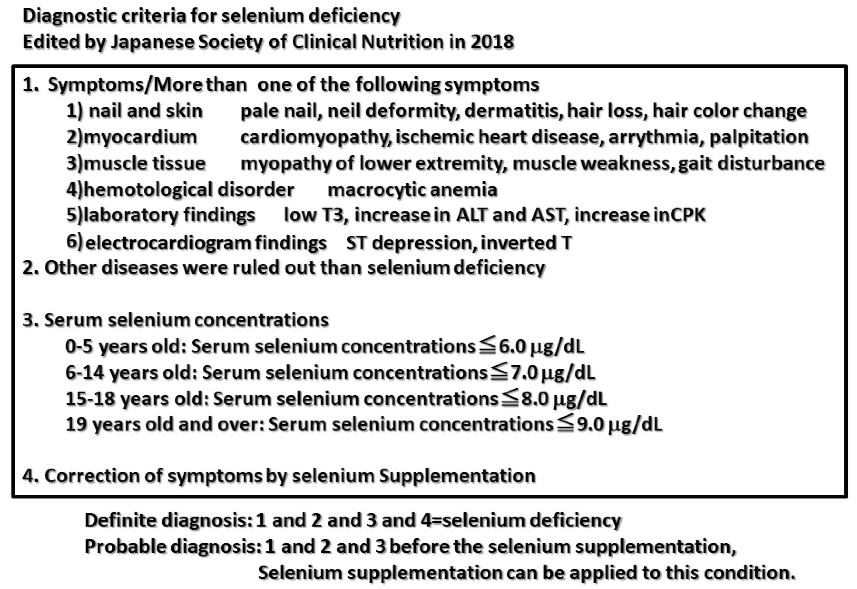
  29. Kodama, H.; Asagiri, M.; Etani, Y.; Koyama, H.; Soh, H.; Ida, S.; Tanaka, Y.; Takayanagi, M.; Funakoshi, M.; Yoshida, M. Clinical guidelines for selenium deficiency. J. Jpn. Soc. Clin. Nutr. 2019, 40, 239–283. [Google Scholar]
  30. Mochizuki, H.; Yokota, S.; Kaneko, K.; Koh, H.; Ishi, J.; Katsuta, M. A case of SEP complicated with dilated cardiomyopathy probably due to selenium deficiency. J. Jpn. Soc. Dial. Ther. 2001, 34, 1095–1099. [Google Scholar] [CrossRef]
  31. Nishida, H.; Abe, H.; Iida, Y.; Toriyama, C. A case of left ventricular hypofunction due to selenium-arginine depletion associated with short bowel syndrome and dialysis. Jpn. J. Intern. Med. 2016, 106, 828–833. [Google Scholar]
  32. Fujishima, Y.; Ohsawa, M.; Itai, K.; Kato, K.; Tanno, K.; Turin, T.C.; Onoda, T.; Endo, S.; Okayama, A.; Fujioka, T. Serum selenium levels are inversely associated with death risk among hemodialysis patients. Transplant 2011, 26, 3331–3338. [Google Scholar] [CrossRef] [PubMed]
  33. Fujishima, Y.; Ohsawa, M.; Itai, K.; Kato, K.; Tanno, K.; Turin, T.C.; Onoda, T.; Endo, S.; Okayama, A.; Fujioka, T. Serum selenium levels in hemodialysis patients are significantly lower than those in healthy controls. Blood Purif. 2011, 32, 43–47. [Google Scholar] [CrossRef] [PubMed]
  34. Tonelli, M.; Wiebe, N.; Bello, A.; Field, C.J.; Gill, J.S.; Hemmelgarn, B.R.; Holmes, D.T.; Jindal, K.; Klarenbach, S.W.; Manns, B.J.; et al. Concentrations of Trace Elements and Clinical Outcomes in Hemodialysis Patients: A Prospective Cohort Study. Clin. J. Am. Soc. Nephrol. 2018, 13, 907–915. [Google Scholar] [CrossRef] [PubMed]
  35. Ruiz, A.A.; Jiménez, E.M.; Bermejo-Barrera, P.; Lozano, R.; Seijas, V.M.-E. Selenium and All-cause Mortality in End-Stage Renal Disease. Retrospective Observational Cohort Study. J. Ren. Nutr. 2020, 30, 484–492. [Google Scholar] [CrossRef] [PubMed]
  36. Zhang, J.; Taylor, E.W.; Bennett, K.; Saad, R.; Rayman, M.P. Association between regional selenium status and reported outcome of COVID-19 cases in China. Am. J. Clin. Nutr. 2020, 111, 1297–1299. [Google Scholar] [CrossRef] [PubMed]
  37. Li, S.; Tang, T.; Guo, P.; Zou, Q.; Ao, X.; Hu, L.; Tan, L. A meta-analysis of randomized controlled trials: Efficacy of selenium treatment for sepsis. Medicine 2019, 98, e14733. [Google Scholar] [CrossRef] [PubMed]
  38. Saint-Georges, M.D.; Bonnefont, D.J.; Bourely, B.A.; Jaudon, M.C.; Cereze, P.; Chaumeil, P.; Gard, C.; D’Auzac, C.L. Correction of selenium deficiency in hemodialyzed patients. Kidney Int. Suppl. 1989, 27, S274–S277. [Google Scholar]
  39. Atakan, A.; Macunluoglu, B.; Kaya, Y.; Ari, E.; Demir, H.; Asicioglu, E.; Kaspar, C. Decreased serum selenium levels are correlated with diminished coronary flow reserve among hemodialysis patients. Biol. Trace Elem. Res. 2013, 155, 333–338. [Google Scholar] [CrossRef]
  40. Yasukawa, M.; Arai, S.; Nagura, M.; Kido, R.; Asakawa, S.; Hirohama, D.; Yamazaki, O.; Tamura, Y.; Fujimaki, M.; Kobayashi, S.; et al. Selenium Associates with Response to Erythropoiesis-Stimulating Agents in Hemodialysis Patients. Kidney Int. Rep. 2022, 7, 1565–1574. [Google Scholar] [CrossRef]
  41. Xu, S.; Zou, D.; Tang, R.; Li, S.; Chen, W.; Wen, L.; Liu, Y.; Liu, Y.; Zhong, X. Levels of trace blood elements associated with severe sleep disturbance in maintenance hemodialysis patients. Sleep Breath 2021, 25, 2007–2013. [Google Scholar] [CrossRef]
  42. Richard, M.J.; Ducros, V.; Forêt, M.; Arnaud, J.; Coudray, C.; Fusselier, M.; Favier, A. Reversal of selenium and zinc deficiencies in chronic hemodialysis patients by intravenous sodium selenite and zinc gluconate supplementation. Biol. Trace Elem. Res. 1993, 39, 149–159. [Google Scholar] [CrossRef] [PubMed]
  43. Napolitano, G.; Bonomini, M.; Bomba, G.; Bucci, I.; Todisco, V.; Albertazzi, A.; Monaco, F. Thyroid function and plasma selenium in chronic uremic patients on hemodialysis treatment. Biol. Trace Elem. Res. 1996, 55, 221–230. [Google Scholar] [CrossRef] [PubMed]
  44. Koenig, J.; Fischer, M.; Bulant, E.; Tiran, B.; Elmadfa, I.; Druml, W. Antioxidant status in patients on chronic hemodialysis therapy: Impact of parenteral selenium supplementation. Wien. Klin. Wochenschr. 1997, 109, 13–19. [Google Scholar] [PubMed]
  45. Temple, K.A.; Smith, A.M.; Cockram, D.B. Selenate-supplemented nutritional formula increases plasma selenium in hemodialysis patients. J. Ren. Nutr. 2000, 10, 16–23. [Google Scholar] [CrossRef]
  46. Zachara, B.A.; Koterska, D.; Manitius, J.; Sadowski, L.; Dziedziczko, A.; Salak, A.; Wasowicz, W. Selenium supplementation on plasma glutathione peroxidase activity in patients with end-stage chronic renal failure. Biol. Trace Elem. Res. 2004, 97, 15–30. [Google Scholar] [CrossRef]
  47. Salehi, M.; Sohrabi, Z.; Ekramzadeh, M.; Fallahzadeh, M.K.; Ayatollahi, M.; Geramizadeh, B.; Hassanzadeh, J.; Sagheb, M.M. Selenium supplementation improves the nutritional status of hemodialysis patients: A randomized, double-blind, placebo-controlled trial. Nephrol. Dial. Transpl. 2013, 28, 716–721. [Google Scholar] [CrossRef]
  48. Tonelli, M.; Network, F.T.A.K.D.; Wiebe, N.; Thompson, S.; Kinniburgh, D.; Klarenbach, S.W.; Walsh, M.; Bello, A.K.; Faruque, L.; Field, C.; et al. Alberta Kidney Disease Network. Trace element supplementation in hemodialysis patients: A randomized controlled trial. BMC Nephrol. 2015, 16, 52. [Google Scholar] [CrossRef]
  49. Shahreki, E.; Kaykhaei, M.A.; Mosallanezhad, Z.; Adineh, Z.; Mokhtari, A.M.; Mohammadi, M.; Hosseini, R.; Bazi, A. Effects of Selenium and/or N-Acetyl-Cysteine Supplementation on Nonthyroidal Illness Syndrome in Hemodialysis Patients: A Factorial Randomized Controlled Trial. Pharmacology 2022, 107, 480–485. [Google Scholar] [CrossRef]
  50. Salimian, M.; Soleimani, A.; Bahmani, F.; Tabatabaei, S.M.H.; Asemi, Z.; Talari, H.R. The effects of selenium administration on carotid intima-media thickness and metabolic status in diabetic hemodialysis patients: A randomized, double-blind, placebo-controlled trial. Clin. Nutr. ESPEN 2022, 47, 58–62. [Google Scholar] [CrossRef]
  51. Atapour, A.; Vahdat, S.; Hosseini, M.; Mohamadian, H. Effect of Selenium on Triglyceride and Total Cholesterol, Weight Gain, and Physical Activity on Hemodialysis Patients: A Randomized Double-Blinded Controlled Trial. Int. J. Prev. Med. 2022, 13, 63. [Google Scholar] [PubMed]
  52. Hurst, R.; Armah, C.N.; Dainty, J.R.; Hart, D.J.; Teucher, B.; Goldson, A.J.; Broadley, M.R.; Motley, A.K.; Fairweather-Tait, S.J. Establishing optimal selenium status: Results of a randomized, double-blind, placebo-controlled trial. Am. J. Clin. Nutr. 2010, 91, 923–931. [Google Scholar] [CrossRef] [PubMed]
  53. Takahashi, A. Role of Zinc and Copper in Erythropoiesis in Patients on Hemodialysis. J. Ren. Nutr. 2022, 32, 650–657. [Google Scholar] [CrossRef] [PubMed]
  54. Higuchi, T.; Matsukawa, Y.; Okada, K.; Oikawa, O.; Yamazaki, T.; Ohnishi, Y.; Fujita, T.; Fukuda, N.; Soma, M.; Matsumoto, K. Correction of copper deficiency improves erythropoietin unresponsiveness in hemodialysis patients with anemia. Intern. Med. 2006, 45, 271–273. [Google Scholar] [CrossRef] [PubMed]
  55. Gaetke, L.M.; Chow-Johnson, H.S.; Chow, C.K. Copper: Toxicological relevance and mechanisms. Arch. Toxicol. 2014, 88, 1929–1938. [Google Scholar] [CrossRef] [PubMed]
  56. Guo, C.-H.; Chen, P.-C.; Yeh, M.-S.; Hsiung, D.-Y.; Wang, C.-L. Cu/Zn ratios are associated with nutritional status, oxidative stress, inflammation, and immune abnormalities in patients on peritoneal dialysis. Clin. Biochem. 2011, 44, 275–280. [Google Scholar] [CrossRef] [PubMed]
  57. Feldman, L.; Beberashvili, I.; Hamad, R.A.; Yakov-Hai, I.; Abramov, E.; Wasser, W.; Gorelik, O.; Rozenberg, R.; Efrati, S. Serum Chromium Levels Are Higher in Peritoneal Dialysis than in Hemodialysis Patients. Perit. Dial. Int. 2019, 39, 330–334. [Google Scholar] [CrossRef]
  58. Jomova, K.; Valko, M. Advances in metal-induced oxidative stress and human disease. Toxicology 2011, 283, 65–87. [Google Scholar] [CrossRef]
  59. Hsu, C.-W.; Weng, C.-H.; Lee, C.-C.; Yen, T.-H.; Huang, W.-H. Association of serum chromium levels with malnutrition in hemodialysis patients. BMC Nephrol. 2019, 20, 302. [Google Scholar] [CrossRef]
  60. Tonelli, M.; Wiebe, N.; Hemmelgarn, B.; Klarenbach, S.; Field, C.; Manns, B.; Thadhani, R.; Gill, J. Alberta Kidney Disease Network. Trace elements in hemodialysis patients: A systematic review and meta-analysis. BMC Med. 2009, 7, 25. [Google Scholar] [CrossRef]
  61. Aschner, J.L.; Aschner, M. Nutritional aspects of manganese homeostasis. Mol. Aspects Med. 2005, 26, 353–362. [Google Scholar] [CrossRef] [PubMed]
  62. Keen, C.; Zidenburg-Cherr, S. Encyclopedia of Food Science, Food Technology, and Nutrition; Academic Press: London, UK, 1993. [Google Scholar]
  63. Tonelli, M.; Wiebe, N.; Bello, A.; Field, C.J.; Gill, J.S.; Hemmelgarn, B.R.; Holmes, D.T.; Jindal, K.; Klarenbach, S.W.; Manns, B.J.; et al. Alberta Kidney Disease Network. Concentrations of Trace Elements in Hemodialysis Patients: A Prospective Cohort Study. Am. J. Kidney Dis. 2017, 70, 696–704. [Google Scholar] [CrossRef]
  64. Gómez de Oña, C.; Martínez-Morillo, E.; Gago González, E.; Vidau Argüelles, P.; Fernández Merayo, C.; Álvarez Menéndez, F.V. Variation of trace element concentrations in patients undergoing hemodialysis in the north of Spain. Scand. J. Clin. Lab. Invest. 2016, 76, 492–499. [Google Scholar] [CrossRef] [PubMed]
  65. Liu, Y.; Hu, J.; Tang, R.; Guo, H.; Chen, Q.; Qiu, J.; Liu, Y.; Tan, R.; Zhong, X. Association between the blood manganese (Mn) and hemoglobin in patients undergoing maintenance hemodialysis. J. Trace Elem. Med. Biol. 2022, 71, 126947. [Google Scholar] [CrossRef]
  66. Vignoli, A.; Tenori, L.; Luchinat, C. An omics approach to study trace metals in sera of hemodialysis patients treated with erythropoiesis stimulating agents. Metallomics 2022, 14, mfac028. [Google Scholar] [CrossRef] [PubMed]
  67. Chen, J.; Peng, H.; Zhang, K.; Xiao, L.; Yuan, Z.; Chen, J.; Wang, Z.; Wang, J.; Huang, H. The insufficiency intake of dietary micronutrients associated with malnutrition-inflammation score in hemodialysis population. PLoS ONE 2013, 8, e66841. [Google Scholar] [CrossRef] [PubMed]
  68. Lin, J.L.; Lin-Tan, D.T.; Hsu, C.W.; Yen, T.H.; Chen, K.H.; Hsu, H.H.; Ho, T.C.; Hsu, K.H. Association of blood lead levels with mortality in patients on maintenance hemodialysis. Am. J. Med. 2011, 124, 350–358. [Google Scholar] [CrossRef] [PubMed]
  69. Lin, J.L.; Lin-Tan, D.T.; Chen, K.H.; Hsu, C.W.; Yen, T.H.; Huang, W.H.; Huang, Y.L. Blood lead levels association with 18-month all-cause mortality in patients with chronic peritoneal dialysis. Nephrol. Dial. Transplant. 2010, 25, 1627–1633. [Google Scholar] [CrossRef]
  70. Feldman, R.G. Urban lead mining: Lead intoxication among deleaders. N. Engl. J. Med. 1978, 298, 1143–1145. [Google Scholar] [CrossRef]
  71. Thomson, R.M.; Parry, G.J. Neuropathies associated with excessive exposure to lead. Muscle Nerve 2006, 33, 732–741. [Google Scholar] [CrossRef]
  72. Huang, W.H.; Hu, C.C.; Yen, T.H.; Hsu, C.W.; Weng, C.H. Blood lead level: An overlooked risk of carpal tunnel syndrome in hemodialysis patients. Ren. Fail. 2019, 41, 786–793. [Google Scholar] [CrossRef] [PubMed]
  73. Danziger, J.; Mukamal, K.J.; Weinhandl, E. Associations of Community Water Lead Concentrations with Hemoglobin Concentrations and Erythropoietin-Stimulating Agent Use among Patients with Advanced CKD. J. Am. Soc. Nephrol. 2021, 32, 2425–2434. [Google Scholar] [CrossRef]
  74. Lu, K.C.; Wu, C.C.; Ma, W.Y.; Chen, C.C.; Wu, H.C.; Chu, P. Decreased blood lead levels after calcitriol treatment in hemodialysis patients with secondary hyperparathyroidism. Bone 2011, 49, 1306–1310. [Google Scholar] [CrossRef] [PubMed]
  75. Ford, M.; Goldfrank, L.; Flomenbaum, N.; Lewin, N.; Howland, M.; Hoffman, R.; Nelson, L. (Eds.) Arsenic. In Goldfrank’s Toxicological Emergencies; McGraw-Hill: New York, NY, USA, 2002; pp. 1183–1199. [Google Scholar]
  76. Schoen, A.; Beck, B.; Sharma, R.; Dube, E. Arsenic toxicity at low doses: Epidemiological and mode of action considerations. Toxicol, Appl, Pharmacol. 2004, 198, 253–267. [Google Scholar] [CrossRef] [PubMed]
  77. Morton, W.E.; Caron, G.A. Encephalopathy: An uncommon manifestation of workplace arsenic poisoning? Am. J. Ind. Med. 1989, 15, 1–5. [Google Scholar] [CrossRef] [PubMed]
  78. Xiang, S.; Jin, Q.; Xu, F.; Yao, Y.; Liang, W.; Zuo, X.; Ye, T.; Ying, C. High serum arsenic and cardiovascular risk factors in patients undergoing continuous ambulatory peritoneal dialysis. J. Trace Elem. Med. Biol. 2019, 52, 1–5. [Google Scholar] [CrossRef]
  79. Ochi, A.; Ishimura, E.; Tsujimoto, Y.; Kakiya, R.; Tabata, T.; Mori, K.; Shoji, T.; Yasuda, H.; Nishizawa, Y.; Inaba, M. Trace elements in the hair of hemodialysis patients. Biol. Trace Elem. Res. 2011, 143, 825–834. [Google Scholar] [CrossRef] [PubMed]
  80. Navarro, J.A.; Granadillo, V.A.; Salgado, O.; Rodríguez-Iturbe, B.; García, R.; Delling, G.; Romero, R.A. Bone metal content in patients with chronic renal failure. Clin. Chim. Acta 1992, 211, 133–142. [Google Scholar] [CrossRef]
  81. Oruc, M.; Mercan, S.; Bakan, S.; Kose, S.; Ikitimur, B.; Trabulus, S.; Altiparmak, M.R. Do trace elements play a role in coronary artery calcification in hemodialysis patients? Int. Urol. Nephrol. 2023, 55, 173–182. [Google Scholar] [CrossRef]
  82. Katko, M.; Kiss, I.; Karpati, I.; Kadar, A.; Matyus, J.; Csongradi, E.; Posta, J.; Paragh, G.; Balla, J.; Kovacs, B.; et al. Relationship between serum nickel and homocysteine concentration in hemodialysis patients. Biol. Trace. Elem. Res. 2008, 124, 195–205. [Google Scholar] [CrossRef]
  83. Schrooten, I.; Elseviers, M.M.; Lamberts, L.V.; De Broe, M.E.; D’Haese, P.C. Increased serum strontium levels in dialysis patients: An epidemiological survey. Kidney Int. 1999, 56, 1886–1892. [Google Scholar] [CrossRef]
  84. D’Haese, P.C.; Schrooten, I.; Goodman, W.G.; Cabrera, W.E.; Lamberts, L.V.; Elseviers, M.M.; Couttenye, M.M.; De Broe, M.E. Increased bone strontium levels in hemodialysis patients with osteomalacia. Kidney Int. 2000, 57, 1107–1114. [Google Scholar] [CrossRef] [PubMed]
Figure 1. Nutrition elements. Five nutritional elements in which minerals are included. Minerals comprise macroelements, microelements, and trace elements.
Kidneydial 03 00025 g001
Figure 2. Diagnostic criteria for zinc deficiency. The criteria were edited by the Japanese Society of Clinical Nutrition in 2018 [1]. Reprinted with permission from [1], in 2018 from Hiroko Kodama.
Kidneydial 03 00025 g002
Figure 3. Diagnostic criteria for selenium deficiency. The criteria were edited by the Japanese Society of Clinical Nutrition in 2018 [29]. Reprinted with permission from [29], in 2018 from Hiroko Kodama.
Kidneydial 03 00025 g003
Table 1. Trace elements and their concentrations in predialysis, hemodialysis, and peritoneal dialysis patients. ↑ represents “higher than normal”, ↓ “lower than normal”, and → “the same as normal”, and ? “no data available”.
Trace ElementNormal RangePredialysisHemodialysisPeritoneal DialysisDeficiency Symptoms
Zinc (Zn)60–121 mg/dLGrowth retardation, wound-healing delay, taste disorder, and sexual dysfunction
Manganese (Mn)0.31–1.04 mg/dL↓ or →Anemia and glucose intolerance
Selenium (Se)18–40 mg/mLCardiac dysfunction, immune disorders, and carcinogenesis
Copper (Cu)68–128 mg/dL→ or ↑Hemolysis, leukocytosis, and metabolic acidosis
Cobalt (Co)0.04–0.40 mg/LCardiac dysfunction and impairment of gluconeogenesis
Chromium (Cr)0.04–0.35 mg/dLLiver dysfunction, renal dysfunction, and carcinogenesis
Molybdenum (Mo)0.27–1.17 mg/L?Amino acid metabolism disorder, arthropathy, and hypercalcemia
Vanadium (V)0.10–1.0 mg/L?Bone disease, dyslipidemia, anemia, and hypertension
Silicon (Si)0.14–0.20 mg/LErythema, bone disease, neuropathy, and Wegener granulomatosis
Nickel (Ni)0.2–0.8 mg/dLCardiac ischemia, anemia, and bone disease
Strontium (Sr)15–30 mg/LOsteomalacia
Bromine (Br)2.19–5.00 mg/LSleep disorders
Cadmium (Cd)2.19–5.00 mg/LGrowth defects, hypertension, and hyperparathyroidism
Rubidium (Rb)0.095–0.272 mg/LDepression and central nervous system dysfunction
Table 2. Clinical trials of zinc.
ModeNumberTreatment RouteCombinationEvaluationSe ConcentrationOutcomeRef.
HD40 dialysis
HBV non-responder
Zn: n = 28
Control: n = 12
60 mg zinc aspartate after each dialysis session for
8 weeks
NoneHBV antibody formation
Titer of HVB antibody
Zn group:
77 ± 2.4 μg/dL
Control group: 63 ± 2.1 μg/dL
HBV antibody formation Zn: 6 out of 28 patients
Control: 2 out of 12 patients
Antibody titer Zn: 0–2364 IE/L
Control: 0–1110 IE/L
[16]
HDRandomized, double-blind, before–after trial
n = 20 (15 women, 5 men)
Zn: 7.7 pmol zinc sulfate (2200 μg) daily
Control: cornstarch placebo capsule daily
90 days
NoneSerum Zn concentration
PCR (protein catabolic rate)
12.2 μmol/L (80 μg/dL) on day 0
to 15.3 μmol/L (100 μg/dL) on day 90
A significant positive correlation (r = 0.61) between PCR and serum zinc concentrations [17]
HDZn group:
34 zinc-deficient HD patients.
Control group:
16 sex- and age-matched normal volunteers
Zn: zinc (20 mg/day)
Control: placebo
3 months
NoneLevels of Zn malondialdehyde (MDA)
osmotic fragility of red blood cells
Zn: 12.5 ± 1.0 to 18.8 ± 3.0 μmol/L
Control: from 12.3 ± 1.0 to 12.1 ± 1.4 μmol/L
Increase in Zn concentration;
Improvement of osmotic fragility;
Decrease in the level of MDA
[18]
HDRandomized, double-blind, before–after trialSupplementation of Zn: 7.7 μmol zinc sulfate/day (50 mg elemental zinc/day)
Control: cornstarch placebo capsule
90 days
NoneSerum zinc,
dietary intake,
HDL, LDL, and TC
Zn: 0.79 μg/mL to 0.96 μg/mLIncrease in serum total cholesterol and LDL;
No change in HDL;
Increase in reported energy intake;
No change in dietary intake of zinc, cholesterol, total fat, or saturated fat
[19]
HDn = 55 hemodialysis patients (32 men and 23 women)Zinc supplementation group (n = 28):
220 mg zinc sulfate capsule
Control group (n = 27): placebo capsule (220 mg corn starch)
42 days
NoneSerum zinc
C-reactive protein levels
Zn group: 57.4 ± 2.4 μg/dL to 88.4 ± 4.8 μg/dLDecrease in serum C-reactive protein:
13.5 ± 3.8 mg/L to 10.5 +/− 3.5 mg/L
[20]
HDDouble-blind
zinc-deficient HD subjects
Total: n = 53
(25 female
and 28 male)
Zn group (n = 27): 220 mg zinc sulfate (50 mg elemental
zinc)
Control group (n = 26):
starch placebo
42 days
NoneSerum concentration of zinc, total cholesterol, HDL and LDL cholesterol, and triglyceridesZn group: 0.53 ± 0.36 μg/mL to 0.86 μg/mL ± 0.42 μg/mL
Control group: 0.52 ± 0.25 μg/mL to 0.64 ± 0.29 μg/mL
Increase in serum total cholesterol,
serum LDL, and HDL cholesterol
serum triglyceride
[21]
HDDouble-blind, randomized, controlled trial
Total: n = 60
Zn group (n = 30): 100 mg/day zinc
Control group (n = 30): placebo
2 months
NoneParaoxonase (PON) enzyme activity
Lipid profile
apolipoprotein AI (Apo-AI) and B (Apo-B) levels
Not measuredNo change in serum levels of TC, TG, or LDL or Apo-B levels
Increase in serum levels of HDL, Apo-AI, and PON activity
[22]
HDDouble-blind, randomized, clinical trial
Total: n = 97 ESRD patients with Zn deficiency
Zn group (n = 50): 50 mg/day Zn
Control group (n = 47): placebo
6 weeks
NoneSeum Zn
Homocysteine (hCys) level
Zn group: 56.9 ± 13.9 to 120.8 ± 26 μg/dL
Control group: 60.9 ± 9.8 to 63.9 ± 13.2 μg/dL
Decrease in serum hCys[23]
HDDouble-blind, randomized, controlled trial
65 HD patients
Group A: placebo
Group B: zinc (100 mg/day)
2 months.
NoneSerum Zn concentration, total antioxidant capacity (TAC), whole blood glutathione peroxidase (GSH) level, superoxide dismutase (SOD) activity, and
malondialdehyde (MDA) level
The levels of serum zinc were increasedIncrease in TAC, GSH, and SOD activity
Decrease in MDA
[24]
HDLong-term HD patients with low plasma Zn concentrations
(<80 mg/dL)
Zn group (n = 40):
daily oral Zn
No supplements (n = 25)
Control (n = 38): age- and sex-matched healthy individuals
8 weeks
NonePlasma concentrations of Zn and Cu, Cu/Zn ratios, oxidative stress, and proinflammatory cytokines
percentages of CD4 and CD19 lymphocytes
CD4/CD8 ratios
The levels of serum Zn were increasedDecrease in Cu, Cu/Zn ratios, oxidative stress status, and inflammatory responses
Increase in percentages of CD4 and CD19 lymphocytes and CD4/CD8 ratios
[25]
HDRandomized, double-blind, and placebo-controlled trial
60 HD patients
Supplemented group (n = 30; male/female: 19/11): 100 mg/day elemental Zn
Control group (n = 30; male/female: 17/13): placebo
60 days
Noneserum zinc
serum Leptin
anthropometric measurements
Supplemented group: male, 81.7 ± 11 μg/dL to 105.5 ± 18 μg/dL;
female, 75.5 ± 11 μg/dL to 106.3 ± 16 μg/dL
Control group: male, 85.8 ± 16 μg/dL to 83.6 ± 9.6 μg/dL; female, 80.7 ± 18.8 μg/dL to 86.5 ± 12.7 μg/dL
Decrease in leptin in women;
Increase in BMI and body weight in men;
Increase in albumin and Hb;
Negative association between serum zinc and leptin levels
[26]
HDProspective clinical trial study
Pediatric HD patients
between 5 and 18 years old
Total: n = 60
Group I (n = 40): 50–100 mg zinc sulfate (equivalent to 11–22 mg elemental zinc)
Group II (n = 20): placebo (cornstarch) twice daily
90 days
Noneserum zinc
serum leptin
anthropometric measurements
Group I: 53.2 ± 8.15 μg/dL to 90.75 ± 12.2 μg/dL
Group II: 55.45 ± 9.1 μg/dL to 55.35 ± 9.15 μg/dL
Decrease in leptin;
Increase in BMI and body weight;
Negative association between serum zinc and leptin levels
[27]
HDPatients on HD with low serum zinc levels (<65 μg/dL)
Total: n = 70
Polaprezinc group (n = 35): polaprezinc, 34 mg/day of zinc
Control group (n = 35):
no supplementation
12 months
Epoetin alphERI (erythropoietin responsiveness index); Weekly ESA dose (units)/dry weight (kg)/hemoglobin (g/dL)Polaprezinc group: 53 ± 6 μg/dL to 80 ± 18 μg/dL
Control group: 55 ± 5 μg/dL to 56 ± 10 μg/dL
Decrease in ESA dosage and ERI;
No changes in Hb;
No Change in serum iron or TSAT;
Decrease in ferritin;
Decrease in copper
[3]
HDRCT
Zn-deficient diabetic HD patients
Total: n = 46
Zn supplement group (n = 21): 220 mg/day Zn sulfate capsule (containing 50 mg Zn)
Control group (n = 25): placebo capsule (220 mg corn starch)
8 weeks
Noneserum levels of copeptin, high-sensitive C-reactive protein (hs-CRP)
glycemic control
anthropometric parameters
renal function
Zn supplement group: 55.9 ± 8.0 μg/dL to 90.6 ± 15.7 μg/dL
Control group: 68.26 ± 6.2 μg/dL to 68.5 ± 6.5 μg/dL
Decrease in serum copeptin, hs-CRP, BUN, Cr, and FBG levels;
Increase in BMI and body weight;
No change in QUICKI (quantitative insulin sensitivity check index), HOMA-IR (homeostasis model assessment—insulin resistance), or serum insulin
[15]
HDBefore–after trial
Patients with serum Zn < 60 μg/dL
Total: n = 21
Zinc acetate hydrate 50 mg
6 months
Noneerythropoietin resistance index (ERI)
ERI = dose (IU) of erythropoiesis-stimulating agent (ESA)/week/body weight (kg)/hemoglobin content (g/dL)
52.4 ± 7.6 μg/dL to 84.1 ± 16.3 μg/dLDecrease in ERI and ESA dose;
No change in Hb
[28]
Zn: zinc, HD: hemodialysis, PCR: protein catabolic rate, BMI: body mass index, ESA: erythropoiesis-stimulating agent, Hb: hemoglobin.
Table 3. The lists of Selenoproteins.
Glutathione peroxidase (GPx)GPx1The biochemical function of glutathione peroxidase (GPx) is to reduce lipid hydroperoxides to their corresponding alcohols and to reduce free hydrogen peroxide to water.
Gpx2
GPx3
Gpx4
Gpx6
Thioredoxin reductases (Txnrd)TrxR1Thioredoxin reductases (TrxR) are the enzymes that catalyze the reduction of thioredoxin; hence, they are a central component in the thioredoxin system. Together with thioredoxin (Trx) and NADPH, this system’s most general description is as a system for reducing disulfide bonds in cells. They contribute to the antioxidant effects.
TrxR2
TrxR3
Iodothyronine deiodinase (DIO)DIO1Iodothyronine deiodinase (DIO) an important enzyme in the activation and deactivation of thyroid hormones. Thyroxine (T4), the precursor of 3,5,3′-triiodothyronine (T3), is transformed into T3 by deiodinase activity.
DIO2
DIO3
SelenoproteinSelHSelenoproteins (Sels) are composed of 13 proteins that contain selenium in the molecule. Selenoprotein P is the most common selenoprotein found in the plasma. It is unusual because in humans, it contains 10 s residues.
SelI
SelK
SelM
Sel15
SelN
SelO
SelP
SelR
SelS
SelT
SelV
SelW
Table 4. Clinical trials of selenium.
SubjectNumberTreatment RouteCombinationEvaluationSe ConcentrationOutcomeRef.
HDSe group: n = 39
Control group: n = 15
500 μg oral administration for 3 months and 200 μg for the next 2 monthsNoneSerum GPx,
GPx in RBC, and muscle volume IVS in echocardiogram
3.83 μg/dL to 9.0–8.0 μg/dLIncrease in serum GPx and GPx in RBC;
Increase in muscle volume;
Decrease in IVS
[38]
HDSe group: n = 650 μg intravenous administration for 5 weeks and 100 μg for the next 15 weeksIntravenous Zn gluconate for 20 weeks Serum GPx, GPx in RBC, serum TBARs, and serum Zn0.45 μmol/L to 0.89 μmol/LIncrease in serum GPx and GPx in RBC;
Decrease in serum TBARS;
No change in serum Zn
[42]
HDSe group: n = 10
Placebo group: n = 5
500 μg oral administration for 3 months and 200 μg for the next 3 monthsNoneSerum Se fT3 and TSHSe group: 7.68 μg/dL
Placebo group: 5.30 μg/L
Increase in serum Se;
Increase in fT3;
Decrease in TSH
[43]
HDSe group: n = 12400 mg Intravenous sodium selenite after HD for 8 weeksNoneSerum Se and α-tocopherol, Se and α-tocopherol in RBC
Serum ascorbic acid, serum retinol, serum glutathione, GPx, SOD activity in RBC, and serum MDA
Serum Se: increased to 8.37 μg/dL 4 weeks after the treatment
Se in RBC: increased to 15.9 μg/dL 4 weeks after the treatment
GPx activity: increase in serum levels and no change in RBC;
Decrease in MDA;
No change in CAT or SOD activity
[44]
HDTotal: n = 793
Divided into 3 groups
Three-affiliation prospective, randomized, single-blind study
Oral selenite Se 28 μg,
Oral selenate Se 28 μg
Control oral Se 7 μg
14 days
Energy intake of 35 kcal/kg/daySerum GPx, GPx in RBC, and Se in RBCSelenite group: 1.4 μmol/L
Selenate group: 1.5 μmol/L
Control group: 1.2 μmol/L
No differences in serum GPx, GPx in RBC, or Se in RBC[45]
HD4 groups
n = 15 in each group
Erythropoietin (EPO) 2000 × 3/week
Se-rich yeast 300 μg × 3/week
  • Placebo
  • EPO
  • Se-rich yeast
  • EPO + Se-rich yeast
Serum GPx and GPx activity in RBCIncreased to 120, 110, and 150 ng/mL in Se concentration in serum, blood, and RBC.
Se concentration in serum and blood plateaued.
Se in RBC increased to 200–250 ng/mL.
Increase in GPx activity in RBC in groups 3 and 4;
No change in serum GPx
[46]
HDTotal: n = 80
Se group: n = 29
Placebo group: n = 36 Randomized, double-blind, placebo-controlled study
200 μg/day oral administration for 12 weeks NonePrimary; SGA Secondary: MDA, IL-6, high-sensitivity CRP, homocysteine, transferrin, ferritin, MIS, and HbNot measuredDecrease in SGA and MIS;
Decrease in MDA;
No change in IL-6, high-sensitivity CRP, homocysteine, transferrin, ferritin, or Hb
[47]
HDTotal: n = 150
Three groups
n = 50 in each group
Randomized, double-blind, active-control study
Se capsule
1. standard supplementation (SS) (vitamins)
2. Low supplementation (LS) SS + vitamin E 250 IU + Zn 25 mg + Se 5 μg
3. Moderate supplementation (MS)
SS + vitamin E 250 IU +Zn 50 mg + Se 75 μg
Standard supplementation;
biotin 300 μg, folic acid 1 mg, nicotinamide 20 mg,
thiamine 1.5 mg
Cyanocobalamin 6 μg,
riboflavin 1.7 mg,
pyrydoxine 10 mg,
ascorbic acid 100 mg
Primary: incidence of low Se and low Zn after 90 days
Secondary: incidence of low Se and low Zn after 180 days
Low Zn: Zn < 815 μg/L
Low Se: Se < 121 μg/L
Day 90:
SS: Se, 13.1 μg/dL
LS: Se, 14.0 μg/dL
MS: Se, 14.6 μg/dL
Day 180:
SS: Se, 13.5 μg/dL
LS: Se, 13.5 μg/dL
MS: Se, 13.0 μg/dL
Primary and secondary outcomes:
No difference among three groups;
No difference in sodium sensitivity or intradialytic body weight gain
[48]
HDTotal: n = 68
Se and
NAC (N-acetylcysteine) treatment
4 groups each n = 17
12 weeks
Group A:placebo
Group B: NAC 600 μg/day
Group C: Se 200 μg/day
Group D: Se 200 μg/day+ NAC 600 μg/day
NAC
N-acetylcysteine
Free tri-iodothyronine (FT3),
free thyroxine (FT4), thyroid-stimulating hormone (TSH), and reverse T3 (rT3)
Not measuredDecrease in rT3 levels in groups B, C, and D;
No change in FT3, FT4, and TSH between the groups;
Good effects on nonthyroidal illness syndrome (NTIS)
[49]
HD53 diabetic HD patients
Randomized, double-blind, placebo-controlled trial
Selenium group (n = 26): 200 μg selenium per day
Placebo group (n = 27) for 24 weeks
NoneCarotid intima-media thickness, FPG, insulin, HOMA-IR, QUICKI, triglycerides, VLDL-C, total-C, LDL-C, HDL-C, CRP, total nitrites, TAC, GSH (total glutathione), and MDA Not measuredDecrease in serum insulin levels, insulin resistance, total cholesterol, LDL cholesterol, and CRP;
Increase in insulin sensitivity, HDL cholesterol, and GSH;
No change in carotid intima-media thickness
[50]
HDTotal: n = 78
Intervention: n = 40
Placebo: n = 38
Double-blind clinical trial
400 μg Oral selenium vs. placebo tablets three times after each hemodialysis session for 3 monthsNoneBlood Se levels, serum triglyceride, total cholesterol, weight, and physical activities (five times sit to stand test)Intervention: 40 to 65 µg/L
Placebo: 45 to 42 µg/L
Increase in Se concentration and physical activity in intervention group;
No change in triglycerides, total cholesterol, or body weight in either group
[51]
HD, hemodialysis; Se, selenium; RBC, red blood cell; Zn, zinc; IVS, interventricular septum; TBARS, thiobarbituric acid reactive substances; GPx, glutathione peroxidase; T3, tri-iodothyronine; SOD, superoxide dismutase; TAC, total antioxidant capacity; GSH, total glutathione; MDA, malondialdehyde; MIS, malnutrition–inflammation score; Hb, hemoglobin; SGA, subjective global assessment.
Disclaimer/Publisher’s Note: The statements, opinions and data contained in all publications are solely those of the individual author(s) and contributor(s) and not of MDPI and/or the editor(s). MDPI and/or the editor(s) disclaim responsibility for any injury to people or property resulting from any ideas, methods, instructions or products referred to in the content.

