Brief Report

Extraction-Free RT-PCR Surveillance Testing and Reporting for SARS-CoV-2

by
Patrick R. Carney
1,2,*,
Tyler Duellman
3,
Jia-Yi Chan
3,
Lauren Wells
3,
Michael Tessmer
4,
Leah Frater-Rubsam
3,
Molly Zeller
3,
Mark Field
4,
James Speers
3,
Kelly Tyrrell
5,
Luke Thompson
6,
Michael Bondurant
4,
Tami Morin
6,
Tamra Dagnon
6,
Brian Goff
6,
Corissa Runde
4,
Sandra Splinter-Bondurant
3,
Charles Konsitzke
3,
Patrick Kelly
6,
Christopher A. Bradfield
1,2,3 and
Joshua Hyman
3
add Show full author list remove Hide full author list
1
Molecular and Environmental Toxicology, School of Medicine and Public Health, University of Wisconsin-Madison, Madison, WI 53706, USA
2
Department of Oncology, School of Medicine and Public Health, University of Wisconsin-Madison, Madison, WI 53706, USA
3
Biotechnology Center, University of Wisconsin-Madison, Madison, WI 53706, USA
4
Division of Information Technology, University of Wisconsin-Madison, Madison, WI 53706, USA
5
University Communications, University of Wisconsin-Madison, Madison, WI 53706, USA
6
University Health Services, University of Wisconsin-Madison, Madison, WI 53706, USA
*
Author to whom correspondence should be addressed.
COVID 2023, 3(7), 1031-1041; https://doi.org/10.3390/covid3070075
Submission received: 1 June 2023 / Revised: 14 July 2023 / Accepted: 16 July 2023 / Published: 21 July 2023

Abstract

:
The COVID-19 pandemic necessitated sensitive, fast, and inexpensive testing for the virus in 2020 prior to the widespread availability of vaccines. Early testing efforts were limited by bottlenecks on reagents, low-throughput testing options, and the slow return of test results. In this paper, we detail the testing pipeline we established at the University of Wisconsin-Madison for rapid, inexpensive, and sensitive surveillance testing for SARS-CoV-2, and we highlight the strengths of the platform that would allow it to be applied to other disease surveillance projects, SARS-CoV-2 variant testing, or future pandemics. This pipeline can be quickly established for further accreditation and clinical application.

1. Introduction

The emergence of SARS-CoV-2 in late 2019, leading to the COVID-19 pandemic, shuttered research laboratories across the country and necessitated innovation in viral testing that was sensitive, fast, and inexpensive for clinical diagnostic and non-clinical surveillance applications [1]. Further, testing needs and capabilities varied dramatically based on local resources. As a biotechnology core on a major public campus with over 45,000 students and staff, our group was uniquely positioned to attempt to address local testing needs.
Initial diagnostic testing recommendations from the Centers for Disease Control and Prevention in early 2020 relied on the detection of two viral nucleocapsid targets (2019-nCoV_N1 and 2019-nCoV_N2) alongside a human positive control (RP), utilizing specific RNA extraction kits and a small number of RT-PCR Mastermix options [2]. The recommendations were also established for use in a 96-well format on Applied Biosystems 7500 Fast Dx Real-time PCR Instruments [2]. Considering the paucity of diagnostics tests in early 2020, we sought to develop a sensitive, fast, and inexpensive surveillance test for local use that avoided some of the common pitfalls of early testing. This included direct sample input to avoid competition with diagnostic labs sourcing RNA extraction reagents, PBS as a sample medium considering the shortage of VTM, and the multiplexing of RT-PCR targets on 384-well instruments to scale up the testing capacity.
Numerous other groups have developed innovative extraction-free testing platforms for SARS-CoV-2 detection and have repeatedly shown the process to be similarly sensitive to extraction-based protocols [3,4,5,6]. Further, other groups have shown the equivalence of reverse-transcriptase loop-mediated isothermal amplification (RT-LAMP) as compared to RT-PCR [7,8,9].
Here, we detail the optimization of non-clinical extraction-free RT-PCR testing for surveillance. Considering the excellent clinical diagnostic testing options that became available in our area, as well as the early, widespread vaccination efforts, our surveillance system was not fully utilized. The infrastructure in place would allow us to rapidly respond to an increased need for testing and can be adapted for other qPCR-based surveillance purposes, such as respiratory illness screening, sexually transmitted infection screening, or as a response to a future potential pandemic agent.

2. Materials and Methods

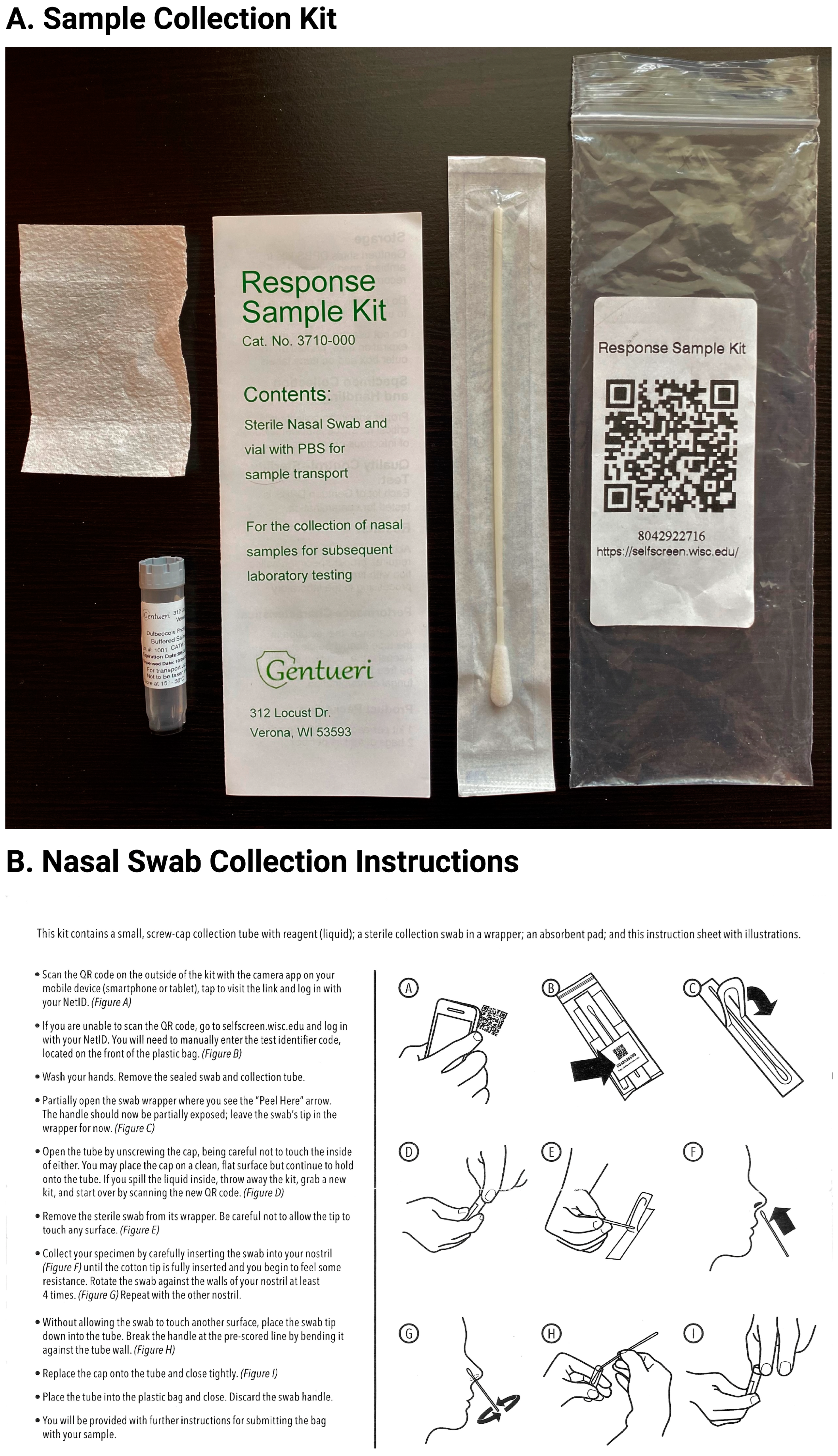
2.1. Sample Collection and Inactivation

Unsupervised self-collection was performed using the Response Sample Kit (Genturi, Verona, WI, USA). Instructions were printed and distributed in the kit, as shown (Figure 1). The kit consists of a flocked nasal swab, a 1.5 mL screw-cap tube pre-filled with 1 mL of sterile PBS, a test kit content insert, and an absorbent pad in the event of sample spilling. Briefly, participants were to wash their hands, unscrew the tube, swab inside of each nostril four times, break the swab off in the tube, and then replace the cap on the tube. The sample is then placed back in the plastic bag. Bags containing samples were opened in BSC, tubes were checked for the presence of a swab and a tight seal, and then they were placed in an autoclavable container with a lid. Once filled and sealed, the container was transferred to a 70-degree incubator for 30 min.

2.2. One-Step RT-PCR

RT-PCR Mastermix was prepared using 3 μL of 4× Taqpath 1-Step Multiplex Mastermix with Mustang Purple (Applied Biosystems, Waltham, MA, USA), 1 μL of N1-FAM Primer–Probe, 1 μL of N2-ABY Primer–Probe, 1 μL of RP-VIC Primer–Probe, and 3.5 μL of Nuclease-Free Water. The N1 and N2 primer–probe sets were designed to replicate the CDC-recommended primer–probes against viral nucleocapsid. Then, 9.5 μL of this Mastermix was added to the 3 μL sample for a total reaction volume of 12.5 μL in 1 well of a 384-well plate. Primer–probe sequences and concentrations can be found in Table 1 and were designed through Thermo Fisher Custom Oligos (Thermo Fisher, Waltham, MA, USA). Cycling was performed on a QuantStudio 7 Pro Real-Time PCR 384-well Instrument (Thermo Fisher, Waltham, MA, USA). Mock samples were prepared using the nCoV_N positive control plasmid at the indicated dilutions (copies/reaction), with the Hs_RPP30 control plasmid (IDT, Coralville, IA, USA) spiked in at 40,000 copies/reaction to ensure the RP human control signal would not diminish the viral target signals. In addition, positive controls in the form of Twist synthetic RNA (Control 2, 102024, Twist Bioscience, San Francisco, CA, USA) and the BEI-inactivated virus (NR-52286 Heat-Inactivated 2019-nCoV/USA-WA1/2020, ATCC, Manassas, VA, USA) were obtained for testing. Therefore, the assay was validated against the plasmid sequence, synthetic RNA, and the inactivated viral material.

2.3. Data Analysis

Data were exported into the Design and Analysis Software Version 2.5 for the QuantStudio 6/7 Pro systems (Thermo Fisher, Waltham, MA, USA). Amplification curve phenotypes were assessed and ΔRn thresholds were set at 1 for N1 and N2 and at 0.3 for RP. The Cq values were exported and analyzed along with “Referral” and “No Action Needed” assessments, as shown in Table 2.

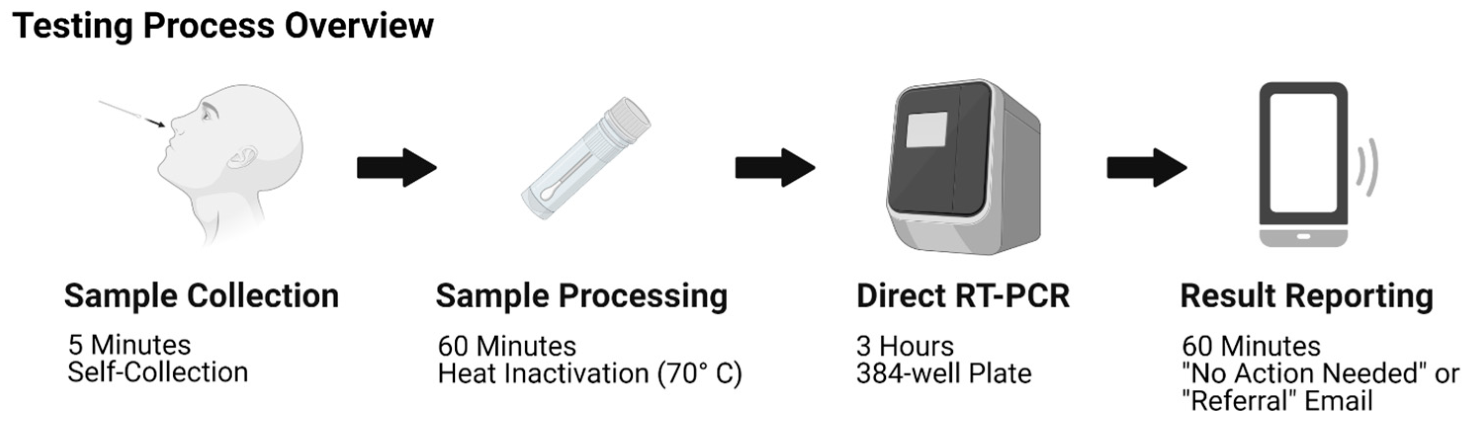
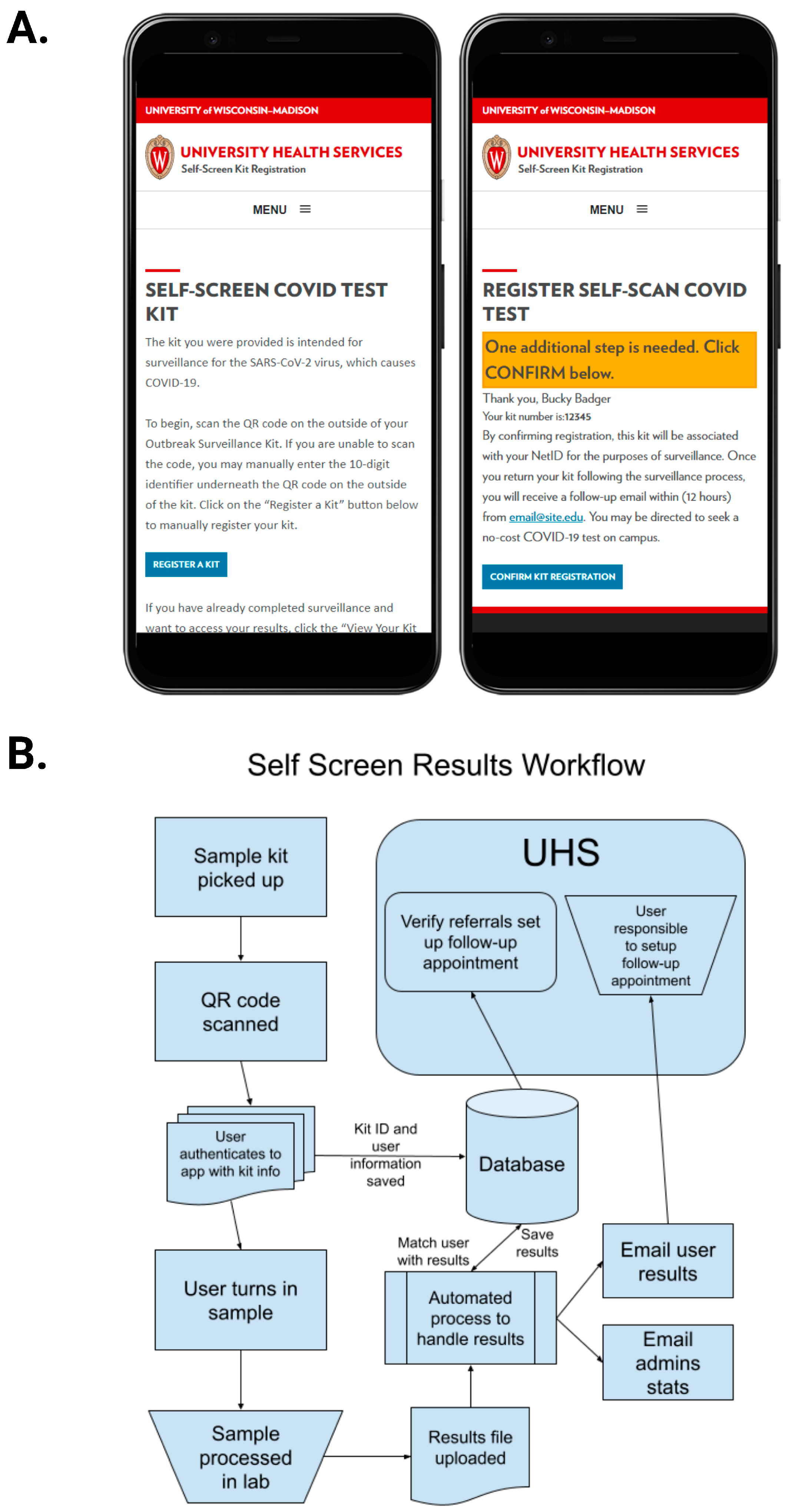
2.4. Test Kits and Testing Pipeline

An overview of the testing process is depicted in Figure 1. Testing kits were purchased locally (Gentueri, Verona, WI, USA) and included a flocked nasal swab in a protective pouch, a barcoded sample tube pre-filled with 1.5 mL of PBS, an absorbent pad, a safety insert, and sampling instructions, all in a plastic bag with a QR code on the front (Figure 2A). One limitation of early SARS-CoV-2 testing was the requirement for supervised collection to perform diagnostic clinical testing. Thus, this test was designed to be self-collected to reduce the need for staffing a collection center and provide ease of access for participants. The self-collection steps are detailed in Figure 2B.
First, participants were instructed to scan the QR code on the bag with their smartphone, which opens a website designed by the UW-Madison Division of Information Technology (DoIT) (Figure 3A). This provides a mechanism for UW-Madison staff and students to automatically link the kit barcode to their campus ID within the University Health Services record system and notifies UHS that the participant is submitting a sample. Participants were also able to use a computer to manually enter a kit ID if they did not have a smart phone or device with a working camera. This process increased the chance of user error by entering the wrong kit number, but steps were taken later during the automated process to try to validate any errors. Participants were to remove the swab from its protective pouch, swab the inside of each nostril four times, and break the flocked swab off into the PBS-filled tube. Finally, each participant sealed the tube, placed the sample in the specimen bag, and submitted it. Self-collection should take roughly five minutes. Samples were then placed in a collection box to be transported to the processing facility that day. Once the samples arrived at the processing facility, they were removed from the specimen bag in a biological safety cabinet and placed in an incubator to be heat-inactivated at 65 °C for half an hour, before being directly transferred to a 384-well plate and combined with the multiplex primer/probe assay and Taqpath Mastermix for qRT-PCR. Following qRT-PCR, the data were analyzed by a member of the testing facility and a .csv file was uploaded, with results indicating “Referral” for samples in which the viral targets were positive or “No Action Needed” for samples in which there was no viral target. This terminology was selected specifically in keeping with this test being utilized for widespread, non-diagnostic surveillance testing, such that the university could rapidly screen a large number of people and recommend follow-up diagnostic testing for a much smaller subset.
The upload location was monitored by an automated process, watching for new .csv files. When a new file was found, it was parsed, and the test results were merged with the matching user. Known controls (guaranteed “referral” or “non-referral”) in the results were validated and the administrators were alerted when a result did not match the expected outcomes (Figure 3B). The automated process then sent out a predefined email message alerting users of their results and notified the University Health Services of basic statistics from that run (e.g., total samples, total referrals, total non-referrals, missing kits, etc.). Users with a referral result were instructed to create a follow-up appointment with UHS and were able to return to the web application to obtain more information on the next steps.

3. Results

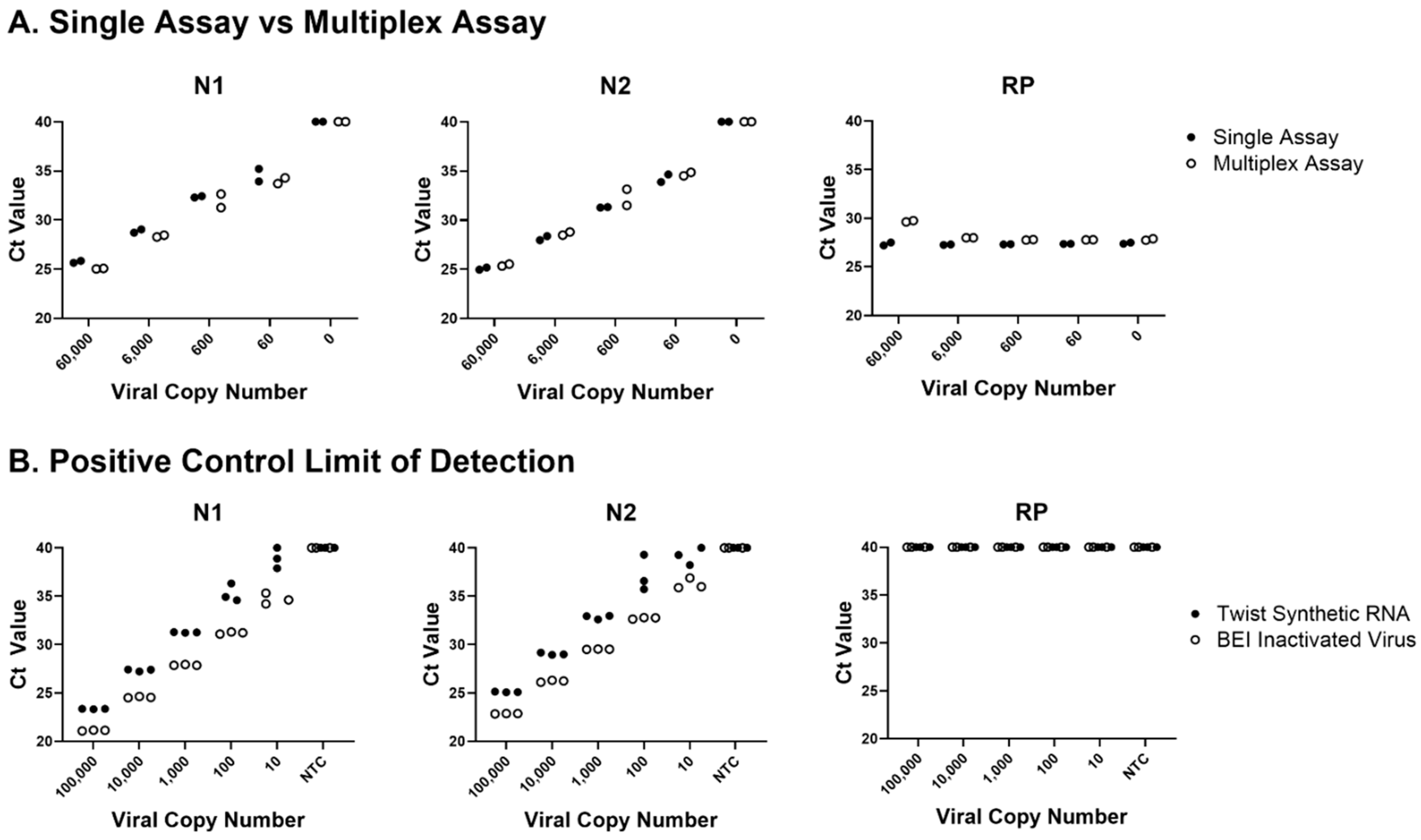
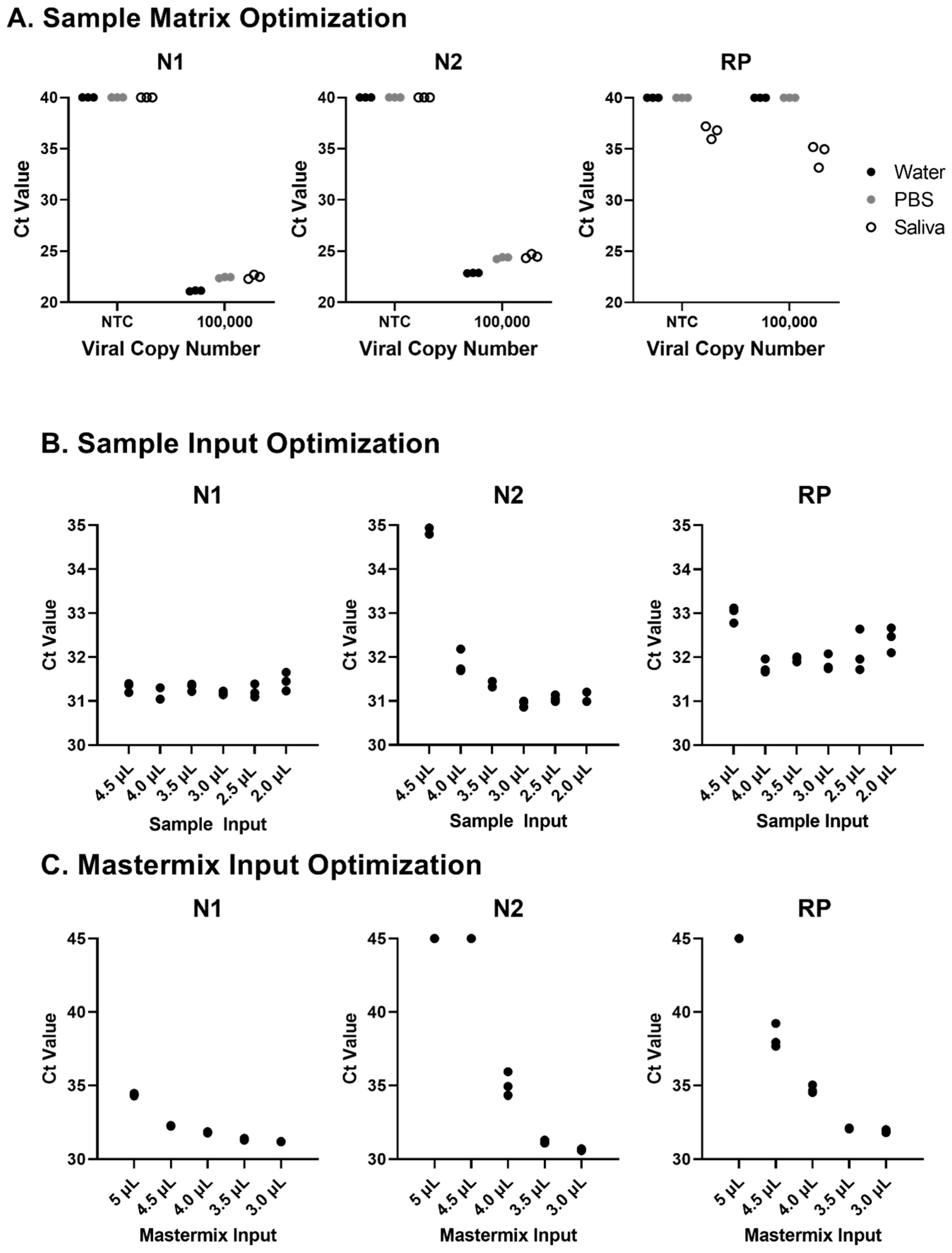
3.1. Development of a Multiplex SARS-CoV-2 qPCR Assay

We developed our own qRT-PCR assay based on CDC-recommended viral targets and controls [2]. To accomplish this, we designed a multiplex qRT-PCR assay for the viral targets (N1 and N2) and the human control (RP) using Thermo Fisher custom assays (Thermo Fisher, Waltham, MA, USA), as detailed in Table 1. Initial testing of these reagents against the CDC-recommended 2019-nCoV_N_Positive Control plasmid (IDT, Coralville, IA, USA) demonstrated similar performance using the probes alone or in combination as a multiplex assay (Figure 4A). The multiplex assay performed similarly to single assays across a dilution series of positive controls and in the presence of a high amount of human control background. Further, we validated the multiplex assay against other positive controls, including Twist synthetic RNA (Control 2, 102024, Twist Bioscience, San Francisco, CA, USA) and the BEI-inactivated virus (NR-52286 Heat-Inactivated 2019-nCoV/USA-WA1/2020, ATCC, Manassas, VA, USA) (Figure 4B). The assay showed similar results when tested against both synthetic RNA and the heat-inactivated virus. It should be noted that dilutions were calculated based on the initial concentration of the product as it arrived, which may account for the variability in Ct values between the positive controls.

3.2. Testing of Direct RT-PCR from a Nasal Swab in PBS Medium

Considering the shortage of clinical testing materials (especially RNA extraction kits and VTM) and our desire to create a rapid, inexpensive surveillance test, we developed a testing platform utilizing nasal swabs in a PBS medium. Initial testing with the BEI-inactivated virus showed similar detection of the assay using water, PBS, and saliva as media (Figure 5A). We elected to proceed with nasal swab testing in a PBS medium based on the ability to source a large number of pre-packaged PBS-filled tubes and nasal swabs (described in more detail later). Following optimization testing, we arrived at 3 μL of the PBS-based sample input and 3 μL of Taqpath Mastermix (Thermo Fisher, Waltham, MA, USA) as optimal volumes for our direct qRT-PCR testing (Figure 5B,C).

4. Discussion

The SARS-CoV-2 pandemic challenged the scientific community to respond and innovate to meet the need for viral testing. Many groups came up with innovative ways to address this problem in late 2020, including non-PCR methods, PCR testing without sample extraction, and multiplexed PCR to increase throughput and decrease the time and cost [10,11,12]. Here, we presented the testing process that our group developed in late 2020. Clinical diagnostic testing in labs with The College of American Pathologists/Clinical Laboratory Improvement Amendments (CAP/CLIA) approval was the early gold standard in testing, but its reliance on RNA extraction reagents, approved nasal swabs, and transport medium, and the need for well-trained personnel to staff the testing labs, meant that the availability of these tests early on was scarce. Other groups evaluated alternative extraction methods, the cost-effectiveness of various tests, and the use of alternative reagents to circumvent the challenges in replicating these clinical-grade tests [13,14,15]. Some groups have even suggested AI-based imaging solutions to detect COVID-19 infection due to the cost and time required to perform RT-PCR [16]. We sought to develop a non-clinical surveillance testing pipeline that bypassed these restrictions for rapid, inexpensive, and sensitive testing results to inform local decision-making. Our results show that our extraction-free multiplex RT-PCR testing platform retained detection while bypassing RNA extraction, providing a testing option that is faster, cheaper, and more scalable than traditional extraction-based clinical diagnostic testing. A limitation of extraction-free testing is that it cannot replace CAP/CLIA-approved clinical-grade testing, and as such, it will rely on those clinical tests for providing information that clinical decisions can be based on.
Our group was specifically tasked with delivering a testing process that can be completed in 6–8 h for rapid turnaround of surveillance test results to inform decision-making on our campus. We accomplished this by partnering with campus healthcare workers (University Health Services) and campus information technology specialists (DoIT) to create our testing pipeline. One major strength of our innovation here is the use of self-collection nasal swabs for obtaining the specimen, which reduced the need to staff and expose testing center personnel. Additionally, the unique QR code linked to a campus ID allows for participants to register their kit safely and securely, such that only campus healthcare information technology staff can link the kit back to them and securely report their test results through email. By creating an extraction-free direct RT-PCR testing process, we estimated that the testing facility could manually process about 1000 samples in 1 day, with a 6–8 h turnaround on those samples using only 2 testing facility staff members working full-time. This process could be dramatically scaled up with the use of robotics for RT-PCR plate preparation.
The major limitation of our study was the inability to collect and analyze real-world data. The data were collected in late 2020, at which time there were no effective vaccines widely available, and as such, the use of human COVID-19 samples was heavily restricted.
The result of this work is the establishment of infrastructure for rapid and inexpensive swab-based testing. Due to the rapid development of vaccines and the decreased need for testing, this method was not applied in a real-world scenario. However, the infrastructure and methods now exist to rapidly implement this process if needed. This testing platform has broader public health implications as it could rapidly be adapted to another outbreak or the emergence of a concerning SARS-CoV-2 variant. One would simply need to generate new primer/probe sets for another pathogen or a SARS-CoV-2 variant and verify that the current swab collection methodology works for that particular disease. In particular, this testing platform would be ideal for another respiratory agent such as influenza or another coronavirus. For example, other groups have published their platforms for COVID-19 testing as a blueprint for simplifying PCR-based clinical tests, particularly during pandemics, where standard testing supply chains are heavily impacted [6].

Author Contributions

Conceptualization, P.R.C., T.D. (Tyler Duellman), L.F.-R. and J.H.; data curation, P.R.C., T.D. (Tyler Duellman) and L.T.; formal analysis, P.R.C., T.D. (Tyler Duellman) and L.F.-R.; funding acquisition, C.K., C.A.B. and J.H.; investigation, P.R.C., T.D. (Tyler Duellman), J.-Y.C., L.W., M.Z. and J.S.; methodology, P.R.C., T.D. (Tyler Duellman) and S.S.-B.; project administration, K.T., T.M., T.D. (Tamra Dagnon), B.G., C.K. and J.H.; software, M.T., M.F. and M.B.; supervision, T.M., T.D. (Tamra Dagnon), B.G., S.S.-B., C.K., P.K., C.A.B. and J.H.; validation, P.R.C. and T.D. (Tyler Duellman); visualization, P.R.C., K.T. and C.R.; writing—original draft, P.R.C.; writing—review and editing, P.R.C. and T.D. (Tyler Duellman). All authors have read and agreed to the published version of the manuscript.

Funding

This work was supported by the National Institutes of Health (R35-ES028377, T32-GM140935, and T32-CA009135).

Institutional Review Board Statement

Not applicable.

Informed Consent Statement

Not applicable.

Data Availability Statement

The data presented in this study are available upon request from the corresponding author.

Acknowledgments

Thank you to Norman Drinkwater and the University of Wisconsin-Madison for their support in the development of this project. Thanks to Troy Slama with DoIT at UW-Madison for creating the graphics in the test inserts. Special thanks to Integrated DNA Technologies for providing variant assays for testing.

Conflicts of Interest

The authors declare no conflict of interest.

References

  1. Naming the Coronavirus Disease (COVID-19) and the Virus That Causes It. Available online: https://www.who.int/emergencies/diseases/novel-coronavirus-2019/technical-guidance/naming-the-coronavirus-disease-(covid-2019)-and-the-virus-that-causes-it (accessed on 21 April 2021).
  2. CDC 2019-Novel Coronavirus (2019-nCoV) Real-Time RT-PCR Diagnostic Panel. Available online: https://www.fda.gov/media/134922/download (accessed on 21 April 2021).
  3. Lübke, N.; Senff, T.; Scherger, S.; Hauka, S.; Andrée, M.; Adams, O.; Timm, J.; Walker, A. Extraction-free SARS-CoV-2 detection by rapid RT-qPCR universal for all primary respiratory materials. J. Clin. Virol. 2020, 130, 104579. [Google Scholar] [CrossRef] [PubMed]
  4. E Dumm, R.; Elkan, M.; Fink, J.; Richard-Greenblatt, M.; E Obstfeld, A.; Harris, R.M. Implementation of an Extraction-Free COVID Real-Time PCR Workflow in a Pediatric Hospital Setting. J. Appl. Lab. Med. 2021, 6, 1441–1451. [Google Scholar] [CrossRef] [PubMed]
  5. Smyrlaki, I.; Ekman, M.; Lentini, A.; De Sousa, N.R.; Papanicolaou, N.; Vondracek, M.; Aarum, J.; Safari, H.; Muradrasoli, S.; Rothfuchs, A.G.; et al. Massive and rapid COVID-19 testing is feasible by extraction-free SARS-CoV-2 RT-PCR. Nat. Commun. 2020, 11, 4812. [Google Scholar] [CrossRef] [PubMed]
  6. Srivatsan, S.; Heidl, S.; Pfau, B.; Martin, B.K.; Han, P.D.; Zhong, W.; van Raay, K.; McDermot, E.; Opsahl, J.; Gamboa, L.; et al. SwabExpress: An End-to-End Protocol for Extraction-Free COVID-19 Testing. Clin. Chem. 2021, 68, 143–152. [Google Scholar] [CrossRef] [PubMed]
  7. Rödel, J.; Egerer, R.; Suleyman, A.; Sommer-Schmid, B.; Baier, M.; Henke, A.; Edel, B.; Löffler, B. Use of the variplexTM SARS-CoV-2 RT-LAMP as a rapid molecular assay to complement RT-PCR for COVID-19 diagnosis. J. Clin. Virol. Off. Publ. Pan Am. Soc. Clin. Virol. 2020, 132, 104616. [Google Scholar] [CrossRef]
  8. Mohon, A.N.; Oberding, L.; Hundt, J.; van Marle, G.; Pabbaraju, K.; Berenger, B.M.; Lisboa, L.; Griener, T.; Czub, M.; Doolan, C.; et al. Optimization and clinical validation of dual-target RT-LAMP for SARS-CoV-2. J. Virol. Methods 2020, 286, 113972. [Google Scholar] [CrossRef] [PubMed]
  9. Thi, V.L.D.; Herbst, K.; Boerner, K.; Meurer, M.; Kremer, L.P.; Kirrmaier, D.; Freistaedter, A.; Papagiannidis, D.; Galmozzi, C.; Stanifer, M.L.; et al. A colorimetric RT-LAMP assay and LAMP-sequencing for detecting SARS-CoV-2 RNA in clinical samples. Sci. Transl. Med. 2020, 12, eabc7075. [Google Scholar] [CrossRef]
  10. Chen, L.; Zhang, G.; Liu, L.; Li, Z. Emerging biosensing technologies for improved diagnostics of COVID-19 and future pandemics. Talanta 2021, 225, 121986. [Google Scholar] [CrossRef] [PubMed]
  11. Maricic, T.; Nickel, O.; Aximu-Petri, A.; Essel, E.; Gansauge, M.; Kanis, P.; Macak, D.; Richter, J.; Riesenberg, S.; Bokelmann, L.; et al. A direct RT-qPCR approach to test large numbers of individuals for SARS-CoV-2. PLoS ONE 2020, 15, e0244824. [Google Scholar] [CrossRef] [PubMed]
  12. Nyaruaba, R.; Li, C.; Mwaliko, C.; Mwau, M.; Odiwuor, N.; Muturi, E.; Muema, C.; Xiong, J.; Li, J.; Yu, J.; et al. Developing multiplex ddPCR assays for SARS-CoV-2 detection based on probe mix and amplitude based multiplexing. Expert Rev. Mol. Diagn. 2021, 21, 119–129. [Google Scholar] [CrossRef] [PubMed]
  13. Perez, V.P.; Pessoa, W.F.B.; Galvão, B.H.A.; Sousa, E.S.S.; Dejani, N.N.; Campana, E.H.; Cavalcanti, M.G.D.S.; Cantarelli, V.V. Evaluation of alternative RNA extraction methods for detection of SARS-CoV-2 in nasopharyngeal samples using the recommended CDC primer-probe set. J. Clin. Virol. Plus 2021, 1, 100032. [Google Scholar] [CrossRef] [PubMed]
  14. Dhakad, M.S.; Gogoi, S.; Kumari, A.; Singh, A.K.; Jais, M.B.; Prakash, A.; Pangtey, G.; Kaur, R. Comparative evaluation of cost effective extraction free molecular technique for detection of SARS-CoV-2 with reference to standard VTM based RT-qPCR method. Iran. J. Microbiol. 2021, 13, 748–756. [Google Scholar] [CrossRef] [PubMed]
  15. Stanton, J.-A.L.; O’Brien, R.; Hall, R.J.; Chernyavtseva, A.; Ha, H.J.; Jelley, L.; Mace, P.D.; Klenov, A.; Treece, J.M.; Fraser, J.D.; et al. Uncoupling Molecular Testing for SARS-CoV-2 From International Supply Chains. Front. Public Health 2022, 9, 808751. [Google Scholar] [CrossRef] [PubMed]
  16. Das, N.N.; Kumar, N.; Kaur, M.; Kumar, V.; Singh, D. Automated Deep Transfer Learning-Based Approach for Detection of COVID-19 Infection in Chest X-rays. Ing. Rech. Bioméd. 2022, 43, 114–119. [Google Scholar] [CrossRef]
Figure 1. Testing process. This schematic depicts the extraction-free surveillance testing workflow we developed. Briefly, the participant registered the testing kit to their campus ID using the QR code provided and their mobile device, self-collected a nasal specimen, placed it in a screw-cap tube containing 1 mL of PBS, and then submitted their test. The samples were handled in a biological safety cabinet and heat-inactivated at 70 °C for 30 min. A 3 µL sample was then added directly to the qRT-PCR Mastermix in a 384-well plate, which was run on a Quantstudio 7 Pro platform. Data analysis was performed, and a .csv file was uploaded with sample ID and an interpretation of “No Action Needed” for negative tests or “Referral” for positive tests, or tests where the internal control failed. The test results were then matched to the campus ID and the email of the participant who registered the test, and the result was sent by email. The entire process was intended to be completed in 6–8 h, and the capacity can be easily increased with the use of robotics for sample handling. The figure was created with BioRender.com.
Covid 03 00075 g001
Figure 2. Sample collection kit. (A) The sample collection kit is pictured. The entire kit was delivered in a plastic bag with a QR code and a unique test ID, which allowed the user to register the kit by going toa designated URL. The kit consists of a flocked nasal swab, a 1.5 mL screw-cap tube pre-filled with 1 mL of sterile PBS, a test kit content insert, and an absorbent pad in the event of sample spilling. (B) The nasal swab collection instructions contain clear imagery and instructions to show the participant exactly how to register the kit and self-collect their nasal specimen. The instructions are also available in other languages. The figure was created with BioRender.com.
Covid 03 00075 g002
Figure 3. Testing kit registration and testing result workflow. (A) A mockup of the self-screen registration page is shown on a mobile device with a welcome screen to register the kit and a confirmation screen to link the scanned kit to the user’s ID, along with preliminary instructions on what follow-up may be required. (B) The self-screen workflow is depicted, showing how samples were linked to user identifiers. The University Health Services (UHS) was notified of the results to verify individuals needing follow-up, and emails were sent to the user with instructions that indicate if follow-up is necessary. The figure was created with BioRender.com.
Covid 03 00075 g003
Figure 4. Development of the multiplex SARS-CoV-2 assay. Our SARS-CoV-2 multiplex assay was developed based on the primer/probe set recommended by the CDC for ease of obtaining Emergency Use Authorization if needed. (A) The primer/probe sets utilized showed similar detection of viral nucleocapsid targets (N1 and N2) and the human control target (RP) over a dilution series of positive control SARS-CoV-2 nucleocapsid plasmid, with a background of 30,000 copies/µL RP plasmid. (B) Repeat testing of the multiplex assay with more physiologically relevant positive controls, including synthetic RNA (Twist Biosciences) and the heat-inactivated virus (ATCC), showed detection of viral genetic material over a dilution series. Of note, we observed that Twist synthetic RNA degraded faster in solution than the inactive viral samples, which may account for the slightly higher Ct values shown.
Covid 03 00075 g004
Figure 5. Matrix testing and optimization of the Mastermix and sample volume. (A) Optimization of the multiplex SARS-CoV-2 involved testing the assay performance in water, PBS, and saliva, showing similar detection in all three solutions at 100,000 copies of heat-inactivated SARS-CoV-2 virus (ATCC). Based on these data, we proceeded with a PBS-based nasal swab test as it maintained similar detection of the sample in water and was easier and safer for us to collect, inactivate, and test. (B) After selecting a PBS-based assay, we optimized the amount of sample added to the qRT-PCR reaction. The N1 primer/probe set performed well across all conditions, but there were some signal issues with the N2 primer/probe set with high sample input. We decided to stick with the 3 µL sample input, as this yielded the lowest average Ct values across primer/probe sets and would allow for plenty of residual sample for repeat testing, variant testing, and sequencing. (C) We also optimized the amount of Taqpath Mastermix added to the reaction and determined that 3 µL of Taqpath produced optimal Ct values across primer/probe sets.
Covid 03 00075 g005
Table 1. SARS-CoV-2 multiplex assay reagents.
Primer/Probe NameSequence (5′→3′)
2019-nCoV_N1-FWDGACCCCAAAATCAGCGAAAT
2019-nCoV_N1-REVTCTGGTTACTGCCAGTTGAATCTG
2019-nCoV_N1-ABYABY-ACCCCGCATTACGTTTGGTGGACC-QSY
2019-nCoV_N2-FWDTTACAAACATTGGCCGCAAA
2019-nCoV_N2-REVGCGCGACATTCCGAAGAA
2019-nCoV_N2-FAMFAM-ACAATTTGCCCCCAGCGCTTCAG-QSY
RP-FWDAGATTTGGACCTGCGAGCG
RP-FEVGAGCGGCTGTCTCCACAAGT
RP-VICVIC-TTCTGACCTGAAGGCTCTGCGCG-QSY
A list of the primer and probe names and sequences as used in the SARS-CoV-2 multiplex assay. N1 and N2 denote nucleocapsid 1 and nucleocapsid 2, respectively, referring to probes against the nucleocapsid sequence. RP denotes RNase P, a common control probe. FWD denotes forward primer. REV denotes reverse primer. ABY, FAM, and VIC denote the fluorescent labels each probe contains.
Table 2. Presence/absence call settings.
Presence TargetsAbsence TargetsCallAssessment
N1, N2, RP PresenceReferral
N1N2, RPPresenceReferral
N2N1, RPPresenceReferral
N1, RPN2PresenceReferral
N2, RPN1PresenceReferral
N1, N2RPPresenceReferral
N1, N2, RPInconclusiveReferral
RPN1, N2AbsenceNo Action Needed
The presence and absence settings used in the design and analysis presence/absence module to make a call and deliver an assessment result. N1 and N2 denote viral nucleocapsid targets, whose presence is determined to mean presence of the virus. RP denotes RNase P, a control target that should be present in all human samples. Any samples that detected N1 or N2 in any combination resulted in a referral for diagnostic testing. Only samples that resulted in a positive RP without N1 or N2 were deemed “No Action Needed”. Samples negative for all three targets were likely poor-quality or uncollected specimens.
Disclaimer/Publisher’s Note: The statements, opinions and data contained in all publications are solely those of the individual author(s) and contributor(s) and not of MDPI and/or the editor(s). MDPI and/or the editor(s) disclaim responsibility for any injury to people or property resulting from any ideas, methods, instructions or products referred to in the content.

