Article

Breaking the Mold: Towards Rapid and Cost-Effective Microbial Contamination Detection in Paints and Cosmetics Using ATP-Bioluminescence

Department of Biotechnology & Food Engineering, MCI—The Entrepreneurial School, Maximilianstraße 2, 6020 Innsbruck, Austria
*
Authors to whom correspondence should be addressed.
Appl. Microbiol. 2024, 4(2), 582-593; https://doi.org/10.3390/applmicrobiol4020040
Submission received: 23 February 2024 / Revised: 18 March 2024 / Accepted: 20 March 2024 / Published: 22 March 2024

Abstract

:
Traditional culture-based methods, though a “gold standard” for bacterial detection in various industrial sectors, do often not fulfill today’s high requirements regarding rapidity, on-site applicability, and cost-efficiency both during operation and evaluation. Here, the feasibility of using an adenosine triphosphate (ATP)-based assay for determining microbial contaminations in paints and cosmetics was investigated and compared with standard plate count techniques and dipslides. Therefore, we initially determined the level of sensitivity and assessed the accuracy and concordance among the different methods via spiking tests using a mix of frequently abundant bacterial species to simulate microbial contamination. Bioluminescence intensity was linearly proportional to log colony counts over five orders of magnitude (R2 = 0.99), indicating a high level of sensitivity. Overall, the accuracy varied depending on the test specimen, most probably due to matrix-related quenching effects. Although the degree of conformity was consistently higher at target concentrations ≥ 105 CFU·mL−1, microbial contaminations were detectable down to 103 CFU·mL−1, thus meeting the high requirements of various industries. ATP-based results tended to be within an order of magnitude lower than the reference. However, bearing that in mind, the developed assay serves as a rapid, real-time alternative for routine quality control and hygiene monitoring.

Graphical Abstract

1. Introduction

Standardized methods (e.g., ISO methods) constitute the reference analytical methods for routine quality and hygiene monitoring across many end-use industries. These standardized methods are foremost comprising traditional culture-based methods, using nutritious broth or agar media to grow, isolate, and enumerate various microorganisms [1]. They have long been considered the gold standard method for bacterial and fungal identification. However, they present certain inherent drawbacks. Amongst others, culture-dependent methods are laborious to perform, encompass time-consuming procedures during operation and data collection, and provide results only retrospectively [1]. Thus, they are not sufficiently sensitive to meet the industry’s requirements in terms of rapidity and on-site applicability [2]. Further limitations include possible preceding enrichment steps and the choice of media and/or incubation conditions that orientate the microbial groups to be cultivated. Most importantly, many microorganisms are not (yet) cultivable or exist in a viable but non-cultivable (VBNC) state, allowing microbial contaminations to arise quickly and without any warning [3]. To comply with those requirements, culture-independent methods have been recognized as a promising alternative to be used in conjunction with, or even supplanting, culture-dependent methods [4]. In this context, direct epifluorescence filter techniques (DEFT) and bioluminescence ATP, next to modern molecular approaches using genomic and proteomic information, are increasingly being advocated as promising alternatives. The latter comprises polymerase chain reaction (PCR), quantitative PCR (qPCR), digital PCR (dPCR), high-throughput next-generation DNA sequencing, matrix-assisted laser desorption ionization-time-of-flight (MALDI-TOF) MS, ion mobility spectrometry (IMS) or determination of biomarker volatile organic compounds (MVOC) by solid-phase microextraction gas chromatography-mass spectrometry [5,6,7,8,9]. Matching with culture-independent methods, we succeeded in employing qPCR and most-probable number techniques for detecting paint-spoiling bacteria in general and sulfate-reducing bacteria in particular in a previous investigation [10]. Alternative methodologies to classical microbiological counting are intended to save materials and time. Although molecular approaches benefit from omitting the culturing step and high specificity, they are sensitive to PCR inhibitors, sequencing errors, and overestimation, along with high costs for consumables, instruments, or analysis software and require technical expertise [9]. ATP assays are easy to use and have a short detection cycle for determining the level of organic matter, and are thus suitable as an early warning system for potential contamination and as a long-term continuous monitoring and sanitation tool that is independent of pre-culturing as well [2,11,12]. Commercially available kits are able to detect femtomoles or less of ATP using luminescence, though they require specialized instrumentation and trained personnel. The method typically involves the addition of an ATP-releasing reagent to lyse cells and release ATP, which, in the presence of luciferase, reacts with the substrate, d-luciferin, to produce light [13]. The intensity of emitted light can be measured as relative light units (RLU), which is interpreted as a measure of ATP concentration and an indicator of the number of viable cells [4]. Alternatively, as the intracellular concentration of ATP per cell has been shown to vary with changing environmental and physiological conditions, it can be used as a proxy to indicate metabolic activity [14].
In view of increasing consumer awareness of the harmful effects of volatile organic compounds (VOCs) present in traditional paints, water-based paints are witnessing significant growth due to their low VOC content; however, they are more susceptible to microbial contamination [15,16,17]. Together with tightening regulations regarding the use of biocides (e.g., BPR, Regulation (EU) 528/2012, refer to https://eur-lex.europa.eu/eli/reg/2012/528/oj, accessed on 23 February 2024), industries have called upon intensified searches for alternative methodologies for rapid presumptive testing of microbial contaminations in paints. Rapid hygiene monitoring tests based on the presence of ATP are a popular choice in the food and beverage industry; however, this market finds increasing applications in various sectors such as water treatment, healthcare, and pharmaceutical manufacturing, both at the formulation development stage (i.e., preservative screening) and for microbiological quality assurance. The objective of the present study was thus to develop a sensitive, high-throughput ATP-luminescence assay using samples from the paint and cosmetic industry (both comprising rather complex, viscous matrices) as test specimens and determine benefits and possible limitations of using a culture-independent method relative to frequently used culture-dependent methods. Therefore, we initially determined the level of sensitivity and subsequently assessed the applicability and ability to detect bacterial abundance and viability via targeted matrix spiking and recovery assays and compared the obtained results with those derived from traditional plate count techniques and dipslide test assays.

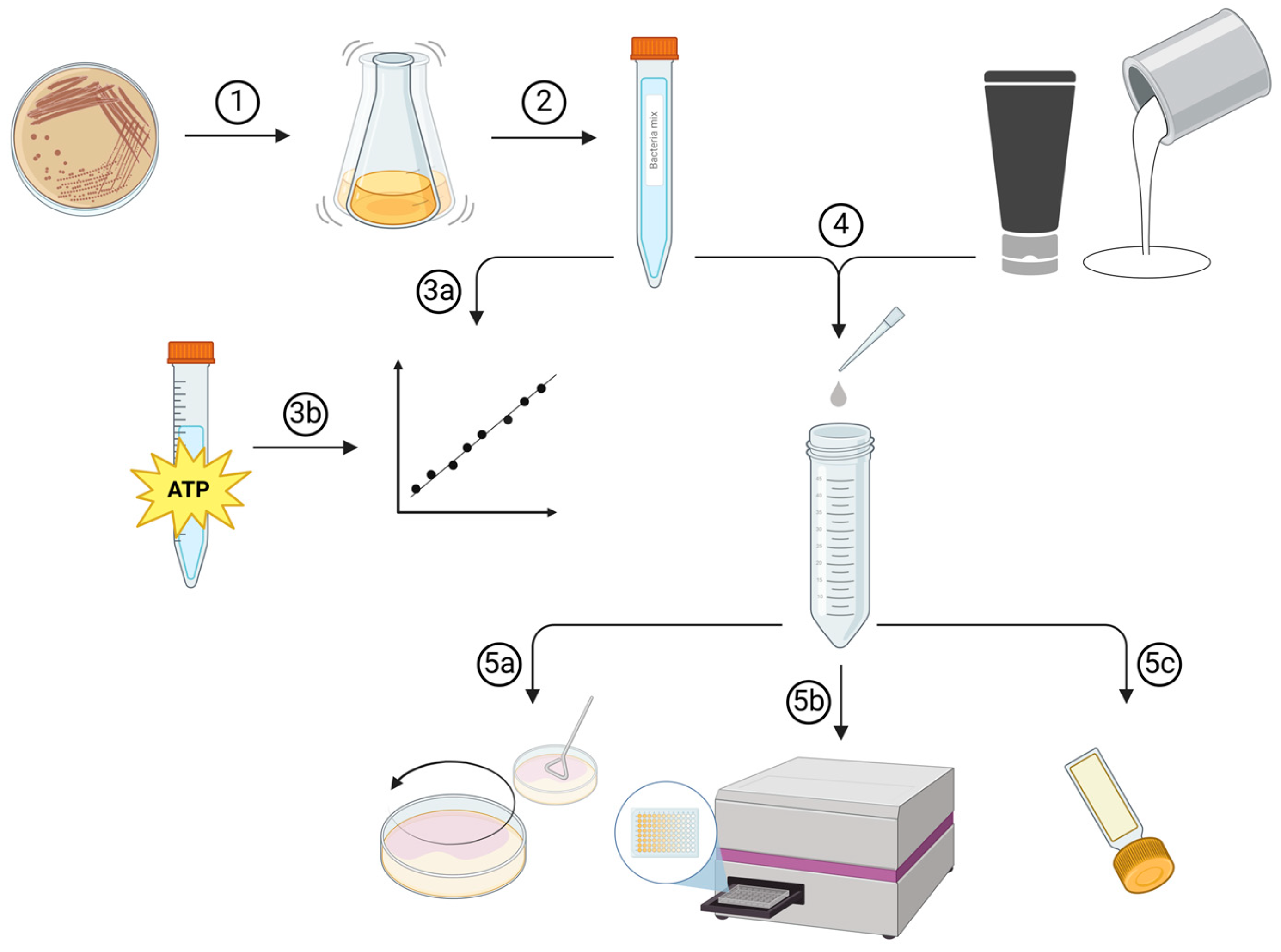
2. Materials and Methods

The experimental setup is graphically displayed in Figure 1, comprising 5 major consecutive steps. Briefly, paint and cosmetic samples were used as test matrices employing four different bacterial species as targets that were commonly found in both product lines. A mixed bacterial culture was used to generate calibration curves and to inoculate the samples for the spiking experiments simulating microbial contamination (mock specimens). Bacterial viability was determined via ATP luminescence assays and compared with traditional cultivation techniques, including aerobic plate count (gold standard) and commercially available contact agar (dipslides, representing easy-to-use culture tests for on-site monitoring of microbial contaminations in industrial environments such as paint manufacturing and to test used liquids and/or creams in cosmetic industries) for technical validation.

2.1. Test Specimens

In the present investigation, four different samples either derived from the cosmetic or paint industry served as test specimens covering a semi-solid water-in-oil (WO) or oil-in-water (OW) emulsion, a water-based acrylic wall paint (WP) and a wood stain (WS). All test products exhibit pH values in the near neutral to alkaline range (cosmetics: pH 6.8–6.9; paints: pH 7.7–8.0) and did not reveal any preload microbiological contaminations.

2.2. Test Organisms, Growth Conditions, and Preparation of Pure and Mixed Inocula

Escherichia coli (DSM 4230), Brevundimonas vesicularis (NCIMB 13293; former Pseudomonas vesicularis), Bacillus subtilis (DSM 10), and Micrococcus luteus (DSM 20030) were used as representative Gram-negative and Gram-positive bacteria in the present investigation that correspond to specified organisms according to the American Society for Testing and Materials (ASTM D5588-97). For the experiments, the bacterial strains were kept in cryovials at −80 °C and streaked on CASO agar plates (Carl Roth GmbH & Co. KG, Karlsruhe, Germany; comprised of enzymatic digests of casein and soybean meal equivalent to TSB). After 24 to 72 h, single colonies were picked to inoculate 50 mL of CASO medium (Carl Roth GmbH & Co. KG, Karlsruhe, Germany). Cultures were incubated in a shaking incubator at 110 rpm and 30 °C until reaching the stationary phase. Subsequently, the cultures were washed three times with phosphate-buffered saline (PBS) by centrifugation at 3000 rpm for 10 min and 4 °C and adjusted to an initial concentration of 1 × 10⁹ CFU mL−1 using a counting chamber (Neubauer improved, Marienfeld). Depending on the following experiment, the suspension was either diluted further or a mixed bacterial culture (co-culture of the four individual strains) with an initial concentration of 1 × 10⁹ CFU mL−1 was prepared as described in detail below.

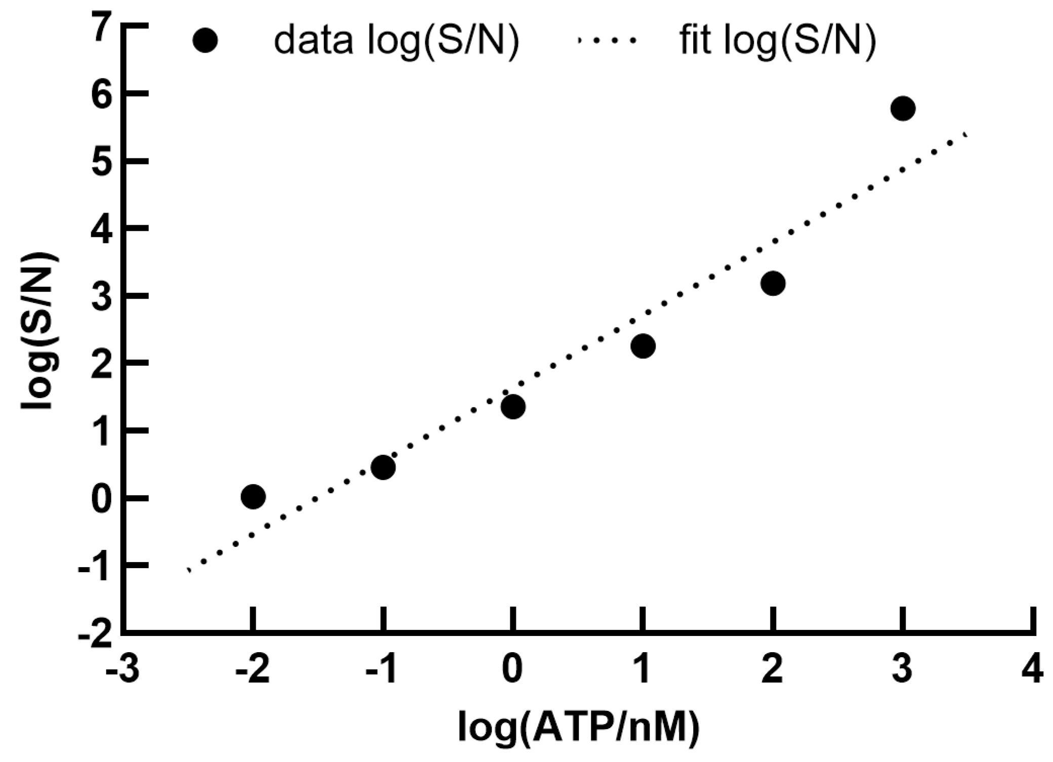
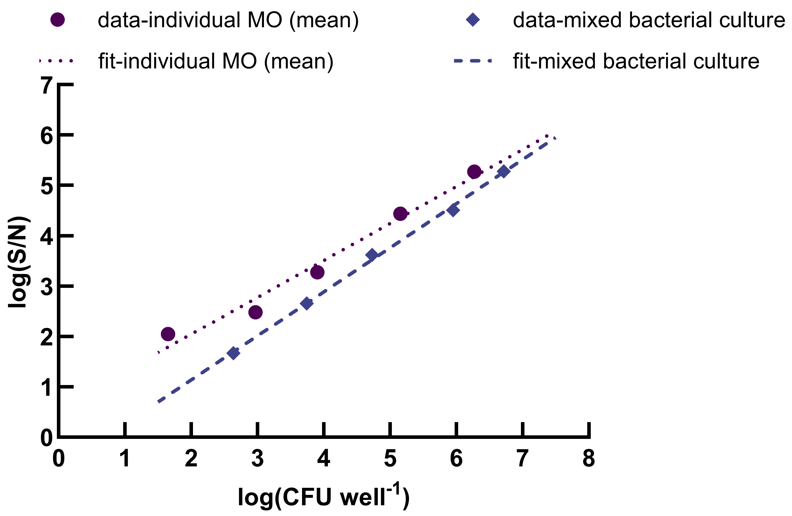
2.3. Calibration Curves
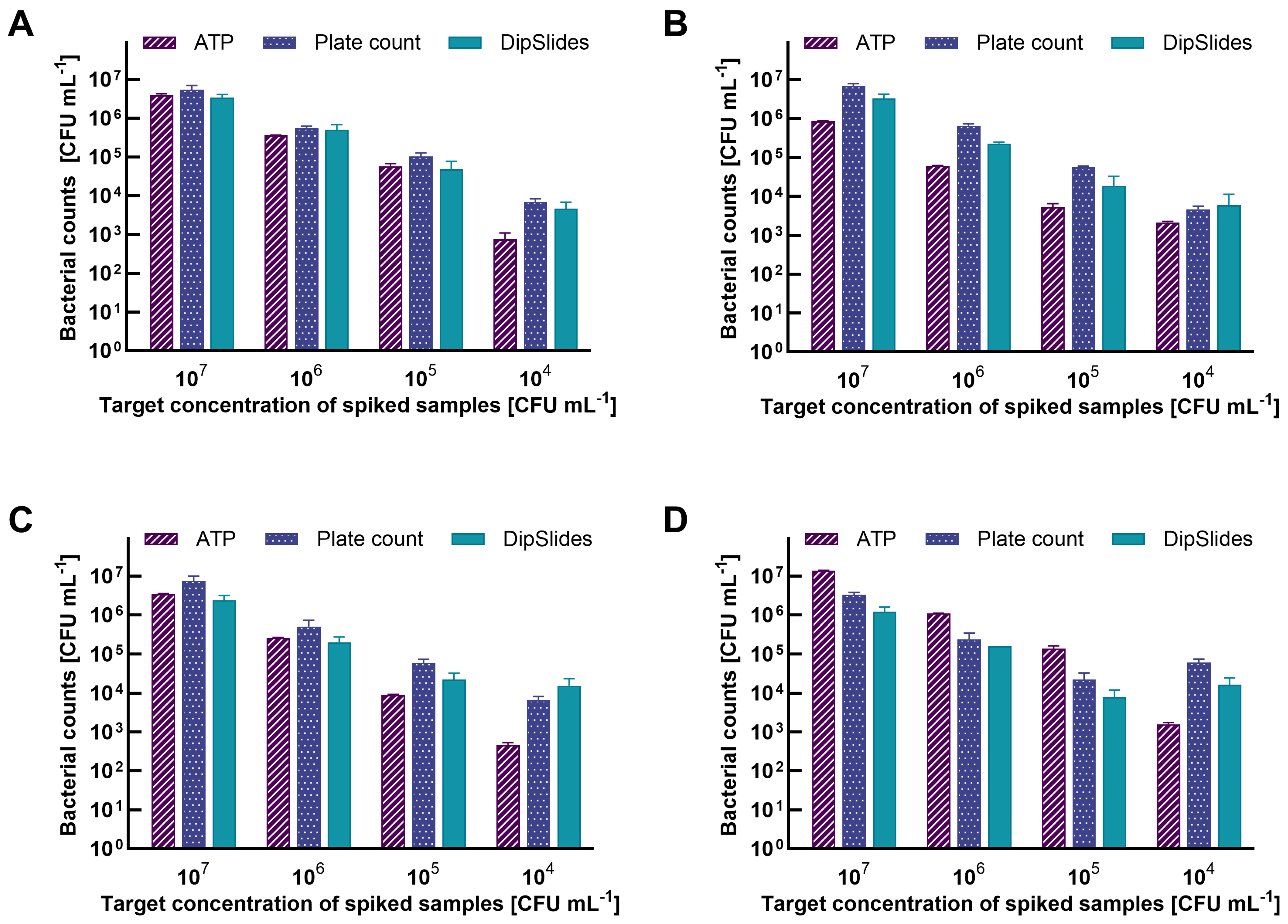
Calibration curves were generated to correlate RLU readings to CFU for enumerations of bacteria and used for linearity and limit of detection (LOD) assessment. Individual calibration curves were generated for each test organism as well as the mixed bacterial culture by serially diluting (10-fold) the suspensions with PBS until a final concentration of 1 × 10² CFU mL−1. Concentrations ranging from 1 × 10⁸ CFU mL−1 to 1 × 10² CFU mL−1 were used in triplicate to measure luminescence and simultaneously verified via plate count (the maximum concentration of 1 × 10⁹ CFU mL−1 was not used as it lies above the upper limit of detection of the microplate reader). Luminescence was recorded using a microplate reader (Mithras LB 940, Berthold Technologies, Bad Wildbad, Germany). Linearity of the ATP standard was evaluated following the manufactural instructions of BacTiter-Glo™ (Promega, Madison, WI, USA). Hence, purified ATP was dissolved in PBS to a final working concentration of 1 μM and then tenfold diluted until 10 fM. Each diluted concentration was measured in triplicate. Measured RLU values from the microplate reader were calculated as signal-to-noise (S/N) ratio according to Equation (1) from Zhang et al. [18]:
S / N = RLU s mean   RLU B S s d B S
where RLUs stands for the relative luminescence unit of the sample, mean RLUBS stands for the mean relative luminescence unit of the background signal, and sdBS stands for the standard deviation from the background signal.
S/N values from the standard curve were plotted against ATP concentrations or determined CFU well−1 in logarithmic mean values. To compare the ATP assay results from the spiking test with the results derived from plate counts and dipslides, S/N values were converted into CFU mL−1 using a linear regression analysis with y-values as S/N and x-values as CFU mL−1 (Equation (2)):
log S / N = k · log ( CFU · mL 1 ) + d
Data from the generated standard curve using the mixed bacterial inoculum were used for linear regression modeling using the reverse function of Equation (2) to calculate CFU mL−1 (Equation (3)):
10 log S / N d k · mL 1 = CFU · mL 1

2.4. Matrix Spiking

Mock specimens were prepared by spiking 40 µL of the prepared mixed bacterial suspensions at four target concentrations comprising 1 × 10⁷, 1 × 10⁶, 1 × 10⁵, and 1 × 10⁴ CFU mL−1. The initial spiking concentrations used as reference were cross-checked via plate count prior to inoculation (Table 1). Undiluted products were used to assess the applicability of ATP-based assays for the screening of complex, highly viscous samples. The spiked samples were then vortexed for homogeneous distribution and immediately processed to avoid both any antimicrobial effects originating from the products and potential biofilm formations of the target organisms that might impede detachment of the cells and thus bias actual cell counts. PBS containing the same concentrations of bacterial inocula served as positive control, while PBS without any inoculation served as negative control. PBS was chosen as it does not exhibit any matrix-induced quenching effects. The average background signal for PBS was 333 ± 191 RLU. Trials showed no significant difference in background noise from complex matrix samples compared with PBS.

2.5. Test Protocol and Method Validation

Subsequently, each mock (spiked) sample was diluted with an appropriate buffer and agitated to detach the cells. For the paint and stain, PBS was used as the diluent, while PBS containing 0.1% Tween® 80 was utilized for the OW and WO emulsions. Each sample was serially diluted (tenfold) prior to performing recovery assays via plate count and ATP luminescence-based methods, as well as dipslide culture tests, as described in detail below. Samples without spiking served as negative controls. Recovery assays were performed in triplicates. The results were evaluated in terms of precision (closeness between independent test results), accuracy (closeness to target value), and concordance among the different methods.

2.5.1. Standard Plate Count

To create bacterial standard curves and to prepare defined inocula for the spiking experiments, spread plate assays were used. For suspensions with low concentrations (target concentration 103 CFU mL−1), pour plate assays were used. Therefore, 100 μL aliquots were either plated on CASO agar plates or 1 mL of the diluted sample was pipetted into the petri dish and covered with tempered liquid CASO agar (according to ISO 4833-1, Austrian Standards International 2022-07-15, https://www.austrian-standards.at, accessed on 23 February 2024). All plates were incubated at 30 °C for 24 to 72 h prior to CFU enumeration.

2.5.2. ATP Bioluminescence Assay

The development of the ATP assay method was carried out according to the manufacturer’s instruction (Promega, Madison, WI, USA) with some minor modifications: the buffer solution and lyophilized BacTiter-Glo™ substrate (Promega, Madison, WI, USA) were equilibrated at room temperature for 24 h before use. After mixing the two components, the ATP reagent was equilibrated at room temperature for at least 10 h to achieve maximum sensitivity. During this “burn-off” process, the background bioluminescence signal decreases as ATP in the reagent, introduced during the reconstitution step, is hydrolyzed (see Supplementary Figure S1).

2.5.3. Dipslides

Dipslides were used according to the manufacturer’s instructions (Carl Roth, Karlsruhe, Germany). Therefore, CASO-TTC/RBCenr dipslides were immersed in triplicate for a few seconds in the desired dilution and the excess sample was carefully drained off. The samples were then incubated at 30 °C for 72 h. The evaluation is semi-quantitative and was performed according to the approximation specified by the manufacturer (Carl Roth, Karlsruhe, Germany). The detection limit is 102 CFU mL−1 solution.

2.6. Statistical Analyses

Statistical analyses were performed using GraphPad Prism v.10.2.1 (GraphPad Software, La Jolla, CA, USA). Results are given as mean (±standard deviation), n = 3 if not specified otherwise.

3. Results and Discussion

In the present study, ATP bioluminescence was produced by using the standard procedure of adopting a standard ATP solution with varying concentrations ranging from 10 fM (lowest detectable concentration) to 1 µM. A typical dose-response curve between bioluminescence intensity (S/N) and ATP concentration (nM) (both log-transformed) is shown in Figure 2. The bioluminescence intensity increased in a dose-dependent manner with the ATP concentration, revealing a linear relationship (R2 = 0.92) for log-transformed data. The analytical sensitivity of the assay (LOD) was determined via serial dilutions. The amount of ATP was further directly proportional to viable cell counts, enabling RLU values to be converted into ATP concentration and subsequently into CFU mL−1 through the approximation of 7 fg cell−1.
The standard bioluminescence assay was further applied to different cell densities of each target organism (Supplementary Figure S2) or a mix of each of them as a source of ATP (Figure 3). The log(S/N) ratios were linearly proportional to log colony counts over five orders of magnitude (102 to 106 CFU mL−1) with a slope of 0.7331 and 0.8746 for individual bacterial suspensions and the bacterial mix, respectively. Similar observations were found by Luo et al. [19], who showed a linear response to pure bacterial cultures at a concentration range from 103 to 108 CFU mL−1. Good linearity between ATP bioluminescence and colony numbers for various microorganisms in clean-room facilities was also reported by Venkateswaran et al. [20], indicating that the ATP extraction efficiency was not affected by the concentration of microorganisms. The literature on microbial contamination detection using ATP assays was foremost performed in the food sector, and data on paints and cosmetics are rather scarce. However, Montanez et al. [21] studied the relationship between the amount of culturable microbial cells and the amount of cellular ATP in four different aqueous polymer emulsions and found a strong correlation with R2 consistently > 0.96. Connolly et al. [22] also found a good correlation between ATP levels and CFU for different bacteria, including Pseudomonas sp., in cosmetics achieved over four orders of magnitude with R2 > 0.85. Actually, pure bacteria are rather rarely encountered but are more commonly found in consortia of different microbial strains equal to the prepared bacterial mix in the present investigation. In this context, our results compare well with those from Narsaiah et al. [23], observing a linear correlation between ATP assay and plate count for a simulated bacteria-mix extracted from mango fruit surfaces. The line of best fit for the bacterial mix taking (log-transformed) ATP (S/N signals) and total plate counts (CFU well−1) into account gives a slope of 0.8746 and an intercept of −0.6138 with a correlation coefficient of R2 = 0.998 (Figure 3), thus indicating a high sensitivity of the assay that allows to capture even tiny alterations in ATP levels within the sample.
Subsequently, spiking experiments were performed to validate the applicability, precision, and accuracy of the proposed ATP assay in detecting bacterial contaminations in real samples. Therefore, paint and cosmetic samples were spiked with four different target concentrations (ranging from 104 to 107 CFU mL−1) and screened for bacterial counts using the developed luminescence assay, traditional plate count techniques, and dipslides simultaneously. Negative samples without spiking were measured concurrently and did not show any positive signal during ATP determination nor reveal any formed colonies both on agar plates and dipslides. In terms of precision, results obtained from the ATP assay showed the lowest dispersion among the three methods and a constant log-linear decline among the different target concentrations. The sensitivity of the applied ATP assay varied depending on the sample matrix (Figure 4), with a high concordance between the different methods using WS as a test sample (Figure 4A). Moreover, the results revealed a surprisingly high degree of conformity to the expected target concentration ranging from 105 to 107 CFU mL−1, indicating a high accuracy (Figure 4A). However, at low inoculation densities of <105 CFU mL−1, the results deviated stronger from the reference, with the determined cell counts being about an order of magnitude lower than the reference (Figure 4A). A similar trend was observed using WP as a mock (spiked) specimen, with the actual bacterial counts obtained with the developed ATP assay being consistently lower (about an order of magnitude) than the expected target concentration (Figure 4B). Overall, the results obtained using traditional plate count techniques tended to be higher than those determined via ATP bioluminescence (Figure 4A,B). In this context, quenching of emitted light is an important factor that can adversely affect microbial ATP determination. Certain inherent paint characteristics and/or compounds can strongly reduce the amount of light measured photometrically, which might make the use of internal standards necessary, as previously shown with skimmed milk [2]. In this regard, the intense white color along with high turbidity, might have masked the luminescence signal, thus leading to lower cell counts than expected.
Similar to the results obtained using WP as a test matrix, the results of the ATP assay using the OW emulsion as a mock specimen showed comparable results with plate count techniques and dipslides at target cell titers > 105 (Figure 4C), with the resulting cell counts among the three different methods being within one order of magnitude. Contrarily, cell counts determined via ATP bioluminescence tended to be lower than those obtained via plate count and the reference at concentrations ≤ 105 (Figure 4C). It is worth noting that different bacteria have different amounts of ATP per cell, with previously reported values varying considerably. Among the factors that affect ATP contents of the cells are medium, metabolic state, and the presence of metabolic inhibitors that impact the relationship between cell counts and luminescence [24]. Regarding the growth phase, Bacillus-related species confront nutrient deprivation at the onset of the stationary growth phase through sporulation [25]. It is well known that stationary-phase cells secrete ATP less efficiently than exponential-phase cells as they may use ATP to maintain cellular functions and prepare for regrowth [26], which was confirmed in an additional experiment comparing cells harvested during the exponential and stationary phase and by relating luminescence to initial cell concentrations (Supplementary Figure S3). Previous studies reported that the measured ATP levels in vegetative cells were at least one order of magnitude higher than in dormant spores [27], which could have led to lower actual cell counts as expected (Figure 4A–C). Bacterial endospores remain in a metabolically inert state that is resistant to manifold environmental stressors, including heat, ultraviolet (UV) light damage, and desiccation, until sensing more favorable conditions and resuming vegetative growth [28], which might further explain why cell counts were about an order of magnitude lower using the ATP assay compared with plate count, as dormant spores might be capable of fully germinating on solid media. The stationary phase was, however, chosen as the release of extracellular ATP, which has to be considered when assessing metabolism or viability. A large percentage of the ATP in a culture can be extracellular, particularly during the exponential growth phase or when exposed to stress and/or disinfectants such as chlorine [29,30], which can ultimately compromise the sensitivity and accuracy of ATP bioluminescence assays. Possible explanations for the lower actual cell counts using ATP-based assays again include matrix-related quenching effects such as the high alcohol content present in OW emulsions with potential impacts on microbial viability and vitality as well as affecting luciferase activity (e.g., folding structure). In this regard, DeLuca et al. [31] reported salts and various organic solvents significantly alter the kinetics of light emission. Interestingly, and contrarily to all other tested matrices, ATP-based cell counts were consistently higher compared with those using plate count techniques or dipslides and were remarkably close to the target cell titers (Figure 4D) using the WO emulsion as the test sample. These results might be related to the high fat content (and thus high viscosity) present in WO emulsions shielding the cells from sufficient oxygen supply, which, in turn, resulted in hostile conditions with a concomitant increase in ATP levels of the cells (Figure 4D). It is well known that ATP concentrations in bacterial cells like E. coli increase in response to certain stresses like osmotic pressure and environmental pH due to an increased demand for energy as new mechanisms are activated to counteract unfavorable environmental conditions [32]. Paciello et al. [33] reported an increase in ATP synthesis due to conditions such as heat shock. In addition, viable cells that lacked the ability to form colonies may contribute to the observed discrepancies between ATP-based numbers of microbial cells and those determined via plate count techniques.
Previous investigations have shown that the most frequent microorganisms found in cosmetics and paints are species affiliated with the phyla Bacillota (Bacillus and Clostridium), Pseudomonadota (Pseudomonas, Escherichia, Klebsiella), and Actinomycetota (Micrococcus, Corynebacterium) [34,35]. Regarding the tested organisms, Cappitelli et al. [36] monitored total bacterial counts in paint samples over a period of 3 months and found Pseudomonas spp. capable of surviving, while Bacillus strains’ capacity to produce endospores ensured longevity and resilience. Moreover, both species were frequently found in fresh and spoiled paint, thus highlighting their ability to tolerate both biocides and a high level of heavy metals [37]. In the cosmetic industry, Pseudomonas spp. have gained increasing scientific interest due to their enhanced resistance to frequently used preservatives. The diverse range of cosmetic products (and paint products), each with its unique composition of ingredients and packaging requirements, thus presents a complex scenario for quality control. Microbial contamination may arise from various sources, including raw materials, manufacturing plant process units, packaging materials, and final containers. Obidi et al. [38] reported total bacterial counts up to 9.5 × 10⁶ CFU g−1 in raw materials, while those present in paint packaging materials ranged from 3.45 × 10⁶–7.65 × 10⁶ CFU g−1. The total bacterial population in fresh and spoilt samples ranged from 1.4 × 10⁶ to 4.4 × 10⁸ and from 2.3 × 10⁶ to 6.8 × 10⁸ CFU mL−1, respectively [37], a range that is well covered by the developed assay by providing accurate results within minutes with low variability. For cosmetics (classified in Category 2), it is generally accepted that the total viable count for aerobic mesophilic microorganisms should not exceed 103 CFU g−1 or 103 CFU mL−1 of the product [39]. Having this in mind, we performed an additional recovery assay using WS and OW as test matrix targeting initial cell titers of 103 CFU mL−1. The results are shown in Supplementary Figure S4A,B and revealed that the ATP assay is sufficiently sensitive even when total ATP levels are low, although the actual counts were again about one order of magnitude lower than expected.

4. Conclusions

Our results demonstrated that the developed ATP assay could be an excellent adjunct in rapid, nonspecific detections of even low microbial loads when keeping in mind that quantifications might be underestimated (about an order of magnitude). Due to potential matrix-related quenching effects that aggravate precise determinations of ATP-based cell count, individual standards adapted to each matrix and typically occurring mixed bacterial cultures, however, seem to be inevitable. Moreover, contamination through external ATP sources must be avoided. Nevertheless, results can be provided within minutes without requiring much human resources and are thus suitable for qualitative and quantitative long-term continuous monitoring—even for rather complex matrices such as paints and cosmetics. With respect to viability and activity, the detection limits of the ATP-reading device could be enhanced via sonication of Gram-negative bacteria like E. coli as it improves cell lysis [40]. In this context, it should also be noted that tube luminometers tend to be more sensitive (about an order of magnitude) than microplate readers [41]. In future studies, adenosine diphosphate (ADP) and adenosine monophosphate (AMP) should also be considered. In this context, it is again important to stress that the amount of ATP is very low in dormant spores compared to their vegetative cell counterpart [20], and germination is required to increase the ATP content [42]. However, spores do have significant levels of adenosine monophosphates (that is interconverted to ADP and/or ATP) and rather significant levels of 3-phosphoglyceric acid (potential rapid ATP sources that are generally significantly higher than those of AMP) [43], that might contribute to increase the accuracy of ATP-based assays, particularly in targeting endospore-forming bacteria.

Supplementary Materials

The following supporting information can be downloaded at https://www.mdpi.com/article/10.3390/applmicrobiol4020040/s1, Figure S1: “Burn-off” process over time. Results are given as mean (±SD), n = 3; Figure S2: Standard curves for each individual target organism including Brevundimonas vesicularis (A), Micrococcus luteus (B), Bacillus subtilis (C) and Escherichia coli (D). Results are given as log-transformed mean values (n = 3); Figure S3: Luminescence (expressed as RLU) depending on the respective growth-phase. Results represent means (±SD), n = 3; Figure S4: Evaluated cell counts (CFU mL−1) using WS (A) and OW (B) as mock specimens targeting an initial cell concentration of 103 CFU mL−1. Results represent means (±SD), n = 3. Asterisks indicate p-values < 0.0001.

Author Contributions

Conceptualization, H.S.; methodology, D.C. and M.M.; validation, D.C., M.M. and H.S.; formal analysis, M.M.; investigation, D.C. and M.M.; resources, H.S.; data curation, D.C. and M.M.; writing—original draft preparation, M.M.; writing—review and editing, D.C. and H.S.; visualization, M.M.; supervision, H.S.; project administration, H.S.; funding acquisition, H.S. All authors have read and agreed to the published version of the manuscript.

Funding

This project was funded by The Austrian Research Promotion Agency FFG (InCanPres, FO999898991 and 46998351).

Data Availability Statement

Data are contained within the article and Supplementary Materials.

Acknowledgments

Daniel Hauptmann is gratefully acknowledged for technical support.

Conflicts of Interest

The authors declare no conflicts of interest.

References

  1. Jasson, V.; Jacxsens, L.; Luning, P.; Rajkovic, A.; Uyttendaele, M. Alternative microbial methods: An overview and selection criteria. Food Microbiol. 2010, 27, 710–730. [Google Scholar] [CrossRef]
  2. Bottari, B.; Santarelli, M.; Neviani, E. Determination of microbial load for different beverages and foodstuff by assessment of intracellular ATP. Trends Food Sci. 2015, 44, 36–48. [Google Scholar] [CrossRef]
  3. Monica, S.; Bancalari, E.; Castellone, V.; Rijkx, J.; Wirth, S.; Jahns, A.; Bottari, B. ATP bioluminescence for rapid and selective detection of bacteria and yeasts in wine. Appl. Sci. 2021, 11, 4953. [Google Scholar] [CrossRef]
  4. Emerson, J.B.; Adams, R.I.; Román, C.M.; Brooks, B.; Coil, D.A.; Dahlhausen, K.; Ganz, H.H.; Hartmann, E.M.; Hsu, T.; Justice, N.B.; et al. Schrödinger’s microbes: Tools for distinguishing the living from the dead in microbial ecosystems. Microbiome 2017, 5, 1–23. [Google Scholar] [CrossRef]
  5. García-Nicolás, M.; Arroyo-Manzanares, N.; Arce, L.; Hernández-Córdoba, M.; Viñas, P. Headspace gas chromatography coupled to mass spectrometry and ion mobility spectrometry: Classification of virgin olive oils as a study case. Foods 2020, 9, 1288. [Google Scholar] [CrossRef]
  6. Alvarez-Rivera, G.; De Miguel, T.; Llompart, M.; Garcia-Jares, C.; Villa, T.G.; Lores, M. A novel outlook on detecting microbial contamination in cosmetic products: Analysis of biomarker volatile compounds by solid-phase microextraction gas chromatography-mass spectrometry. Anal. Methods 2013, 5, 384–393. [Google Scholar] [CrossRef]
  7. Celeiro, M.; Varela, E.; Rodriguez, R.; Penedo, M.; Lores, M. Tracking bacterial spoilage in cosmetics by a new bioanalytical approach: API-SPME-GC-MS to monitor MVOCs. Cosmetics 2020, 7, 38. [Google Scholar] [CrossRef]
  8. Ahn, Y.; Gibson, B.; Williams, A.; Alusta, P.; Buzatu, D.A.; Lee, Y.J.; LiPuma, J.J.; Hussong, D.; Marasa, B.; Cerniglia, C.E. A comparison of culture-based, real-time PCR, droplet digital PCR and flow cytometric methods for the detection of Burkholderia cepacia complex in nuclease-free water and antiseptics. J. Ind. Microbiol. Biotech. 2020, 47, 475–484. [Google Scholar] [CrossRef]
  9. Cao, Y.; Yu, M.; Dong, G.; Chen, B.; Zhang, B. Digital PCR as an emerging tool for monitoring of microbial biodegradation. Molecules 2020, 25, 706. [Google Scholar] [CrossRef]
  10. Mutschlechner, M.; Walter, A.; Bach, K.; Schöbel, H. Beyond Cultivation: Combining Culture-Dependent and Culture-Independent Techniques to Identify Bacteria Involved in Paint Spoilage. Coatings 2023, 13, 1055. [Google Scholar] [CrossRef]
  11. Sanna, T.; Dallolio, L.; Raggi, A.; Mazzetti, M.; Lorusso, G.; Zanni, A.; Farruggia, P.; Leoni, E. ATP bioluminescence assay for evaluating cleaning practices in operating theatres: Applicability and limitations. BMC Infect. Dis. 2018, 18, 1–7. [Google Scholar] [CrossRef]
  12. Lane, K.; McLandsborough, L.A.; Autio, W.R.; Kinchla, A.J. Efficacy of ATP monitoring for measuring organic matter on postharvest food contact surfaces. J. Food Prot. 2020, 83, 1829–1837. [Google Scholar] [CrossRef]
  13. Martens, R. Estimation of ATP in soil: Extraction methods and calculation of extraction efficiency. Soil Biol. Biochem. 2001, 33, 973–982. [Google Scholar] [CrossRef]
  14. Braissant, O.; Astasov-Frauenhoffer, M.; Waltimo, T.; Bonkat, G. A review of methods to determine viability, vitality, and metabolic rates in microbiology. Front. Microbiol. 2020, 11, 547458. [Google Scholar] [CrossRef]
  15. Pandey, P.; Kiran, U.V. Degradation of paints and its microbial effect on health and environment. J. Crit. Rev. 2020, 7, 4879–4884. [Google Scholar]
  16. Jiménez-López, A.M.; Hincapié-Llanos, G.A. Identification of factors affecting the reduction of VOC emissions in the paint industry: Systematic literature review-SLR. Prog. Org. Coat. 2022, 170, 106945. [Google Scholar] [CrossRef]
  17. La Rosa, F.R.; Giese, E.C.; Dekker RF, H.; Pelayo, J.S.; Barbosa, A.M. Microbiological contamination of water-based paints from an industry in the state of Paraná, Brazil. Braz. J. Environ. Sci. (RBCIAMB) 2008, 29, 85–92. [Google Scholar]
  18. Zhang, J.H.; Chung, T.D.; Oldenburg, K.R. A simple statistical parameter for use in evaluation and validation of high throughput screening assays. J. Biomol. Screen. 1999, 4, 67–73. [Google Scholar] [CrossRef]
  19. Luo, J.; Liu, X.; Tian, Q.; Yue, W.; Zeng, J.; Chen, G.; Cai, X. Disposable bioluminescence-based biosensor for detection of bacterial count in food. Anal. Biochem. 2009, 394, 1–6. [Google Scholar] [CrossRef]
  20. Venkateswaran, K.; Hattori, N.; La Duc, M.T.; Kern, R. ATP as a biomarker of viable microorganisms in clean-room facilities. J. Microbiol. Methods 2003, 52, 367–377. [Google Scholar] [CrossRef]
  21. Montanez, G.; Passman, F.; Machtiger, N.; Montemayor, R.; Whalen, P. Bioluminescence testing for rapid detection of microbial contamination in aqueous polymer emulsions. Int. Biodeterior. Biodegrad. 2016, 114, 216–221. [Google Scholar] [CrossRef]
  22. Connolly, P.; Bloomfield, S.F.; Denyer, S.P. A study of the use of rapid methods for preservative efficacy testing of pharmaceuticals and cosmetics. J. Appl. Bacteriol. 1993, 75, 456–462. [Google Scholar] [CrossRef]
  23. Narsaiah, K.; Jha, S.N.; Jaiswal, P.; Singh, A.K.; Gupta, M.; Bhardwaj, R. Estimation of total bacteria on mango surface by using ATP bioluminescence. Sci. Hortic. 2012, 146, 159–163. [Google Scholar] [CrossRef]
  24. Bakke, M. A comprehensive analysis of ATP tests: Practical use and recent progress in the total adenylate test for the effective monitoring of hygiene. J. Food Prot. 2022, 85, 1079–1095. [Google Scholar] [CrossRef]
  25. Phillips, Z.E.V.; Strauch, M.A. Bacillus subtilis sporulation and stationary phase gene expression. Cell. Mol. Life Sci. 2002, 59, 392–402. [Google Scholar] [CrossRef]
  26. Hironaka, I.; Iwase, T.; Sugimoto, S.; Okuda, K.I.; Tajima, A.; Yanaga, K.; Mizunoe, Y. Glucose triggers ATP secretion from bacteria in a growth-phase-dependent manner. Appl. Environ. Microbiol. 2013, 79, 2328–2335. [Google Scholar] [CrossRef]
  27. Fajardo-Cavazos, P.; Nicholson, W. Bacillus endospores isolated from granite: Close molecular relationships to globally distributed Bacillus spp. from endolithic and extreme environments. Appl. Environ. Microbiol. 2006, 72, 2856–2863. [Google Scholar] [CrossRef]
  28. Nicholson, W.L.; Fajardo-Cavazos, P.; Rebeil, R.; Slieman, T.A.; Riesenman, P.J.; Law, J.F.; Xue, Y. Bacterial endospores and their significance in stress resistance. Antonie Van Leeuwenhoek 2002, 81, 27–32. [Google Scholar] [CrossRef]
  29. Mempin, R.; Tran, H.; Chen, C.; Gong, H.; Kim Ho, K.; Lu, S. Release of extracellular ATP by bacteria during growth. BMC Microbiol. 2013, 13, 1–13. [Google Scholar] [CrossRef]
  30. Nescerecka, A.; Juhna, T.; Hammes, F. Behavior and stability of adenosine triphosphate (ATP) during chlorine disinfection. Water Res. 2016, 101, 490–497. [Google Scholar] [CrossRef]
  31. DeLuca, M.; Wannlund, J.; McElroy, W.D. Factors affecting the kinetics of light emission from crude and purified firefly luciferase. Anal. Biochem. 1979, 95, 194–198. [Google Scholar] [CrossRef]
  32. Zhang, W.; Chen, X.; Sun, W.; Nie, T.; Quanquin, N.; Sun, Y. Escherichia coli increases its ATP concentration in weakly acidic environments principally through the glycolytic pathway. Genes 2020, 11, 991. [Google Scholar] [CrossRef]
  33. Paciello, L.; Falco, F.C.; Landi, C.; Parascandola, P. Strengths and weaknesses in the determination of Saccharomyces cerevisiae cell viability by ATP-based bioluminescence assay. Enzyme Microb. Technol. 2013, 52, 157–162. [Google Scholar] [CrossRef]
  34. Phulpoto, A.H.; Maitlo, M.A.; Kanhar, N.A. Culture-dependent to culture-independent approaches for the bioremediation of paints: A review. Int. J. Environ. Sci. Technol. 2021, 18, 241–262. [Google Scholar] [CrossRef]
  35. Halla, N.; Fernandes, I.P.; Heleno, S.A.; Costa, P.; Boucherit-Otmani, Z.; Boucherit, K.; Rodrigues, A.E.; Ferreira, I.C.F.R.; Barreiro, M.F. Cosmetics preservation: A review on present strategies. Molecules 2018, 23, 1571. [Google Scholar] [CrossRef]
  36. Cappitelli, F.; Principi, P.; Pedrazzani, R.; Toniolo, L.; Sorlini, C. Bacterial and fungal deterioration of the Milan Cathedral marble treated with protective synthetic resins. Sci. Total Environ. 2007, 385, 172–181. [Google Scholar] [CrossRef]
  37. Olayide, O.F.; Ayomikun, K.E.; Temitope, F.O. A first report on the identification of a novel archaea, Methanospirillum lacunae from spoilt paints in Lagos, Nigeria using a metagenomic approach. Sci. Afr. 2022, 15, e01029. [Google Scholar] [CrossRef]
  38. Obidi, O.F.; Aboaba, O.O.; Makanjuola, M.S.; Nwachukwu SC, U. Microbial evaluation and deterioration of paints and paint-products. J. Environ. Biol. 2009, 30, 835. [Google Scholar]
  39. Bernauer, U.; Bodin, L.; Chaudhry, Q.; Coenraads, P.J.; Dusinska, M.; Ezendam, J.; Gaffet, E.; Galli, C.L.; Granum, B.; Panteri, E.; et al. The SCCS Notes of Guidance for the testing of cosmetic ingredients and their safety evaluation, 11th revision, 30–31 March 2021, SCCS/1628/21. Regul. Toxicol. Pharmacol. 2021, 127, 105052. [Google Scholar] [CrossRef]
  40. Turner, D.E.; Daugherity, E.K.; Altier, C.; Maurer, K.J. Efficacy and limitations of an ATP-based monitoring system. Lab. Anim. Sci. 2010, 49, 190–195. [Google Scholar]
  41. Kawabe, S.; Uchiho, Y. Development of a highly sensitive microplate luminometer using ATP bioluminescence. Luminescence 2020, 35, 1195–1198. [Google Scholar] [CrossRef] [PubMed]
  42. Tehri, N.; Kumar, N.; Raghu, H.V.; Vashishth, A. Biomarkers of bacterial spore germination. Ann. Microbiol. 2018, 68, 513–523. [Google Scholar] [CrossRef]
  43. Ghosh, S.; Korza, G.; Maciejewski, M.; Setlow, P. Analysis of metabolism in dormant spores of Bacillus species by 31P nuclear magnetic resonance analysis of low-molecular-weight compounds. J. Bacteriol. 2015, 197, 992–1001. [Google Scholar] [CrossRef] [PubMed]
Figure 1. A brief overview of the experimental setup. Different bacterial species were spread on agar plates and a single colony was used to prepare overnight cultures (1). The suspension was then adjusted to defined concentrations (2) and used to generate standard curves (3a). ATP standard curves were generated using pure ATP (3b). For the spiking experiments, the bacterial suspension was used to inoculate the complex matrix samples (4). The cell viability was determined via standard aerobic plate counts (5a), ATP assays (5b) and dipslides (5c). Created with BioRender.com, accessed on 23 February 2024.
Applmicrobiol 04 00040 g001
Figure 2. ATP dose-response curve using signal-to-noise ratios (S/N) from RLU and ATP concentrations (nM) (both log-transformed).
Applmicrobiol 04 00040 g002
Figure 3. Standard curves for individual bacteria and the mixed bacterial culture. Data represent log-transformed mean values.
Applmicrobiol 04 00040 g003
Figure 4. Spiking experiments using wood stain (A), wall paint (B), oil in water (C), and water in oil emulsion (D) as test matrices. Different initial target concentrations were evaluated using ATP assays, plate count techniques, and dipslides. Results represent means (±SD), n = 3.
Applmicrobiol 04 00040 g004
Table 1. Initial spiking concentrations determined via conventional plate counting.
Target
Concentration
Actual Spiking Concentration
(CFU mL−1)
1074.35 × 106–1.99 × 10⁷
1066.97 × 105–1.59 × 10⁶
1055.02 × 104–2.99 × 10⁵
1046.53 × 104–7.67 × 10⁴
Disclaimer/Publisher’s Note: The statements, opinions and data contained in all publications are solely those of the individual author(s) and contributor(s) and not of MDPI and/or the editor(s). MDPI and/or the editor(s) disclaim responsibility for any injury to people or property resulting from any ideas, methods, instructions or products referred to in the content.

