Article

Deficiency in DNA Damage Repair Proteins Promotes Prostate Cancer Cell Migration through Oxidative Stress

by
Philippa Lantwin
1,
Adam Kaczorowski
1,
Cathleen Nientiedt
2,†,
Constantin Schwab
3,
Martina Kirchner
3,
Viktoria Schütz
4,
Magdalena Görtz
4,
Markus Hohenfellner
4,
Anette Duensing
4,5,
Albrecht Stenzinger
3 and
Stefan Duensing
1,4,*
1
Molecular Urooncology, Department of Urology, University Hospital Heidelberg, Im Neuenheimer Feld 517, D-69120 Heidelberg, Germany
2
Department of Medical Oncology, University Hospital Heidelberg, National Center for Tumor Diseases (NCT), Im Neuenheimer Feld 460, D-69120 Heidelberg, Germany
3
Institute of Pathology, University Hospital Heidelberg, Im Neuenheimer Feld 224, D-69120 Heidelberg, Germany
4
Department of Urology, University Hospital Heidelberg, National Center for Tumor Diseases (NCT), Im Neuenheimer Feld 420, D-69120 Heidelberg, Germany
5
Precision Oncology of Urological Malignancies, Department of Urology, University Hospital Heidelberg, Im Neuenheimer Feld 517, D-69120 Heidelberg, Germany
*
Author to whom correspondence should be addressed.
Present address: Department of Hematology, Oncology, and Stem Cell Transplantation, University Hospital Freiburg, Hugstetter Str. 55, D-79106 Freiburg im Breisgau, Germany.
Onco 2024, 4(2), 56-67; https://doi.org/10.3390/onco4020005
Submission received: 30 January 2024 / Revised: 13 March 2024 / Accepted: 25 March 2024 / Published: 28 March 2024

Abstract

:

Simple Summary

A subgroup of men with prostate cancer have defects in genes that mediate the repair of DNA damage. These men also suffer from a more rapid disease progression and early metastatic dissemination. The underlying cause of this finding is incompletely understood. In the present study, we show that deficiency in DNA damage repair proteins is associated with an enhanced prostate cancer cell motility. The enhanced motility involves oxidative stress, since the antioxidant N-acetylcysteine was found to abrogate this effect. Our results underscore that DNA damage repair protein deficiency may be more directly involved in prostate cancer cell dissemination than previously thought.

Abstract

Introduction: DNA damage repair gene deficiency defines a subgroup of prostate cancer patients with early metastatic progression and unfavorable disease outcome. Whether deficiency in DNA damage repair genes directly promotes metastatic dissemination is not completely understood. Methods: The migratory behavior of prostate cancer cells was analyzed after siRNA-mediated knockdown of DNA damage repair and checkpoint proteins, including BRCA2, ATM, and others, using transwell migration assays, scratch assays and staining for F-actin to ascertain cell circularity. Cells deficient in BRCA2 or ATM were tested for oxidative stress by measuring reactive oxygen species (ROS). The effects of ROS inhibition on cell migration were analyzed using the antioxidant N-acetylcysteine (NAC). The correlation between BRCA2 deficiency and oxidative stress was ascertained via immunohistochemistry for methylglyoxal (MG)-modified proteins in 15 genetically defined primary prostate cancers. Results: Prostate cancer cells showed a significantly increased migratory activity after the knockdown of BRCA2 or ATM. There was a significant increase in ROS production in LNCaP cells after BRCA2 knockdown and in PC-3 cells after BRCA2 or ATM knockdown. Remarkably, the ROS scavenger NAC abolished the enhanced motility of prostate cancer cells after the knockdown of BRCA2 or ATM. Primary prostate cancers harboring genetic alterations in BRCA2 showed a significant increase in MG-modified proteins, indicating enhanced oxidative stress in vivo. Conclusions: Our results indicate that DNA damage repair gene deficiency may contribute to the metastatic dissemination of prostate cancer through enhanced tumor cell migration involving oxidative stress.

1. Introduction

Prostate cancer is the leading non-cutaneous cancer in men [1]. While localized prostate cancer can be cured via surgery or radiotherapy, locally advanced or metastatic disease is associated with a poor prognosis and often a lethal disease outcome [2,3]. Therefore, the ability of prostate cancer cells to migrate, to invade the surrounding tissue, and to colonize distant metastatic niches has direct consequences for patient prognosis.
Although the mechanisms underlying tumor cell migration and invasion have been extensively studied [4], relatively little is known about whether, and to what extent, recurrent genetic alterations modulate these activities. In prostate cancer, there is compelling evidence that mutations in genes involved in the homologous recombination-mediated repair (HRR) of DNA double-strand breaks, most notably BRCA2, define a subset of patients with a therapeutic vulnerability to PARP inhibition and platinum compounds [5,6,7]. This subgroup of men with prostate cancer also shows distinct clinical characteristics, including a higher rate of lymph node and distant metastasis and poorer patient survival outcomes [8,9,10]. Moreover, there is evidence that prostate cancers harboring BRCA2 mutations not only show enhanced genomic instability but are also predisposed to castration resistance [11]. The development of castration resistance, i.e., tumor progression despite androgen-deprivation therapy and circulating testosterone at the castrate level, is a multifactorial process that involves intrinsic and extrinsic factors, including oxidative stress [12]. More rapid disease progression and unfavorable patient survival are not limited to BRCA1/2 germline variants but are also found in patients with somatic mutations [10,13,14].
Among the main functions of BRCA2 is the regulation of the activity of RAD51 during the error-free HRR of DNA double-strand breaks. Besides BRCA2, there are a number of other genes involved in HRR that are recurrently altered in prostate cancer, such as BRCA1 or ATM. It is noteworthy that despite their intricately coordinated action during HRR, their gene products vary greatly in terms of function. While BRCA2 physically interacts with single-stranded DNA and RAD51 [15], BRCA1 is a highly multifunctional protein with multiple interaction partners and ubiquitin–ligase activity [16,17,18]. ATM coordinates DNA damage repair though its function as a protein kinase [17]. Genes involved in the error-prone non-homologous end joining pathway of DNA double-strand break repair are not commonly altered in prostate cancer [19]. Other examples of DNA damage repair proteins found to be mutated in prostate cancer are involved in DNA mismatch repair such as MSH2 or MSH6 [19,20,21,22] or perform functions at the interface of DNA replication, repair, and recombination, such as the BLM [23], WRN [20], or RECQL4 helicases [24]. Lastly, TP53, the central tumor suppressor gene involved in DNA damage checkpoint control, is mutated in a substantial fraction of prostate cancers, which typically show a more unfavorable clinical course of disease [10,20,25].
Whether, and to what extent, DNA damage repair gene deficiency can promote the metastatic spread of prostate cancer is incompletely understood. In the present study, we interrogate the role of a number of frequently altered DNA damage repair and checkpoint proteins in prostate cancer cell migration, a crucial first step in tumor cell dissemination.

2. Materials and Methods

2.1. Cell Lines and Transfections

LNCaP and PC-3 cells were obtained from LGC (Teddington, UK). LNCaP cells were maintained in RPMI 1640 (Life Technologies, Darmstadt, Germany), and PC-3 were maintained in F-12K (LGC, Wesel, Germany). Media were supplemented with 0.2% amphotericin B (Life Technologies), 0.5% streptomycin/penicillin (Sigma-Aldrich, Taufkirchen, Germany), and 10% fetal bovine serum (Life Technologies). Cells were cultured at 37 °C and 5% CO2. For gene knockdown, 1 × 10⁵ LNCaP or PC-3 cells were plated and transfected after 24 h with siRNAs (Qiagen, Hilden, Germany) using the DharmaFECT® 3 transfection reagent (Life Technologies) according to the manufacturer’s recommendations. The siRNA target sequences were ATM: GCAAAGCCCUAGUAACAUA; BRCA1: CCAAAGCGAGCAAGAGAAU: BRCA2: GAAGAAUGCAGGUUUAAUA; MSH6 CCACAUGGAUGCUCUUAUU; RECQL4: GCGACCACCUAUACCCAUU; and TP53 GUGCAGCUGUGGGUUGAUU. A non-targeting sequence (UGGUUUACAUG UCGACUAA) was used as a control. The knockdown of each protein was verified via immunoblot analysis (Supplementary Figure S1). Immunoblotting was performed as previously described [26]. Antibodies were directed against ATM (MAT4G10/8, Sigma-Aldrich, 1:1000), BRCA1 (MS110, Millipore, Burlington, MA, USA, 1:1000), BRCA2 (5.23, Millipore, 1:200), MSH6 (44/MSH6, Becton Dickinson Biosciences, Temse, Belgium, 1:1000), RECQL4 (Novus Biologicals, Centennial, CO, USA, cat: 25470002, 1:2000), p53 (DO-1, Santa Cruz, Santa Cruz, CA, USA, 1:1000), Tubulin (DM1A, Cell Signaling, Beverly, MA, USA, 1:1000), or GAPDH (0411, Santa Cruz, 1:500).

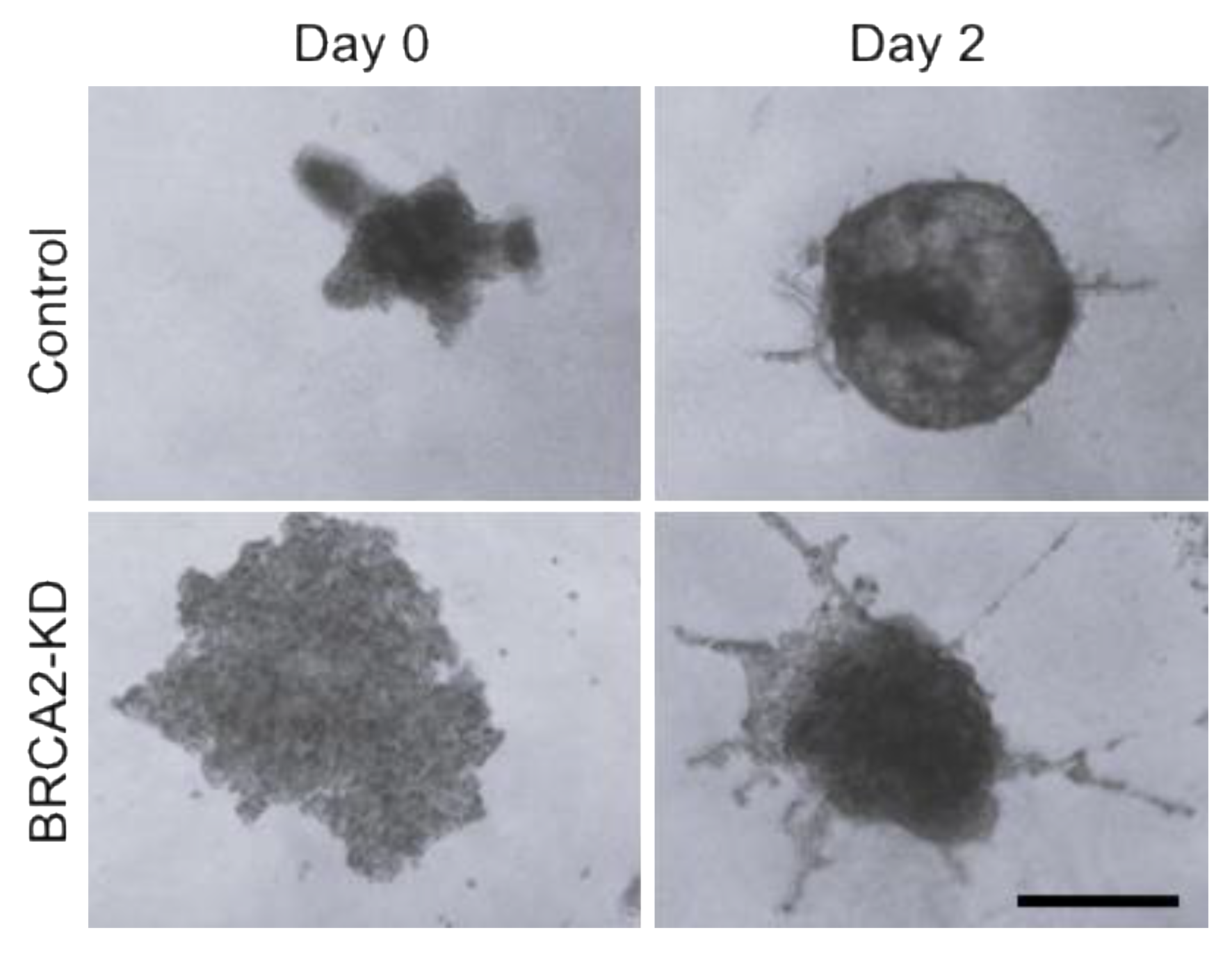
2.2. Wound Healing, Transwell Migration, and 3D Spheroid Invasion Assays

LNCaP or PC-3 cells grown to near confluency were scratched using a 10 µL pipette tip followed by replacement of cell culture media. Digital images of the scratches were obtained every 3 h for up to 18 h (PC-3 cells) or every 6 h for up to 96 h (LNCaP cells). Scratched areas were evaluated with the TScratch (version 1.0) (https://github.com/cselab/TScratch (accessed on 21 May 2019)) software [27]. For the transwell migration assays, Nunc® polycarbonate cell culture inserts with 8 µm pore size (Life Technologies) were used. N-acetylcysteine (NAC; Abcam, Rozenburg, The Netherlands) was added at a 100 µM concentration inside the inserts for the duration of incubation. For quantification, cells were fixed with 4% paraformaldehyde (PFA, Sigma-Aldrich) and stained with 0.1% crystal violet (Sigma-Aldrich). The cells were dissolved in 2% SDS (Sigma-Aldrich), and optical density was measured at 560 nm. The cell invasion assay was performed using the Cultrex 3D Spheroid Basement Membrane Extract Cell Invasion Assay (96-well, R&D Systems, Minneapolis, MN, USA) according to the manufacturer’s instructions. PC-3 or LNCaP cells were seeded in 1x Spheroid Formation ECM 24 h after siRNA-mediated knockdown and incubated 72 h at 37 °C before the start of the assay to allow for spheroid formation. Afterward, an invasion matrix was added and, after gel formation, overlaid with cell culture media containing FBS. Spheroids were incubated at 37 °C, and images were taken every 24 h with a Leica (Wetzlar, Germany) EC3 camera.

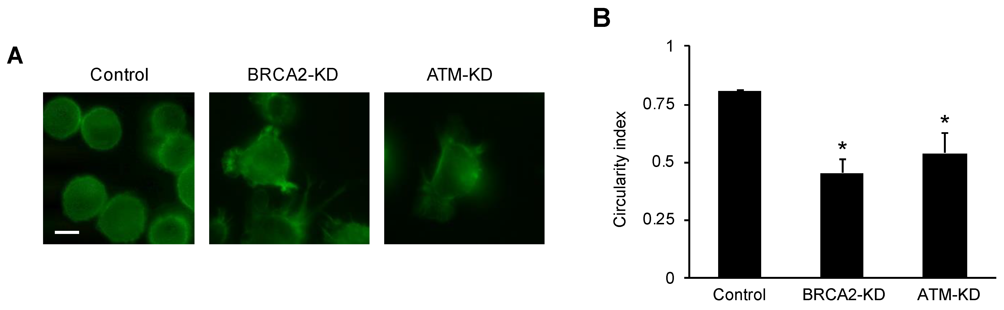
2.3. Fluorescence Microscopy and Cell Circularity Measurement

LNCaP or PC-3 cells were seeded onto cover slips and incubated overnight. They were then fixed with 1% PFA (Sigma-Aldrich) and permeabilized with 0.1% Triton™ X-100 (Sigma-Aldrich). To visualize cytoskeletal structures, Alexa-Fluor™ 488 Phalloidin (Life Technologies) was added to the cells. Afterward, the cover slips were mounted in Vectashield® containing DAPI (Biozol, Eching, Germany) and photographed with a Leica DFC425C camera under a Leica DM5000B epifluorescence microscope. At least 30 cells per sample were photographed from two cover slips. To determine cell circularity, circumference and cell area were evaluated using ImageJ (version 1.52p) (https://imagej.nih.gov/ij/ (accessed on 20 August 2019)) and the circularity index K was determined, defined as K = 4 π A C ² [28], where A is the area and C is the circumference of the cell. K is 1 for a perfectly round cell, and 0 is for an infinitely elongated polygon.

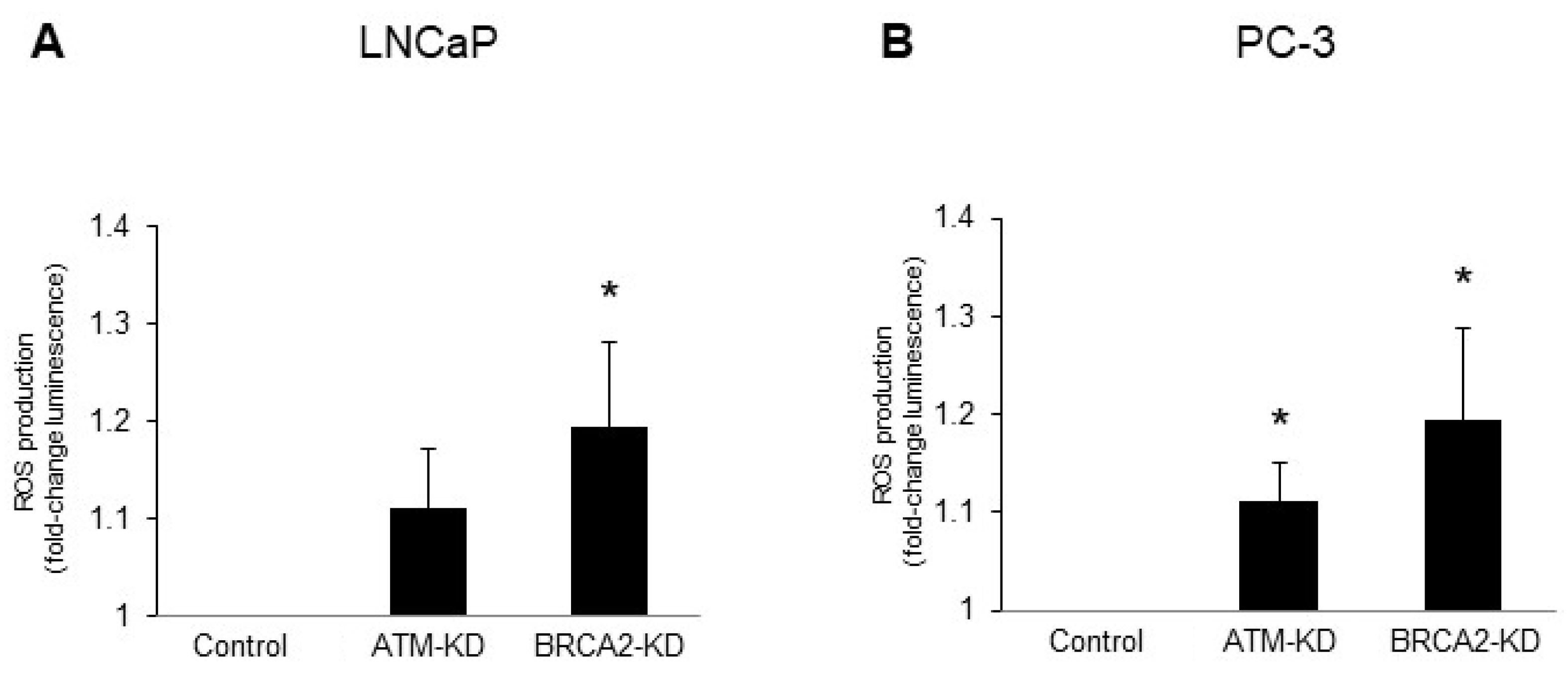
2.4. Measurement of Reactive Oxygen Species (ROS)

ATM or BRCA2 were knocked down by siRNA in LNCaP or PC-3 cells. After 72 h, cell culture medium was harvested and ROS were detected with the ROS-GloTM H2O2 Assay (Promega, Mannheim, Germany) according to the manufacturer’s instructions. Luminescence was recorded using a GloMax®-Multi+ Detection System (Promega, Mannheim, Germany).

2.5. Immunohistochemistry

Formalin-fixed, paraffin-embedded tumor specimens from a total of 15 patients with known BRCA1/2 status were obtained through the tissue bank of the National Center for Tumor Diseases (NCT) Heidelberg in accordance with the regulations of the tissue bank and the approval of the Ethics Committee of the Medical Faculty of the University of Heidelberg (206/2005, 207/2005, S-864/2019). Written informed consent was provided by all patients for the use of their tissue for research and publication. Targeted next-generation sequencing to detect BRCA1/2 alterations was performed as part of a prospective biomarker study, as previously reported [29]. Experimental protocols and methods of this study were approved by the Ethics Committee of the University of Heidelberg School of Medicine (vote S-051/2017). All experiments were carried out in accordance with the Declaration of Helsinki in its latest revised version. The biomarker study is registered under registration number DRKS00015159 in the German Clinical Trials Register (DRKS), an approved primary register of the WHO fulfilling the requirements of the International Committee of Medical Journal Editors (ICMJE). Immunohistochemistry for methylglyoxal (MG)-modified proteins was performed as described previously [26] using a monoclonal antibody (9E7, BioTechne, Wiesbaden, Germany) at a 1:100 dilution. Staining results were quantified using an immunoreactivity score (IRS; staining intensity multiplied by quantity). The staining intensity was scored as follows: 0 = negative, 1 = low, 1.5 = low–moderate, 2 = moderate, 2.5 = moderate–strong, and 3 = strong. The quantity of positive cells was scored as follows: 0 = negative staining, 1 = 1–9%, 2 = 10–49%, 3 = 50–89%, and 4 = 90–100%.

2.6. Statistical Analysis

Statistical significance was ascertained using the Mann–Whitney-U test or Student’s t-test, two-tailed. A p-value of 0.05 was considered significant. The statistical analyses were performed using Microsoft Excel, VassarStats (http://vassarstats.net/ (accessed on 29 March 2019)) and IBM® SPSS® (version 27).

3. Results

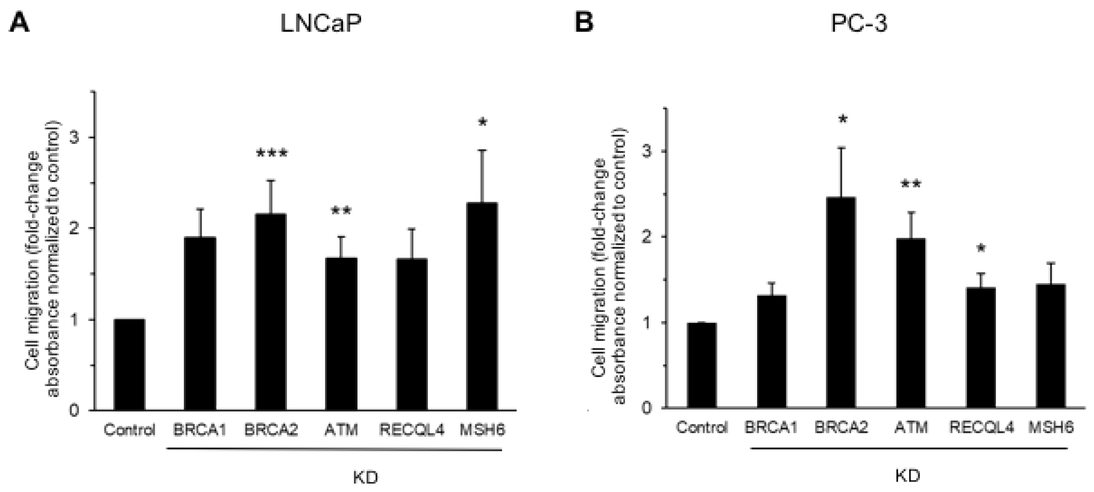
3.1. DNA Damage Repair Protein Deficiency Promotes Prostate Cancer Cell Migration

In a previous study from our group, BRCA2, ATM, RECQL4, and MSH6 were identified among the most frequently mutated DNA damage repair genes in a cohort of 64 patients with treatment-naïve prostate cancer [29].
To analyze the effects of deficiency in these genes, including BRCA1, on tumor cell migration, LNCaP or PC-3 prostate cancer cells were transiently transfected with siRNA oligonucleotides to knock down gene expression. Protein knockdown was confirmed via immunoblot analysis (Supplementary Figure S1).
Using a transwell migration assay (Figure 1), we observed a significant increase in tumor cell migration in LNCaP cells following the siRNA-mediated knockdown of BRCA2 (2.2-fold, p ≤ 0.0005), ATM (1.7-fold, p ≤ 0.005), or MSH6 (2.3-fold, p ≤ 0.05; Figure 1A). In PC-3 cells, a significant increase in tumor cell migration was detected after the knockdown of BRCA2 (2.4-fold, p ≤ 0.05), ATM (2.0-fold, p ≤ 0.005), or RECQL4 (1.4-fold, p ≤ 0.05; Figure 1B).
Since only BRCA2 and ATM knockdown led to an increased migration in both cell lines, we focused on these two proteins in subsequent experiments.
The results were corroborated using a wound-healing assay (Figure 2). In LNCaP cells, the cell-free area after 96 h was significantly reduced following the siRNA-mediated knockdown of ATM (33%) or BRCA2 (25%) in comparison to controls (57%; p ≤ 0.05; Figure 2A). In PC-3 cells, siRNA-mediated knockdown, likewise, led to an accelerated wound closure after 12 h with a 38% cell-free area in the controls, 12% after the knockdown of ATM, and 16% after the knockdown of BRCA2 (p ≤ 0.05; Figure 2B).
In order to test whether BRCA2 or ATM deficiency also affects cell invasion, a 3D spheroid invasion assay was performed (Figure 3). Whereas an enhanced invasion was found following the siRNA-mediated knockdown of BRCA2 in PC-3 cells (Figure 3), no such effect was detected after the knockdown of ATM in PC-3 cells. As expected, the downregulation of BRCA2 or ATM expression in LNCaP cells did not result in changes in tumor cell invasion [30].
We next determined the cellular circularity as a surrogate marker for cytoskeletal remodeling during cell migration (Figure 4). Following the knockdown of BRCA2 or ATM by siRNA in PC-3 cells, the cytoskeletal protein F-actin was visualized using fluorescent phalloidin (Figure 4A). The circularity index was 0.81 in controls, 0.45 in BRCA2-deficient cells, and 0.54 in ATM-deficient cells (p ≤ 0.05; Figure 4B).
Collectively, these results indicate that deficiency in DNA damage repair protein expression, namely BRCA2 and ATM, promotes cytoskeletal remodeling, as well as the migration and invasion (BRCA2 deficiency in PC-3 cells) of prostate cancer cells.

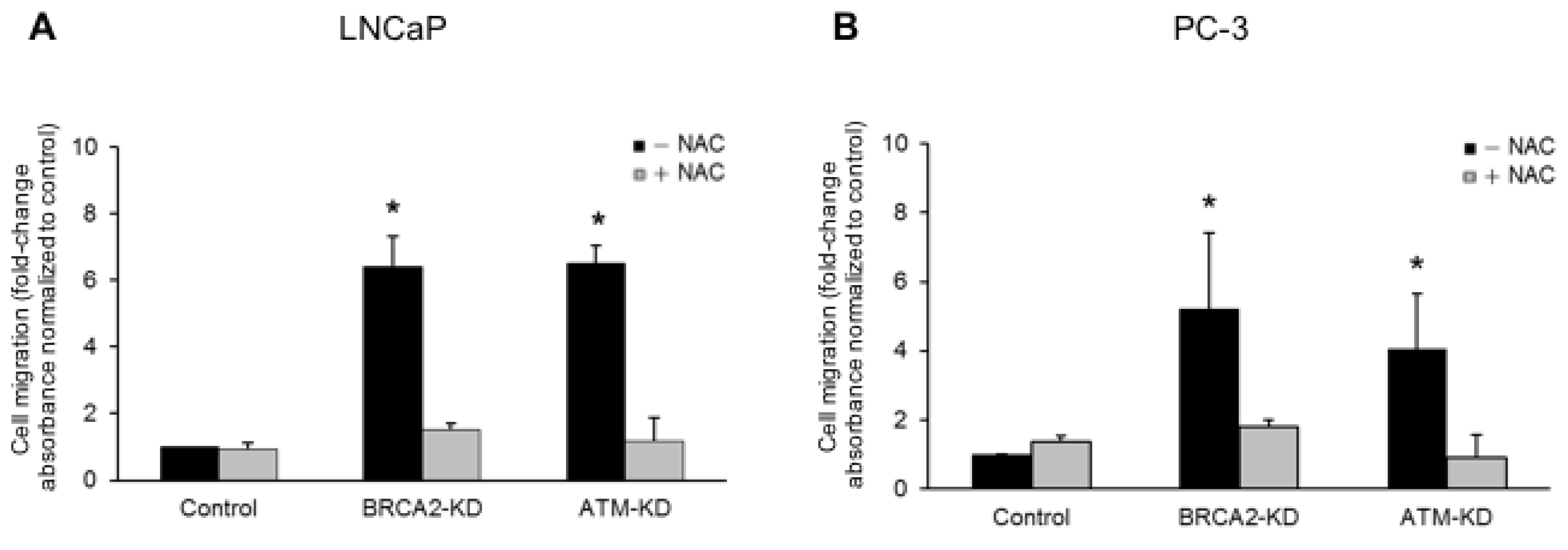
3.2. ATM or BRCA2 Deficiency Promotes Prostate Cancer Cell Migration through the Induction of Oxidative Stress

We next sought to investigate the underlying mechanisms of the enhanced migratory activity of prostate cancer cells following the induction of DNA damage repair gene deficiency, with a focus on oxidative stress.
LNCaP or PC-3 cells transiently transfected with siRNA to knock down ATM or BRCA2 expression showed a significant increase in the level of reactive oxygen species (ROS; Figure 5). In LNCaP cells, a 1.2-fold increase in ROS was detected following BRCA2 knockdown (p ≤ 0.05; Figure 5A). In PC-3 cells, a 1.1-fold increase in ROS was detected after ATM knockdown and a 1.2-fold increase was measured after BRCA2 knockdown (p ≤ 0.05; Figure 5B).
To further analyze the role of oxidative stress in the enhanced migratory properties of prostate cancer cells, we repeated the transwell migration assays in the presence of N-acetylcysteine (NAC), an ROS scavenger. Remarkably, NAC abolished the increased migration of LNCaP and PC-3 cells after the knockdown of ATM or BRCA2 (Figure 6).
These results underscore that oxidative stress plays a crucial role in the increased migration of prostate cancer cells with impaired DNA damage repair gene expression.

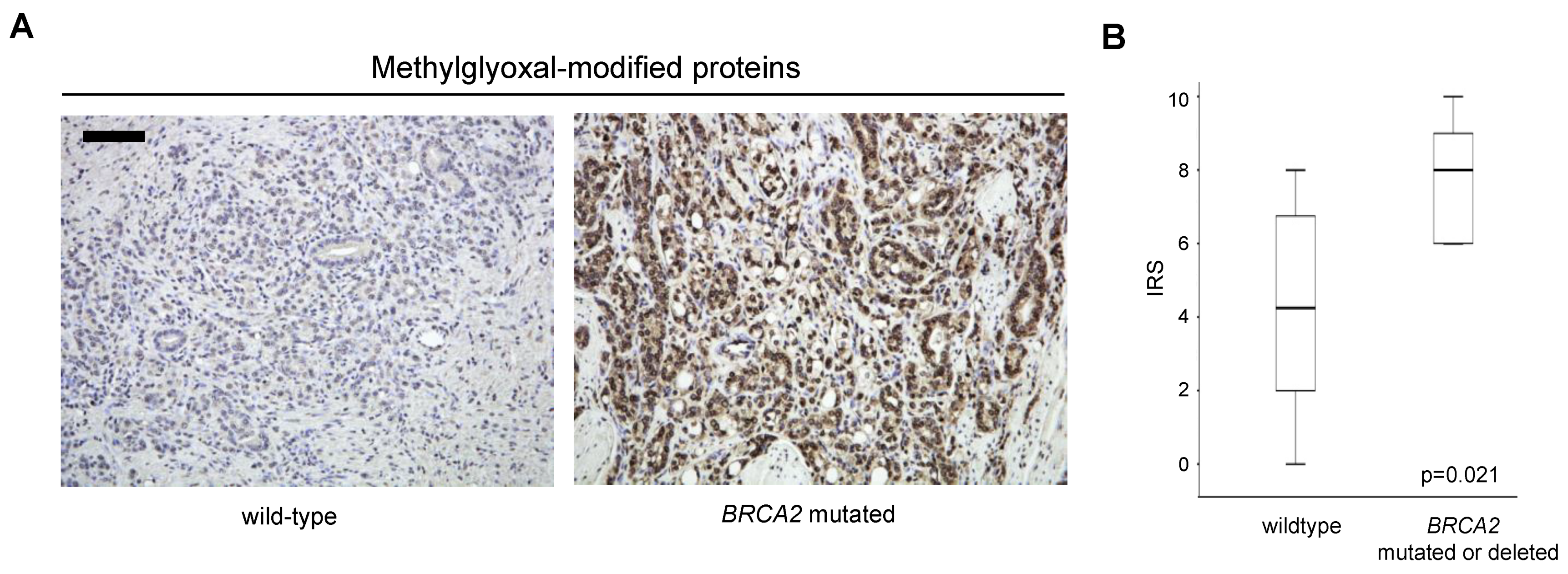
3.3. Increased Oxidative Stress in Prostate Cancer with BRCA2 Inactivation

We next sought to determine whether primary prostate cancers with DNA damage repair gene alterations showed signs of increased oxidative stress (Figure 7). To this end, tissue specimens from seven patients with known BRCA2 alterations (pathogenic frameshift mutations, n = 5; pathogenic point mutation, n = 1; BRCA2 whole-gene deletion, n = 1) were compared to eight patients that were BRCA1/2 wildtype, as previously determined via targeted next-generation sequencing [29]. Tissue specimens were stained via immunohistochemistry for MG-modified proteins as a marker for oxidative stress, and an immunoreactivity score (IRS) was calculated (Figure 7A). There was a significant increase in MG-modified proteins in tumors with BRCA2 inactivation (median IRS was 8; range was 6–10) when compared to BRCA1/2 wild-type prostate cancers (median IRS was 4.25; range was 0–8; p = 0.021; Figure 7B).
These results indicate enhanced oxidative stress in BRCA2-deficient primary prostate cancer.

4. Discussion

Defects in DNA damage repair genes, most notably genes involved in the HRR of DNA double-strand breaks, define a subgroup of men with prostate cancer. These patients have a therapeutic vulnerability to PARP inhibitors and platinum compounds but are also prone to earlier metastatic dissemination and castration resistance [7,8,9,10,31,32]. In the present study, we provide evidence that deficiency in the expression of DNA damage repair and checkpoint genes including BRCA2 or ATM can enhance the migratory activity of prostate cancer cells through increased oxidative stress. Signs of enhanced oxidative stress were also found in primary prostate cancers harboring a deleterious BRCA2 mutation or whole-gene deletion.
There are a number of studies suggesting that the loss of DNA damage repair proteins can play a direct role in cell migration. For example, Gau and colleagues were able to show that BRCA1 deficiency promoted the motility of ovarian cancer cells through a downregulation of the cytoskeletal protein profilin1 [33]. The loss of ATM has been shown to induce enhanced cell migration through an ROS-mediated increase in the activity of Rac1 GTPase [34]. The knockdown of BRCA2 in PC-3 prostate cancer cells has been shown to induce both an increased tumor cell motility and invasiveness. The latter was found to involve an upregulation of MMP9 and the activation of PI3K/AKT [35]. Renaudin et al. were able to show that BRCA2 deficiency led to ROS accumulation and impaired mitochondrial DNA maintenance through increased R-loop formation [36]. An increase in ROS formation has also been shown in cells deficient in PALB2, a direct interaction partner of BRCA2 [37]. Our results confirm an increase in prostate cancer cell motility and invasiveness following BRCA2 knockdown. However, we also observed increased tumor cell motility following the knockdown of MSH6 or REQL4, which may point to a more general role of DNA damage repair defects in tumor cell migration. A causative relationship between DNA damage and ROS production has been reported for a number of defective DNA damage repair and checkpoint genes and may involve a leakage of DNA fragments into the cytoplasm, thereby triggering an ROS-producing innate immune response [38]. However, other mechanisms are very likely to contribute, as well, since it has been shown that MSH6 knockdown does not lead to an immediate increase in genomic instability [39]. MSH6, together with MSH2, plays a role in transcriptional silencing after DNA damage [40]. This process is critical to avoiding transcription-replication conflicts and can lead to replication stress when undermined [41]. Replication stress has been shown to stimulate enhanced ROS levels [42]. More experiments are needed, of course, to prove this notion experimentally in prostate cancer. It needs to be emphasized that enhanced migratory activity is only one factor contributing to metastatic dissemination among many others [43]. Therefore, the role of oxidative stress in metastatic progression may be multifaceted; i.e., it may promote or impair this process in a context-dependent manner [38,43].
The results of the present study confirm and extend these findings by showing signs of oxidative stress in primary prostate cancer with BRCA2 inactivation. Remarkably, our study shows that the ROS scavenger NAC can abolish the enhanced migration of prostate cancer cells following BRCA2 or ATM knockdown.
Most clinical trials do not support the use of antioxidants for the prevention or treatment of cancer [38]. There are, in fact, results suggesting that in individuals with a high cancer risk, antioxidants may even increase cancer incidence [38]. The translational relevance of our finding that NAC can abolish enhanced prostate cancer cell migration following BRCA2 knockdown is, therefore, difficult to fathom. It has been suggested that approaches to exacerbate oxidative stress may be more suitable for cancer treatment [38], a notion that remains to be tested. In the context of prostate cancer, oxidative stress has been shown to upregulate the androgen receptor, and ROS were found to be increased after androgen deprivation [12]. Thus, oxidative stress may contribute to the development of castration resistance. Our finding that prostate cancers with genetic alterations in BRCA2 show signs of enhanced oxidative stress may, hence, lend further support to the notion that BRCA2 deficiency promotes the development of castration resistance [11].
The identification of oxidative stress in tissue relies primarily on the antibody-based detection of modified DNA or protein. Oxidative stress not only induces modified DNA but also leads to protein oxidation or lipid peroxidation. The latter results in the formation of MG, among other reactive carbonyl species, which modify biomacromolecules, including proteins [44]. A number of antigens have been proposed for the detection of ROS exposure using immunohistochemistry [45]. We have tested several of these and found the best signal-to-noise ration with an antibody against MG-modified proteins. The increase in MG-modified proteins in BRCA2-deficient tumors not only indicates enhanced oxidative stress but also an increased rate of aerobic glycolysis [46,47]. Enhanced glycolysis in DNA-damage-repair-deficient tumor cells has been reported for BRCA1 [48] but not for BRCA2. MG stress has been linked to increased tumor cell migration, invasion, and metastasis in breast cancer [49]. Whether MG stress also contributes to the metastatic dissemination in BRCA2-mutated prostate cancer remains to be determined.

5. Conclusions

Collectively, our results underscore that deficiency in genes that are commonly mutated in prostate cancer can promote tumor cell migration through enhanced oxidative stress. Moreover, prostate cancers that harbor alterations in BRCA2 show signs of oxidative stress, specifically MG stress, which may potentially promote metastatic progression and castration resistance. Our findings underscore the need for the individualized management of men with prostate cancer and DNA damage repair gene defects that may include a more intensified and/or (neo)adjuvant therapy.

Supplementary Materials

The following supporting information can be downloaded at: https://www.mdpi.com/article/10.3390/onco4020005/s1, Figure S1: Immunoblot analyses of siRNA-mediated knockdown of DNA damage repair proteins.

Author Contributions

Conceptualization, P.L. and S.D.; Data curation, P.L. and C.N.; Formal analysis, P.L. and S.D.; Funding acquisition, M.H. and S.D.; Investigation, P.L., A.K., C.N., C.S., M.K., V.S., M.G., A.D., A.S. and S.D.; Methodology, P.L., A.K. and S.D.; Project administration, P.L.; Resources, C.N., M.H., A.D., A.S. and S.D.; Software, P.L.; Supervision, M.H., A.D., A.S. and S.D.; Validation, P.L., A.K. and S.D.; Visualization, P.L. and S.D.; Writing—original draft, P.L.; Writing—review and editing, P.L., A.K., C.N., C.S., M.K., V.S., M.G., M.H., A.D., A.S. and S.D. All authors have read and agreed to the published version of the manuscript.

Funding

This work was supported by the Medical Faculty Heidelberg and the Wilhelm Sander-Stiftung.

Institutional Review Board Statement

The study was conducted according to the guidelines of the Declaration of Helsinki and approved by the Ethics Committee of the University of Heidelberg School of Medicine (vote 206/2005, approval date 21 October 2005; vote 207/2005, approval date 21 October 2005; vote S-051/2017, approval date 1 March 2017; vote S-864/2019, approval date 9 January 2020).

Informed Consent Statement

Written informed consent was obtained from all subjects involved in the study for the use and publication of data. Genetic testing of tumor samples by targeted next generation sequencing was performed as part of a prospective biomarker study, as previously reported [29]. The experimental protocols and methods were approved by the Ethics Committee of the University of Heidelberg School of Medicine (S-051/2017). The prospective biomarker study is registered under registration number DRKS00015159 in the German Clinical Trials Register (DRKS), an approved primary register of the WHO, fulfilling the requirements of the International Committee of Medical Journal Editors (ICMJE). All experiments were carried out in accordance with the Declaration of Helsinki in its last revised version.

Data Availability Statement

The data presented in this study are available within the article.

Acknowledgments

We would like to thank the Tissue Bank of the National Center for Tumor Diseases Heidelberg for tissue procurement.

Conflicts of Interest

The authors declare no conflict of interest.

References

  1. Siegel, R.L.; Miller, K.D.; Jemal, A. Cancer statistics, 2020. CA Cancer J. Clin. 2020, 70, 7–30. [Google Scholar] [CrossRef] [PubMed]
  2. Attard, G.; Parker, C.; Eeles, R.A.; Schröder, F.; Tomlins, S.A.; Tannock, I.; Drake, C.G.; de Bono, J.S. Prostate cancer. Lancet 2016, 387, 70–82. [Google Scholar] [CrossRef] [PubMed]
  3. Sartor, O.; de Bono, J.S. Metastatic Prostate Cancer. N. Engl. J. Med. 2018, 378, 1653–1654. [Google Scholar] [CrossRef]
  4. Weiss, F.; Lauffenburger, D.; Friedl, P. Towards targeting of shared mechanisms of cancer metastasis and therapy resistance. Nat. Rev. Cancer 2022, 22, 157–173. [Google Scholar] [CrossRef] [PubMed]
  5. De Bono, J.; Mateo, J.; Fizazi, K.; Saad, F.; Shore, N.; Sandhu, S.; Chi, K.N.; Sartor, O.; Agarwal, N.; Olmos, D.; et al. Olaparib for Metastatic Castration-Resistant Prostate Cancer. N. Engl. J. Med. 2020, 382, 2091–2102. [Google Scholar] [CrossRef] [PubMed]
  6. Lord, C.J.; Ashworth, A. PARP inhibitors: Synthetic lethality in the clinic. Science 2017, 355, 1152–1158. [Google Scholar] [CrossRef] [PubMed]
  7. Nientiedt, C.; Duensing, A.; Zschäbitz, S.; Jäger, D.; Hohenfellner, M.; Stenzinger, A.; Duensing, S. PARP inhibition in prostate cancer. Genes Chromosom. Cancer 2021, 60, 344–351. [Google Scholar] [CrossRef] [PubMed]
  8. Bednarz, N.; Eltze, E.; Semjonow, A.; Rink, M.; Andreas, A.; Mulder, L.; Hannemann, J.; Fisch, M.; Pantel, K.; Weier, H.-U.G.; et al. BRCA1 loss preexisting in small subpopulations of prostate cancer is associated with advanced disease and metastatic spread to lymph nodes and peripheral blood. Clin. Cancer Res. 2010, 16, 3340–3348. [Google Scholar] [CrossRef]
  9. Castro, E.; Goh, C.; Olmos, D.; Saunders, E.; Leongamornlert, D.; Tymrakiewicz, M.; Mahmud, N.; Dadaev, T.; Govindasami, K.; Guy, M.; et al. Germline BRCA mutations are associated with higher risk of nodal involvement, distant metastasis, and poor survival outcomes in prostate cancer. J. Clin. Oncol. 2013, 31, 1748–1757. [Google Scholar] [CrossRef]
  10. Nientiedt, C.; Budczies, J.; Endris, V.; Kirchner, M.; Schwab, C.; Jurcic, C.; Behnisch, R.; Hoveida, S.; Lantwin, P.; Kaczorowski, A.; et al. Mutations in TP53 or DNA damage repair genes define poor prognostic subgroups in primary prostate cancer. Urol. Oncol. Semin. Orig. Investig. 2022, 40, 8.e11–8.e18. [Google Scholar] [CrossRef]
  11. Taylor, R.A.; Fraser, M.; Livingstone, J.; Espiritu, S.M.G.; Thorne, H.; Huang, V.; Lo, W.; Shiah, Y.-J.; Yamaguchi, T.N.; Sliwinski, A.; et al. Germline BRCA2 mutations drive prostate cancers with distinct evolutionary trajectories. Nat. Commun. 2017, 8, 13671. [Google Scholar] [CrossRef] [PubMed]
  12. Shiota, M.; Yokomizo, A.; Tada, Y.; Inokuchi, J.; Kashiwagi, E.; Masubuchi, D.; Eto, M.; Uchiumi, T.; Naito, S. Castration resistance of prostate cancer cells caused by castration-induced oxidative stress through Twist1 and androgen receptor overexpression. Oncogene 2010, 29, 237–250. [Google Scholar] [CrossRef] [PubMed]
  13. Decker, B.; Karyadi, D.M.; Davis, B.W.; Karlins, E.; Tillmans, L.S.; Stanford, J.L.; Thibodeau, S.N.; Ostrander, E.A. Biallelic BRCA2 Mutations Shape the Somatic Mutational Landscape of Aggressive Prostate Tumors. Am. J. Hum. Genet. 2016, 98, 818–829. [Google Scholar] [CrossRef] [PubMed]
  14. Jonsson, P.; Bandlamudi, C.; Cheng, M.L.; Srinivasan, P.; Chavan, S.S.; Friedman, N.D.; Rosen, E.Y.; Richards, A.L.; Bouvier, N.; Selcuklu, S.D.; et al. Tumour lineage shapes BRCA-mediated phenotypes. Nature 2019, 571, 576–579. [Google Scholar] [CrossRef] [PubMed]
  15. Holloman, W.K. Unraveling the mechanism of BRCA2 in homologous recombination. Nat. Struct. Mol. Biol. 2011, 18, 748–754. [Google Scholar] [CrossRef] [PubMed]
  16. Venkitaraman, A.R. Cancer suppression by the chromosome custodians, BRCA1 and BRCA2. Science 2014, 343, 1470–1475. [Google Scholar] [CrossRef]
  17. Venkitaraman, A.R. Functions of BRCA1 and BRCA2 in the biological response to DNA damage. J. Cell Sci. 2001, 114, 3591–3598. [Google Scholar] [CrossRef] [PubMed]
  18. Wu, W.; Koike, A.; Takeshita, T.; Ohta, T. The ubiquitin E3 ligase activity of BRCA1 and its biological functions. Cell Div. 2008, 3, 1. [Google Scholar] [CrossRef] [PubMed]
  19. Abeshouse, A.; Ahn, J.; Akbani, R.; Ally, A.; Amin, S.; Andry, C.D.; Annala, M.; Aprikian, A.; Armenia, J.; Arora, A.; et al. The Molecular Taxonomy of Primary Prostate Cancer. Cell 2015, 163, 1011–1025. [Google Scholar] [CrossRef]
  20. Mateo, J.; Seed, G.; Bertan, C.; Rescigno, P.; Dolling, D.; Figueiredo, I.; Miranda, S.; Nava Rodrigues, D.; Gurel, B.; Clarke, M.; et al. Genomics of lethal prostate cancer at diagnosis and castration resistance. J. Clin. Investig. 2020, 130, 1743–1751. [Google Scholar] [CrossRef]
  21. Robinson, D.; Van Allen, E.M.; Wu, Y.-M.; Schultz, N.; Lonigro, R.J.; Mosquera, J.-M.; Montgomery, B.; Taplin, M.-E.; Pritchard, C.C.; Attard, G.; et al. Integrative clinical genomics of advanced prostate cancer. Cell 2015, 161, 1215–1228. [Google Scholar] [CrossRef] [PubMed]
  22. Pritchard, C.C.; Mateo, J.; Walsh, M.F.; De Sarkar, N.; Abida, W.; Beltran, H.; Garofalo, A.; Gulati, R.; Carreira, S.; Eeles, R.; et al. Inherited DNA-Repair Gene Mutations in Men with Metastatic Prostate Cancer. N. Engl. J. Med. 2016, 375, 443–453. [Google Scholar] [CrossRef] [PubMed]
  23. Ledet, E.M.; Antonarakis, E.S.; Isaacs, W.B.; Lotan, T.L.; Pritchard, C.; Sartor, A.O. Germline BLM mutations and metastatic prostate cancer. Prostate 2020, 80, 235–237. [Google Scholar] [CrossRef] [PubMed]
  24. Luong, T.T.; Bernstein, K.A. Role and Regulation of the RECQL4 Family during Genomic Integrity Maintenance. Genes 2021, 12, 1919. [Google Scholar] [CrossRef]
  25. Abida, W.; Cyrta, J.; Heller, G.; Prandi, D.; Armenia, J.; Coleman, I.; Cieslik, M.; Benelli, M.; Robinson, D.; Van Allen, E.M.; et al. Genomic correlates of clinical outcome in advanced prostate cancer. Proc. Natl. Acad. Sci. USA 2019, 116, 11428–11436. [Google Scholar] [CrossRef] [PubMed]
  26. Kaczorowski, A.; Tolstov, Y.; Falkenstein, M.; Vasioukhin, V.; Prigge, E.-S.; Geisler, C.; Kippenberger, M.; Nientiedt, C.; Ratz, L.; Kuryshev, V.; et al. Rearranged ERG confers robustness to prostate cancer cells by subverting the function of p53. Urol. Oncol. Semin. Orig. Investig. 2020, 38, 736.e1–736.e10. [Google Scholar] [CrossRef] [PubMed]
  27. Gebäck, T.; Schulz, M.M.P.; Koumoutsakos, P.; Detmar, M. TScratch: A Novel and Simple Software Tool for Automated Analysis of Monolayer Wound Healing Assays. BioTechniques 2009, 46, 265–274. [Google Scholar] [CrossRef] [PubMed]
  28. Cox, E.P. A method of assigning numerical and percentage values to the degree of roundness of sand grains. J. Paleontol. 1927, 1, 179–183. [Google Scholar]
  29. Nientiedt, C.; Endris, V.; Jenzer, M.; Mansour, J.; Pour Sedehi, N.T.; Pecqueux, C.; Volckmar, A.-L.; Leichsenring, J.; Neumann, O.; Kirchner, M.; et al. High prevalence of DNA damage repair gene defects and TP53 alterations in men with treatment-naïve metastatic prostate cancer –Results from a prospective pilot study using a 37 gene panel. Urol. Oncol. Semin. Orig. Investig. 2020, 38, 637.e17–637.e27. [Google Scholar] [CrossRef]
  30. Aslan, M.; Hsu, E.-C.; Liu, S.; Stoyanova, T. Quantifying the invasion and migration ability of cancer cells with a 3D Matrigel drop invasion assay. Biol. Methods Protoc. 2021, 6, bpab014. [Google Scholar] [CrossRef]
  31. Lord, C.J.; Ashworth, A. BRCAness revisited. Nat. Rev. Cancer 2016, 16, 110–120. [Google Scholar] [CrossRef] [PubMed]
  32. Castro, E.; Goh, C.; Leongamornlert, D.; Saunders, E.; Tymrakiewicz, M.; Dadaev, T.; Govindasami, K.; Guy, M.; Ellis, S.; Frost, D.; et al. Effect of BRCA Mutations on Metastatic Relapse and Cause-specific Survival After Radical Treatment for Localised Prostate Cancer. Eur. Urol. 2015, 68, 186–193. [Google Scholar] [CrossRef]
  33. Gau, D.M.; Lesnock, J.L.; Hood, B.L.; Bhargava, R.; Sun, M.; Darcy, K.; Luthra, S.; Chandran, U.; Conrads, T.P.; Edwards, R.P.; et al. BRCA1 deficiency in ovarian cancer is associated with alteration in expression of several key regulators of cell motility—A proteomics study. Cell Cycle 2015, 14, 1884–1892. [Google Scholar] [CrossRef] [PubMed]
  34. Tolbert, C.E.; Beck, M.V.; Kilmer, C.E.; Srougi, M.C. Loss of ATM positively regulates Rac1 activity and cellular migration through oxidative stress. Biochem. Biophys. Res. Commun. 2019, 508, 1155–1161. [Google Scholar] [CrossRef] [PubMed]
  35. Moro, L.; Arbini, A.A.; Yao, J.L.; di Sant’agnese, P.A.; Marra, E.; Greco, M. Loss of BRCA2 promotes prostate cancer cell invasion through up-regulation of matrix metalloproteinase-9. Cancer Sci. 2008, 99, 553–563. [Google Scholar] [CrossRef] [PubMed]
  36. Renaudin, X.; Lee, M.; Shehata, M.; Surmann, E.-M.; Venkitaraman, A.R. BRCA2 deficiency reveals that oxidative stress impairs RNaseH1 function to cripple mitochondrial DNA maintenance. Cell Rep. 2021, 36, 109478. [Google Scholar] [CrossRef] [PubMed]
  37. Ma, J.; Cai, H.; Wu, T.; Sobhian, B.; Huo, Y.; Alcivar, A.; Mehta, M.; Cheung, K.L.; Ganesan, S.; Kong, A.-N.T.; et al. PALB2 interacts with KEAP1 to promote NRF2 nuclear accumulation and function. Mol. Cell. Biol. 2012, 32, 1506–1517. [Google Scholar] [CrossRef] [PubMed]
  38. Gill, J.G.; Piskounova, E.; Morrison, S.J. Cancer, Oxidative Stress, and Metastasis. Cold Spring Harb. Symp. Quant. Biol. 2016, 81, 163–175. [Google Scholar] [CrossRef]
  39. Evensen, N.A.; Madhusoodhan, P.P.; Meyer, J.; Saliba, J.; Chowdhury, A.; Araten, D.J.; Nersting, J.; Bhatla, T.; Vincent, T.L.; Teachey, D.; et al. MSH6 haploinsufficiency at relapse contributes to the development of thiopurine resistance in pediatric B-lymphoblastic leukemia. Haematologica 2018, 103, 830–839. [Google Scholar] [CrossRef]
  40. Ding, N.; Bonham, E.M.; Hannon, B.E.; Amick, T.R.; Baylin, S.B.; O’Hagan, H.M. Mismatch repair proteins recruit DNA methyltransferase 1 to sites of oxidative DNA damage. J. Mol. Cell Biol. 2016, 8, 244–254. [Google Scholar] [CrossRef]
  41. Lalonde, M.; Trauner, M.; Werner, M.; Hamperl, S. Consequences and Resolution of Transcription–Replication Conflicts. Life 2021, 11, 637. [Google Scholar] [CrossRef]
  42. Andrs, M.; Stoy, H.; Boleslavska, B.; Chappidi, N.; Kanagaraj, R.; Nascakova, Z.; Menon, S.; Rao, S.; Oravetzova, A.; Dobrovolna, J.; et al. Excessive reactive oxygen species induce transcription-dependent replication stress. Nat. Commun. 2023, 14, 1791. [Google Scholar] [CrossRef] [PubMed]
  43. Pani, G.; Galeotti, T.; Chiarugi, P. Metastasis: Cancer cell’s escape from oxidative stress. Cancer Metastasis Rev. 2010, 29, 351–378. [Google Scholar] [CrossRef] [PubMed]
  44. Moldogazieva, N.T.; Zavadskiy, S.P.; Astakhov, D.V.; Terentiev, A.A. Lipid peroxidation: Reactive carbonyl species, protein/DNA adducts, and signaling switches in oxidative stress and cancer. Biochem. Biophys. Res. Commun. 2023, 687, 149167. [Google Scholar] [CrossRef]
  45. Liou, G.-Y.; Storz, P. Detecting reactive oxygen species by immunohistochemistry. Methods Mol. Biol. 2015, 1292, 97–104. [Google Scholar]
  46. de Bari, L.; Scirè, A.; Minnelli, C.; Cianfruglia, L.; Kalapos, M.P.; Armeni, T. Interplay among Oxidative Stress, Methylglyoxal Pathway and S-Glutathionylation. Antioxidants 2020, 10, 19. [Google Scholar] [CrossRef] [PubMed]
  47. Degenhardt, T.P.; Thorpe, S.R.; Baynes, J.W. Chemical modification of proteins by methylglyoxal. Cell. Mol. Biol. 1998, 44, 1139–1145. [Google Scholar]
  48. Privat, M.; Radosevic-Robin, N.; Aubel, C.; Cayre, A.; Penault-Llorca, F.; Marceau, G.; Sapin, V.; Bignon, Y.-J.; Morvan, D. BRCA1 induces major energetic metabolism reprogramming in breast cancer cells. PLoS ONE 2014, 9, e102438. [Google Scholar] [CrossRef]
  49. Nokin, M.-J.; Bellier, J.; Durieux, F.; Peulen, O.; Rademaker, G.; Gabriel, M.; Monseur, C.; Charloteaux, B.; Verbeke, L.; van Laere, S.; et al. Methylglyoxal, a glycolysis metabolite, triggers metastasis through MEK/ERK/SMAD1 pathway activation in breast cancer. Breast Cancer Res. 2019, 21, 11. [Google Scholar] [CrossRef]
Figure 1. Increased prostate cancer cell migration after the knockdown of DNA damage repair gene expression. Transwell migration assay using LNCaP (A) or PC-3 (B) cells following siRNA-mediated knockdown (KD) of the DNA damage repair proteins indicated. Each bar represents mean and standard error of the fold-change absorbance normalized to control from three independent experiments. * p ≤ 0.05; ** p ≤ 0.005; *** p ≤ 0.0005.
Onco 04 00005 g001
Figure 2. Enhanced wound closure in BRCA2- or ATM-deficient prostate cancer cells. Scratch assay using LNCaP (A) or PC-3 (B) cells after siRNA-mediated knockdown (KD) of BRCA2 or ATM. Images of scratches at the time points indicated (left panels) and quantification of the percentage of cell-free area (right panels) are shown. Each bar represents mean and standard error from three independent experiments. Scale bar = 10 μm. * p ≤ 0.05.
Onco 04 00005 g002
Figure 3. Enhanced invasion after knockdown of BRCA2 in PC-3 prostate cancer cells. 3D spheroid invasion assays following knockdown (KD) of BRCA2 by siRNA. Light microscopic images were taken at the time indicated. Scale bar = 500 μm.
Onco 04 00005 g003
Figure 4. Decreased cell circularity in BRCA2- or ATM-deficient prostate cancer cells. Fluorescent phalloidin staining of PC-3 cells after siRNA-mediated knockdown (KD) of BRCA2 or ATM to visualize F-actin (A) and quantification of the cellular circularity (B) from two independent experiments with at least 2 × 30 cells measured per experiment. Each bar represents mean and standard error. Scale bar = 10 µm. * p ≤ 0.05.
Onco 04 00005 g004
Figure 5. ATM or BRCA2 deficiency increases ROS levels in prostate cancer cells. Quantification of reactive oxygen species (ROS) in LNCaP (A) or PC-3 (B) cells after knockdown (KD) of ATM or BRCA2 using a luminescence-based assay. Each bar represents mean and standard error of the fold-change in luminescence from four independent experiments. * p ≤ 0.05.
Onco 04 00005 g005
Figure 6. ROS inhibition abolishes increased prostate cancer cell migration after ATM or BRCA2 knockdown. Transwell migration assay using LNCaP (A) or PC-3 (B) cells after knockdown (KD) of BRCA2 or ATM and with or without treatment with the ROS scavenger N-acetylcysteine (NAC). Each bar represents mean and standard error of the fold-change absorbance normalized to control from three independent experiments. * p ≤ 0.05.
Onco 04 00005 g006
Figure 7. Increased signs of oxidative stress in BRCA2-deficient primary prostate cancer. Immunohistochemistry for methylglyoxal-modified proteins to visualize oxidative stress in wild-type and BRCA2-mutated prostate cancer (A). Box plot of the immunoreactivity scores (IRSs) of methylglyoxal-modified proteins in wildtype (n = 8) and BRCA2-mutated (n = 6) or BRCA2-deleted (n = 1) primary prostate cancers (B). Scale bar = 50 μm.
Onco 04 00005 g007
Disclaimer/Publisher’s Note: The statements, opinions and data contained in all publications are solely those of the individual author(s) and contributor(s) and not of MDPI and/or the editor(s). MDPI and/or the editor(s) disclaim responsibility for any injury to people or property resulting from any ideas, methods, instructions or products referred to in the content.

