Article

UV-Induced Benzyloxy Rotamerization in an Ortho OH-Substituted Aryl Schiff Base

1
Department of Physics, Bitlis Eren University, 13000 Bitlis, Turkey
2
CQC-IMS, Department of Chemistry, University of Coimbra, 3004-535 Coimbra, Portugal
3
MTA-ELTE Lendület Laboratory Astrochemistry Research Group, Institute of Chemistry, ELTE Eötvös Loránd University, H-1518 Budapest, Hungary
4
Department of Chemistry, Eskişehir Technical University, 26470 Eskişehir, Turkey
*
Authors to whom correspondence should be addressed.
Photochem 2022, 2(2), 376-389; https://doi.org/10.3390/photochem2020026
Submission received: 26 April 2022 / Revised: 10 May 2022 / Accepted: 12 May 2022 / Published: 25 May 2022

Abstract

:
A new benzyloxy containing ortho hydroxyl-substituted aryl Schiff base, trans 2-((2-(benzyloxy)benzylidene) amino)phenol (abbreviated as BBAP), was synthesized and characterized by 1H-, 13C-NMR and infrared spectroscopic techniques and elemental analysis. The conformational landscape of the compound, as well as its infrared spectra in argon and N2 cryogenic matrices (10 K) were investigated, followed by the study of the effects of in situ UV irradiation of the matrix-isolated compound. The structural information was obtained through an extensive series of quantum chemical calculations performed at the DFT(B3LYP)/6-311++G(d,p) level of theory, which enabled to identify 3 low-energy OH···N intramolecularly H-bonded conformers of the molecule that were later found to be present in the as-deposited cryogenic matrices. The 3 experimentally relevant conformers of BBAP differ in the geometry of the benzyloxy substituent, and were discovered to interconvert upon in situ UV irradiation (λ = 230 nm) of the matrix-isolated compound. This is the first report on UV-induced conformational changes taking place in a benzyloxy fragment for a matrix-isolated compound.

1. Introduction

Schiff bases are common organic structures (also known as azomethines; R1R2C = NR3, with R1, R2 = alkyl, aryl or H, and R3 = alkyl or aryl) that can usually be easily synthesized through a one-step synthetic procedure, which involves the reaction of primary amines with carbonyl compounds [1,2]. Aryl Schiff bases have their azomethine characteristic group connected to a benzene ring, and are the most relevant members of this family of compounds due to their wide spectrum of applications, particularly as corrosion inhibitors [3], catalyst carriers [4,5], thermo-stable materials [6,7,8], metal ion complexing agents [9], optical data storage systems [10,11,12], molecular switches [13,14], and sensors [15,16]. These compounds also have many uses in molecular electronics, computing, [17,18] and medicine [19,20,21].
In aryl Schiff bases, the presence of a hydroxyl group in the ortho position the azomethine group leads to the formation of an intramolecular resonance-stabilized O–H···N hydrogen bond, which has a positive effect on the thermodynamic stability of the whole molecule [22,23]. In addition, in these compounds, enol-imine/keto-amine tautomerism, as well as cis/trans (or E/Z) isomerism around the azomethine C=N bond, are, in general, easily accessible, particularly as a result of their photo-excitation. The photo-induced enol-imine/keto-amine tautomerism in ortho hydroxyl-substituted aryl Schiff bases has been the subject of many studies and is known to take place via excited-state intramolecular proton transfer (ESIPT) [24,25,26,27,28,29].
We have been investigating the structure and reactivity of ortho hydroxyl-substituted aryl Schiff bases using a concerted approach that included the experimental study of the molecules of the compounds isolated in low-temperature inert matrices and the theoretical evaluation of their conformational landscape by means of quantum chemical theoretical methods [28,29,30]. Emphasis has been given to the evaluation of the photochemical behavior of the matrix-isolated compounds when subjected to ultraviolet (UV) irradiation, as, for example, the UV-induced methoxy rotamerization of a methoxy-substituted ortho-hydroxy aryl Schiff base and the E-enol/Z-enol and E-enol/E-keto photo-isomerizations undergone by N-salicylidene-p-carboxyaniline [28,29], as well as to quantum mechanical H-atom tunneling reactions taking place in the cryogenic matrices [30].
In the present study, we have investigated trans 2-((2-(benzyloxy)benzylidene)amino)phenol (BBAP), which is an ortho-OH substituted aryl Schiff base bearing a benzyloxy substituent (Scheme 1). This substituent is a conformationally flexible moiety, with three internal rotations that may lead to different conformers (δ, ε and ζ in Scheme 1). The work focused on the characterization of the conformational space of the molecule, through the identification of its lowest-energy conformers, the study of the infrared spectrum of BBAP isolated in cryogenic Ar and N2 matrices, and the evaluation of the processes that took place upon UV irradiation of the matrix-isolated compound. Extensive quantum chemical calculations were performed within the density functional theory (DFT) framework, in order to characterize the molecule conformationally and to identify the most relevant intramolecular interactions that determine the nature of the lowest-energy forms of BBAP. As it is shown in the next sections of this article, the calculations render 3 low-energy conformers of the compound, with energies within ca. 4 kJ mol−1, which could be trapped from the gas phase into the cryogenic matrices and characterized vibrationally. These conformers differ in the geometry of the benzyloxy moiety, and are stabilized by the characteristic stabilizing intramolecular O–H···N hydrogen bond established between the phenolic O–H group (as H-donor) and the azomethine nitrogen atom (as acceptor), and by the absence of significant steric hindrance. In situ narrowband UV irradiation of the matrix-isolated compound at λ = 230 nm was found to induce interconversions between the low-energy conformers initially present in the matrices (i.e., it was found to lead to rotamerization within the benzyloxy group) but no evidence was found either for transcis (C=N) isomerization or enol-imine/keto-amine tautomerization. To the best of our knowledge, this is the first report on the UV-induced conformational changes that take place in a benzyloxy fragment of a matrix-isolated compound.

2. Experimental and Computational Methods

2.1. Synthesis Procedure and Characterization

2-((2-(Benzyloxy)benzylidene)amino)phenol (BBAP) was synthesized from 2-aminophenol and 2-(benzyloxy)benzaldehyde (Scheme 2), following the general procedure previously reported [25,26,27,28,29,30]. 2-Aminophenol (1.091 g, 0.01 mol) and 2-(benzyloxy)benzaldehyde (2.12 g, 0.01 mol) were first dissolved in 25 mL of methanol by heating, and the solutions were then added slowly. After the substance precipitated, the mixture was stirred for another 1–2 h, at T = 60 °C. The precipitated product was filtered, purified by recrystallization in methanol and dried in a vacuum desiccator at room temperature. The infrared (IR) spectrum of the purified crystalline material (in a KBr pellet, at room temperature) and the 1H- and 13C-NMR spectra in deuterated dimethylsulfoxide (DMSO-d6) solution were found to be compatible with the desired product. These are provided as supporting materials (Figures S1–S3). The synthesis exclusively yielded the E (C=N) enol-imine isomeric form of the compound, with no evidence of the formation of either the Z (C=N) isomer or of the keto-amine tautomeric species.
The IR spectrum was recorded by using a Perkin Elmer FTIR 100 spectrometer, and the 1H- and 13C-NMR spectra were obtained in a Bruker Biospin UltrashieldTM NMR spectrometer, at room temperature. The melting point (m.p.) of the compound was measured using a Gallenkamp Sanyo Heater device. Elemental analysis was performed using a CNHS-932 LECO apparatus.
IR (KBr disc, ν cm−1): 3369 (O–H); 3097–3006 (C–H, aromatic); 2928–2843 (C–H, aliphatic); 1621 (C=N); 1587–1448 (C=C, aromatic); 1027 (CH2–O–C). 1H-NMR (300 MHz, DMSO-d6, δ ppm): 9.02 (s, 1H); 9.00 (s, 1H); 8.21 (dd, J = 7.7, 1.7 Hz, 1H); 7.61–6.69 (m, 13H); 5.26 (s, 2H). 13C-NMR (75 MHz, DMSO-d6, δ ppm): 158.67 (s); 155.04 (s); 151.30 (s); 139.26 (s); 137.31 (s); 133.27 (s); 129.01 (s); 128.40 (s); 128.01 (s); 127.53 (s); 125.14 (s); 121.33 (s); 120.05 (s); 119.97 (s); 116.48 (s); 113.87 (s). Elemental analysis: C20H17NO2, Calc. (Obs.): C, 79.19 (79.05); H, 5.65 (5.79); N, 4.62 (4.70). m. p.: 83–84 °C.

2.2. Matrix-Isolation and UV-Irradiation Experiments

To obtain the cryogenic matrices of the compound, a solid sample of BBAP was sublimated by using a homemade Knudsen cell, connected to the vacuum chamber of the cryostat (APD Cryogenics closed-cycle helium refrigeration system, with a DE-202A expander) and co-deposited with a large excess of argon (Ar) or nitrogen (N2) (both obtained from Air Liquide) onto a cold (10.0 ± 0.1 K) CsI substrate, mounted at the cold tip of the cryostat. The solute:matrix ratio was ~1:1000, to provide the adequate isolation of the compound.
The infrared spectra of the matrices were collected using a Nicolet 6700 FTIR spectrometer, equipped with a mercury cadmium telluride (MCT) detector and a KBr beam splitter, in the 4000–400 cm−1 range and with 0.5 cm−1 resolution. The instrument was purged by a stream of dry/CO2-filtered air in order to avoid interference from atmospheric H2O and CO2.
UV irradiation of the matrix-isolated BBAP was performed at λ = 230 nm, through the outer KBr window of the cryostat, using UV light provided by a Spectra Physics MOPO-SL optical parametric oscillator (OPO), pumped by a pulsed (pulse duration 10 ns, repetition rate 10 Hz) Quanta Ray Pro-Series Nd-YAG laser.

2.3. Quantum Chemical Calculations

All calculations were carried out using the GAUSSIAN 09 (Revision C.01) program [31]. The ground-state geometries, relative energies, and infrared spectra of the different forms of BBAP were obtained at the DFT(B3LYP)/6-311++G(d,p) level of theory [32,33,34,35,36,37], the calculated harmonic vibrational frequencies were subsequently scaled by the factor 0.955, above 1800 cm−1, and by the factor 0.983, below 1800 cm−1, to correct them for the effect of basis set limitation, for the neglected part of electron correlation and, mainly, for the anharmonicity effects.
Assignment of the vibrational spectra was carried out with the help of the animation module of ChemCraft (version 1.8) [38]. In the simulated spectra presented in the figures, the IR bands were broadened by Lorentzian profiles (fwhm = 2 cm−1), centered at the calculated (scaled) wavenumbers.
The UV spectrum of BBAP in the gas phase was calculated within the time-dependent-DFT (TD-DFT) theoretical framework, with the same functional and basis set used in the performed ground-state calculations [39,40].

3. Results and Discussion

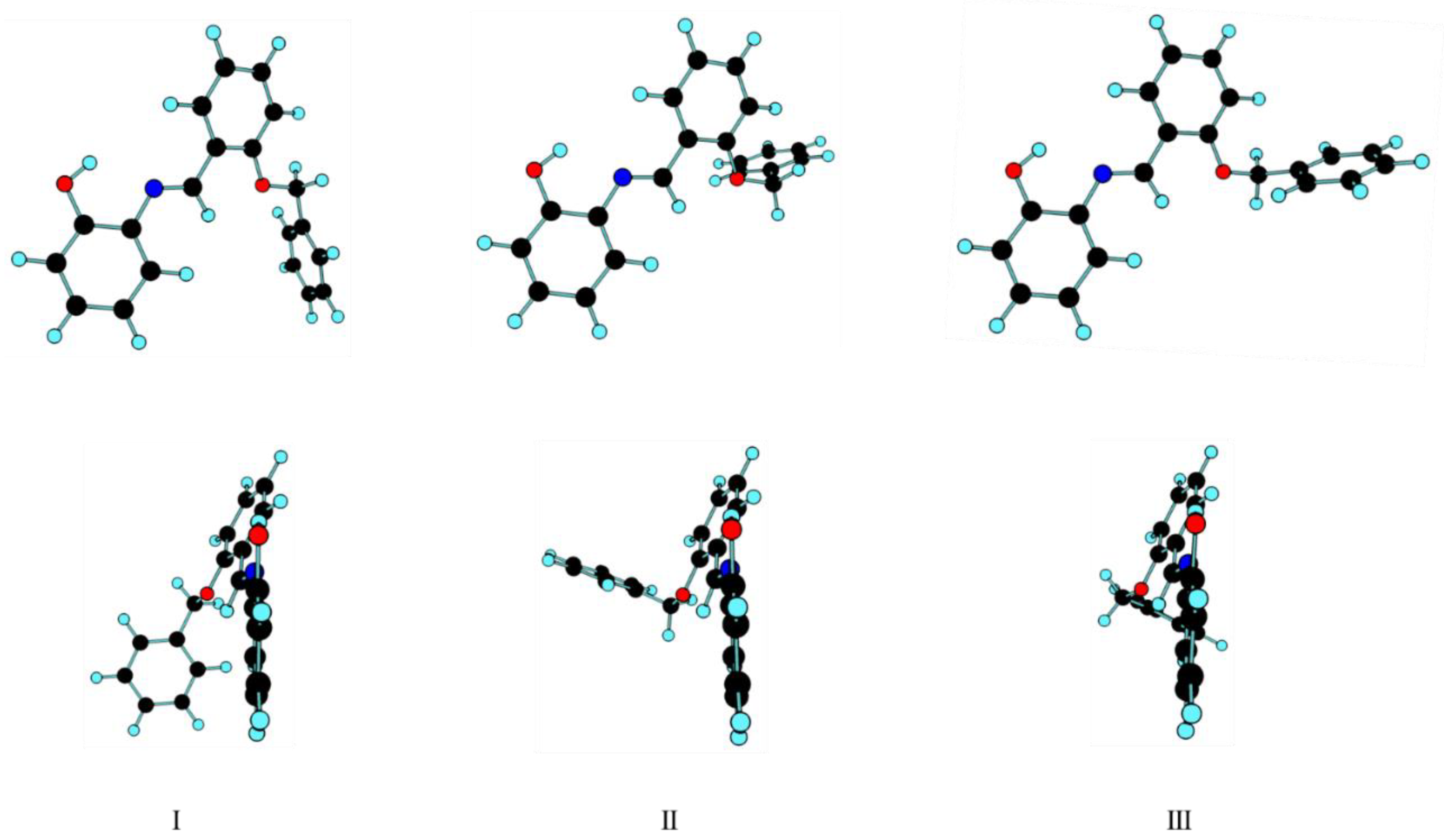
3.1. Geometrical Features and Energies of BBAP Isomeric Structures

The BBAP molecule possesses six internal degrees of conformational freedom, besides cis and trans isomerism around the C=N azomethine bond. However, all the low-energy conformers of the synthesized trans C=N isomer (E isomer) have the geometry of the phenolic moiety determined by the stabilizing intramolecular O–H···N hydrogen bond established between the phenolic O–H group (as H-donor) and the azomethine nitrogen atom (as acceptor), as it is characteristic for ortho OH-substituted aryl Schiff bases [22,23,28,29,30], which was pointed out in the introduction. The calculations for different arrangements around the C–O(H) and C–N bonds led to conformers whose energies are 28–51 kJ mol−1 higher than that of the most stable conformer (see Table S1 and Figure S4) and are irrelevant in practical terms. In this way, the number of significant conformational degrees of freedom was reduced to four, which correspond to the internal rotations around the two exocyclic C–C bonds and the two ether C–O bonds (see Scheme 1). A systematic search on the potential energy surface of the molecule considering these four internal rotations as scanning coordinates allowed us to identify 11 different conformers (all having an equivalent-by-symmetry form), of which three are low-energy forms, with relative energies within ca. 4 kJ mol−1, and the remaining have energies higher than that of the most stable conformer, by 13–29 kJ mol−1 (Table 1).
The structures of the three lowest-energy conformers of BBAP are shown in Figure 1. The calculated relative electronic energies (with and without the inclusion of the zero-point correction) and standard Gibbs energies (at 298.15 K) of the 11 conformers of BBAP, bearing the intramolecular O–H···N hydrogen bond, and their estimated populations in the room-temperature (RT) gas-phase equilibrium are shown in Table 1. Relevant dihedral angles for these conformers are given in Table 2.
All three of the most stable conformers of BBAP (I, II and III: see Figure 1) share a common geometry, except that of the benzyloxy substituent. In the most stable form (I), the C–O–CH2–C dihedral angle (ε) angle is ~180° (anti arrangement), while in conformers II and III, this dihedral defines a gauche arrangement, being –78.7 and 79.2°, respectively. On the other hand, the O–CH2–C–C (ζ) dihedral angles in II and III correspond to a geometry where one of the methylene C–H bonds is nearly perpendicular to the phenyl ring of the benzyloxy substituent, while, in the case of conformer I, it is the C–O bond that is approximately perpendicular to the ring. As a whole, the geometric arrangements defined by the ε and ζ axes in the different conformers make conformer II a more open structure, relative to forms I and III; the phenyl ring of the benzyloxy moiety in II points away from the main plane of the molecule, while in III it points towards this plane. Form I presents an intermediate geometry regarding this structural feature (see Figure 1).
Conformers II and III have similar energies, which are higher than that of conformer I by 3.52 and 3.65 kJ mol−1 (3.91 and 4.16 kJ mol−1, when the zero-point correction is considered). Since the OH···H hydrogen bond distance was predicted by the calculations to be ~2.06 Å in all three conformers, their energy difference should be attributed to the specific geometric arrangement of their benzyloxy substituent. In conformer I, the ether O atom was better placed than in both forms II and III to allow for its lone electron pairs to conjugate with the π-electron cloud of the phenyl ring, which might justify its lower energy.
The standard Gibbs energies (at 298.15 K) of conformers II and III are 6.83 and 7.13 kJ mol−1 higher than that of conformer I, which led to estimated populations for I, II and III in the gas-phase equilibrium (at room temperature: RT) of ca. 89%, 6% and 5%, respectively. It is interesting to note that the calculated entropy (at RT) for conformer I was ~634 J mol−1 K−1, being larger than those estimated for conformers II and III (~623 J mol−1 K−1), indicating that the conformational flexibility of the benzyloxy group in conformer I is higher than in conformers II and III. This is in agreement with the relative values of the calculated torsional frequencies around the ζ axis for the three conformers: 11 cm−1 for I, 33 cm−1 for II, and 29 cm−1 for III, which demonstrates that the potential energy profile for this torsional mode is considerably flatter around the minimum I, compared to II and III.
The structures of the remaining conformers bearing the stabilizing intramolecular OHN hydrogen bond (IV–XI) are shown in Figure S5 (supporting information). Forms IV and V (with relative energies of 13.29 and 15.23 kJ mol−1, respectively) are related to conformers II and III, respectively, regarding the value of the ε dihedral angle, but have a different arrangement about the ζ axis, which leads to an increased steric hindrance between the two nearby-located phenyl rings, thus accounting for their higher relative energy compared to II and III. On the other hand, forms VI–XI have the N=C–C–CH (γ) dihedral angle anti (contrarily to forms I–V, which have a syn arrangement). The energies of these conformers are higher than that of the most stable conformer, I, by ~13–29 kJ mol−1, as a result of the destabilizing interactions between the benzyloxy and phenol moieties (see Figure S4). In the two lowest energy conformers of this group (VI and VII), a weak O–HO intramolecular hydrogen bond (~2.80–2.90 Å) exists between the phenol group and the benzyloxy oxygen atom, which justifies their lowest-energy compared to the remaining conformers (VIII-XI), where such interaction is absent.
Figure 2 shows the potential energy profile for interconversion between the three lowest-energy conformers of BBAP. In this figure, the symmetry-equivalent forms of I, II and III are designated as I’, II’ and III’, respectively. The scanning coordinate is the C–O–CH2–C dihedral angle (ε) and the starting geometries for the different scans that were used to build the figure were those of I’, II and III. In the figure, the values assumed by the O–CH2–C–C angle (ζ, where the ring carbon atom is that defining the smallest dihedral angle among the two possible in the starting conformer) along the scans are also shown, since, at certain critical geometries along the scans, the arrangement of the phenyl group, as defined by the ζ angle, changed in order to relief steric hindrance.
In starting the scan at the geometry of II, towards the geometry of III (i.e., increasing the value of ε: the blue line in Figure 2), the energy raises until the transition structure, which stayed 25 kJ mol−1 higher in energy than conformer II, and then starts to decrease. At ε ~90°, there is an abrupt decrease of energy, which is determined by the change of the orientation of the phenyl group, as seen in the change of the ζ angle to the characteristic value of conformer III (ca. –150°). This change results from the steric hindrance between the two phenyl groups. The interesting point to notice is that this change of orientation of the phenyl group takes place along the scan for a value of the scanning coordinate, ε, that is larger than that of conformer III (the ζ angle is initially that of conformer II, ca. –30°, and decreases smoothly till ca. –120°, the point at which the ring flip takes place) so that this latter form is not effectively reached in this way. Nevertheless, after the flip of the phenyl group, the molecule is taken to the potential energy well of conformer III, which meant that, in practical terms, the conversion II→III is achieved after the relaxation of the structure obtained after the change of orientation of the phenyl group.
When the scan starts at the geometry of conformer III, for increasing values of ε (the red line in Figure 2), conformer I is reached after the reorientation of the phenyl group at ε ~150°, which also implies a fast decrease of energy around this geometry. The energy barrier separating III from I is about 8.5 kJ mol−1 (12 kJ mol−1 in the reverse direction). The continuation of the scan towards II allowed to reach this latter conformer via a barrier of 12 kJ mol−1 (~8.5 kJ mol−1 in the reverse direction). These low-energy barriers demonstrate the easiness of the processes for interconversion between the three conformers.
The performed scan that starts from the geometry of I’ (black line in Figure 2), for increasing values of ε, yields form III’ (through an energy barrier of ~11 kJ mol−1) after the reorientation of the phenyl group at ε ~270°, which then converts to form II’ through a barrier of ca. 30 kJ mol−1. The slightly higher barrier when the scan goes from III’ to II’, compared to that obtained when the scan goes from II to III, results from the slightly different interactions at the geometries at which the flip of the phenyl group takes place when the scan is performed in the two different directions.
The most important conclusions that can be extracted from the results shown in Figure 2 are, on the one side, that the energy barriers separating the different forms are large enough to allow their trapping from the gas phase into the cryogenic matrices without significant conformational cooling during matrix deposition [41,42], and, on the other side, that they are sufficiently low to provide the molecule with substantial conformational flexibility around this coordinate in the gas phase. One can also expect that the interconversion barriers in the excited states are also not very large.

3.2. Infrared Spectrum of Matrix-Isolated BBAP

Monomers of BBAP were isolated from the gas phase into cryogenic (10 K) Ar and N2 matrices, as described in Section 2.2. The experimental IR spectra obtained in both matrices are presented in Figure 3, alongside the simulated IR spectrum, that was constructed by adding the DFT(B3LYP)/6-311++G(d,p) calculated spectra of conformers I, II and III, weighted by their estimated populations in the gas phase prior to deposition. As stated before, these three conformers were the only forms of BBAP expected to be significantly populated in the gas phase, with the lowest energy conformer, I, strongly dominating (see Table 1). Since the calculations also indicated that the energy barriers separating these conformers from each other would be large enough to prevent their interconversion during matrix deposition, the presence of these three conformers in the as-deposited matrices was expected. The agreement between the experimental and the simulated spectra, shown in Figure 3 is excellent, what is in consonance with the presence in the as-deposited matrices of the 3 conformers in relative amounts similar to those estimated theoretically. However, the expected amounts of conformers II and III present in the matrices were considerably smaller than that of conformer I (6 and 5% vs. 89%), which, together with the fact that the spectra of the three conformers were very much similar (in particular, those of II and III, that were very much coincident regarding their frequencies and that differed mostly in the relative band intensities) due to their structural resemblance, made the identification of marker bands for conformers II and III very difficult. Band assignments are provided in Table S2 (supporting information), where no assignments to individual conformers are given. It should, however, be highlighted that the results of the performed UV irradiation of the matrices, which are presented in the next section, clearly show that all three conformers are, in fact, present in the matrices, though the assignment of the bands to individual conformers could not be done either based on those results, as explained below.

3.3. Narrowband UV-Induced Rotamerization of Phenyl Ring

The effects of in situ UV irradiation of matrix-isolated BBAP in both Ar and N2 matrices were investigated. The matrices were irradiated at λ = 230 nm for 50 min, the irradiation wavelength being chosen taking into account the TD-DFT-calculated UV spectrum of the compound (see Figure S6, in the supporting information).
The different spectra obtained by subtracting the IR spectra of the as-deposited matrices from those of the UV-irradiated matrices are shown in Figure 4.
The first point that we observed was the fact that irradiation did not result in the appearance of new bands in the spectra but led only to changes in the relative intensities of the bands. This observation indicated that no new species were being generated upon the performed irradiations, in particular, that neither the transcis (C=N) isomerization nor the enol-imine/keto-amine tautomerization were taking place. Otherwise, the observed redistribution of the band intensities revealed that the amount of the BBAP conformers that were initially present in the matrices were changing—i.e., that UV irradiation resulted in rotamerization within the benzyloxy group.
Detailed comparison of the experimental results with the calculated spectra of conformers I, II and III allowed us to conclude that the amount of conformer II present in the matrices after irradiation diminished, while the amount of conformers I and III increased. A simulated IR difference spectrum defined as a1 × SIR(I) + a3 × SIR(III)−SIR(II), where SIR(x) represents the calculated spectrum of conformer x, and the coefficients ax apply to the intensities (a1 + a3 = 1), was then obtained by adjusting the coefficients a1 and a3 to reproduce, as best as possible, the experimental difference spectra. A good solution was obtained for a1 = 0.43 and a3 = 0.57 (see Figure 4), which indicated that, upon irradiation, the population of conformer III increased somewhat more than that of conformer I.
Unfortunately, the assignment of individual bands to specific conformers could not be done, even taking into account the results of the UV irradiation experiments. As mentioned in Section 3.2, the spectra of the three conformers are very similar, in particular, those of conformers II and III. Hence, since irradiation led to the consumption of conformer II and the formation of conformer III (and I, which also has most of its bands coincident with those of conformers II and III: see Table S2), the appearance of negative bands in the difference spectra, shown in Figure 4, does not mean that these bands are only ascribable to conformer II; in the same way, the fact that the bands appear as positive in the difference spectra does not imply that this conformer does not also contribute to their intensity. Nevertheless, the presence of the three conformers in the matrices is beyond doubt, as is the fact that the UV irradiation of the matrices leads to their interconversion.
The detailed explanation for the specific observations resulting from the UV irradiation of the matrices escapes the scope of this study, since it requires high-level excited-states theoretical calculations and also a detailed study of the constraints imposed by the matrix morphology to the involved transformations. Nevertheless, a general picture can be presented, based on simple considerations that are in agreement with the observations. The first consideration relies on the fact that all three of the conformers—I, II and III—have similar UV absorbance spectra. This means that they were simultaneously excited by the performed irradiation of the matrices, and the system should tend to a photostationary state. Thus, the observed changes in the relative populations of the conformers were a reflex of the occurrence of the different photo-induced interconversions between the three conformers towards the photo-stationary state. In the absence of rigorous theoretical data on the different reaction parameters for each individual isomerization process, one cannot say anything regarding the relative efficiency and extension of the different processes. Assuming that they have probabilities that are intrinsically identical, then the final result should be dependent largely on matrix effects. The critical variable in this case would be the volume available in the primary matrix cage for the conformational changes within the benzyloxy fragment to take place. In this regard, conformers I, II and III of BBAP are significantly different, with conformer II being a considerably more open structure than conformers I and III and, thus, requiring a much larger embedment volume in the matrices to accommodate the benzyloxy fragment, which is, in this case, more deviated from the plane of the main fragment of the molecule (see Figure 1). On the other hand, conformer III is the most closed form, requiring the smallest volume to properly accommodate the benzyloxy fragment among the three conformers. The conformational changes would take place without important changes of the original matrix-site morphology, which would cost a large amount of energy for matrix rearrangement. Under these circumstances, one would expect that volume constraints to the efficiency of the conformational conversions were maximized for those leading to the formation of conformer II and minimized for those leading to the formation of conformer III, thus justifying the observed changes in the relative populations of the conformers. We could also expect that the photoisomerization in Ar and N2 would show some difference due to the difference in lattice parameters for these two host crystals. Nevertheless, no striking differences were observed in the results obtained in the two matrices (though around the 750 cm−1, the difference spectra, obtained in the two matrices and shown in Figure 4, differ somewhat from each other). The apparent relative lack of high-sensitivity of the observed photoisomerization to the matrix host may be due to the comparatively large volume of the rotating group compared to the host-matrix particles (either Ar atom or N2 molecule).

4. Conclusions

In this study, the conformational space of the molecule of benzyloxy substituted ortho-OH substituted aryl Schiff base trans 2-((2-(benzyloxy)benzylidene)amino)phenol (BBAP) has been investigated by an extensive series of quantum chemical calculations, undertaken within the density functional theory framework. Thirty-one distinct conformers of the compound were identified, with the three most stable conformers (I–III) having considerably lower energies than all the remaining forms. The most relevant intramolecular interactions in BBAP were evaluated and used to explain the relative energies of the different conformers, with emphasis given to the lowest-energy forms, whose relative energies stay within ca. 4 kJ mol−1. These conformers differ in the geometry of the benzyloxy moiety, and are stabilized by an intramolecular O–H···N hydrogen bond, established between the phenolic O–H group (as H-donor) and the azomethine nitrogen atom (as acceptor), and by the absence of significant steric hindrance.
The compound was synthesized from 2-aminophenol and 2-(benzyloxy)benzaldehyde and isolated in cryogenic argon and N2 matrices. The three lowest-energy conformers (I–III) predicted theoretically have been successfully trapped into the low-temperature matrices (10 K), and the IR spectra of the compound in both solid Ar and N2 have been assigned. Subsequently, in situ UV irradiation (λ = 230 nm) of the matrix-isolated compound was carried on. It was concluded that the performed irradiation induced interconversions between the 3 low-energy conformers of BBAP initially present in the matrices, i.e., rotamerization within the benzyloxy group, while no evidence was found both for transcis (C=N) isomerization and enol-imine/keto-amine tautomerization. An explanation for the observed conformational population changes upon UV irradiation was proposed, which considered the attainment of a photo-stationary state upon irradiation, together with the different primary cages occupied by the reactant molecules. Conformer II has a considerably more open structure than conformers I and III and, thus, requires a much larger embedment volume in the matrices to accommodate the benzyloxy fragment, while conformer III requires the smallest volume among the three conformers. Since the photo-induced conformational changes shall take place without important changes of the original matrix-site morphology (which would cost a large amount of energy), volume constraints to the efficiency of the conformational conversions are maximized for those leading to formation of conformer II and minimized for those leading to conformer III, thus justifying the observed changes in the relative populations of the conformers, with that of II reducing and those of I and III increasing, with the last being the one increasing the most. To the best of our knowledge, this is the first report on UV-induced conformational changes taking place in a benzyloxy fragment of a matrix-isolated compound.

Supplementary Materials

The following supporting information can be downloaded at: https://www.mdpi.com/article/10.3390/photochem2020026/s1. Figures S1–S7 with IR, 1H- and 13C-NMR spectra of BBAP, pictures of higher-energy conformers of BBAP, and TD-DFT(B3LYP)/6-311++G(d,p) calculated and experimental UV-vis spectrum of BBAP. Tables S1 and S2 with DFT(B3LYP)/6-311++G(d,p) calculated relative energies of the high-energy conformers of BBAP and proposed band assignments for the IR spectra of matrix-isolated BBAP (in Ar and N2 matrices).

Author Contributions

H.B. synthesized the compound, performed its characterization by IR and NMR spectroscopies, and elemental analysis. Y.G.S. performed the DFT calculations. İ.S. and S.G. collected the experimental infrared spectra, performed the initial analysis of the data, and wrote the first draft of the manuscript. R.F. consolidated the data interpretation, and wrote the final draft of the manuscript. All authors have read and agreed to the published version of the manuscript.

Funding

This research received no external funding.

Acknowledgments

The authors greatly appreciate Bitlis Eren University Research Foundation for financial support (Projects BEBAP-2014.05 and BEBAP-2013.04) and for the computational facilities, the Portuguese Science Foundation (“Fundação para a Ciência e a Tecnologia”—FCT; Projects CQC-IMS UIDB/00313/2020 and UIDP/00313/2020—National Funds), and the Chemistry Department, Faculty of Sciences, Eskişehir Technical University.

Conflicts of Interest

The authors declare no conflict of interest.

References

  1. Borisova, N.E.; Reshetova, M.D.; Ustynyuk, Y.A. Metal-Free Methods in the Synthesis of Macrocyclic Schiff Bases. Chem. Rev. 2007, 107, 46–79. [Google Scholar] [CrossRef] [PubMed]
  2. Vigato, P.A.; Tamburini, S.; Bertolo, L. The development of compartmental macrocyclic Schiff bases and related polyamine derivatives. Coord. Chem. Rev. 2007, 251, 1311–1492. [Google Scholar] [CrossRef]
  3. Emregül, K.C.; Düzgün, E.; Atakol, O. The application of some polydentate Schiff base compounds containing aminic nitrogens as corrosion inhibitors for mild steel in acidic media. Corros. Sci. 2006, 48, 3243–3260. [Google Scholar] [CrossRef]
  4. Drozdzak, R.; Allaert, B.; Ledoux, N.; Dragutan, I.; Dragutan, V.; Verpoort, F. Ruthenium complexes bearing bidentate Schiff base ligands as efficient catalysts for organic and polymer syntheses. Coord. Chem. Rev. 2005, 249, 3055–3074. [Google Scholar] [CrossRef]
  5. Sessler, J.L.; Melfi, P.J.; Pantos, G.D. Uranium complexes of multidentate N-donor ligands. Coord. Chem. Rev. 2006, 250, 816–843. [Google Scholar] [CrossRef]
  6. Yang, C.-J.; Jenekhe, S.A. Conjugated aromatic polyimines. 2. Synthesis, structure, and properties of new aromatic polyazomethines. Macromolecules 1995, 28, 1180–1196. [Google Scholar] [CrossRef]
  7. Destri, S.; Khotina, I.A.; Porzio, W. 3-Hexyl Tetra-Substituted Sesquithienylene- Phenylene Polyazomethines with High Molecular Weight. Mechanistic Considerations. Macromolecules 1998, 31, 1079–1086. [Google Scholar] [CrossRef]
  8. Grigoras, M.; Catanescu, O.; Simionescu, C.I. Poly(azomethine)s. Rev. Roum. Chim. 2001, 46, 927–939. [Google Scholar]
  9. Kaya, I.; Vilayetoglu, A.R.; Mart, H. The synthesis and properties of oligosalicylaldehyde and its Schiff base oligomers. Polymer 2001, 42, 4859–4865. [Google Scholar] [CrossRef]
  10. Wu, P.; Bhamidipati, M.; Coles, M.; Rao, D. Biological nano-ceramic materials for holographic data storage. Chem. Phys. Lett. 2004, 400, 506–510. [Google Scholar] [CrossRef]
  11. Yuan, W.; Sun, L.; Tang, H.; Wen, Y.; Jiang, G.; Huang, W.; Jiang, L.; Song, Y.; Tian, H.; Zhu, D. A novel thermally stable spironaphthoxazine and its application in rewritable high density optical data storage. Adv. Mater. 2005, 17, 156–160. [Google Scholar] [CrossRef]
  12. Yanez, C.O.; Andrade, C.D.; Yao, S.; Luchita, G.; Bondar, M.V.; Belfield, K.D. Photosensitive polymeric materials for two-photon 3D WORM optical data storage systems. ACS Appl. Mater. Interfaces 2009, 1, 2219–2229. [Google Scholar] [CrossRef] [PubMed]
  13. Melloni, A.; Rossi Paccani, R.; Donati, D.; Zanirato, V.; Sinicropi, A.; Parisi, M.L.; Martin, E.; Ryazantsev, M.; Ding, W.J.; Frutos, L.M. Modeling, preparation, and characterization of a dipole moment switch driven by Z/E photoisomerization. J. Am. Chem. Soc. 2010, 132, 9310–9319. [Google Scholar] [CrossRef]
  14. Staykov, A.; Watanabe, M.; Ishihara, T.; Yoshizawa, K. Photoswitching of conductance through salicylidene methylamine. J. Phys. Chem. C 2014, 118, 27539–27548. [Google Scholar] [CrossRef]
  15. Dalapati, S.; Jana, S.; Guchhait, N. Anion recognition by simple chromogenic and chromo-fluorogenic salicylidene Schiff base or reduced-Schiff base receptors. Spectrochim. Acta A Mol. Biomol. Spectrosc. 2014, 129, 499–508. [Google Scholar] [CrossRef] [PubMed]
  16. Zhang, X.; Yin, J.; Yoon, J. Recent advances in development of chiral fluorescent and colorimetric sensors. Chem. Rev. 2014, 114, 4918–4959. [Google Scholar] [CrossRef]
  17. Hadjoudis, E.; Mavridis, I.M. Photochromism and thermochromism of Schiff bases in the solid state: Structural aspects. Chem. Soc. Rev. 2004, 33, 579–588. [Google Scholar] [CrossRef]
  18. Amimoto, K.; Kawato, T. Photochromism of organic compounds in the crystal state. J. Photochem. Photobiol. C Photochem. Rev. 2005, 6, 207–226. [Google Scholar] [CrossRef]
  19. Larkin, D.R. The role of catalysts in the air oxidation of aliphatic aldehydes. J. Org. Chem. 1990, 55, 1563–1568. [Google Scholar] [CrossRef]
  20. Vančo, J.; Švajlenová, O.; Račanská, E.; Muselík, J.; Valentová, J. Antiradical activity of different copper (II) Schiff base complexes and their effect on alloxan-induced diabetes. J. Trace Elem. Med. Biol. 2004, 18, 155–161. [Google Scholar] [CrossRef]
  21. Jarząbek, B.; Kaczmarczyk, B.; Sęk, D. Characteristic and spectroscopic properties of the Schiff-base model compounds. Spectrochim. Acta. A. Mol. Biomol. Spectrosc. 2009, 74, 949–954. [Google Scholar] [CrossRef] [PubMed]
  22. Raczuk, E.; Dmochowska, B.; Samaszko-Fiertek, J.; Madaj, J. Different Schiff Bases—Structure, Importance and Classification. Molecules 2022, 27, 787. [Google Scholar] [CrossRef] [PubMed]
  23. Król-Starzomska, I.; Filarowski, A.; Rospenk, M.; Koll, A.; Melikova, S. Proton Transfer Equilibria in Schiff Bases with Steric Repulsion. J. Phys. Chem. A 2004, 108, 2131–2138. [Google Scholar] [CrossRef]
  24. Minkin, V.I.; Tsukanov, A.V.; Dubonosov, A.D.; Bren, V.A. Tautomeric Schiff bases: Iono-, solvato-, thermo-and photochromism. J. Mol. Struct. 2011, 998, 179–191. [Google Scholar] [CrossRef]
  25. Sıdır, Y.G.; Pirbudak, G.; Berber, H.; Sıdır, İ. Study on the electronic and photophysical properties of the substitute-((2-phenoxybenzylidene) amino) phenol derivatives: Synthesis, solvatochromism, electric dipole moments and DFT calculations. J. Mol. Liq. 2017, 242, 1096–1110. [Google Scholar] [CrossRef]
  26. Sıdır, Y.G.; Berber, H.; Sıdır, İ. The Dipole Moments and Solvatochromism of ((4-(Benzyloxy) benzylidene) amino) phenol Compounds as Solvatochromic Materials. J. Solut. Chem. 2019, 48, 775–806. [Google Scholar] [CrossRef]
  27. Sıdır, Y.G.; Aslan, C.; Berber, H.; Sıdır, İ. The electronic structure, solvatochromism, and electric dipole moments of new Schiff base derivatives using absorbance and fluorescence spectra. Struct. Chem. 2019, 30, 835–851. [Google Scholar] [CrossRef]
  28. Avadanei, M.; Cozan, V.; Shova, S.; Paixão, J.A. Solid state photochromism and thermochromism of two related N-salicylidene anilines. Chem. Phys. 2014, 444, 43–51. [Google Scholar] [CrossRef]
  29. Sıdır, İ.; Sıdır, Y.G.; Góbi, S.; Berber, H.; Ildiz, G.O.; Fausto, R. UV-induced –OCH3 rotamerization in a matrix-isolated methoxy-substituted ortho-hydroxyaryl Schiff base. Photochem. Photobiol. Sci. 2022. [Google Scholar] [CrossRef]
  30. Sıdır, İ.; Gülseven Sıdır, Y.; Góbi, S.; Berber, H.; Fausto, R. Structural Relevance of Intramolecular H-Bonding in Ortho-Hydroxyaryl Schiff Bases: The Case of 3-(5-bromo-2-hydroxybenzylideneamino) Phenol. Molecules 2021, 26, 2814. [Google Scholar] [CrossRef]
  31. Frisch, M.; Trucks, G.W.; Schlegel, H.B.; Scuseria, G.E.; Robb, M.A.; Cheeseman, J.R.; Scalmani, G.; Barone, V.; Mennucci, B.; Petersson, G.A.; et al. Gaussian 09; Gaussian, Inc.: Wallingford, CT, USA, 2009. [Google Scholar]
  32. Becke, A.D. Density-functional exchange-energy approximation with correct asymptotic behavior. Phys. Rev. A 1988, 38, 3098–3100. [Google Scholar] [CrossRef] [PubMed]
  33. Lee, C.; Yang, W.; Parr, R.G. Development of the Colle-Salvetti correlation-energy formula into a functional of the electron density. Phys. Rev. B Condens. Matter 1988, 37, 785–789. [Google Scholar] [CrossRef] [PubMed] [Green Version]
  34. Vosko, S.H.; Wilk, L.; Nusair, M. Accurate spin-dependent electron liquid correlation energies for local spin density calculations: A critical analysis. Can. J. Phys. 1980, 58, 1200–1211. [Google Scholar] [CrossRef] [Green Version]
  35. McLean, A.D.; Chandler, G.S. Contracted Gaussian basis sets for molecular calculations. I. Second row atoms, Z = 11–18. J. Chem. Phys. 1980, 72, 5639–5648. [Google Scholar] [CrossRef]
  36. Krishnan, R.; Binkley, J.S.; Seeger, R.; Pople, J.A. Self-consistent molecular orbital methods. XX. A basis set for correlated wave functions. J. Chem. Phys. 1980, 72, 650–654. [Google Scholar] [CrossRef]
  37. Frisch, M.J.; Pople, J.A.; Binkley, J.S. Self-consistent molecular orbital methods 25. Supplementary functions for Gaussian basis sets. J. Chem. Phys. 1984, 80, 3265–3269. [Google Scholar] [CrossRef]
  38. Chemcraft—Graphical Program for Visualization of Quantum Chemistry Computations. Available online: https://www.chemcraftprog.com/ (accessed on 3 January 2022).
  39. Bauernschmitt, R.; Ahlrichs, R. Treatment of electronic excitations within the adiabatic approximation of time dependent density functional theory. Chem. Phys. Lett. 1996, 256, 454–464. [Google Scholar] [CrossRef]
  40. Stratmann, R.E.; Scuseria, G.E.; Frisch, M.J. An efficient implementation of time-dependent density-functional theory for the calculation of excitation energies of large molecules. J. Chem. Phys. 1998, 109, 8218–8224. [Google Scholar] [CrossRef]
  41. Reva, I.D.; Stepanian, S.G.; Adamowicz, L.; Fausto, F. Missing Conformers: Comparative Study of Conformational Cooling in Cyanoacetic Acid and Methyl Cyanoacetate Isolated in Low Temperature Inert Gas Matrixes. Chem. Phys. Lett. 2003, 374, 631–638. [Google Scholar] [CrossRef] [Green Version]
  42. Rosado, M.T.S.; Lopes Jesus, A.J.; Reva, I.D.; Fausto, R.; Redinha, J.S. Conformational Cooling Dynamics in Matrix-Isolated 1,3-butanediol. J. Phys. Chem. A 2009, 113, 7499–7507. [Google Scholar] [CrossRef] [Green Version]
Scheme 1. Structure of BBAP, highlighting the conformationally relevant internal rotation axes (designated by the Greek letters).
Photochem 02 00026 sch001
Scheme 2. Synthesis of BBAP.
Photochem 02 00026 sch002
Figure 1. B3LYP/6-311++G(d,p) optimized structures for the three lowest energy conformers (I, II and III) of BBAP (two views). Each conformer has a symmetry-equivalent form. See Table 1 for relative energies, and Table 2 for values of the conformationally relevant dihedral angles.
Photochem 02 00026 g001
Figure 2. Potential energy profiles for interconversion between conformers I, II and III (and their symmetry-related forms I’, II’ and III’), obtained by performing relaxed scans (shown in different colors) with the C–O–CH2–C (ε) dihedral angle as scanning coordinates. The initial geometries chosen for the scans and the direction of the scans are indicated by the arrows.
Photochem 02 00026 g002
Figure 3. As-deposited IR spectra of BBAP, isolated in Ar (top panel) and N2 (middle panel) matrices (10 K), and simulated IR spectrum, built using the calculated infrared spectra of conformers I, II and III, weighted by their estimated populations in the matrices (lower panel). Calculated wavenumbers were scaled as described in Section 2.3. The experimental bands at ca. 667 cm−1 were due to traces of atmospheric CO2.
Photochem 02 00026 g003
Figure 4. Top and middle panels: IR difference spectra (spectrum obtained after 50 min. of UV irradiation at 230 nm minus as-deposited spectrum) obtained in Ar and in N2 (10 K). Lower panel: Simulated infrared difference spectrum was built based on the calculated spectra of BBAP conformers I, II and III, with intensities scaled as 0.43 × I + 0.57 × III−II. Calculated wavenumbers were scaled as described in Section 2.3. The experimental bands at ca. 667 cm−1 were due to traces of atmospheric CO2.
Photochem 02 00026 g004
Table 1. B3LYP/6-311++G(d,p) calculated relative electronic energies (ΔEel), zero-point corrected electronic energies (ΔE(0)), and standard Gibbs energies (ΔG298.15) for the low-energy conformers of trans BBAP, and their estimated room temperature gas-phase equilibrium conformational populations (P298.15) a.
ConformerΔEelΔE(0)ΔG298.15P298.15 (%)
I0.000.000.0089
II3.523.916.836
III3.654.167.135
IV13.2913.0114.63
V15.2314.5814.09
VI12.8312.2314.09
VII14.3714.1416.80
VIII21.6421.1725.05
IX21.8820.5724.27
X22.0920.7622.50
XI28.7427.1827.92
a Energies in kJ mol−1, relative to conformer I. See Figure 1 and Figure S5 for structures of the conformers.
Table 2. B3LYP/6-311++G(d,p) optimized conformationally-relevant dihedral angles (°) of the low-energy conformers of trans BBAP a.
C–N=C–CH–O–C–CNCH–C–N=CN=C–C–CHCH–C–O–CC–O–CH2–CO–CH2–C–C
Conformer αβγδεζ
I–177.4–2.721.76.7–0.7–179.580.1/–98.9
II–177.0–3.123.48.35.6–78.7–31.5/151.4
III–177.5–2.922.44.9–6.779.329.5/–153.3
IV–176.4–3.222.813.6–95.874.268.6/–111.2
V–179.1–2.316.4–3.087.0–77.8–64.6/115.5
VI–179.3–3.216.2179.3–0.3–179.3–84.2/95.1
VII–179.2–1.512.4177.12.4–77.6–30.8/152.1
VIII179.9–1.725.9–175.3–105.474.268.7/–111.0
IX–179.4–3.928.1179.5–98.5174.9–27.3/154.7
X–179.7–3.229.7–177.1–100.1170.5–67.4/112.8
XI–177.9–2.130.9–152.672.2–169.488.6/–90.4
a See Figure 1 and Figure S5 for structures of the conformers.
Publisher’s Note: MDPI stays neutral with regard to jurisdictional claims in published maps and institutional affiliations.

