Article

The Toxicological and Pharmacological Evaluation of the Anacyclus pyrethrum Aqueous Extract: Implications for Medicinal and Therapeutic Applications

1
Laboratory of Pharmacology, Neurobiology, Anthropobiology and Environment, Department of Biology, Faculty of Sciences Semlalia, Cadi Ayyad University, Marrakech 40000, Morocco
2
Laboratory of Biological Engineering, Faculty of Sciences and Technology, Sultan Moulay Slimane University, Beni Mellal 23000, Morocco
3
Higher Institute of Nursing and Health Techniques, Ministry of Health and Social Protection, Rabat 10000, Morocco
4
Laboratory of Biochemistry, Department of Applied Biological Chemistry, Faculty of Agriculture, University of Niigata, Niigata 950-2181, Japan
*
Author to whom correspondence should be addressed.
Stresses 2024, 4(1), 79-93; https://doi.org/10.3390/stresses4010005
Submission received: 12 October 2023 / Revised: 4 November 2023 / Accepted: 3 January 2024 / Published: 9 January 2024
(This article belongs to the Collection Feature Papers in Human and Animal Stresses)

Abstract

:
Plants have long been valued for their medicinal and nutritional contributions to human life. Anacyclus pyrethrum, a member of the Asteraceae family, has attracted increasing attention as a source of natural products with diverse applications. In this study, we explored the toxicity and pharmacological properties of the aqueous extract of A. pyrethrum (AEAP). The acute toxicity study involved groups of mice subjected to oral administration of varying doses of AEAP, with immediate post-administration observations to detect any signs of toxicity or mortality. Comprehensive biochemical and hematological analyses encompassed assessments of renal function. The pharmacological profile was assessed by evaluating antinociceptive, anxiolytic, and antidepressant effects, which were measured using the hot plate test, elevated plus maze, open field test, and forced swim test, respectively. Different doses (100, 200, 400, and 800 mg/kg) were administered to rats via gavage for this assessment. The results revealed that the acute toxicity demonstrated the safety of AEAP at the tested doses, with no observed mortality or significant alterations. Moreover, it revealed that AEAP possesses an LD50 value greater than 5000 mg/kg. The pharmacological properties of AEAP demonstrated anxiolytic and antidepressant activities at a dose of 200 mg/kg, while no antinociceptive effect was observed. These findings underscore the potential of A. pyrethrum as a natural source of bioactive compounds with therapeutic applications. Further research is needed to explore long-term and chronic effects for a comprehensive assessment.

1. Introduction

For numerous years, plants have played a vital role in human life, offering both medicinal benefits and nutritional value. Naturally derived products from plants are garnering increasing interest as essential raw materials in various sectors, including pharmaceuticals and agri-food. Plants encompass a wide array of bioactive compounds with positive effects on human health and well-being [1]. Today, the significance of aromatic and medicinal plants (AMPs) extends beyond their traditional use in disadvantaged communities in developing countries. They have evolved into valuable sources of highly sought-after bioactive molecules for the pharmaceutical, agri-food, cosmetic, and perfume industries [2]. AMPs are prominently featured in both modern and traditional medicine, with ca. 50% of prescribed medicine having a natural origin [3].
The resurgence of interest in AMPs is driven by several factors. Firstly, concerns about the adverse effects of chemical compounds present in certain medications have propelled this renewed focus [4]. Secondly, economic considerations, including the high cost associated with synthetic drugs, have contributed to the growing interest [5]. In this context, AMPs offer a promising alternative as a natural source of bioactive compounds. According to Van Wyk [6], despite over 5400 plant species being used in traditional African medicine, only 10% of them have been commercially developed to some extent.
This renewed emphasis on AMP is particularly pertinent in Mediterranean ecosystems, such as those in Morocco, which are renowned for their rich and diverse flora shaped by ecological diversity [7]. Local communities in these areas possess extensive traditional knowledge and have driven studies and inventories of plants used in phytotherapy [8]. These efforts have identified nearly 570 aromatic and/or medicinal plant species, spanning 98 families and 486 genera, representing ca. 8.1% of Morocco’s total flora [8].
Anacyclus pyrethrum belongs to the Asteraceae family and the Anacyclus genus. It is known as Pellitory, Aqar Qarha, Oud El Attas, and tigandizt. This plant is native to Morocco, Algeria, and Spain [9,10]. A. pyrethrum is a medicinal plant with a history of traditional therapeutic uses, serving as a stimulant, cordial, and rubefacient [11]. It is often recommended for gargling to alleviate symptoms of conditions like rhinitis, neuralgia, rheumatic discomfort, and musculoskeletal pain [9]. Additionally, the root has demonstrated promising results in the treatment of conditions such as sciatica, paralysis, hemiplegia, and amenorrhea [12]. In Morocco, A. pyrethrum roots are renowned in traditional medicine for their beneficial effects in treating various conditions, including stomach diseases, stomatitis, cysts, articular rheumatism, and dental pain [13].
Numerous experimental studies have revealed the extensive biological effects associated with A. pyrethrum, including antioxidant, anti-inflammatory, analgesic properties [14], anti-epileptic effects [15], immunostimulatory capabilities [16], and insecticidal activity [17]. Notably, A. pyrethrum has also shown the ability to reduce insulin requirements in individuals with insulin-dependent diabetes mellitus and lower plasma glucose and serum cholesterol levels after oral administration for 3-6 weeks [18]. These attributes stem from a diverse array of phytochemical compounds, with over a hundred distinct compounds identified thus far, including phenolic compounds, flavonoids, and alkaloids [9,19]. A recent study using HPLC–PDA–MS/MS has provided valuable insights into the specific secondary metabolites present in the aqueous extract of A. pyrethrum (AEAP). This analysis identified 24 secondary metabolites, with pellitorine and 3,4-dihydroxybenzoic acid, also known as protocatechuic acid, emerging as the most abundant components [20]. Particularly, pellitorine plays a significant role within the extract and has previously been recognized as a key contributor to the diverse beneficial activities associated with A. pyrethrum. These findings underscore the plant’s multifaceted biological and pharmacological properties [20,21,22].
Given the extensive chemical characterization outlined, our attention shifts toward the global use of analgesics, antidepressants, and anxiolytics. According to the World Health Organization (WHO), these medications have widespread global use, making them the most commonly prescribed category of drugs. However, many of the currently available anxiolytics and antidepressants have undesirable side effects, resulting in poor patient compliance [23,24]. This highlights the importance of exploring alternative sources, such as medicinal plants, in the search for new drug leads [25].
Expanding the therapeutic applications of A. pyrethrum could enrich treatment options, especially in regions where traditional medicine plays a crucial role in healthcare. This endeavor could provide empirical support for the traditional use of this plant, potentially leading to the development of new herbal remedies or pharmaceutical innovations. In this context, our study aims to contribute to the valorization of A. pyrethrum. Our objective is to uncover novel therapeutic prospects while substantiating previous research. We conducted a comprehensive investigation into the AEAP, exploring its toxicity and pharmacological attributes.

2. Results

2.1. Acute Toxicity

2.1.1. Lethal Dose 50 Estimation (LD50)

All tested doses of AEAP at 1000, 2000, and 5000 mg/kg were found to be safe (non-toxic). Over a 14-day observation period following AEAP administration, no instances of mortality were recorded, and there were no significant changes in both body and organ weights (p > 0.05), as shown in Table 1. This emphasizes that the oral LD50 (lethal dose that affects 50% of the subjects) of AEAP exceeds 5000 mg/kg, indicating a low level of toxicity in accordance with the Organization for Economic Cooperation and Development (OECD) guidelines.

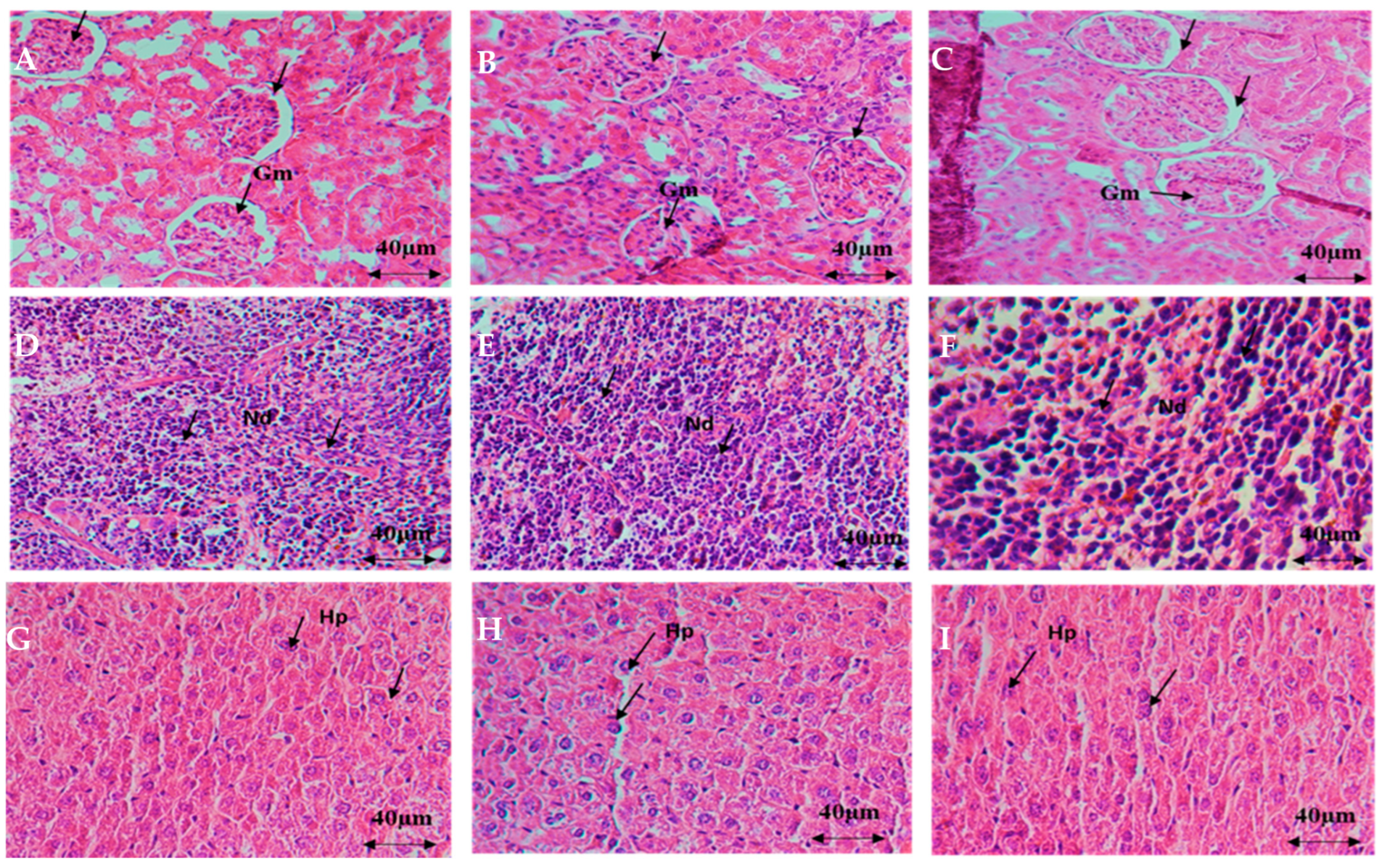
2.1.2. Histopathological Examination

The microscopic examination of kidney, liver, and spleen tissues revealed a normal appearance similar to the control group, indicating the absence of deleterious changes or morphological disturbances due to oral AEAP administration. Additionally, this study determined that oral AEAP administration at doses up to 5000 mg/kg did not elicit any visible signs or symptoms of toxicity in mice. There were no instances of mortality, and no significant shifts in body or organ weights were observed after 14 days of AEAP administration. The median lethal dose (LD50) values for AEAP were found to exceed 5000 mg/kg, indicating its low toxicity (Figure 1).

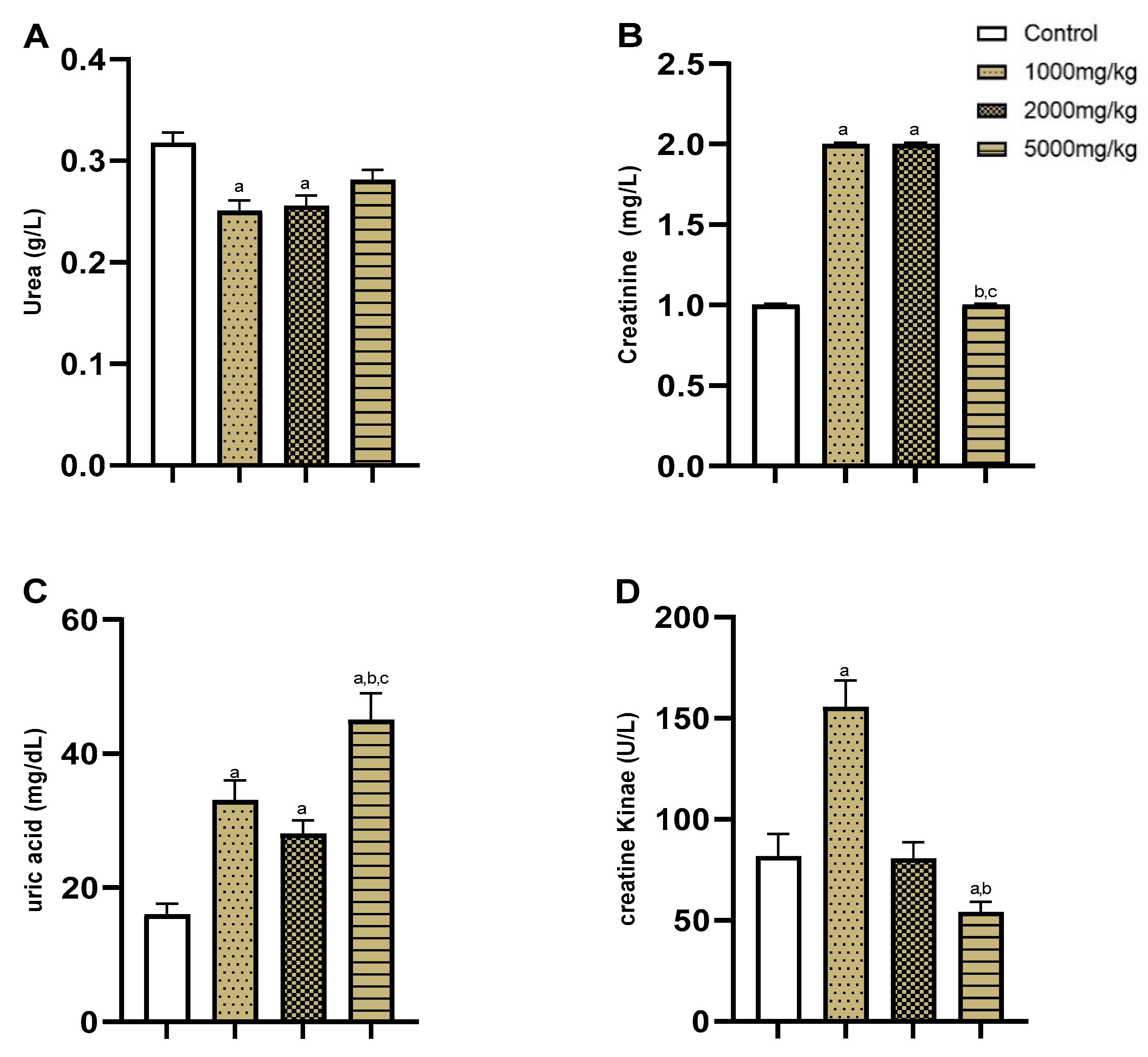
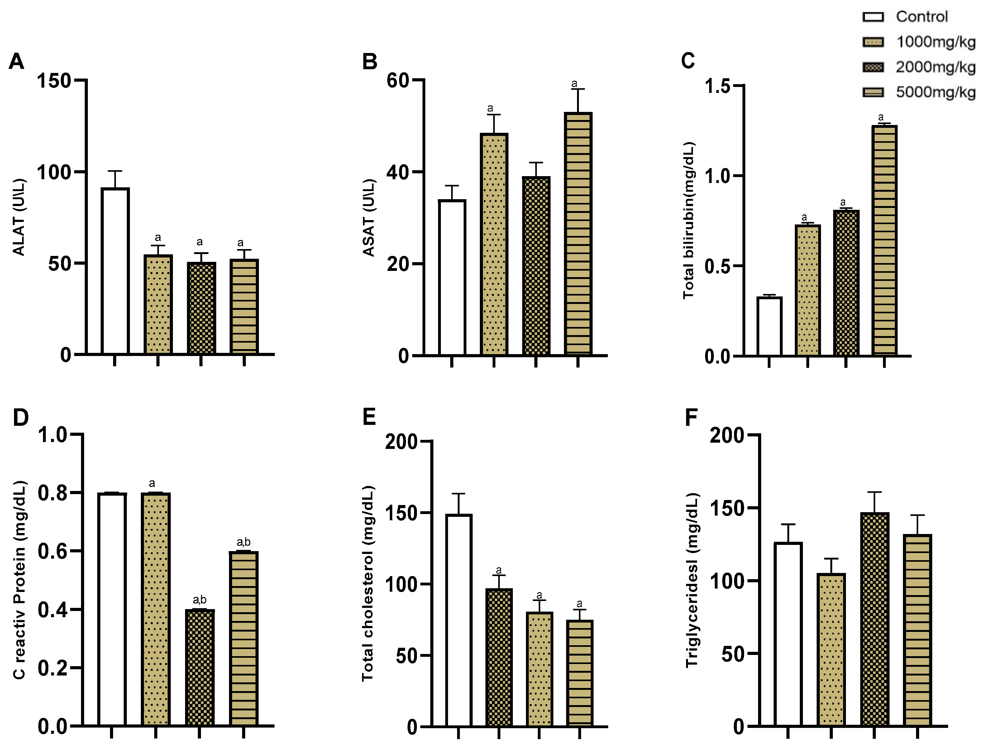
2.1.3. Biochemical Analyses

Next, we assessed the AEAP toxicity on renal and liver functions. In terms of renal function markers, including urea, creatinine, uric acid, and creatine kinase levels, the control group exhibited standard levels: urea (0.3 g/L), creatinine (1 mg/L), uric acid (16 mg/L), and creatine kinase (80 U/L). The administration of AEAP at varying doses (1000, 2000, and 5000 mg/kg) led to slight variations in these markers, with slightly reduced urea levels (0.25, 0.25, and 0.28 g/L, respectively) and slightly elevated creatinine levels (2, 2, and 1 mg/L, respectively). Meanwhile, uric acid levels increased in groups treated with AEAP (33, 28, and 45 mg/dL, respectively), and the 1000 mg/kg AEAP-treated group showed slightly higher creatine kinase levels (155 U/L) compared to those of 2000 mg/kg (80 U/L) and 5000 mg/kg (54 U/L) AEAP-treated groups. These findings indicated that AEAP administration did not yield abnormal values or toxic effects on renal function (Figure 2).
The liver function assessment encompassed the analysis of ALAT, ASAT, total bilirubin, C-reactive protein, total cholesterol, and triglyceride levels among AEAP-treated groups in comparison to the control group (Figure 3). The control group displayed an ALAT concentration of 91 ± 9.0 U/L. Notably, groups treated with AEAP at doses of 1000, 2000, and 5000 mg exhibited lower ALAT levels (54, 50, and 52 U/I, respectively) (Figure 3A), suggesting a statistically significant (F(3,20) = 9.77, p < 0.001) potential benefit of AEAP on ALAT levels and liver function improvement. A similar trend was observed for ASAT levels (Figure 3B), where the control group displayed a concentration of 33±3.0 U/L, and AEAP-treated groups at doses of 1000 mg/kg, 2000 mg/kg, and 5000 mg/kg showed statistically (F(3,20)) = 5.26, p < 0.01) higher ASAT levels (48 U/L, 39 U/L, and 53U/L, respectively).
The total bilirubin levels (Figure 3C) in the control group were 0.3 ± 0.01 mg/dL, while, strikingly, AEAP-treated groups exhibited higher total bilirubin levels 0.7 mg/dL (1000 mg AEAP), 0.8 mg/dL (2000 mg AEAP), and 1.2 mg/dL (5000 mg AEAP), with significant statistical differences (F(3,20) = 151.9, p < 0.001). In terms of C-reactive protein levels (Figure 3D), the control group had a concentration of 0.8 mg/dL. Remarkably, AEAP-treated groups at doses of 2000 mg/kg and 5000 mg/kg displayed lower C-reactive protein levels (0.4 and 0.6 mg/dL, respectively) (p < 0.05), whereas the 1000 mg/kg AEAP-treated group exhibited a similar level (0.8 mg/dL) to the control group. These differences suggest a potential beneficial effect of AEAP on reducing C-reactive protein levels. The control group exhibited a total cholesterol concentration of 149 mg/dL (Figure 3E). Notably, the three groups treated with AEAP displayed lower total cholesterol levels (97, 80, and 75 mg/dL at 1000, 2000, and 5000 mg/kg AEAP dose, respectively) (F(3,20) = 11.73, p < 0.001). Interestingly, the groups treated with AEAP at doses of 1000 mg/kg (105 mg/dL) and 5000 mg (132 mg/dL) exhibited similar triglyceride levels to those in the control group (126 mg/dL), while the 2000 mg/kg AEAP-treated group displayed higher levels (146 mg/dL) (Figure 3F).

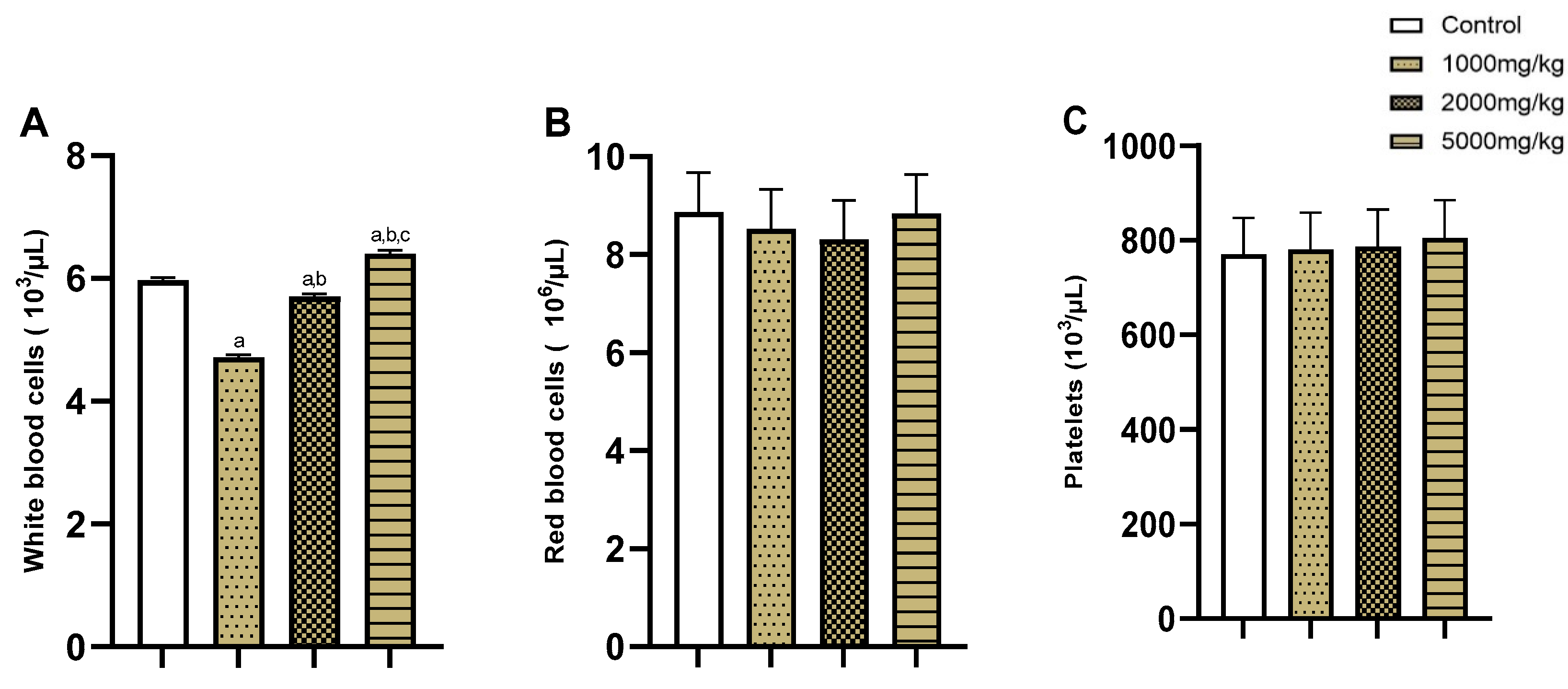
2.1.4. Hematological Analyses

Subsequently, we conducted a hematological analysis to assess the levels of white and red blood cells, as well as platelets, among various groups subjected to AEAP treatments (Figure 4). The control group displayed a white blood cell count of 5 × 10³/μL. The administration of AEAP resulted in a slight increase in white blood cell levels at doses of 2000 mg/kg (5.7 × 10³/μL) and 5000 mg/kg (6.4 × 10³/μL) and a slight decrease at 1000 mg/kg (4.72 × 10³/μL) (Figure 4A). Conversely, no statistically significant differences were observed in the red blood cell and platelet levels following AEAP administration compared to the control groups (Figure 4B,C).

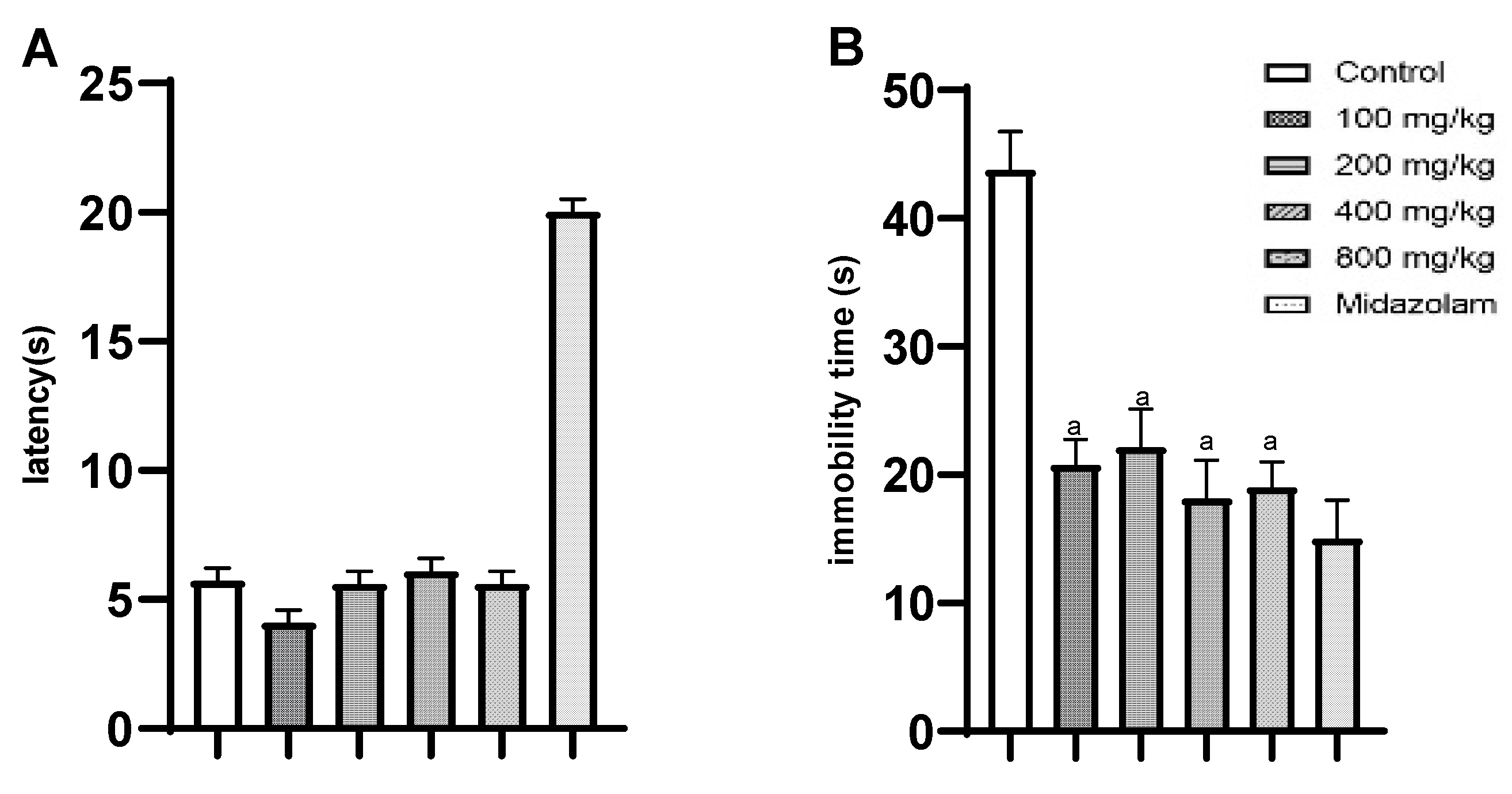
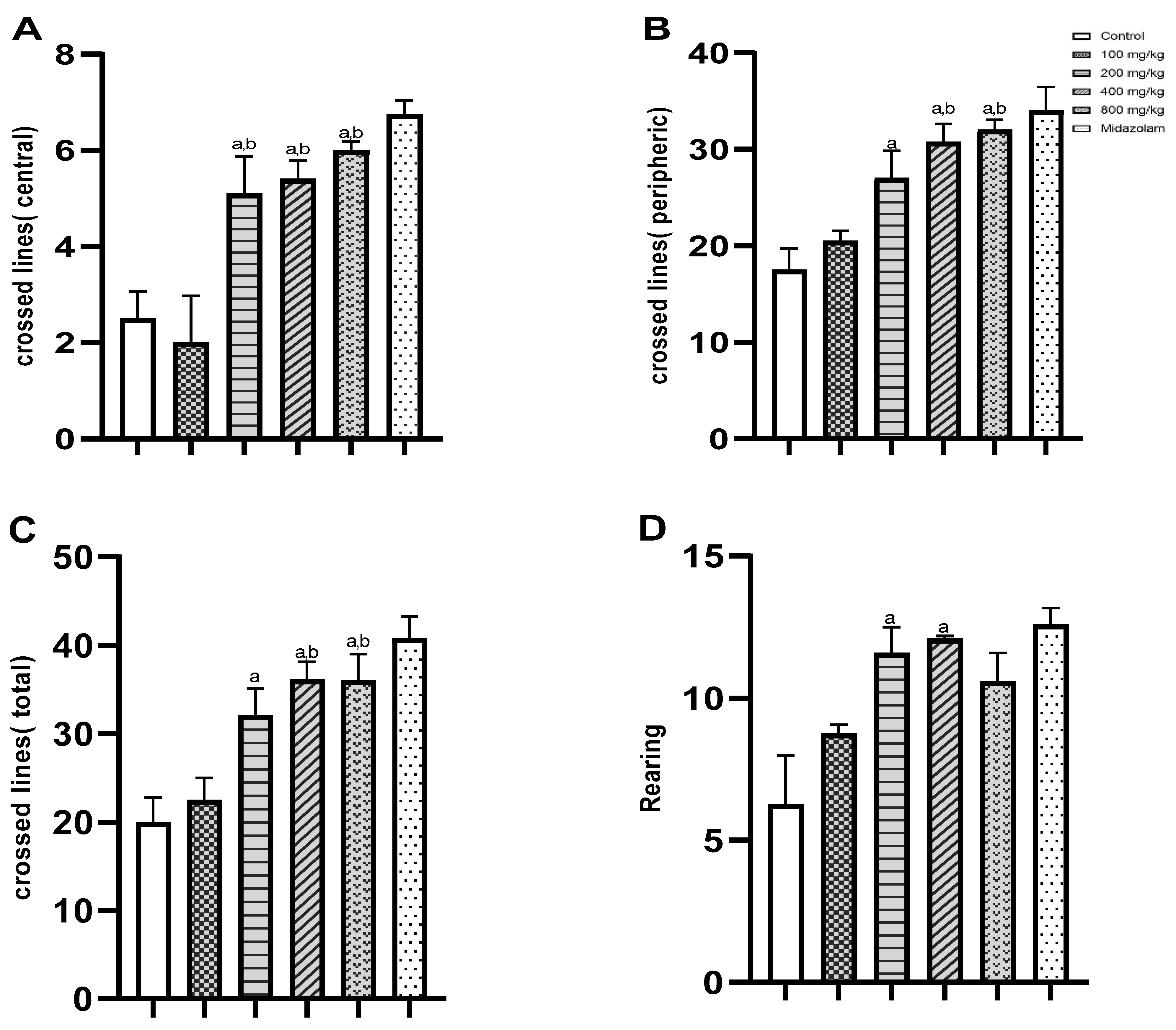
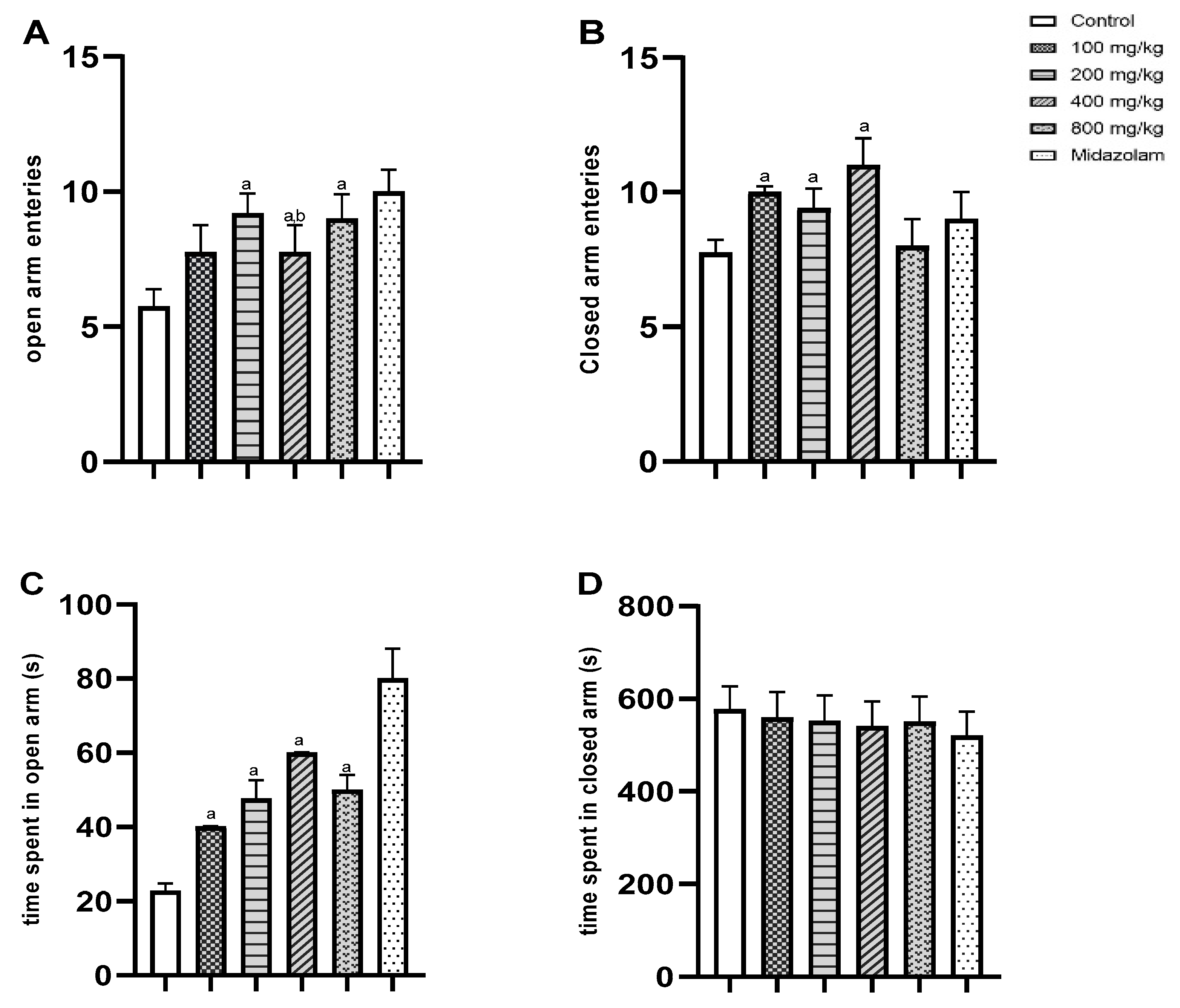
2.2. Pharmacological Effects

Next, we proceeded to examine the pharmacological effects of AEAP using a series of tests to evaluate its potential antinociceptive, antidepressant, and anxiolytic activities. To assess the antinociceptive effect, we employed the hot plate test (HPT), measuring the latency time in response to a thermal stimulus. The control group exhibited a latency time of 6 ± 0.5 s, while the groups treated with varying doses of AEAP showed slightly lower latency times ranging from 4 (at 100 mg/kg) to 6 (at 400 mg/kg) seconds. However, these differences were not statistically significant (Figure 5A). Additionally, the antidepressant activity of AEAP was evaluated using the FST, which measures immobility time as an indicator of depressive-like behavior. As shown in Figure 5B, the control group exhibited an immobility time of 43 ± 2 seconds. Notably, the groups treated with AEAP at different doses displayed significantly lower immobility times, ranging from 18 to 22 s (F(5,30) = 14.73, p < 0.001).
We assessed the anxiolytic effect of AEAP using the open field test (OFT), focusing on central crossed lines (Figure 6A), peripheral crossed lines (Figure 6B), total crossed line (Figure 6C), and rearing behavior (Figure 6D). The control group showed 2.5 ± 0.8 crossings in the central crossed lines analysis, while the groups treated with AEAP displayed higher numbers (5.1, 5.4, and 6.0 crossings at 200, 400, and 800 mg/kg compared to the control group, respectively; F(5,30) = 10.48, p < 0.001). Similarly, in peripheral crossed lines, the control group had 17.5 ± 2.2 crossings, while the AEAP-treated groups exhibited progressively higher counts ranging from 20 (at 100 mg/kg) to 32 (at 800 mg/kg) crossings, which was significant compared to the control group (F(5,30) = 6.62, p < 0.001). Furthermore, the rearing behavior analysis revealed that the control group exhibited 6.2 ± 0.9 rearing, whereas AEAP administration increased the ranging counts (8.75, 11.6, and 12.1 rearing at 100, 200, and 400 mg/kg, respectively; F(5,30) = 10.47, p < 0.05) (Figure 6).
We further investigated the anxiolytic effect using the elevated plus maze (EPM) test, specifically measuring open/closed arm entries (Figure 7A,B) and time spent in each arm (Figure 7C,D). The administration of 200, 400, and 800 mg/kg AEAP significantly increased the open arm entries (9.2, 7.75, and 9.00 entries, respectively) compared to the control group (5.75 ± 0.64) (F(5,30) = 3.07, p < 0.02). Similarly, in terms of time spent in open arms, the control group had a duration of 22.8 ± 2.0 s, while the AEAP-treated groups demonstrated progressively longer durations from 47.6 s at 200 mg/kg to 50 s at 800 mg/kg (F(5,30) = 20.28, p < 0.001).

3. Discussion

The objective of this study was to provide valuable insights into the toxicity and pharmacological properties of the AEAP. We explore the implications of our findings, their significance in the context of medicinal plants, and their potential applications in various fields. The acute toxicity assessment revealed the safety of AEAP at the tested doses, as no mortality or significant alterations in the body and organ weights of male Swiss mice were observed. The data indicate that AEAP possesses an LD50 value greater than 5000 mg/kg, signifying low acute toxicity. These results are consistent with previous findings, which reported an LD50 higher than 5000 mg/kg [14]. Following the guidelines for chemical labeling and classification of acute systemic toxicity provided by the OECD, it is evident that AEAP falls into the least toxic classification category. These findings support the conclusions drawn in earlier studies [13,26]. Importantly, our study aligns with the findings by Jawhari et al., who concluded that various parts of A. pyrethrum, including roots, leaves, seeds, and capitulas, did not induce nephrotoxicity or lead to changes in the renal biomarkers levels [27].
The administration of AEAP did not result in significant deviations in renal and liver function markers, suggesting that it does not exert adverse effects on kidney function. In fact, several variations observed in these parameters could fall within the normal range of biological variability [9]. Strikingly, AEAP demonstrated potential benefits in reducing C-reactive protein levels, a marker of inflammation, and in improving liver function by lowering levels of liver enzymes, such as ALAT and ASAT. Additionally, the observed reduction in total cholesterol levels in the AEAP-treated groups further indicates its potential to manage lipid profiles. Furthermore, the hematological analysis indicated that AEAP did not cause significant alterations in white and red blood cells or platelet levels, suggesting that AEAP does not adversely affect blood parameters, which aligns with the findings of Sharma et al. [28]. Their study also demonstrated the potent hematopoietic activity of A. pyrethrum. The research conducted by Oanh et al. [29] echoes these insights, indicating that medicinal plants like A. pyrethrum not only lack toxicity but also have the potential to positively impact hematological and biochemical attributes. These findings contribute to the overall safety profile of AEAP.
The histopathological examination conducted herein confirmed the absence of morphological disturbances, signifying the absence of toxic effects in the kidney, liver, and spleen tissues. This finding is consistent with previous studies conducted by Bezza et al. [15] and Manouze et al. [14]. In contrast, the research by Jawhari et al. [27] reported no abnormalities in these organs among subjects administered various extracts at lower doses. However, it is essential to note that their high-dose groups (2000 mg/kg) exhibited histopathological alterations in the liver, kidneys, and spleen, marked by hepatic distress, inflammatory infiltration, focal tubular necrosis, vascular congestion, and lymphoid hyperplasia.
The data indicated that AEAP did not exhibit significant central antinociceptive effects, as evaluated by the HPT, suggesting limited pain-relieving properties. This outcome contrasts with the findings of Manouze et al., demonstrating dose-dependent antinociceptive effects following oral treatment with both aqueous and methanolic extracts of A. pyrethrum, hinting at a potential central mechanism of action. According to the same study, A. pyrethrum exhibited peripheral antinociceptive effects attributed to alkylamides [14]. In our study, we observed robust antidepressant activity in the FST via reduced immobility times compared to the control group, indicating significant antidepressant potential. Similar findings have been reported earlier [21,30], indicating that the ethanolic extract of A. pyrethrum also acts as an antidepressant in mice. The observed decrease in immobility is comparable to the effects observed with a reference antidepressant, further supporting the consistency of our results with other studies. Moreover, it has been proposed that the root extract of A. pyrethrum may exert an antidepressant effect by interacting with the adrenergic or dopaminergic system, resulting in elevated norepinephrine and dopamine levels [31]. Additionally, in behavioral tests such as the OFT and the EPM, AEAP displayed anxiolytic potential by increasing exploratory and locomotor activities. This was evidenced by elevated central crossed lines, peripheral crossed lines, and rearing behavior in the OFT, as well as increased exploration of open arms and higher open arm entries in the EPM. These observed anxiolytic effects are consistent with results from another study [28] investigating the effects of the ethanol extract of A. pyrethrum in mice. This research revealed an increase in the time spent in the light compartment and alterations in the number of shuttle crossings, indicating its anxiolytic activity. These anxiolytic effects may be attributed to the compound agonistic effects on the gamma-aminobutyric acid (GABA)/benzodiazepine receptor complex and/or the 5-HT1A receptor and/or its ability to antagonize the 5-HT1B receptor [32,33].
The observed anxiolytic and antidepressant effects in AEAP can, at least partly, be attributed to the presence of bioactive compounds, especially alkylamides, within the extract. It has been shown that an increase in GABA levels in the brain could potentially trigger these effects, as GABA is recognized for its anxiety-reducing and relaxation-promoting properties [34]. Badhe et al. [31] suggested that the root extract of A. pyrethrum could potentially exert an antidepressant effect by interacting with either the adrenergic or dopaminergic system, ultimately leading to elevated levels of norepinephrine and dopamine. The anxiolytic effects observed in the OFT and EPM tests are particularly noteworthy, suggesting that AEAP may have applications in the management of anxiety-related disorders [34].
The identification of polyphenols in the extract, including hydroxybenzoic acid quinyl ester, dihydrocaffeic acid, feruloylquinic acid, caffeic acid, p-coumaric acid, p-coumaroylquinic acid, hydroxycoumarin, along with the alkylamide pellitorine is of significant importance, as recently reported [20]. These compounds have been well-documented for their diverse range of beneficial activities in various studies. In fact, hydroxybenzoic acid quinyl ester is associated with antioxidant properties [35], while dihydrocaffeic acid and feruloylquinic acid have demonstrated anti-inflammatory and neuroprotective effects [36]. Caffeic acid and p-coumaric acid are known for their antioxidant and anti-inflammatory properties [37], contributing to their potential to support overall health. Furthermore, p-coumaroylquinic acid and hydroxycoumarin have shown promise in their roles as antioxidants and potential cardiovascular health promoters [38]. The alkylamide pellitorine, which is characteristic of A. pyrethrum, has exhibited various bioactivities, including anti-inflammatory and analgesic properties [39,40]. The presence of these compounds in the AEAP extract not only supports the traditional uses of A. pyrethrum in herbal medicine but also underscores the rich pharmacological potential of this plant.
The dose–response relationship serves as a fundamental framework for understanding the intricate connection between exposure attributes and the spectrum of effects. This established correlation relies on the measurement of responses in relation to incremental dosages, providing valuable insights into the interplay between exposure and health outcomes. Our results unequivocally pinpointed the 200 mg dose of AEAP as the most efficacious in generating the observed pharmacological effects. This specific dosage consistently yielded optimal results across a spectrum of assessment parameters, underscoring its critical role in achieving the desired pharmacological responses.
The potential applications of A. pyrethrum in both the pharmaceutical industry and traditional medicine are indeed of great importance. The findings suggest that A. pyrethrum holds significant potential for use in herbal medicine and may offer alternative, natural approaches for managing anxiety-related disorders and depressive symptoms, with profound implications for human health and well-being. Furthermore, the identification of specific bioactive compounds within the extract provides a foundation for future research into their isolation and potential development into pharmaceutical agents or herbal remedies. This exploration could yield novel treatments with reduced side effects compared to conventional medications, ultimately benefiting human health.
Building on the robust findings of this study demonstrating notable antidepressant and anxiolytic effects, future research should aim to uncover the molecular pathways and receptor interactions responsible for the observed antidepressant and anxiolytic effects, addressing the current lack of detailed mechanistic insight. Ongoing research will refine our understanding of A. pyrethrum’s therapeutic potential, unravel its mechanisms of action, and explore broader applications in both preclinical and clinical contexts.

4. Materials and Methods

4.1. Plant Samples and Extraction

The roots of Anacyclus pyrethrum were collected from the Bin El Ouidan region, Morocco (32° 7′48″ latitude N/6° 27′36″ longitude W), and the voucher specimen MARK-1003 was deposited in the herbarium of the Department of Biology, Faculty of Sciences Semlalia, Cadi Ayyad University, Marrakech, Morocco. The root powder was obtained by crushing the washed and dried roots, which were then extensively extracted with distilled water (1 g/10 mL) under agitation for 12 h. The solvent used for the extraction was distilled water (650 mL). The bottle containing the root powder (50 g) and sterile water were sealed with parafilm to prevent solvent evaporation and maintain optimal extraction conditions. The aqueous macerate was subsequently centrifuged (1200 rpm), filtered, and lyophilized using a Christ instrument. The weight of the concentrated extract was 9 g, resulting in a yield of approximately 18%. The extract underwent microbial study, and there was no microbial contamination (Escherichia coli, Salmonella, Staphylococcus aureus, or Pseudomonas aeruginosa) in the extract. The lyophilized dry powder was sealed in amber bottles and kept at 4 °C until its use.

4.2. Drugs

Midazolam (Synthemedic, Casablanca, Morocco) and Fentanyl (MAPHAR, Casablanca, Morocco) were employed as reference molecules in this study. Midazolam (1 mg/kg) was utilized as a reference for the assessment of antidepressant and anxiolytic properties [41], while Fentanyl (20 μg/kg) was employed as a reference for evaluating analgesic properties [42].

4.3. Animals

Adult male Swiss mice (25–35 g) and Male Sprague Dawley rats (180–230 g) were sourced from the animal facility of the Faculty of Sciences Semlalia, Cadi Ayyad University, Marrakech, Morocco. These animals were housed in individual chambers under controlled conditions: temperature of 22 ± 2 °C, a 12:12 h light/dark cycle, and transparent cages containing up to five animals. They were provided with ad libitum access to water and food.
This study rigorously adhered to established ethical guidelines and principles, ensuring the welfare and dignity of the animal subjects, including those outlined in the Institutional Animal Care and Use Committee (IACUC), and in compliance with national and international regulations (EU2010/63). All animal procedures were designed with the utmost care to minimize potential harm or distress to the subjects. Prior to the commencement of the study, proper ethical approvals were obtained from the Institutional Review Board of the Faculty of Sciences, Cadi Ayyad University, Marrakech, Morocco (protocol code FCR-CS-07/2023-001, date of approval: January 2023). Measures were taken to optimize animal housing conditions, including appropriate nutrition, temperature control, and minimizing social isolation. The research team remained vigilant in monitoring the animals’ well-being throughout the study, promptly addressing any signs of discomfort or distress.

4.4. Acute Toxicity

The limit test dose (5000 mg/kg) in the acute toxicity study was conducted in accordance with the OECD guideline no. 423 [43]. Four groups of mice, each comprising six animals, were used. Three groups received an oral administration of AEAP at doses of 1000, 2000, and 5000 mg/kg, while the remaining group (negative control) received distilled water. The administration was performed at a rate of 10 mL/kg. The mice were observed for signs of toxicity and mortality during the initial two hours following extract administration. The mice’s body weights were recorded daily for a period of 14 days.

4.4.1. Biochemical and Hematological Analyses

At the end of the behavioral tests, the animals were sacrificed via cervical dislocation, and blood samples were expeditiously collected and centrifuged for serum. The renal (urea, creatinine, uric acid, and protein kinase), liver (ALAT, ASAT, C reactive protein, total bilirubin, total cholesterol, and triglycerides), and hematologic (white blood cells, red blood cells, and platelets) functions were evaluated. The measurements adhered to standard techniques utilizing a biochemical machine (Cobas 6000, Roche, Basel, Switzerland).

4.4.2. Histological Study

For the histopathological examination, vital organs such as the liver, kidneys, and spleen were dissected and immersed in a 10% formalin solution for overnight fixation after biochemical and hematological analyses. Subsequently, the organs underwent dehydration using graded alcohol solutions and were embedded in paraffin wax. Thin sections of 4–10 μm thickness were prepared from paraffin blocks and stained with hematoxylin and eosin, following standard staining protocol. The stained sections were subjected to microscopic examination for pathological analysis [44].

4.5. Pharmacological Effects

To assess the pharmacological effects, six groups of rats, each comprising six animals, were used. Four groups received an oral administration of AEAP at doses of 100, 200, 400, and 800 mg/kg. A positive control group received the reference molecules (Fentanyl/Midazolam), while the remaining group (negative control) received distilled water. All substances were administered 40 min before each test.

4.5.1. Forced Swim Test (FST)

The rats were individually subjected to immobility in an open cylinder (21 × 50 cm diameter × tall) filled with 25 cm of water maintained at 25 ± 1 °C, and the immobility time was recorded for 10 min. Immobility was defined as the period during which the rats remained motionless in the water with no active behaviors such as jumping, diving, or swimming and making only movements to keep their head above water. Prolonged immobility indicates a depressant-like effect in the behavioral profile [45].

4.5.2. Open Field Test (OFT)

An OFT was conducted to assess exploratory behavior and overall locomotor activity [46]. The test apparatus consisted of a white arena measuring 80 × 80 × 40 cm, divided into 25 equal squares. Each rat was placed individually in the arena for 10 min, and the number of squares crossed using all four legs and the frequency of rearing was recorded. After each session, the arena floor was cleaned with 10% ethanol to eliminate residual odors or markings.

4.5.3. Elevated Plus Maze (EPM)

The EPM apparatus comprises a raised platform 100 cm above the floor, with two open arms and two enclosed arms, each measuring 50 × 10 cm (length × width). The test began by positioning the rat in the central zone (10 × 10 cm) of the maze facing the intersection and recording the exploratory behavior in the maze for 10 min. The number of entries into open and closed arms (four legs on the arm) and the time spent in these arms were recorded. These parameters indicate anxiety-like behaviors, increased time spent in closed arms, and fewer entries into open arms suggest higher anxiety levels [47]. After each test, the EPM was cleaned with 10% ethanol to eliminate the possibility of introducing pheromonal cues or markings.

4.5.4. Hot Plate Test

Nociceptive responses were evaluated using the method previously described [48]. Animals were placed on a glass cylinder atop a heated metal plate set at 55 ± 1 °C. The reaction time was measured as the latency to responses, including licking, shaking a paw, or jumping, indicative of discomfort After AEAP and Fentanyl administration, latencies to nociceptive responses were measured 40 min later. This method gauged sensitivity to painful stimuli, providing insights into AEAP’s potential analgesic effects.

4.6. Statistical Analyses

Data were analyzed and presented as mean ± standard error of the mean (SEM) using GraphPad Prism 09 (San Diego, CA, USA). One-way analysis of variance (ANOVA) was performed, followed by post hoc Tukey’s tests to assess the differences among groups. A p-value < 0.05 was considered statistically significant.

5. Conclusions

In conclusion, our study established that aqueous A. pyrethrum roots are non-toxic substances, adding to the growing body of evidence supporting their safety for potential use in pharmacological interventions. Furthermore, we observed that at specific dosage levels, AEAP displayed significant anxiolytic and antidepressant properties, offering promise for addressing anxiety- and depression-related disorders. These findings underscore the potential of A. pyrethrum as a natural product of bioactive compounds with therapeutic applications. While our study provides compelling evidence of the beneficial effects of AEAP, it is essential to recognize that the precise molecular mechanisms underlying the observed activities warrant further investigation. The complex interplay of secondary metabolites within A. pyrethrum may contribute to these effects, but detailed studies are required to isolate and elucidate the specific pharmacologically active compounds and their molecular pathways. As we continue to unravel the mechanisms of action, these findings lay the groundwork for future research, ultimately contributing to the development of novel therapeutic Interventions rooted in A. pyrethrum’s bioactive potential.

Author Contributions

Conceptualization, A.B. and M.B.; methodology, A.B. and H.K.; software, A.B. and R.A.; validation, M.B., S.B. and A.C.; formal analysis, A.B. and M.B.; investigation, A.B. and M.B.; resources, R.A. and S.B.; data curation, A.B. and A.C.; writing—original draft preparation, A.B. and M.B.; writing—review and editing, A.B., S.B. and M.B.; visualization, R.A. and A.C.; supervision, M.B., S.B. and A.C.; project administration, S.B. and A.C. All authors have read and agreed to the published version of the manuscript.

Funding

This research received no external funding.

Institutional Review Board Statement

This study adhered rigorously to established ethical guidelines and principles, ensuring the welfare and dignity of the animal subjects, including those outlined in the Institutional Animal Care and Use Committee (IACUC), and in compliance with national and international regulations (EU2010/63). All animal procedures were designed with the utmost care to minimize potential harm or distress to the subjects. Prior to the commencement of the study, proper ethical approvals were obtained from the Institutional Review Board of the Faculty of Sciences, Cadi Ayyad University, Marrakech, Morocco (protocol code FCR-CS-07/2023-001 date of approval January 2023). Measures were taken to optimize animal housing conditions, including appropriate nutrition, temperature control, and minimizing social isolation. The research team remained vigilant in monitoring the animals’ well-being throughout the study, promptly addressing any signs of discomfort or distress.

Data Availability Statement

Data are contained within the article.

Acknowledgments

The authors wish to thank Abderrazak Regragui (Cadi Ayyad University) for the animal handling and Ilham Hamdaz and Taoufik Hakkoum (Laboratory of Biochemistry, Mohamed VI Teaching Hospital) for the excellent technical assistance during the biochemical analyses. M.B. acknowledges the technical and administrative support of the Faculty of Sciences Semlalia (Marrakech, Morocco).

Conflicts of Interest

The authors declare that the research was conducted in the absence of any commercial or financial relationships that could be construed as a potential conflict of interest.

References

  1. Samtiya, M.; Aluko, R.E.; Dhewa, T.; Moreno-Rojas, J.M. Potential Health Benefits of Plant Food-Derived Bioactive Components: An Overview. Foods 2021, 10, 839. [Google Scholar] [CrossRef]
  2. Atanasov, A.G.; Zotchev, S.B.; Dirsch, V.M.; Supuran, C.T. Natural products in drug discovery: Advances and opportunities. Nat. Rev. Drug. Discov. 2021, 20, 200–216. [Google Scholar] [CrossRef]
  3. Lee, E.L.; Barnes, J. Prevalence of Use of Herbal and Traditional Medicines. In Pharmacovigilance for Herbal and Traditional Medicines: Advances, Challenges and International Perspectives; Barnes, J., Ed.; Springer International Publishing: Cham, Switzerland, 2022; pp. 15–25. [Google Scholar] [CrossRef]
  4. Hamilton, A.C. Medicinal plants, conservation and livelihoods. Biodivers. Conserv. 2004, 13, 1477–1517. [Google Scholar] [CrossRef]
  5. Newmaster, S.G.; Grguric, M.; Shanmughanandhan, D.; Ramalingam, S.; Ragupathy, S. DNA barcoding detects contamination and substitution in North American herbal products. BMC Med. 2013, 11, 222. [Google Scholar] [CrossRef]
  6. Van Wyk, B.E. A review of commercially important African medicinal plants. J. Ethnopharmacol. 2015, 176, 118–134. [Google Scholar] [CrossRef]
  7. Bellakhdar, J. Contribution à l’étude de la Pharmacopée Traditionnelle au Maroc: La Situation Actuelle, les Produits, les Sources du Savoir (Enquête Ethnopharmacologique de Terrain Réalisée de 1969 à 1992); Université Paul Ver-laine: Metz, France, 1997; Available online: https://hal.univ-lorraine.fr/tel-01752084 (accessed on 3 August 2023).
  8. Ouarghidi, A.; Martin, G.J.; Powell, B.; Esser, G.; Abbad, A. Botanical identification of medicinal roots collected and traded in Morocco and comparison to the existing literature. J. Ethnobiol. Ethnomedicine 2013, 9, 59. [Google Scholar] [CrossRef]
  9. Elazzouzi, H.; Fadili, K.; Cherrat, A.; Amalich, S.; Zekri, N.; Zerkani, H.; Tagnaout, I.; Hano, C.; Lorenzo, J.M.; Zair, T. Phytochemistry, Biological and Pharmacological Activities of the Anacyclus pyrethrum (L.) Lag: A Systematic Review. Plants 2022, 11, 2578. [Google Scholar] [CrossRef]
  10. Anacyclus pyrethrum (L.) Link. Available online: https://www.gbif.org/fr/species/3148578 (accessed on 20 October 2023).
  11. Kalim, M.D.; Bhattacharyya, D.; Banerjee, A.; Chattopadhyay, S. Oxidative DNA damage preventive activity and antioxidant potential of plants used in Unani system of medicine. BMC Complement. Altern. Med. 2010, 10, 77. [Google Scholar] [CrossRef]
  12. Manouze, H.; Bouchatta, O.; Bennis, M.; Sokar, Z.; Ba-M’hamed, S. Anticonvulsive and neuroprotective effects of aqueous and methanolic extracts of Anacyclus pyrethrum root in kainic acid-induced-status epilepticus in mice. Epilepsy Res. 2019, 158, 106225. [Google Scholar] [CrossRef]
  13. Kumar, V.K.; Lalitha, K.G. Pharmacognostical Studies on the Root of Anacyclus pyrethrum DC; NISCAIR-CSIR: Delhi, India, 2012. [Google Scholar]
  14. Manouze, H.; Bouchatta, O.; Gadhi, A.C.; Bennis, M.; Sokar, Z.; Ba-M’hamed, S. Anti-inflammatory, Antinociceptive, and Antiox-idant Activities of Methanol and Aqueous Extracts of Anacyclus pyrethrum Roots. Front. Pharmacol. 2017, 8, 598. [Google Scholar] [CrossRef]
  15. Bezza, K.; Gabbas, Z.E.; Laadraoui, J.; Laaradia, M.A.; Oufquir, S.; Chait, A. Ameliorative potential of Anacyclus pyrethrum ex-tract in generalized seizures in rat: Possible cholinergic mediated mechanism. Bangladesh J. Pharmacol. 2019, 14, 188–195. [Google Scholar] [CrossRef]
  16. Bendjeddou, D.; Lalaoui, K.; Satta, D. Immunostimulating activity of the hot water-soluble polysaccharide extracts of Ana-cyclus pyrethrum, Alpinia galanga and Citrullus colocynthis. J. Ethnopharmacol. 2003, 88, 155–160. [Google Scholar] [CrossRef] [PubMed]
  17. Kushwaha, M.N.; Jatav, V.S.; Pandey, S. Plant Anacyclus pyrethrum—A review. Res. J. Pharmacogn. Phytochem. 2012, 4, 164–170. [Google Scholar]
  18. Khare, C.P. Indian Medicinal Plants: An Illustrated Dictionary; Springer Science & Business Media: Berlin/Heidelberg, Germany, 2008. [Google Scholar]
  19. Boonen, J.; Sharma, V.; Dixit, V.K.; Burvenich, C.; Spiegeleer, B.D. LC-MS N-alkylamide Profiling of an Ethanolic Anacyclus py-rethrum Root Extract. Planta Med. 2012, 80, 1787–1795. [Google Scholar]
  20. Baslam, A.; Aitbaba, A.; Aboufatima, R.; Agouram, F.; Boussaa, S.; Chait, A.; Baslam, M. Phytochemistry, Antioxidant Potential, and Antibacterial Activities of Anacyclus pyrethrum: Promising Bioactive Compounds. Horticulturae 2023, 9, 1196. [Google Scholar] [CrossRef]
  21. Usmani, A.; Khushtar, M.; Arif, M.; Siddiqui, M.A.; Sing, S.P.; Mujahid, M. Pharmacognostic and phytopharmacology study of Anacyclus pyrethrum: An insight. J. Appl. Pharm. Sci. 2016, 6, 144–150. [Google Scholar] [CrossRef]
  22. Ee, G.C.L.; Lim, C.M.; Rahmani, M.; Shaari, K.; Bong, C.F.J. Pellitorine, a Potential Anti-Cancer Lead Compound against HL60 and MCT-7 Cell Lines and Microbial Transformation of Piperine from Piper Nigrum. Molecules 2010, 15, 2398–2404. [Google Scholar] [CrossRef]
  23. David, D.J.; Gourion, D. Antidepressant and tolerance: Determinants and management of major side effects. L’Encephale 2016, 42, 553–561. [Google Scholar] [CrossRef]
  24. Slee, A.; Nazareth, I.; Bondaronek, P.; Liu, Y.; Cheng, Z.; Freemantle, N. Pharmacological treatments for generalised anxiety dis-order: A systematic review and network meta-analysis. Lancet 2019, 393, 768–777. [Google Scholar] [CrossRef]
  25. Fajemiroye, J.O.; da Silva, D.M.; de Oliveira, D.R.; Costa, E.A. Treatment of anxiety and depression: Medicinal plants in retro-spect. Fundam. Clin. Pharmacol. 2016, 30, 198–215. [Google Scholar] [CrossRef]
  26. Doudach, L.; Meddah, B.; Alnamer, R.; Chibani, F.; Cherrah, Y. In vitro antibacterial activity of the methanolic and aqueous ex-tracts of Anacyclus pyrethrum used in Moroccan traditional medicine. Int. J. Pharm. Pharm. Sci. 2012, 4, 4. [Google Scholar]
  27. Jawhari, F.Z.; El Moussaoui, A.; Imtara, H.; Mechchate, H.; Es-Safi, I.; Bouhrim, M.; Kharchoufa, L.; Miry, A.; Bousta, D.; Bari, A. Evaluation of the acute toxicity of the extracts of Anacyclus pyrethrum var. pyrethrum (L.) and Anacyclus pyrethrum var. depressus Maire in Swiss mice. Veter-World 2021, 14, 457–467. [Google Scholar] [CrossRef] [PubMed]
  28. Sharma, V.; Thakur, M.; Chauhan, N.S.; Dixit, V.K. Immunomodulatory activity of petroleum ether extract of Anacyclus pyrethrum. Pharm. Biol. 2010, 48, 1247–1254. [Google Scholar] [CrossRef]
  29. Oanh, N.C.; Lam, T.Q.; Tien, N.D.; Hornick, J.L.; Ton, V.D. Effects of medicinal plants mixture on growth performance, nutrient digestibility, blood profiles, and fecal microbiota in growing pigs. Veter-World 2021, 14, 1894–1900. [Google Scholar] [CrossRef]
  30. Richelson, E. Pharmacology of antidepressants. Mayo Clin. Proc. 2001, 76, 511–527. [Google Scholar] [CrossRef] [PubMed]
  31. Badhe, S.R.; Badhe, R.V.; Ghaisas, M.M.; Chopade, V.V.; Deshpande, A.D. Evaluations of antidepressant activity of Anacyclus pyrethrum root extract. IJGP 2010, 4. Available online: https://www.greenpharmacy.info/index.php/ijgp/article/view/124 (accessed on 25 August 2023). [CrossRef]
  32. Millan, M.J.; Hjorth, S.; Samanin, R.; Schreiber, R.; Jaffard, R.; De Ladonchamps, B.; Veiga, S.; Goument, B.; Peglion, J.L.; Spedding, M.; et al. S 15535, a novel benzodioxopiperazine ligand of serotonin (5-HT)1A receptors: II. Modulation of hippocampal serotonin release in relation to potential anxiolytic properties. J. Pharmacol. Exp. Ther. 1997, 282, 148–161. [Google Scholar]
  33. Nishikawa, H.; Hata, T.; Itoh, E.; Funakami, Y. A role for corticotropin-releasing factor in repeated cold stress-induced anxiety-like behavior during forced swimming and elevated plus-maze tests in mice. Biol. Pharm. Bull. 2004, 27, 352–356. [Google Scholar] [CrossRef]
  34. Baslam, A.; Aitbaba, A.; Lamrani Hanchi, A.; Tazart, Z.; Aboufatima, R.; Soraa, N.; Ait-El-Mokhtar, M.; Boussaa, S.; Baslam, M.; Chait, A. Modulation of Gut Microbiome in Ecstasy/MDMA-Induced Behavioral and Biochemical Impairment in Rats and Potential of Post-Treatment with Anacyclus pyrethrum L. Aqueous Extract to Mitigate Adverse Effects. Int. J. Mol. Sci. 2023, 24, 9086. [Google Scholar] [CrossRef]
  35. Farhoosh, R.; Johnny, S.; Asnaashari, M.; Molaahmadibahraseman, N.; Sharif, A. Structure–antioxidant activity relationships of o-hydroxyl, o-methoxy, and alkyl ester derivatives of p-hydroxybenzoic acid. Food Chem. 2016, 194, 128–134. [Google Scholar] [CrossRef]
  36. Na Yin, Z.; Wu, W.J.; Sun, C.Z.; Liu, H.F.; Chen, W.B.; Zhan, Q.P.; Lei, Z.G.; Xin, X.; Ma, J.J.; Yao, K.; et al. Antioxidant and Anti-inflammatory Capacity of Ferulic Acid Released from Wheat Bran by Solid-state Fermentation of Aspergillus niger. Biomed. Environ. Sci. 2019, 32, 11–21. [Google Scholar]
  37. Sarı, A.; Şahin, H.; Özsoy, N.; Özbek Çelik, B. Phenolic compounds and in vitro antioxidant, anti-inflammatory, antimicrobial activities of Scorzonera hieraciifolia Hayek roots. S. Afr. J. Bot. 2019, 125, 116–119. [Google Scholar] [CrossRef]
  38. Kilani-Jaziri, S.; Mokdad-Bzeouich, I.; Krifa, M.; Nasr, N.; Ghedira, K.; Chekir-Ghedira, L. Immunomodulatory and cellular anti-oxidant activities of caffeic, ferulic, and p-coumaric phenolic acids: A structure–activity relationship study. Drug. Chem. Toxicol. 2017, 40, 416–424. [Google Scholar] [CrossRef] [PubMed]
  39. Elufioye, T.O.; Habtemariam, S.; Adejare, A. Chemistry and Pharmacology of Alkylamides from Natural Origin. Rev. Bras. Farmacogn. Orgao. Soc. Bras. Farmacogn. 2020, 30, 622–640. [Google Scholar] [CrossRef] [PubMed]
  40. Mijangos-Ramos, I.F.; Zapata-Estrella, H.E.; Ruiz-Vargas, J.A.; Escalante-Erosa, F.; Gómez-Ojeda, N.; García-Sosa, K.; Cechinel-Filho, V.; Meira-Quintão, N.L.; Peña-Rodríguez, L.M. Bioactive dicaffeoylquinic acid derivatives from the root extract of Calea urticifolia. Rev. Bras. Farmacogn. 2018, 28, 339–343. [Google Scholar] [CrossRef]
  41. Miao, Y.L.; Guo, W.Z.; Shi, W.Z.; Fang, W.W.; Liu, Y.; Liu, J.; Li, B.W.; Wu, W.; Li, Y.F. Midazolam Ameliorates the Behavior Deficits of a Rat Posttraumatic Stress Disorder Model through Dual 18 kDa Translocator Protein and Central Benzodiazepine Receptor and Neuroster-oidogenesis. PLoS ONE 2014, 9, e101450. [Google Scholar] [CrossRef] [PubMed]
  42. Laboureyras, E.; Boujema, M.B.; Mauborgne, A.; Simmers, J.; Pohl, M.; Simonnet, G. Fentanyl-induced hyperalgesia and analgesic tolerance in male rats: Common underlying mechanisms and prevention by a polyamine deficient diet. Neuropsychopharmacology 2022, 47, 599–608. [Google Scholar] [CrossRef] [PubMed]
  43. OECD. Guidelines for the Testing of Chemicals. Acute Oral Toxicity—Acute Toxic Cl Method Test No-423; Organisation for Economic Co-Operation and Development (OECD): Paris, France, 2001. [Google Scholar]
  44. Malatesta, M. Histological and Histochemical Methods—Theory and Practice. Eur. J. Histochem. 2016, 60, 2639. [Google Scholar] [CrossRef]
  45. Porsolt, R.D.; Anton, G.; Blavet, N.; Jalfre, M. Behavioural despair in rats: A new model sensitive to antidepressant treatments. Eur. J. Pharmacol. 1978, 47, 379–391. [Google Scholar] [CrossRef]
  46. Gould, T.D.; Dao, D.T.; Kovacsics, C.E. The Open Field Test. In Mood and Anxiety Related Phenotypes in Mice: Characterization Using Behavioral Tests; Gould, T.D., Ed.; Humana Press: Totowa, NJ, USA, 2009; pp. 1–20. [Google Scholar]
  47. Rodgers, R.J.; Dalvi, A. Anxiety, defence and the elevated plus-maze. Neurosci. Biobehav. Rev. 1997, 21, 801–810. [Google Scholar] [CrossRef]
  48. Okolo, C.O.; Johnson, P.B.; Abdurahman, E.M.; Abdu-Aguye, I.; Hussaini, I.M. Analgesic effect of Irvingia gabonensis stem bark extract. J. Ethnopharmacol. 1995, 45, 125–129. [Google Scholar] [CrossRef] [PubMed]
Figure 1. Histopathological examinations of mice organs (liver, spleen, and kidneys) in acute toxicity: (A) Kidney tissue: control, (B) AEAP at 2000 mg/kg, and (C) AEAP at 5000 mg/kg. (D) Spleen tissue: control, (E) AEAP at 2000 mg/kg, and (F) AEAP at 5000 mg/kg. (G) Liver tissue: control, (H) AEAP at 2000 mg/kg, and (I) AEAP at 5000 mg/kg. Sections were stained with H&E (×20).
Stresses 04 00005 g001
Figure 2. Biochemical characterization of the effect of Anacyclus pyrethrum aqueous extract (AEAP) on renal function: (A) urea, (B) creatine, (C) uric acid, and (D) creatine kinase. Results are expressed as mean ± SEM (n = 6 per group). Data were analyzed by one-way ANOVA followed by Tukey’s post hoc test, “a” vs. control, “b” vs. 1000 mg, and “c” vs. 2000 mg. Letters a, b, or c indicate p < 0.05.
Stresses 04 00005 g002
Figure 3. Biochemical characterization of the effect of Anacyclus pyrethrum aqueous extract (AEAP) on liver function: (A) ALAT, (B) ASAT, (C) total bilirubin, (D) C reactive protein, (E) total cholesterol, and (F) triglycerides. Results are expressed as mean ± SEM (n = 6 per group). Data were analyzed by one-way ANOVA followed by Tukey’s post hoc test, “a” vs. control, “b” vs. 1000 mg. Letters a or b indicate p < 0.05.
Stresses 04 00005 g003
Figure 4. Hematological characterization of the effect of Anacyclus pyrethrum aqueous extract (AEAP) on (A) white blood cells, (B) red blood cells, and (C) platelets. Results are expressed as mean ± SEM (n = 6 per group). Data were analyzed by one-way ANOVA followed by Tukey’s post hoc test, “a” vs. control, “b” vs. 1000 mg, and “c” vs. 2000 mg. Letters a, b, or c indicate p < 0.05.
Stresses 04 00005 g004
Figure 5. Pharmacological characterization of the effect of Anacyclus pyrethrum aqueous extract (AEAP) on (A) latency time in hot plate test, (B) immobility time in forced swim test. Results are expressed as mean ± SEM (n = 6 per group). Data were analyzed by one-way ANOVA followed by Tukey’s post hoc test, “a” vs. control. Letter “a” indicates p < 0.05.
Stresses 04 00005 g005
Figure 6. Pharmacological characterization of Anacyclus pyrethrum aqueous extract (AEAP) effects on: (A) central crossed lines, (B) peripheric crossed lines, (C) total crossed lines, and (D) rearing in open field test. Results are expressed as mean ± SEM (n = 6 per group). Data were analyzed by one-way ANOVA followed by Tukey’s post hoc test, “a” vs. control, “b“ vs. 100 mg/kg, a or b indicates p < 0.05.
Stresses 04 00005 g006
Figure 7. Pharmacological characterization of the effect of Anacyclus pyrethrum aqueous extract (AEAP) on (A) open arm entries, (B) closed arm entries, (C) time spent in open arms, and (D) time spent in closed arms in elevated plus maze. Results are expressed as mean ± SEM (n = 6 per group). Data were analyzed by one-way ANOVA followed by Tukey’s post hoc test, “a” vs. control, “b” vs. 100 mg/kg, a or b indicates p < 0.05.
Stresses 04 00005 g007
Table 1. Effects of a single oral dose of Anacyclus pyrethrum aqueous extract (AEAP) on mice body weight and relative organ weights.
Control1000 mg/kg2000 mg/kg5000 mg/kg
Body weight (g)30.4 ± 0.830.8 ± 1.328.9 ± 1.031.5 ± 1.2
Relative organ weights (g/100 b.wt)
Liver8.07 ± 0.57.00 ± 0.89.10 ± 0.67.46 ± 0.7
Kidney0.58 ± 0.10.52 ± 0.20.48 ± 0.00.63 ± 0.2
Brain1.30 ± 0.11.43 ± 0.11.50 ± 0.21.40 ± 0.1
Spleen0.33 ± 0.020.33 ± 0.00.34 ± 0.00.35 ± 0.0
Data are expressed as mean ± SEM.
Disclaimer/Publisher’s Note: The statements, opinions and data contained in all publications are solely those of the individual author(s) and contributor(s) and not of MDPI and/or the editor(s). MDPI and/or the editor(s) disclaim responsibility for any injury to people or property resulting from any ideas, methods, instructions or products referred to in the content.

