Review

Orbital Inflammation in Thyroid Eye Disease: Stress Responses and Their Implications

by
Tracy Aoun
1,†,
Diana Danielova Gueorguieva
1,† and
Kevin Y. Wu
2,*
1
Faculty of Medicine, University of Montreal, Montreal, QC H3T 1J4, Canada
2
Department of Surgery, Division of Ophthalmology, University of Sherbrooke, Sherbrooke, QC J1G 2E8, Canada
*
Author to whom correspondence should be addressed.
These authors contributed equally to this work.
Stresses 2024, 4(1), 54-78; https://doi.org/10.3390/stresses4010004
Submission received: 30 October 2023 / Revised: 29 November 2023 / Accepted: 3 January 2024 / Published: 8 January 2024
(This article belongs to the Section Animal and Human Stresses)

Abstract

:
Thyroid Eye Disease (TED) is a debilitating autoimmune condition characterized by significant inflammation of orbital tissues, including the extraocular muscles and adipose tissues. The pathological mechanisms underlying this inflammation involve a complex interplay of stress responses at the cellular and molecular level. This review aims to critically evaluate and synthesize existing literature on the mechanisms of orbital inflammation in TED. We discuss the role of autoantibodies, cytokines, and reactive oxygen species (ROS) in the initiation and propagation of the inflammatory process. Additionally, we explore how stress responses triggered by these elements affect the integrity of orbital tissues and contribute to its remodeling. Our review underscores the need for continued research in this field, which may pave the way for novel therapeutic strategies for TED.

1. Introduction

Thyroid eye disease (TED), also known as Graves’ ophthalmopathy (GO) or Graves’ orbitopathy (GO), is a rare autoimmune disease most associated with Graves’ disease (GD), an autoimmune disorder of the thyroid gland resulting in the overproduction of thyroid hormones (hyperthyroidism). Studies suggest that around 40% of patients with GD will experience TED [1,2]. TED is a multifactorial disease characterized by an inflammation of orbital tissues such as extraocular muscles, connective tissues, and adipose tissues, which can be disfiguring and sight-threatening for those affected.
TED is a complex ophthalmological autoimmune disease. Disease severity ranges from mild to severe to sight-threatening, affecting both clinical presentation and management [3]. Clinically, ophthalmic manifestations can range from dry eye and itching in the mild form, to compressive optic neuropathy which can lead to irreversible vision loss in its most severe form. Development of the disease is often characterized by proptosis, strabismus, impaired visual function, and ocular surface inflammation [4].
The disease’s detrimental impact on patients’ quality of life is well established [4,5,6]. Evidence has shown that TED patients experience a poorer quality of life than those with other chronic conditions. Disfiguring proptosis, for instance, one of the main symptoms of TED, can result in impaired self-perception and social isolation [4]. Evidence has shown they also experience significant occupational disability [7]. Patients must not only cope with the disease’s functional impairment but also with significant emotional distress, which is higher than in other chronic autoimmune ophthalmic conditions [7,8].
TED is also a significant public health burden [7]. Given its impact on patients’ wellbeing and its complexity, as well as recent therapeutic advances, we aim to provide an overview of the disease, with a focus on current understanding of the role of cytokines and oxidative stress and their implication in the physiopathology of the disease. We also briefly highlight potential therapeutic avenues and advances related to these topics.

2. Epidemiology, Risk Factors and Clinical Understanding of TED

2.1. Epidemiology

The onset of TED is mostly associated with Graves’ Disease, an auto-immune condition in which antibodies target the thyrotropin receptors (TSHR) on the thyroid gland, causing excessive thyroid hormones production and hyperthyroidism. More than 90% of TED patients have in fact concomitant GD [9]. However, about 10% of patients are hypothyroid or euthyroid [10,11] when developing TED.
It is estimated that 1 to 2% of the adult population is affected by GD [12]. While it was reported that 40% of GD patients develop TED, its overall prevalence is likely between 25% and 40% [1,10].
The disease usually presents in three phases: an initial phase, characterized by inflammation and swelling (lasting for an average of two years), is then followed by active TED (inflammatory phase), which commonly progresses to stable and inactive TED, within 1–3 years [13,14]. Among patients living with TED, moderate-to-severe forms of the disease account for 2% to 5–6% [10,15].
The disease’s incidence is estimated to be 16 cases/100,000 population-year for women and 2.9 cases/100,000 population-year for men [16]. However, no recent cohort study has followed to confirm TED’s current incidence in North America. Recent European prospective studies from Sweden and Denmark suggest a decreasing incidence trend [10,17,18]. Disease incidence may potentially be slowed with improved understanding and management of risk factors such as a decreasing smoking pattern, a well-established risk factor in TED [10]. Improved GD diagnosis and management and increased awareness might also explain this association. The prevalence of TED seems to range between 90 and 300/100,000 population, without much ethnic variation [10].
Age and sex are relevant factors in the occurrence and severity of TED. The disease’s onset typically occurs between 30 and 50 years of age [19]. TED, like other auto-immune diseases, more commonly affects women, with a ratio of women to men found to be between 2.5 and 6:1 [10,20]. The disease tends to be more severe in male patients and those first diagnosed after their fifth decade of life [21]. A recent multi-center cohort study confirmed the influence of male sex on disease severity [22]. Despite some controversies on this link, this correlation seems generally accepted [10].

2.2. Risk Factors

As a multifactorial disease, many factors have been associated with the development and progression of TED: autoimmune, genetic, and environmental risk factors seem to be involved.
The most significant environmental risk factor remains cigarette smoking [23]. Solid evidence has established its association with severity and poorer treatment outcomes [24,25]. While smoking has been studied extensively, the potential effect of e-cigarettes as an independent risk factor in TED’s development remains unsettled. In fact, an in vitro study examining the effects of e-cigarettes and heated tobacco products on TED patients’ orbital fibroblasts reported inconclusive findings [26]. Other known risk factors include radioiodine therapy, late diagnosis of GD, and prolonged dysthyroid state [5,27].
Oxidative stress, selenium deficiency, hypercholesterolemia, and amounts of TSH receptor antibodies have also been linked to occurrence and progression of the disease [10]. Diabetes is also a potential risk factor [24]. Stress as an environmental risk factor has also been associated with TED [28]. More recently, it has been shown that a disbalance in gut flora may be a potential risk factor [29]. Novel documented risk factors may include higher serum creatinine levels and lower platelet count [30]. Also, statins were found to be associated with lower risk of TED among GD patients [14].
Mitigation of modifiable risk factors, such as through smoking cessation counselling, remains crucial in the therapeutic approach to TED. There may be value in increased patient education regarding management of comorbidities such as Graves’ disease and hypercholesterolemia, as well as increased awareness of risk factors.
The current knowledge of risk factors linked to TED is well researched in the literature [10,24]. However, to date, the literature pertaining to ethnicity’s impact on the development of TED remains controversial; some studies have reported a sixfold risk for Europeans compared to Asians, while a more recent study found a higher prevalence in Asians [1]. There is a paucity of data examining ethnic influences on TED, such that the precise impact in disease presentation remains unclear [10]. A meta-analysis and systematic review by Chin et al. examined the prevalence of TED among GD patients with a focus on ethnic and regional differences and reported the lowest prevalence in North America (27%), whilst it found it to be highest in Oceania (58%), followed by Asia (44%) and Europe (38%) [1]. However, further data are needed before determining whether ethnic and geographic factors play a role. Moreover, a gap in the literature relating to the prevalence of TED in African and Indigenous populations is noted, which warrants further studies.
Sex-specific risk factors, however, had not been studied until a recent Korean nationwide population-based retrospective cohort study, which found a relationship between younger age, hypercholesterolemia, and low BMI at the time of GD diagnosis for development of TED in women, while risk factors of young age, low income, and heavy drinking were identified in men [31]. As it is the only study assessing sex-related risk factors in TED, more work is required to better grasp the implications of these findings and potential causal links. Studies within other demographics should be conducted as well.

2.3. Signs and Symptoms

Typical presenting symptoms include redness, ocular discomfort, diplopia, visual disturbances, tearing, and photophobia. Patients can also complain of a foreign-body sensation in their eyes or non-specific dry eyes [24].
The most common signs are eyelids retraction (Dalrymple’s sign) accompanied by upper eyelid lag on downgaze (von Graefe’s sign), as well as lid edema [14].
This disease is also the most common cause of unilateral or bilateral exophthalmos or proptosis, which refers to the eyes’ forward protrusion [14]. The involvement of proptosis and eyelids retraction may produce in the patient a staring appearance (white sclera is exposed above and below the limbus), referred to as thyroid stare. Moreover, eyelid and periorbital edema may be present due to increased orbital pressure and venous congestion [32,33].
Extraocular muscles involvement is common, with muscles usually affected in the following order: inferior rectus, medial rectus, superior rectus, elevator, lateral rectus, and oblique. Usually, a single muscle is involved [20,34,35].
TED has a heterogenous presentation among patients. These symptoms are not exhaustive, and numerous factors influence its clinical presentation of the disease, such as sex, age, and smoking [36]. Smoking usually increases the severity of TED symptoms [23]. In the elderly, disease can also more often present unilaterally or asymmetrically [36].
Moreover, potential ethnic differences exist in the clinical manifestations, with evidence suggesting that Asian patients may show milder phenotypic features, with less proptosis and extraocular muscle restriction and more frequent lower lid retraction [24,37]. It is to be noted that the literature on clinical findings in the African and Indigenous communities is nonexistent; studies should be conducted in this regard for a better understanding and care of all ethnic groups. Due to its varying clinical presentation, a thorough differential diagnosis is crucial. TED remains a challenging diagnosis as many orbital inflammatory diseases can resemble its signs and symptoms [38].

2.4. Complications

Complications arising from TED range from mild to severe. Exposure keratopathy can occur due to proptosis as eyelids are unable to properly close, which causes dryness and damages the cornea. This can lead to further complications, such as corneal ulcerations, infections, scarring, and vision loss, which may require surgical interventions [14]. Other complications, such as restrictive myopathies and strabismus, may occur with extraocular muscles involvement and may require surgical treatment. Dysthyroid optic neuropathy, though rare (occurring in about 4–10% of TED patients), is an ophthalmic emergency and the most dreaded complication of TED; it is characterized by impairments to optic nerve function which can cause irreversible vision loss [14,16]. Although a number of etiologies have been suggested for its development in TED patients, it most commonly occurs because of optic nerve compression, caused by enlarged extraocular muscles, along with orbital fat expansion and interstitial edema [32,39]. Associated symptoms include decreased visual acuity, dyschromatopsia, and constricted visual fields. Orbital decompression surgery is often indicated, along with orbital radiation therapy, to reduce muscles’ inflammatory swelling. Surgical treatment may also be required if globe subluxation or corneal decompensation occur [14].

2.5. Diagnostic Criteria

Diagnosis is clinical, based upon history, anamnesis, laboratory, and imaging tests, as well as clinical findings following a complete ophthalmic assessment. Diagnosis is made when two of three findings occur among ocular signs, immune-related thyroid dysfunction and/or extraocular muscle enlargement evidenced through radiology [14]. To date, there exists no single laboratory test that is pathognomonic for TED [5].
The use of imaging methods for diagnosis and management of TED should be briefly underlined. Imaging studies can orient and confirm diagnosis and exclude other potential causes such as cancers [5]. They are useful in identifying active inflammatory phase vs. fibrotic end stage, assessment in follow-up after treatment is started, and can guide surgical strategies like orbital decompression surgery [24]. Magnetic resonance imaging of the orbit remains the gold-standard assessment method since it allows for identification of soft-tissue and orbital fat expansion, as well as extraocular muscle enlargement [5,24].
As identification of mild forms remains a challenge, improved interprofessional education as well as patient education is needed [40]. Improved detection and diagnosis are crucial to improve prognosis and limit inflammation, which highlights the importance of continued research in this area. To date, no specific biomarker exists to distinguish TED from GD. This limits potential earlier diagnosis and highlights the importance of continued research in this area.

2.6. Clinical Evaluation

The clinical evaluation of TED focuses upon determination of clinical activity and severity by assessing visual acuity, pupils, color vision, extraocular movements, visual field, exophthalmometry, external eyelid evaluation, slit-lamp examination, and dilated fundus examination [5].
A few classification guidelines are available to the clinician to evaluate activity and the severity of the disease, which guide therapeutic management. The Clinical Activity Score (CAS) is a useful tool to assess patients for TED activity. This 7-point scale attempts to characterize the inflammation process (active or inactive) in the disease by listing signs and symptoms associated with inflammation. A point is allocated for each of these findings: ocular pain, pain on eye movement, eyelid swelling, conjunctival redness, eyelid edema, chemosis (scleral edema) or caruncle swelling. An inactive form of TED is considered when a score of lower than 3 is found, while a score of 3 or greater defines active TED (see Table 1) [41].
The NO SPECS classification can be used for evaluation of disease severity, usually classified as mild, moderate-to-severe, or sight-threatening (see Table 2). Meanwhile, to assess for both activity and severity of the disease, the VISA (vision, inflammation, strabismus, and appearance), as well as the EUGOGO (European Group on Graves’ Orbitopathy), guidelines are commonly used [42].
It is important to stress that if a patient shows any significant signs of TED or complains of ocular pain, the indicated course of action is prompt referral to an ophthalmologist.

2.7. Treatment

Treatment varies with the level of activity and severity of the disease [24]. Management is supportive, medical, and surgical, with general treatment principles focused on restoration of a stable thyroid function, on limitation of inflammation, and smoking cessation [14].
Mild cases are usually managed with selenium supplementation and monitoring. First-line treatment for the active form of the disease is corticosteroid therapy, usually followed by other immunosuppressive agents in cases of non-responsiveness. Current evidence also supports the use of Teprotumumab for the medical management of the active moderate-to-severe form of TED [44]. Surgical correction is envisioned for more severe or sight-threatening cases [24]. A comprehensible treatment algorithm can be found in Table 3.
EUGOGO’s clinical practice guideline for the medical management of TED is the most recent updated consensus statement pertaining to clinical management [45].

3. Pathophysiology of TED

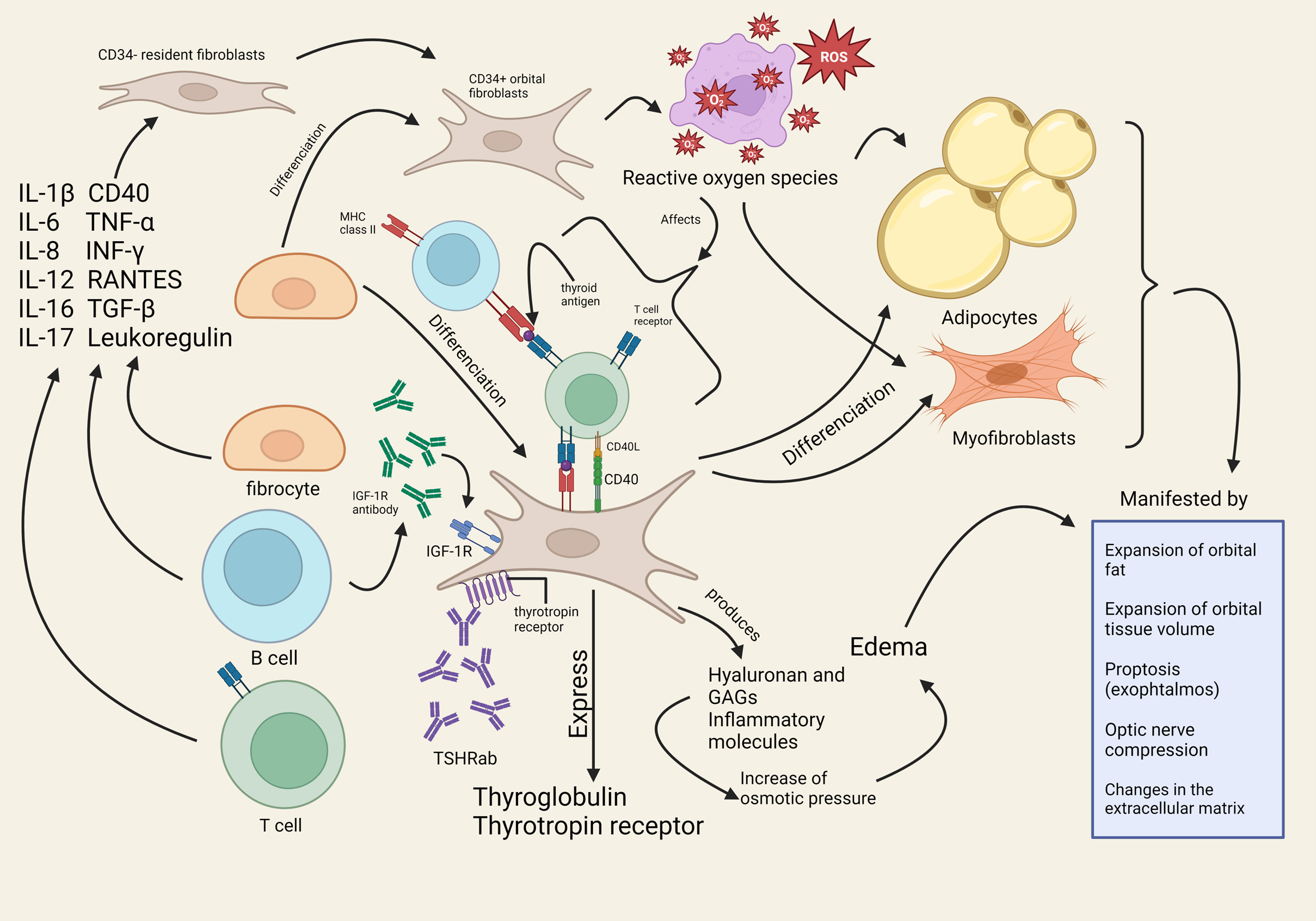
3.1. Overview of Pathophysiological Changes in TED and Its Implication

TED is a multifactorial autoimmune disease, and its pathophysiology is not yet fully understood, despite significant research in the field.
Summarily, an autoimmune process is involved which targets the ocular tissue. Activation and binding of autoantibodies to the thyrotropin receptors (TSHR) expressed on the surface of orbital fibroblasts is found to be the main event that initiates an inflammatory cascade [46]. It is now well-established that concentration of TSHR antibodies positively correlates with the clinical activity and severity of the disease [47,48]. In fact, orbital fibroblasts, which are the main components of the ocular fibrous tissue, have been found to overexpress these receptors in TED patients [47].
The underlying immune process involves an infiltration of activated B and T cells, with a release of inflammatory cytokines and chemokines including interferon gamma (IFN-y), tumor necrosis factor alpha (TNF-α), interleukin-1β (IL-1β), interleukin-2 (IL-2), interleukin-6 (IL-6), and leukoregulin [24]. This leads to inflammation and promotes orbital fibroblasts’ proliferation, adipogenesis, and production of glycosaminoglycans (GAG) (see Figure 1 and Section 4 for a discussion on cytokines) [49].
Recent research suggests that onset of orbital inflammation may be characterized by early infiltration of macrophage cells in the orbital region, which induce TSHR antibodies, followed by a subsequent infiltration of CD8+-specific T-cells and increase in brown adipose tissue. A study by Philipp et al. examined cellular and immunological parameters in the process of orbital inflammation to understand the kinetics of the disease using a mouse model of TED [50].
B cells, T cells, and CD34+ fibrocytes infiltrate the orbit. Fibrocytes originating from the bone marrow differentiate into CD34+ fibroblasts. These fibroblasts have the ability to differentiate into either adipocytes or myofibroblasts. CD34+ fibroblasts and CD34- residential fibroblasts cohabit within the orbit. Cytokines are then secreted by all these different cells. The molecules produced include interleukin 1 β , 6, 8, 12, 16, and 17, as well as tumor necrosis factor α (TNF- α ), interferon γ (IFN- γ ), RANTES (regulated on activation, normal T-cell expressed and secreted), TGF- β , CD40, and Leukoregulin. These molecules activate orbital fibroblasts (CD34+) which in turn start expressing thyroglobulin and thyrotropin receptors. Thyroid-stimulating antibodies activate the receptor complex made up of both the insulin-like growth factor 1 receptor and the thyrotropin receptor, resulting in the expression of inflammatory molecules and glycosaminoglycans (GAGs). In addition, antibodies targeting the IGF-1R induce hyaluronan secretion by orbital fibroblast, resulting in the expansion of orbital tissue. Adipogenesis occurs with promotion of differentiation of orbital fibroblasts into adipocytes, causing orbital fat tissue expansion. Reactive oxygen species (ROS) are also involved, affecting immunocompetent cells, as well as adipocytes and myofibroblasts, and promoting inflammation and cell damage.
This immune cascade activates orbital fibroblasts, which then proliferate, and induces their local production of glycosaminoglycans (GAG), including hyaluronan (HA), a component of the extra-cellular matrix [51]. Spilling of HA in the interstitial space leads to tissue edema: HA is very hydrophilic, which leads to increased osmotic pressure, and venous and lymphatic circulations may be compromised [24]. Consequent swelling of connective tissue and extraocular muscles results from GAG accumulation and overproduction and increases the intraocular pressure [52]. Fibrosis, in the long-run, explains the restrictive myopathy found in the disease.
Moreover, activated orbital fibroblasts can also differentiate into adipocytes [53]. The fibrocyte, a precursor cell of the fibroblast found within the bone marrow, seems to have a role in the pathogenesis of TED [19]. The activation of the immune cascade described previously has been found to activate CD34+ fibrocytes, which can differentiate into myofibrocytes and adipocytes [24]. Since fibrocytes over-express TSHR, a potential role for TSHR antibody in its stimulation is proposed [19].
Anatomically, these processes cause an increased intra-orbital pressure and fibrosis. Clinically, this leads to forward protrusion of the eyeballs along with eyelid retraction, which is objectified as exophthalmos [24]. In the long course, inflammation is damaging to nearby tissue, and remodeling and fibrosis of muscles accounts for diplopia and restrictive strabismus [5,24]. Briefly, inflammation, expansion, and remodeling of the orbital compartment explain disease manifestations.

3.2. Role of IGF-1R and Potential IGF-1R Autoantibody

Insulin-like growth factor 1 (IGF-1) is a protein that regulates and stimulates cell growth. It is mainly secreted by the liver under stimulation by the growth hormone (GH). IGF-1 has a role in immune function and can influence the development of diseases like cancers.
IGF-1 receptors (IGF-1R) are tyrosine kinase proteins; they are present on the surface of cell membranes and can bind both IGF-1 and IGF-2, as well as insulin, though they display highest affinity towards IGF-1 [54]. Both B-cells (involved in humoral immunity) and T-cells (involved in cellular immunity) express IGF-1 receptors on their surface, such that they can produce and respond to IGF-1; because of this, researchers believe there may be a possible immune loop involving both autocrine and paracrine signaling [55].
Though first hypothesized by Weightman et al. that specific IGF-1R autoantibodies may come into play in the pathogenesis of TED, the current literature remains inconclusive on the matter, with some works confirming and others denying presence of these antibodies or an implication [24,56,57]. The existence of specific antibodies against IGF-1R has been hypothesized, with a potential implication in the pathogenesis of the disease. However, research pertaining to its existence is contradictory as of now. Minich et al. and Krieger et al.’s works did not detect specific IGF-1R antibodies [56,58,59]. A possible reason for such differences may be explained by variance in experimental designs and conditions, as compared to the original work of Pritchard et al. [60]. As such, a potential role remains unclear, with controversial hypotheses on the matter. Minich et al. have suggested they may act as IGF-1 antagonists by inhibition of the IGF-1-induced signaling and reducing cell growth. For them, they did not stimulate autophosphorylation of IGF-1R [56]. Varewijck et al., for their part, detected an IGF-1R stimulating antibody activity [61].
It is now suggested that the IGF-1 receptor might be involved in crosstalk with the TSH receptor, participating in the inflammatory pathway. Its exact implication remains unknown; thus, more research is required to elucidate this [59,62]. Studies have shown that the IGF-1 and thyroid-stimulating hormone (TSH) pathways influence one another [63]. Ingbar et al., in their in vitro study, found increased TSH and thyroid-stimulating immunoglobulin (TSI) activity in cultured cells inducted by IGF-1 and insulin [64]. Tsui et al. found that the use of an antibody against IGF-1R was able to reduce the signaling of TSHR, confirming an association or interplay between these two pathways. IGF-1 receptors and TSH receptors were found to interact both physically and functionally in human orbital fibroblasts and thyroid epithelium. When looking at patients with TED, Tsui et al. found the presence of both receptors on orbital fibroblasts and human thyroid epithelial cells, as well as orbital fat [65].
Weightman et al. were the first to state the possible influence of IGF-1R in TED using orbital fibroblasts from tissues explanted from extraocular muscle. Their study demonstrated that IgGs from patients with GD, with or without TED, interacted with and altered orbital fibroblasts’ IGF-1 binding sites, while normal patients’ IgGs did not [57]. Furthermore, B-cells and T-cells were found to overexpress IGF-1R in patients with GD [60,66]. Pritchard et al. then found that IGF-1R was the main binding site for IGF-1 on orbital fibroblasts and that GD-IgGs were also able to show a high affinity for the same binding site. GD-IgGs binding to the IGF-1R resulted in the same effects caused by IGF-1, thus providing a possible analogous property of GD-IgGs [67]. They also found that signaling from IgGs of GD patients led to an important expression of IL-16 and RANTES-two T-cell chemoattractants, a process that did not occur in healthy individuals [60]. It was also found that binding of IgGs to IGF-1 receptors could induce the production of hyaluronan (HA) in TED orbital fibroblasts, a finding that was not seen with healthy subjects’ orbital fibroblasts [68]. When working in synergy, IGF-1R and TSHR were found to produce hyaluronan and cytokines, leading to inflammation and expansion of muscle and adipose tissues, as seen in TED [69].
Among the various cell types involved in TED, fibrocytes and orbital fibroblasts have been found to overexpress IGF-1 receptors, especially in orbital fat and extraocular muscles [70]. In vitro, the IGF-1 receptor antibody Teprotumumab diminished IGF-1 and TSH activity in both fibrocytes and orbital fibroblasts, resulting in much smaller amounts of proinflammatory cytokines [63]. Its use also reduced level of diplopia and proptosis in patients with GD [55].
The presence of IGF-1R autoantibodies in TED patients appears to be inconsistent and is further complicated by potential limitations in experimental techniques used to identify them. Minich et al.’s work, for instance, has shown prevalence of these autoantibodies of only 14% when testing in a serum bank of GD patients, which were also detected in 11% of controls’ sera [56]. Many limitations were acknowledged, namely the inability to identify lower antibody concentrations with the experimental design used in the study.
Currently, Lanzolla et al. propose a potential protective role in the disease, finding an increased concentration of these antibodies in patients with GD without TED when compared to patients with TED [71]. This seems plausible in light of Minich’s work suggesting a potential inhibitory effect on the IGF-1 pathway; it may suggest a role in limiting growth and proliferation of orbital fibroblasts. In light of the potential crosstalk involved between IGF-1R and TSH-R, a potential role in inhibiting indirectly the TSHR pathway can be envisioned, leading to reduced inflammation. The literature has also shown that an IGF-1R-blocking antibody is involved in halting GD-IgG, immunoglobulins that play a role in the cascade involving cytokines production, and hyaluronan production.
Continued research to better understand the role and interaction of these antibodies within the inflammatory process that takes place in TED is needed, notably to determine whether a potential inhibitory effect exists, how it might interact with the IGF-1R and TSHR crosstalk, and whether it acts on orbital adipose tissue, since these have not yet been explored. Also, as many binding proteins to IGF, as well as subtypes of IGF, exist, future work should aim to distinguish them to elucidate potential implications [72]. As TED is known to clinically correlate with the concentration of TSHR antibodies, it would be interesting to see if such findings could potentially exist for IGF-1R antibodies within subsets of patients that express it [71].

3.3. Implications: Understanding Mechanisms of Orbital Inflammation Can Guide Development of Novel Therapies

Increased understanding of TED’s physiopathology at the molecular and cellular level has encouraged a growing body of therapeutic research [9].
The well-known role of TSHR in the physiopathological mechanism of TED has led to continued therapeutic research. TSHR-targeted treatments are well explained in Neumann’s et al.’s work [63]. Although multiple blocking agents have been described in the literature, a new TSHR antagonist named SYD5115 has received increased attention for its ability to stop thyroxine from stimulating antibody synthesis [64]. Taylor et al.’s work also provides a good review of mechanistic insights from clinical trials conducted between 2017 and 2020, which are pertinent for the development of future therapies [11].
Neumann et al. have proposed a model of TSHR/IGF-1R crosstalk in which simultaneous signaling occurs to activate the disease’s signaling cascade, leading to HA production, suggesting that development of therapies targeting both receptors may reduce therapeutic dose and/or compensate for potential efficacy loss of IGF-1R antagonists [62].
More recently, Philipps et al.’s research evaluated the chronology of orbital inflammation in TED in murine TED models and found a potential early-onset role of macrophages in the inflammation process, which were found to stimulate TSHR antibodies [50]. Further studies are needed to confirm their findings, but potential therapeutic implications of targeting macrophages in early orbital inflammation stage could be envisioned.
The key role of IGF-1R in the pathological mechanisms of TED led to the development and commercialization of Teprotumumab, but research on its long-term effects remains to be conducted. However, Linsitinib, an inhibitor of both IGF-1R and insulin receptor (IR), was studied by Gulbins et al. through administration to murine models with active and inactive TED. They found Linsitinib to be efficient in slowing disease progression by slowing morphological changes and reducing T-cell infiltration, while in the disease’s late stage, it was rather the orbit that was saved by the drug [73].

4. A Molecular Perspective on TED’s Pathophysiology

4.1. Cytokines and Chemokines: Role in the Inflammation Process and Current Research

Cytokines are proteins that are produced and secreted by various cells, both immune (macrophages, B cells, T cells, monocytes, NK cells) and non immune (such as fibroblasts and endothelial cells). By interacting with the cells, these molecules influence cell-to-cell communication [74]. Cells can affect changes on themselves when they secrete cytokines that in turn bind to their own cell surface receptors (autocrine). Cytokines can also mediate communication to a surrounding cell (paracrine) or produce effects on a distant cell after traveling in the bloodstream (endocrine); they can act on cell proliferation, differentiation, or activation [75].
Initially, in TED, T-cells invade the soft tissues in the orbit and produce cytokines, which activate orbital fibroblasts. Specifically, Th1 cells have been found to be the predominant T-cells involved at this stage: they release various cytokines such as IL-2, TNF- α , IFN- γ , and IL-1 β , that act by inducing inflammation but also increasing the production of glycosaminoglycans (GAG). Later on in the disease process, Th2 cells take over, releasing their own set of cytokines which have been found to induce orbital fibroblasts’ differentiation into either adipocytes or myofibroblasts [76].
It is clear that pro-inflammatory cytokines play a crucial role in the inflammatory and autoimmune process; IL-1 β , IL-6, IL-8, IL-18, IL-38, IFN- γ , and TNF- α are the main ones involved. When overexpression of these cytokines occurs during a prolonged period of time, chronic inflammation happens. Evidence of a link between chronic inflammation and autoimmune disorders is well-established [77,78]. Moreover, a different set of cytokines seem to be involved in auto-immune vs. non-autoimmune disorders: for instance, in auto-immune hyperthyroidism (Graves’ disease), it is mainly IL-18 and IL-6 that have been found to be elevated, while in non-autoimmune hyperthyroidism, IL-8, TNF- α , and IL-6 were most involved [79,80].

4.1.1. Interleukins

Interleukins are cytokines that regulate cell development and regulate immune response. Many interleukins are involved in the pathogenesis and inflammatory response present in TED, among which there is IL-1 β , IL-6, IL-8, IL-18, and IL-38.
IL-6 promotes the differentiation of naïve T cells into Th17 cells when regulated by IL-23 and TGF- β . Some studies have shown increased levels of IL-6 in patients with GD and even higher ones in patients with TED [81]. Some suggest that this may be explained by TSH and TSI stimulation of orbital fibroblasts and fibrocytes [82]. Paik et al. found that palmitate, an abundant free fatty acid present in plasma, can promote inflammation by inducing the secretion of proinflammatory cytokines, noting that palmitate induced the secretion of IL-6 by orbital fibroblasts in patients affected by TED [83]. Tocilizumab, a monoclonal antibody that binds to IL-6 receptors and attacks them, has been found to neutralize and reduce IL-6 activity, proving itself as a promising candidate to treat autoimmune disorders such as GD. Indeed, a study conducted by Pérez-Moreiras et al. treated 18 patients with corticosteroid-resistant TED, of which 13 patients saw their proptosis diminish [84]. Furthermore, Sànchez-Bilbao et al. treated glucocorticoid-resistant TED patients with the molecule and they found a decrease in disease activity [85].
Both IL-1α and IL-1 β are proinflammatory cytokines and bind to the same receptor; IL-1R [86]. A comparison of orbital fibroblasts from TED patients and controls showed an important difference in how the IL-1Ra gene was expressed and regulated; the concentration of IL-1Ra was significantly lower in TED patients [87]. Li and Smith were the first ones to demonstrate a clear link between the TSH and IL-1 pathways. Their study found that when stimulated by TSH, fibrocytes and orbital fibroblasts of patients with GD produced IL-1Ra [88]. Because of the lower concentration of IL-1Ra in the orbital fibroblasts of TED patients compared to their fibrocytes, the orbital fibroblasts reacted more significantly to IL-1 β than the fibrocytes did [89]. A study conducted by Boutet et al. showed that IL-38, a known antagonist of IL-1Ra, could only have an anti-inflammatory effect at high concentrations [90]. This anti-inflammatory property was also shown in vitro [91]. Levels of IL-38 seem to be diminished in the bloodstream of TED patients, as well as in their orbital connective tissues, while Pan et al. observed that inflammation in TED patients’ orbital fibroblasts was halted by an increase in IL-38, leading researchers to believe that IL-38 could be a target for future therapies [92].
IL-8 plays an important role in the migration of both neutrophils and T lymphocytes. It also helps immune cells adhere to the endothelial surface. This proinflammatory cytokine has been found to be involved in the pathogenesis of TED [93]. Both IL-1 and TNF can induce its production. Weetman et al. found that IFN- γ , TNF-a, and IL-1a all induced thyroid follicular cells to secrete IL-8 [94]. Gu et al. found higher IL-8 levels in TED patients compared to controls. They believe IL-8 impacts the disease development by influencing both immune and gene regulation. A downside of this study was that it only focused on GD in a Chinese population. Therefore, further studies are needed to confirm if this IL-8 polymorphism seen within the Chinese population is present in other ethnicities [93].
Responsible for chemokine and cytokine release and involved in both innate and adaptative immune responses, IL-18 is known for its proinflammatory characteristics. While not many studies focus on the role of IL-18 in TED specifically, researchers are led to believe it is involved in the development of the disease. Myśliwiec et al. found higher levels of IL-18 in TED patients compared to healthy individuals, while those levels were significantly lowered by a corticosteroid treatment [95]. Furthermore, IL-18 was found to be elevated in TED patients’ tears [96]. A study conducted by Zhang et al. also found elevated seric concentrations of IL-27 and IL-35 and decreased concentrations of seric IL-12 in patients with TED, which they believe could be used as biomarkers for the disease [97].
A variety of interleukins have been linked to the pathogenesis of TED, hence the importance of continued research on the matter for an even better understanding of their individual involvement.

4.1.2. Tumor Necrosis Factors (TNF)

TNF- α is an inflammatory cytokine found in both acute and chronic inflammation. Its presence and influence in autoimmune diseases has been noted in many studies [98]. An overexpression of this cytokine’s mRNA has been found in the orbital connective tissues of patients affected by TED [99]. TNF- α acts in many ways to stimulate inflammation resulting in the remodeling of the surrounding tissues. For instance, orbital fibroblasts have been seen to express ICAM-1 at their surface when stimulated by the cytokine, leading to an increase in inflammatory cells [100]. This cytokine also appears to induce the expansion of surrounding tissue since orbital fibroblasts have been shown to produce more glycosaminoglycans (GAG) when stimulated by TNF- α   [101].
Targeting TNF- α has been one of the treatment avenues at the moment. Indeed, in a study conducted by Ayabe et al., the monoclonal antibody Adalimumab, able to bind to TNF- α receptors, was proven effective in reducing periorbital inflammation [102]. Another anti-TNF- α antibody, named Infliximab, was proven to be effective against steroid and surgical-resistant TED. Indeed, a patient with severe TED was successfully treated using this drug [103]. Additionally, a decrease in inflammation and an improvement in visual acuity was reported following one dose of the treatment [104]. Even though its efficacy in improving patients’ symptoms has been reported, TNF- α inhibitors still need to undergo randomized trials in the future and studies with bigger cohorts are still needed.

4.1.3. Interferons

For IFN- γ specifically, studies have proved its implication in autoimmune diseases. Using multiomic data on a murine model, Bae et al. showed that a low but chronic expression of IFN- γ altered cecal microbiota, showing that this cytokine may be involved in the pathogenesis of autoimmune diseases [77]. Furthermore, a pre-clinical study showed that mice with a chronic expression of IFN- γ developed an IFN- γ -induced autoimmune disease [105]. Recent work has determined its presence in the early phase of the inflammatory process of TED, along with TNF-a [50].
All in all, many studies have shown how molecules such as IFN- γ could induce the secretion of proinflammatory cytokines, among which CXCL10 has particularly been noted because of its expression by fibroblasts and preadipocytes in TED patients and not in healthy controls, thus highlighting its invovement in the pathogenesis of TED.

4.1.4. Chemokines

Chemokines are small signaling proteins secreted by cells that are implicated in chemotaxis. They activate other cells and stimulate leucocyte migration toward an inflammatory site. Chemokines link to transmembrane receptors coupled with G-proteins on the surface of the targeted cells. Four subtypes of chemokines exist: C, CC, CXC, and CX3C. The predominant chemokines in the pathogenesis of the active phase of TED are Th1-chemokines such as CXCL9, CXCL10, and CXCL11, as well as a receptor called CXCR3, In the stable phase, Th2 becomes more prevalent than Th1 in the immune response, causing a switch from a cell-mediated response to a humoral response [106]. When bound to the CXCR3 receptor, Th1-chemokines attract Th1 lymphocytes, resulting in increased production of cytokines and thus creating a feedback loop [107]. As mentioned previously, the active phase is dominated by a Th1 response, which helps promote cell-mediated immunity, thus resulting in secretion of IFN- γ , TNF- α , IL-1 β , and IL-2. All these molecules stimulate fibroblasts and GAG production such as hyaluronan (HA). Fibroblasts are then stimulated by IFN- γ to produce Th1 chemokines. IFN- γ also causes lymphocytes to migrate to the needed region [108]. Not only does IFN- γ stimulate GAG synthesis but so does IL-1 β [109]. This cytokine will also stimulate orbital fibroblasts to produce CCL2, CCL5, IL-6, IL-16, and IL-8 [110]. All this inflammation influences a switch in prevalence from Th1 to Th2 lymphocytes, leading to a secretion of various cytokines such as IL-13, IL-10, IL-5, and IL-4. Fibrosis and tissue remodeling, which is the mainstay of the inactive phase of TED, occurs with the release of these molecules and with the concurrence of IgGs secreted by Th2 lymphocytes [76].
It has been suggested that fibroblasts and preadipocytes could be involved in the pathogenesis of TED through chemokines’ induction. A study by Antonelli et al. showed higher levels of CXCL10 in patients with active TED. When stimulated by IFN- γ alone and by IFN- γ with TNF- α , retrobulbar fibroblasts and retrobulbar preadipocytes were induced to secrete CXCL10, with an even higher concentration when IFN- γ and TNF- α were combined. This activity was absent when these same thyroid follicular cells from TED patients were grown in basal conditions. This shows how cytokines induced thyrocytes to produce Th1 chemokine and thus involved thyrocytes in the development of inflammation [111]. Antonelli et al. also researched extraocular muscle cells’ reaction to cytokine stimulation. They noted that once again, when stimulated by IFN- γ alone and by a combination of IFN- γ and TNF- α , extraocular muscle cells from TED patients released CXCL10, while primary cells did not [112]. Furthermore, Methimazole, a drug given for treatment of hyperthyroidism, was shown to have an immuno-modulatory effect as it decreased serum concentration of CXCL10. Thyroidectomy or radioactive iodine also was found to decrease levels of CXCL10, proving once again that the thyroid gland’s cells are involved in secretion of this chemokine [111].

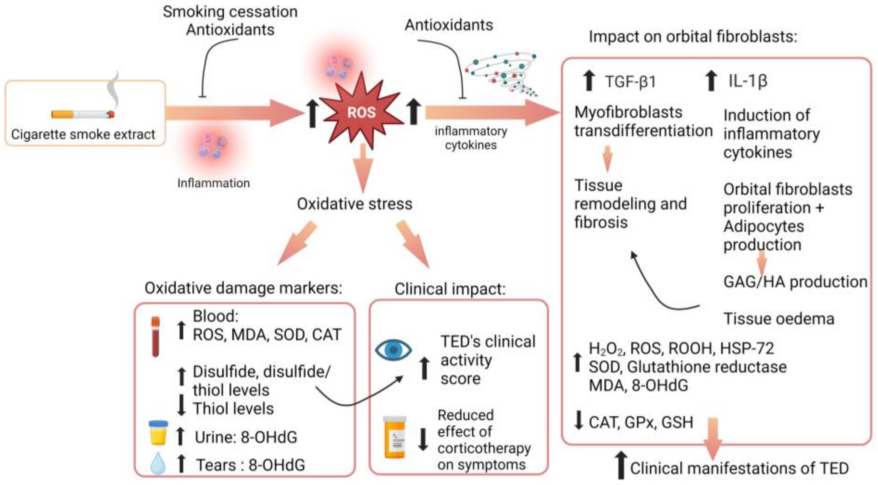
5. Oxidative Stress and TED’s Pathophysiology

5.1. Overview of Oxidative Stress

Oxidative stress occurs when there is an imbalance between the generation of reactive oxygen species (ROS) and cells’ ability to effectively counter them [113]. Since cells engage in oxidative respiration to generate energy, they reduce molecular oxygen to water and create as byproducts reactive oxygen species. ROS are highly reactive molecules: structurally, they have unpaired electrons, which render them unstable [114]. Different forms of ROS exist, such as hydrogen peroxide (H2O2), hydroxyl radicals (OH), and superoxide anions (O2-). They cause cellular and tissue damage by altering the cell’s structural integrity and interacting with membranes and intracellular components (lipids, proteins, and DNA), while also having the ability to mediate a variety of cell-death responses [114,115].
Under physiological conditions, cells counter the deleterious effects of ROS through an antioxidant defensive system. This system is composed mainly of enzymes (notably, superoxide dismutase (SOD), catalase (CAT), glutathione peroxidase (GPx)), vitamins (vitamin A, C, E), and minerals (see Table 4). As such, the disruption between pro-oxidant and anti-oxidant systems leads to oxidative stress and cellular damage [114]. Inflammation also leads to production of these free radicals and so do many environmental stressors such as UV, ionizing radiation, pollutants, and heavy metals [116].

5.2. Current Role of Oxidative Stress in TED

Increased oxidative stress occurs in Graves’ disease (GD), but also in TED, and a role for oxidative stress in the pathogenesis of TED is accepted in the literature [119,120,121]. A summary of main studies which have examined the role of oxidative damage in TED is presented at the end of this section (see Table 5).
An association between hyperthyroidism and overproduction of ROS is recognized in the literature [114,122]. However, since most TED patients have concomitant hyperthyroidism as they suffer from Graves’ disease, it was important to determine whether TED could independently impact the production of ROS. Bednarek et al.’s study aimed to determine levels of oxidative stress parameters in hyperthyroid but also in euthyroid patients with TED [123]. They found increased oxidative stress parameters in the serum of hyperthyroid patients, as could be expected from previous findings, but more importantly, increased oxidative stress also occurred in euthyroid patients with active TED. These oxidative damage markers were stabilized only in non-TED patients. This study implied that TED could be an independent factor leading to overproduction of ROS. It was posited that TED’s implication in oxidative stress generation was likely achieved through orbital inflammation [122,124]. Further research confirms the association of inflammation, TED, and oxidative stress, with levels of oxidative stress being increased in TED patients when compared to GD patients without TED [121].
Oxidative stress seems to play a role in the proliferation of orbital fibroblasts. In fact, it has been found that when orbital fibroblasts derived from TED patients are stimulated with a super-oxide anion (O2−, which is a ROS), they proliferate in a dose-dependent manner, a finding that does not occur in control orbital fibroblasts [125]. Later studies have confirmed these findings, showing that oxidative stress could enhance the proliferation of orbital fibroblasts [113]. Reversal of this effect was also possible with antioxidants or free-radical scavenging molecules like methimazole [125]. To explain orbital fibroblasts’ heightened response to ROS and their induced proliferation, it has been proposed that hypersensitivity of TED orbital fibroblasts to oxidative stress may be involved in the pathogenesis of the disease [126]. Further studies are needed to better understand whether such a mechanism is in play. Elucidating potential signaling pathways involving ROS may allow to halt oxidative stress-mediated orbital fibroblast proliferation.
Though it emerges from the current literature that ROS can trigger orbital fibroblast proliferation in TED patients, the pathways through which this occurs remain to be clarified. It most likely involves a complex interaction among ROS and pro-inflammatory cytokines [127]. To that effect, Tsai et al.’s work has shown that ROS are able to induce the expression of the proinflammatory cytokine IL-1 β in TED orbital fibroblasts [113]. IL-1 β is known to stimulate glycosaminoglycans (GAG) and hyaluronan production [128]. As such, an indirect contributory role for ROS in the production of these connective tissue components seems to emerge from the literature. Further research examining how ROS impact cells’ proliferation and whether they impact GAG production directly is needed. Assessing and comparing whether a proliferative response occurs in other types of fibroblasts, such as retro-orbital fibroblasts, would also be pertinent.
Previous work by Lu et al. had also shown that IL-1 β increases production of ROS in retro-ocular fibroblasts in a dose-dependent manner [128]. This confirms an interaction between IL-1 β and ROS in the disease pathway and points to a potential synergistic effect with cross-activation and amplification, which potentially adds to the understanding of how sustained inflammation can occur in TED. IL-1 β , released by activated macrophages in the orbital space, is an important mediator of inflammation [121]. Research comparing the effects of ROS and IL-1 β has not differentiated between active and inactive TED patients, and future work in that area should be conducted to further understand these implications.
ROS such as H2O2 can also induce the expression of TGF- β 1, which is a potent fibrogenic cytokine involved in tissue fibrosis that occurs in TED [113]. The precise mechanisms involving their interaction remain to be elucidated. It may be suggested that the inflammation and auto-immune process in TED leads to increased ROS and oxidative stress, along with depletion of antioxidant systems, which in turn exacerbate inflammation and lead to increases in numerous proinflammatory cytokines, including TGF- β 1. Indirect effects through induction of IL-1 β by ROS and its interaction with TGF- β 1 may also be involved. Oxidative stress pathways and interactions remain, however, to be fully understood [119].
Targeting ROS through novel antioxidant therapies may contribute to reducing the disease’s progression by limiting inflammation, along with orbital fibroblasts’ proliferation, IL-1 β -induced GAG production, and fibrosis, by limiting its effect on TGF- β 1 [125,128,129]. Research examining natural compounds’ effects on ROS and inflammation in TED has been led by looking into their anti-inflammatory and antioxidant properties [127,130]. Notably, selenium has been shown to have suppressing actions against the generation of ROS, while also being able to inhibit fibroblasts proliferation, hyaluronan production, and the release of proinflammatory cytokines [131,132]. Selenium-based therapy is recommended in the treatment of mild TED after its proven effect in reducing symptoms and slowing down of the disease course in a dedicated clinical trial [133,134]. Reduction in ROS was also possible with resveratrol, a natural compound found in wine, and quercetin, which can be found in fruits and vegetables [135,136]. Thus, the role of oxidative stress in the disease explains the principle of adding antioxidants as a treatment for TED, and research shows promising potential in slowing disease progression and reducing inflammation [114,129,137].
Oxidative stress has further been studied to evaluate its correlation with the disease’s clinical activity score, leading to findings of a positive correlation [138]. Tsai et al. have investigated the relationship of oxidative stress with clinical activity score but also with smoking by measuring the concentration of a urinary marker of oxidative DNA damage, 8-OHdG, in TED patients, in control patients, and in TED never-smoking patients. Significant levels of oxidative damage were found in TED patients, which correlate to CAS, and increased levels were also found in smokers compared to never-smokers [139]. Later studies have confirmed this positive correlation among oxidative damage markers and CAS, this time with the serum marker MDA, a marker of lipid peroxidation, and also with tear levels of MDA and 8-OHdG in TED patients [140,141].
Regarding smoking, Tsai et al.’s study implies that smoking-induced oxidative stress may contribute to TED and further lead to oxidative damage [139]. This is consistent with the literature already establishing a role of smoking in ROS generation [142]. These findings are clinically pertinent since smoking is a known risk factor for TED, affecting its development and progression. This provides additional proof to counsel smoking cessation to improve patients’ quality of life and limit clinical activity of TED by reducing oxidative damage and inflammation caused by tobacco. In line with this work, a recent study examined imbalance in thiol-disulfide homeostasis, an indicator of oxidative stress, in plasma of TED patients, but also in smoking patients and controls [143]. They found that increased disbalance in favor of disulfides, which are pro-oxidant molecules [144], occurs in TED patients compared to controls, but also in active compared to inactive TED patients, as well as in smokers [143]. Also, imbalance in thiol-disulfide homeostasis positively correlates with clinical activity score and is associated with proptosis, confirming a role for ROS in the clinical presentation of the disease [143,144]. Potential use of this marker in monitoring TED’s activity may be envisioned in the future for management of TED, though further research is required, notably to establish whether a link between this homeostasis disbalance marker and clinical activity score in the context of corticosteroid treatment or with the use of anti-inflammatory compounds.
These studies highlight again a link between tobacco, oxidative stress, and inflammation, while further pointing to the fact that smoking decreases the antioxidant systems’ homeostasis and is linked to clinical activity score and proptosis. Smoking cessation will thus help reduce clinical activity score as well as treatment outcomes, notably by reducing ROS and inflammation [25].
To conclude, the role for ROS in the inflammation process of TED seems clear. The literature suggests ROS are involved in the disease’s physiopathology but also in its severity and clinical activity. It also shows that ROS, IL-1, and TGF-B play critical roles in inflammation and influence each other. Targeting oxidative stress early on may have potential implications for disease management by decreasing inflammation in the retro-orbital and ocular space, preventing buildup of GAG and orbital fibroblasts’ expansion, reducing cell homeostasis disbalance, and limiting oxidative damage parameters, altogether improving clinical activity score and symptoms such as proptosis [144]. A better understanding of the orbital inflammation process and its interaction with oxidative stress, as well as increased understanding of the complex physiopathology of TED, may pave the way for novel therapeutic approaches and improved disease management. Figure 2 below provides an overview of potential implications of oxidative stress in TED.
Table 5. Summary of main studies involving ROS in TED.
AuthorYearConclusion
Heufelder et al. [145]1992H2O2 induced the expression of a heat shock protein-72 (which has a role in antigen recognition and T-cells recruitment).
Burch et al. [125]1997Superoxide anions trigger, in patients with TED:
Retro-orbital fibroblasts proliferation,
Glycosaminoglycan production,
Pro-inflammatory cytokines production.
Lu et al. [128]1999IL-1β increases ROS production in TED patients and control-derived retro-orbital fibroblasts;
IL-1β increases production of GAG in a dose-dependent manner in all retro-orbital fibroblasts;
ROS were expressed in retro-orbital fibroblasts from TED patients but not in controls.
Bednarek et al. [123]2005Orbital inflammation contributes to increased oxidative stress parameters in hyperthyroid and euthyroid TED patients;
Stabilization of oxidative stress parameters was achieved only in non-TED patients.
Tsai et al. [139]2007There are increased levels of urinary 8-OHdG (a marker of oxidative DNA damage) in patients with active TED.
Hondur et al. [137]2008Decreased activity of superoxide dismutase, GPx, and glutathione peroxidase occurs in orbital fibroblasts from TED patients.
Tsai et al. [138]2009Oxidative stress perpetuates oxidative damage to DNA, as seen with increased urinary levels of 8-OHdG in TED patients;
Oxidative DNA damage positively correlates to clinical activity of TED;
Higher levels of urinary 8-OHdG are seen in smokers compared to never-smokers.
Tsai et al. [113]2010Oxidative stress increases lipid peroxidation and oxidative DNA damage in TED orbital fibroblasts;
TED orbital fibroblasts accumulate higher amounts of intracellular ROS (such as superoxide anions and H2O2) compared to those of normal controls.
Tsai et al. [126]2011H2O2 exacerbates elevation of ROS in TED orbital fibroblasts;
Potential hypersensitivity of TED orbital fibroblasts to oxidative stress may be involved in the pathogenesis of TED.
Tsai et al. [129]2013H2O2 induces expression of intracellular pro-inflammatory cytokines TGF- β and IL-1 β in TED orbital fibroblasts;
H2O2 (at low level) induced proliferation of orbital fibroblasts;
Antioxidants (such as vitamin C) protect against these peroxide-induced effects.
Akarsu et al. [140] 2011There are increased MDA levels and decreased GSH levels in sera of GD patients with TED compared to GD patients without TED and controls;
MDA levels positively correlate with the disease’s clinical activity score;
GC therapy decreases serum MDA levels, whether taken orally or IV.
Marique et al. [146]2015Increased oxidative stress activity was found in extraocular muscle and adipocytes from TED patients, along with upregulation of antioxidants;
Serum TSHR antibody levels are related to the expression of oxidative stress.
Choi et al. [141]2018There are increased markers of oxidative stress (MDA and 8-OHdG levels) in tear film of TED patients;
Concentration of these markers correlates with disease severity.
Yuksel et al. [143]2019Imbalance in thiol-disulfide homeostasis (TDH) indicates presence of oxidative stress in moderate-to-severe TED;
Imbalance is significant in TED patients compared to controls, in active TED patients compared to inactive TED patients, and in smokers;
TDH imbalance correlates with clinical activity score.
Acibucu et al. [144]2019Thiol-disulfide homeostasis (TDH) disbalance is associated with proptosis in TED patients.
TED: thyroid eye disease; GD: Graves’ disease; GC: glucocorticoid; MDA: malondialdehyde; 8-OHdG: 8-hydroxy-2′-deoxyguanosine.

6. Implications for Treatment

The present review aimed to review and synthesize the current understanding of TED’s pathogenesis, particularly highlighting inflammatory and stress responses and their implications. In this section, we aim to provide a brief overview of how increased understanding of TED’s histopathology has oriented new research and therapies by showcasing therapeutic avenues that are currently explored in relation to cytokines and oxidative stress.
Because of the involvement of ROS in the pathophysiology of TED, oxidative stress markers in tears have received researchers’ attention in recent years. Notably, levels of oxidative stress markers 8-OHdG and MDA were studied in tears of active TED patients and increased levels were reported. Concentration of these markers also seems to correlate with the disease’s clinical activity and severity [141]. MDA levels in tears also decreased with glucocorticoid treatment [140]. More studies examining oxidative stress markers in tears, but also natural compounds’ effects on tear composition, should be conducted given their antioxidant and anti-inflammatory role, along with their proven ability to reduce ROS.
Since cytokines are also involved in the pathogenesis of TED, research aimed at examining differences in tear cytokine profiles is also underway. A study by Kishazi et al. found increased levels of IL-6 and TNF- α in TED patients’ tears when compared to those of healthy individuals, with levels notably higher in TED patients with CAS equal to or higher than 3. This is consistent with previous studies, like that of which examined tear cytokine profiles in TED patients, in GD patients without TED, and in healthy control patients [96]. The expression of seven cytokines across these groups were examined, and a significant up-regulation of IL-6, TNF- α , IL-1 β , IL-13, IL-17A, and IL-18 and RANTES in TED patients compared with controls was found. However, no significant differences were found in tears’ cytokine profile among TED and GD patients. They also examined the release of plasminogen activator inhibitor-1 (PAI-1) in these groups and found increased levels of PAI-1 in TED and GD patients compared with the control group, with PAI-1 being significantly higher in TED patients than in GD patients. A significant positive correlation between tear levels of IL-6 and PAI-1 and CAS score was also noted. More studies examining tear composition among GD patients with TED and GD patients without TED are needed to better distinguish tear profiles, evaluate whether differences in other cytokines’ expression exist, such as in VEGF, and to further understand the implications of PAI-1 and its potential role as a biomarker.
Cai et al. found that patients with inactive TED had higher levels of IL-7 when compared to healthy controls and active TED patients, respectively [147]. Tear levels of IL-15 and IL-17 have also been found to be increased in active TED compared to inactive TED [148]. Huang et al. not only confirmed the previous findings but also found positive correlations between levels of IL-1 β , IL-6, and IL-17A and clinical activity score, which points to a potential future use in assessing disease’s severity [149]. This shows that differences in tear films’ composition have been found among patients with active and inactive TED, compared to healthy patients [150].
A recent study further examined tears’ inflammatory cytokines profile in active TED patients following administration of corticosteroids, finding significantly decreased concentrations of IL-1 β , IL-6 IL-8, TNF- α , and VEGF after a 12 week treatment course. They also confirmed the previously noted positive correlation between IL-6 and CAS, while also finding a correlation among IL-8 levels and CAS [151]. Future work should also examine the impact of corticosteroid therapy on tears’ biomarkers in inactive TED patients to assess whether differences exist, but also on PAI-1 expression, which has been linked to disease activity score, as seen previously.
Although further research is needed, increased evidence suggests a potential role for tears as a source of biomarkers, highlighting a potential role as a diagnostic and clinical management tool [152]. Overall, these findings point to a potential role for tears as a biomarker fluid which could shape the future management of TED. Increased understanding of tears’ composition in TED and how it may be altered through oxidative stress and use of therapeutics may allow for a closer follow-up of patients’ treatment, along with prompt and targeted reduction in oxidative stress. More studies, notably on the sensitivity and specificity of these potential biomarkers may help to uncover suitable markers in the diagnosis and prognosis of TED [141,148].
To date, most studies focusing on TED’s pathophysiology have involved orbital tissues’ sampling. Research focusing on tear composition may prove interesting by helping to understand TED’s pathogenesis and develop new therapeutic targets, while also having the advantage of being less invasive than surgical tissue sampling and more accessible [141].

7. Conclusions

Increased understanding of the disease’s physiopathology has improved patient care and given way to new treatment options, showing the implications of research from lab to bedside. Despite increased knowledge on the interplay of immune factors, orbital fibroblasts, and oxidative stress in the development of TED, further studies are needed to elucidate the complex interactions of these factors and their effect on disease severity.
Notably, more research to elucidate the role of IGF-1R autoantibodies is required, as well as on the precise involvement and mechanisms of ROS in orbital inflammation and its relationship with proinflammatory cytokines. Future work on potential therapeutic approaches such as tear biomarkers is underway and could pave the way for increased disease management and follow-up. Research exploring tools for increased diagnosis, monitoring, and prediction of the disease’s severity and progression is also needed.

Author Contributions

Conceptualization—K.Y.W.; writing—original draft preparation, T.A. and D.D.G.; writing—review and editing, T.A., D.D.G., and K.Y.W.; figures, T.A. and D.D.G.; reference and formatting, T.A.; supervision and final review, K.Y.W. All authors have read and agreed to the published version of the manuscript.

Funding

This research received no external funding.

Data Availability Statement

Not applicable.

Conflicts of Interest

The authors declare no conflicts of interest.

References

  1. Chin, Y.H.; Ng, C.H.; Lee, M.H.; Koh, J.W.H.; Kiew, J.; Yang, S.P.; Sundar, G.; Khoo, C.M. Prevalence of thyroid eye disease in Graves’ disease: A meta-analysis and systematic review. Clin. Endocrinol. 2020, 93, 363–374. [Google Scholar] [CrossRef] [PubMed]
  2. Mohyi, M.; Smith, T.J. IGF1 receptor and thyroid-associated ophthalmopathy. J. Mol. Endocrinol. 2018, 61, T29–T43. [Google Scholar] [CrossRef] [PubMed]
  3. Bartalena, L.; Baldeschi, L.; Dickinson, A.; Eckstein, A.; Kendall-Taylor, P.; Marcocci, C.; Mourits, M.; Perros, P.; Boboridis, K.; Boschi, A.; et al. Consensus statement of the European Group on Graves’ orbitopathy (EUGOGO) on management of GO. Eur. J. Endocrinol. 2008, 158, 273–285. [Google Scholar] [CrossRef] [PubMed]
  4. Bruscolini, A.; Sacchetti, M.; La Cava, M.; Nebbioso, M.; Iannitelli, A.; Quartini, A.; Lambiase, A.; Ralli, M.; de Virgilio, A.; Greco, A. Quality of life and neuropsychiatric disorders in patients with Graves’ Orbitopathy: Current concepts. Autoimmun. Rev. 2018, 17, 639–643. [Google Scholar] [CrossRef]
  5. Weiler, D.L. Thyroid eye disease: A review. Clin. Exp. Optom. 2017, 100, 20–25. [Google Scholar] [CrossRef] [PubMed]
  6. Wiersinga, W.M. Quality of life in Graves’ ophthalmopathy. Best Pract. Res. Clin. Endocrinol. Metab. 2012, 26, 359–370. [Google Scholar] [CrossRef] [PubMed]
  7. Ponto, K.A.; Merkesdal, S.; Hommel, G.; Pitz, S.; Pfeiffer, N.; Kahaly, G.J. Public health relevance of Graves’ orbitopathy. J. Clin. Endocrinol. Metab. 2013, 98, 145–152. [Google Scholar] [CrossRef]
  8. Coulter, I.; Frewin, S.; Krassas, G.E.; Perros, P. Psychological implications of Graves’ orbitopathy. Eur. J. Endocrinol. 2007, 157, 127–131. [Google Scholar] [CrossRef]
  9. Taylor, P.N.; Zhang, L.; Lee, R.W.J.; Muller, I.; Ezra, D.G.; Dayan, C.M.; Kahaly, G.J.; Ludgate, M. New insights into the pathogenesis and nonsurgical management of Graves orbitopathy. Nat. Rev. Endocrinol. 2020, 16, 104–116. [Google Scholar] [CrossRef]
  10. Bartalena, L.; Piantanida, E.; Gallo, D.; Lai, A.; Tanda, M.L. Epidemiology, Natural History, Risk Factors, and Prevention of Graves’ Orbitopathy. Front. Endocrinol. 2020, 11, 615993. [Google Scholar] [CrossRef]
  11. Cyranska-Chyrek, E.; Olejarz, M.; Szczepanek-Parulska, E.; Stajgis, P.; Pioch, A.; Ruchala, M. Severe unilateral orbitopathy in a patient with Hashimoto’s thyroiditis—A case report. BMC Ophthalmol. 2019, 19, 9. [Google Scholar] [CrossRef]
  12. Perros, P.; Neoh, C.; Dickinson, J. Thyroid eye disease. BMJ 2009, 338, b560. [Google Scholar] [CrossRef]
  13. Shah, S.S.; Patel, B.C. Thyroid Eye Disease. In StatPearls; StatPearls Publishing LLC.: Treasure Island, FL, USA, 2023. [Google Scholar]
  14. Hoang, T.D.; Stocker, D.J.; Chou, E.L.; Burch, H.B. 2022 Update on Clinical Management of Graves Disease and Thyroid Eye Disease. Endocrinol. Metab. Clin. N. Am. 2022, 51, 287–304. [Google Scholar] [CrossRef]
  15. Perros, P.; Hegedüs, L.; Bartalena, L.; Marcocci, C.; Kahaly, G.J.; Baldeschi, L.; Salvi, M.; Lazarus, J.H.; Eckstein, A.; Pitz, S.; et al. Graves’ orbitopathy as a rare disease in Europe: A European Group on Graves’ Orbitopathy (EUGOGO) position statement. Orphanet J. Rare Dis. 2017, 12, 72. [Google Scholar] [CrossRef]
  16. Bartley, G.B. The epidemiologic characteristics and clinical course of ophthalmopathy associated with autoimmune thyroid disease in Olmsted County, Minnesota. Trans. Am. Ophthalmol. Soc. 1994, 92, 477–588. [Google Scholar]
  17. Abraham-Nordling, M.; Byström, K.; Törring, O.; Lantz, M.; Berg, G.; Calissendorff, J.; Nyström, H.F.; Jansson, S.; Jörneskog, G.; Karlsson, F.A.; et al. Incidence of hyperthyroidism in Sweden. Eur. J. Endocrinol. 2011, 165, 899–905. [Google Scholar] [CrossRef] [PubMed]
  18. Laurberg, P.; Berman, D.C.; Bülow Pedersen, I.; Andersen, S.; Carlé, A. Incidence and clinical presentation of moderate to severe graves’ orbitopathy in a Danish population before and after iodine fortification of salt. J. Clin. Endocrinol. Metab. 2012, 97, 2325–2332. [Google Scholar] [CrossRef]
  19. Douglas, R.S.; Gupta, S. The pathophysiology of thyroid eye disease: Implications for immunotherapy. Curr. Opin. Ophthalmol. 2011, 22, 385–390. [Google Scholar] [CrossRef]
  20. Şahlı, E.; Gündüz, K. Thyroid-associated Ophthalmopathy. Turk. J. Ophthalmol. 2017, 47, 94–105. [Google Scholar] [CrossRef]
  21. Perros, P.; Crombie, A.L.; Matthews, J.N.; Kendall-Taylor, P. Age and gender influence the severity of thyroid-associated ophthalmopathy: A study of 101 patients attending a combined thyroid-eye clinic. Clin. Endocrinol. 1993, 38, 367–372. [Google Scholar] [CrossRef]
  22. Manji, N.; Carr-Smith, J.D.; Boelaert, K.; Allahabadia, A.; Armitage, M.; Chatterjee, V.K.; Lazarus, J.H.; Pearce, S.H.; Vaidya, B.; Gough, S.C.; et al. Influences of age, gender, smoking, and family history on autoimmune thyroid disease phenotype. J. Clin. Endocrinol. Metab. 2006, 91, 4873–4880. [Google Scholar] [CrossRef] [PubMed]
  23. Thornton, J.; Kelly, S.P.; Harrison, R.A.; Edwards, R. Cigarette smoking and thyroid eye disease: A systematic review. Eye 2007, 21, 1135–1145. [Google Scholar] [CrossRef] [PubMed]
  24. Gontarz-Nowak, K.; Szychlińska, M.; Matuszewski, W.; Stefanowicz-Rutkowska, M.; Bandurska-Stankiewicz, E. Current Knowledge on Graves’ Orbitopathy. J. Clin. Med. 2020, 10, 16. [Google Scholar] [CrossRef]
  25. O’Dell, J.M.; Mussatto, C.C.; Chu, R.L.; Al-Sabbagh, M.Q.; Timoney, P.J.; Sokol, J.A. Effects of Smoking on Outcomes of Thyroid Eye Disease Treated with Teprotumumab: A Retrospective Cohort Study. Kans. J. Med. 2023, 16, 62–64. [Google Scholar] [CrossRef]
  26. Aranyosi, J.K.; Galgoczi, E.; Erdei, A.; Katko, M.; Fodor, M.; Ujhelyi, Z.; Bacskay, I.; Nagy, E.V.; Ujhelyi, B. Different Effects of Cigarette Smoke, Heated Tobacco Product and E-Cigarette Vapour on Orbital Fibroblasts in Graves’ Orbitopathy; a Study by Real Time Cell Electronic Sensing. Molecules 2022, 27, 3001. [Google Scholar] [CrossRef] [PubMed]
  27. Bartalena, L.; Marcocci, C.; Bogazzi, F.; Manetti, L.; Tanda, M.L.; Dell’Unto, E.; Bruno-Bossio, G.; Nardi, M.; Bartolomei, M.P.; Lepri, A.; et al. Relation between therapy for hyperthyroidism and the course of Graves’ ophthalmopathy. N. Engl. J. Med. 1998, 338, 73–78. [Google Scholar] [CrossRef] [PubMed]
  28. Mizokami, T.; Wu Li, A.; El-Kaissi, S.; Wall, J.R. Stress and thyroid autoimmunity. Thyroid 2004, 14, 1047–1055. [Google Scholar] [CrossRef]
  29. Cao, J.; Su, Y.; Chen, Z.; Ma, C.; Xiong, W. The risk factors for Graves’ ophthalmopathy. Graefe’s Arch. Clin. Exp. Ophthalmol. 2022, 260, 1043–1054. [Google Scholar] [CrossRef]
  30. Zawadzka-Starczewska, K.; Stasiak, B.; Wojciechowska-Durczyńska, K.; Lewiński, A.; Stasiak, M. Novel Insight into Non-Genetic Risk Factors of Graves’ Orbitopathy. Int. J. Environ. Res. Public Health 2022, 19, 16941. [Google Scholar] [CrossRef]
  31. Lee, J.; Kang, J.; Ahn, H.Y.; Lee, J.K. Sex-specific risk factors associated with graves’ orbitopathy in Korean patients with newly diagnosed graves’ disease. Eye 2023, 37, 3382–3391. [Google Scholar] [CrossRef]
  32. Khong, J.J.; McNab, A.A.; Ebeling, P.R.; Craig, J.E.; Selva, D. Pathogenesis of thyroid eye disease: Review and update on molecular mechanisms. Br. J. Ophthalmol. 2016, 100, 142–150. [Google Scholar] [CrossRef] [PubMed]
  33. Khoo, T.K.; Bahn, R.S. Pathogenesis of Graves’ ophthalmopathy: The role of autoantibodies. Thyroid 2007, 17, 1013–1018. [Google Scholar] [CrossRef] [PubMed]
  34. American Academy of Ophtalmology. Thyroid Eye Disease. Available online: https://eyewiki.aao.org/Thyroid_Eye_Disease#:~:text=The%20most%20commonly%20affected%20muscle,adducted%20i.e.,%20double%20elevator%20palsy (accessed on 2 October 2023).
  35. Bartley, G.B.; Fatourechi, V.; Kadrmas, E.F.; Jacobsen, S.J.; Ilstrup, D.M.; Garrity, J.A.; Gorman, C.A. Clinical features of Graves’ ophthalmopathy in an incidence cohort. Am. J. Ophthalmol. 1996, 121, 284–290. [Google Scholar] [CrossRef] [PubMed]
  36. Kendler, D.L.; Lippa, J.; Rootman, J. The initial clinical characteristics of Graves’ orbitopathy vary with age and sex. Arch. Ophthalmol. 1993, 111, 197–201. [Google Scholar] [CrossRef] [PubMed]
  37. Chng, C.L.; Seah, L.L.; Khoo, D.H. Ethnic differences in the clinical presentation of Graves’ ophthalmopathy. Best Pract. Res. Clin. Endocrinol. Metab. 2012, 26, 249–258. [Google Scholar] [CrossRef] [PubMed]
  38. Marinò, M.; Ionni, I.; Lanzolla, G.; Sframeli, A.; Latrofa, F.; Rocchi, R.; Marcocci, C. Orbital diseases mimicking graves’ orbitopathy: A long-standing challenge in differential diagnosis. J. Endocrinol. Investig. 2020, 43, 401–411. [Google Scholar] [CrossRef] [PubMed]
  39. Kennerdell, J.S.; Rosenbaum, A.E.; El-Hoshy, M.H. Apical optic nerve compression of dysthyroid optic neuropathy on computed tomography. Arch. Ophthalmol. 1981, 99, 807–809. [Google Scholar] [CrossRef] [PubMed]
  40. Perros, P.; Dayan, C.M.; Dickinson, A.J.; Ezra, D.; Estcourt, S.; Foley, P.; Hickey, J.; Lazarus, J.H.; MacEwen, C.J.; McLaren, J.; et al. Management of patients with Graves’ orbitopathy: Initial assessment, management outside specialised centres and referral pathways. Clin. Med. 2015, 15, 173–178. [Google Scholar] [CrossRef]
  41. Mourits, M.P.; Koornneef, L.; Wiersinga, W.M.; Prummel, M.F.; Berghout, A.; van der Gaag, R. Clinical criteria for the assessment of disease activity in Graves’ ophthalmopathy: A novel approach. Br. J. Ophthalmol. 1989, 73, 639–644. [Google Scholar] [CrossRef]
  42. Barrio-Barrio, J.; Sabater, A.L.; Bonet-Farriol, E.; Velázquez-Villoria, Á.; Galofré, J.C. Graves’ Ophthalmopathy: VISA versus EUGOGO Classification, Assessment, and Management. J. Ophthalmol. 2015, 2015, 249125. [Google Scholar] [CrossRef]
  43. Werner, S.C. Modification of the classification of the eye changes of Graves’ disease. Am. J. Ophthalmol. 1977, 83, 725–727. [Google Scholar] [CrossRef] [PubMed]
  44. Bartalena, L. Role of teprotumumab in the treatment of active moderate-to-severe Graves’ orbitopathy. Eur. Thyroid J. 2022, 11, e220185. [Google Scholar] [CrossRef] [PubMed]
  45. Bartalena, L.; Kahaly, G.J.; Baldeschi, L.; Dayan, C.M.; Eckstein, A.; Marcocci, C.; Marinò, M.; Vaidya, B.; Wiersinga, W.M. The 2021 European Group on Graves’ orbitopathy (EUGOGO) clinical practice guidelines for the medical management of Graves’ orbitopathy. Eur. J. Endocrinol. 2021, 185, G43–G67. [Google Scholar] [CrossRef]
  46. Diana, T.; Ponto, K.A.; Kahaly, G.J. Thyrotropin receptor antibodies and Graves’ orbitopathy. J. Endocrinol. Investig. 2021, 44, 703–712. [Google Scholar] [CrossRef] [PubMed]
  47. Gerding, M.N.; van der Meer, J.W.; Broenink, M.; Bakker, O.; Wiersinga, W.M.; Prummel, M.F. Association of thyrotrophin receptor antibodies with the clinical features of Graves’ ophthalmopathy. Clin. Endocrinol. 2000, 52, 267–271. [Google Scholar] [CrossRef] [PubMed]
  48. Sarić Matutinović, M.; Kahaly, G.J.; Žarković, M.; Ćirić, J.; Ignjatović, S.; Nedeljković Beleslin, B. The phenotype of Graves’ orbitopathy is associated with thyrotropin receptor antibody levels. J. Endocrinol. Investig. 2023, 46, 2309–2317. [Google Scholar] [CrossRef] [PubMed]
  49. Selter, J.H.; Gire, A.I.; Sikder, S. The relationship between Graves’ ophthalmopathy and dry eye syndrome. Clin. Ophthalmol. 2015, 9, 57–62. [Google Scholar] [CrossRef]
  50. Philipp, S.; Horstmann, M.; Hose, M.; Daser, A.; Görtz, G.E.; Jesenek, C.; Flögel, U.; Hansen, W.; Bechrakis, N.; Banga, J.P.S.; et al. An Early Wave of Macrophage Infiltration Intertwined with Antigen-Specific Proinflammatory T Cells and Browning of Adipose Tissue Characterizes the Onset of Orbital Inflammation in a Mouse Model of Graves’ Orbitopathy. Thyroid 2022, 32, 283–293. [Google Scholar] [CrossRef]
  51. Dik, W.A.; Virakul, S.; van Steensel, L. Current perspectives on the role of orbital fibroblasts in the pathogenesis of Graves’ ophthalmopathy. Exp. Eye Res. 2016, 142, 83–91. [Google Scholar] [CrossRef]
  52. Pappa, A.; Jackson, P.; Stone, J.; Munro, P.; Fells, P.; Pennock, C.; Lightman, S. An ultrastructural and systemic analysis of glycosaminoglycans in thyroid-associated ophthalmopathy. Eye 1998, 12 Pt 2, 237–244. [Google Scholar] [CrossRef]
  53. Kumar, S.; Nadeem, S.; Stan, M.N.; Coenen, M.; Bahn, R.S. A stimulatory TSH receptor antibody enhances adipogenesis via phosphoinositide 3-kinase activation in orbital preadipocytes from patients with Graves’ ophthalmopathy. J. Mol. Endocrinol. 2011, 46, 155–163. [Google Scholar] [CrossRef] [PubMed]
  54. Smith, T.J. The insulin-like growth factor-I receptor and its role in thyroid-associated ophthalmopathy. Eye 2019, 33, 200–205. [Google Scholar] [CrossRef] [PubMed]
  55. Janssen, J.; Smith, T.J. Lessons Learned from Targeting IGF-I Receptor in Thyroid-Associated Ophthalmopathy. Cells 2021, 10, 383. [Google Scholar] [CrossRef] [PubMed]
  56. Minich, W.B.; Dehina, N.; Welsink, T.; Schwiebert, C.; Morgenthaler, N.G.; Köhrle, J.; Eckstein, A.; Schomburg, L. Autoantibodies to the IGF1 receptor in Graves’ orbitopathy. J. Clin. Endocrinol. Metab. 2013, 98, 752–760. [Google Scholar] [CrossRef] [PubMed]
  57. Weightman, D.R.; Perros, P.; Sherif, I.H.; Kendall-Taylor, P. Autoantibodies to IGF-1 binding sites in thyroid associated ophthalmopathy. Autoimmunity 1993, 16, 251–257. [Google Scholar] [CrossRef] [PubMed]
  58. Krieger, C.C.; Neumann, S.; Place, R.F.; Marcus-Samuels, B.; Gershengorn, M.C. Bidirectional TSH and IGF-1 receptor cross talk mediates stimulation of hyaluronan secretion by Graves’ disease immunoglobins. J. Clin. Endocrinol. Metab. 2015, 100, 1071–1077. [Google Scholar] [CrossRef]
  59. Krieger, C.C.; Place, R.F.; Bevilacqua, C.; Marcus-Samuels, B.; Abel, B.S.; Skarulis, M.C.; Kahaly, G.J.; Neumann, S.; Gershengorn, M.C. TSH/IGF-1 Receptor Cross Talk in Graves’ Ophthalmopathy Pathogenesis. J. Clin. Endocrinol. Metab. 2016, 101, 2340–2347. [Google Scholar] [CrossRef]
  60. Pritchard, J.; Horst, N.; Cruikshank, W.; Smith, T.J. Igs from patients with Graves’ disease induce the expression of T cell chemoattractants in their fibroblasts. J. Immunol. 2002, 168, 942–950. [Google Scholar] [CrossRef]
  61. Varewijck, A.J.; Boelen, A.; Lamberts, S.W.; Fliers, E.; Hofland, L.J.; Wiersinga, W.M.; Janssen, J.A. Circulating IgGs may modulate IGF-I receptor stimulating activity in a subset of patients with Graves’ ophthalmopathy. J. Clin. Endocrinol. Metab. 2013, 98, 769–776. [Google Scholar] [CrossRef]
  62. Neumann, S.; Krieger, C.C.; Gershengorn, M.C. Targeting TSH and IGF-1 Receptors to Treat Thyroid Eye Disease. Eur. Thyroid J. 2020, 9, 59–65. [Google Scholar] [CrossRef]
  63. Girnita, L.; Smith, T.J.; Janssen, J. It Takes Two to Tango: IGF-I and TSH Receptors in Thyroid Eye Disease. J. Clin. Endocrinol. Metab. 2022, 107, S1–S12. [Google Scholar] [CrossRef] [PubMed]
  64. Tramontano, D.; Cushing, G.W.; Moses, A.C.; Ingbar, S.H. Insulin-like growth factor-I stimulates the growth of rat thyroid cells in culture and synergizes the stimulation of DNA synthesis induced by TSH and Graves’-IgG. Endocrinology 1986, 119, 940–942. [Google Scholar] [CrossRef] [PubMed]
  65. Tsui, S.; Naik, V.; Hoa, N.; Hwang, C.J.; Afifiyan, N.F.; Sinha Hikim, A.; Gianoukakis, A.G.; Douglas, R.S.; Smith, T.J. Evidence for an association between thyroid-stimulating hormone and insulin-like growth factor 1 receptors: A tale of two antigens implicated in Graves’ disease. J. Immunol. 2008, 181, 4397–4405. [Google Scholar] [CrossRef] [PubMed]
  66. Douglas, R.S.; Gianoukakis, A.G.; Kamat, S.; Smith, T.J. Aberrant expression of the insulin-like growth factor-1 receptor by T cells from patients with Graves’ disease may carry functional consequences for disease pathogenesis. J. Immunol. 2007, 178, 3281–3287. [Google Scholar] [CrossRef] [PubMed]
  67. Pritchard, J.; Han, R.; Horst, N.; Cruikshank, W.W.; Smith, T.J. Immunoglobulin activation of T cell chemoattractant expression in fibroblasts from patients with Graves’ disease is mediated through the insulin-like growth factor I receptor pathway. J. Immunol. 2003, 170, 6348–6354. [Google Scholar] [CrossRef] [PubMed]
  68. Smith, T.J.; Hoa, N. Immunoglobulins from patients with Graves’ disease induce hyaluronan synthesis in their orbital fibroblasts through the self-antigen, insulin-like growth factor-I receptor. J. Clin. Endocrinol. Metab. 2004, 89, 5076–5080. [Google Scholar] [CrossRef]
  69. Smith, T.J.; Janssen, J. Insulin-like Growth Factor-I Receptor and Thyroid-Associated Ophthalmopathy. Endocr. Rev. 2019, 40, 236–267. [Google Scholar] [CrossRef]
  70. Matos, K.; Manso, P.G.; Marback, E.; Furlanetto, R.; Alberti, G.N.; Nosé, V. Protein expression of VEGF, IGF-1 and FGF in retroocular connective tissues and clinical correlation in Graves’ ophthalmopathy. Arq. Bras. Oftalmol. 2008, 71, 486–492. [Google Scholar] [CrossRef]
  71. Lanzolla, G.; Ricci, D.; Nicolì, F.; Sabini, E.; Sframeli, A.; Brancatella, A.; Mantuano, M.; Dottore, G.R.; Bucci, I.; Figus, M.; et al. Putative protective role of autoantibodies against the insulin-like growth factor-1 receptor in Graves’ Disease: Results of a pilot study. J. Endocrinol. Investig. 2020, 43, 1759–1768. [Google Scholar] [CrossRef]
  72. Smith, T.J. Is IGF-I receptor a target for autoantibody generation in Graves’ disease? J. Clin. Endocrinol. Metab. 2013, 98, 515–518. [Google Scholar] [CrossRef]
  73. Gulbins, A.; Horstmann, M.; Daser, A.; Flögel, U.; Oeverhaus, M.; Bechrakis, N.E.; Banga, J.P.; Keitsch, S.; Wilker, B.; Krause, G.; et al. Linsitinib, an IGF-1R inhibitor, attenuates disease development and progression in a model of thyroid eye disease. Front. Endocrinol. 2023, 14, 1211473. [Google Scholar] [CrossRef] [PubMed]
  74. Liu, C.; Chu, D.; Kalantar-Zadeh, K.; George, J.; Young, H.A.; Liu, G. Cytokines: From Clinical Significance to Quantification. Adv. Sci. 2021, 8, e2004433. [Google Scholar] [CrossRef] [PubMed]
  75. Liu, X.; Faes, L.; Kale, A.U.; Wagner, S.K.; Fu, D.J.; Bruynseels, A.; Mahendiran, T.; Moraes, G.; Shamdas, M.; Kern, C.; et al. A comparison of deep learning performance against health-care professionals in detecting diseases from medical imaging: A systematic review and meta-analysis. Lancet Digit. Health 2019, 1, e271–e297. [Google Scholar] [CrossRef] [PubMed]
  76. Mikoś, H.; Mikoś, M.; Obara-Moszyńska, M.; Niedziela, M. The role of the immune system and cytokines involved in the pathogenesis of autoimmune thyroid disease (AITD). Endokrynol. Pol. 2014, 65, 150–155. [Google Scholar] [CrossRef] [PubMed]
  77. Bae, H.R.; Leung, P.S.C.; Hodge, D.L.; Fenimore, J.M.; Jeon, S.M.; Thovarai, V.; Dzutsev, A.; Welcher, A.A.; Boedigheimer, M.; Damore, M.A.; et al. Multi-omics: Differential expression of IFN-γ results in distinctive mechanistic features linking chronic inflammation, gut dysbiosis, and autoimmune diseases. J. Autoimmun. 2020, 111, 102436. [Google Scholar] [CrossRef] [PubMed]
  78. Serra, M.B.; Barroso, W.A.; da Silva, N.N.; Silva, S.D.N.; Borges, A.C.R.; Abreu, I.C.; Borges, M. From Inflammation to Current and Alternative Therapies Involved in Wound Healing. Int. J. Inflam. 2017, 2017, 3406215. [Google Scholar] [CrossRef] [PubMed]
  79. Salvi, M.; Vannucchi, G.; Currò, N.; Introna, M.; Rossi, S.; Bonara, P.; Covelli, D.; Dazzi, D.; Guastella, C.; Pignataro, L.; et al. Small dose of rituximab for graves orbitopathy: New insights into the mechanism of action. Arch. Ophthalmol. 2012, 130, 122–124. [Google Scholar] [CrossRef] [PubMed]
  80. Siddiqi, A.; Monson, J.P.; Wood, D.F.; Besser, G.M.; Burrin, J.M. Serum cytokines in thyrotoxicosis. J. Clin. Endocrinol. Metab. 1999, 84, 435–439. [Google Scholar] [CrossRef]
  81. Molnár, I.; Balázs, C. High circulating IL-6 level in Graves’ ophthalmopathy. Autoimmunity 1997, 25, 91–96. [Google Scholar] [CrossRef]
  82. Raychaudhuri, N.; Fernando, R.; Smith, T.J. Thyrotropin regulates IL-6 expression in CD34+ fibrocytes: Clear delineation of its cAMP-independent actions. PLoS ONE 2013, 8, e75100. [Google Scholar] [CrossRef]
  83. Paik, J.S.; Cho, W.K.; Oh, E.H.; Lee, S.B.; Yang, S.W. Palmitate induced secretion of IL-6 and MCP-1 in orbital fibroblasts derived from patients with thyroid-associated ophthalmopathy. Mol. Vis. 2012, 18, 1467–1477. [Google Scholar] [PubMed]
  84. Pérez-Moreiras, J.V.; Alvarez-López, A.; Gómez, E.C. Treatment of active corticosteroid-resistant graves’ orbitopathy. Ophthalmic Plast. Reconstr. Surg. 2014, 30, 162–167. [Google Scholar] [CrossRef] [PubMed]
  85. Sánchez-Bilbao, L.; Martínez-López, D.; Revenga, M.; López-Vázquez, Á.; Valls-Pascual, E.; Atienza-Mateo, B.; Valls-Espinosa, B.; Maiz-Alonso, O.; Blanco, A.; Torre-Salaberri, I.; et al. Anti-IL-6 Receptor Tocilizumab in Refractory Graves’ Orbitopathy: National Multicenter Observational Study of 48 Patients. J. Clin. Med. 2020, 9, 2816. [Google Scholar] [CrossRef] [PubMed]
  86. Migliorini, P.; Italiani, P.; Pratesi, F.; Puxeddu, I.; Boraschi, D. The IL-1 family cytokines and receptors in autoimmune diseases. Autoimmun. Rev. 2020, 19, 102617. [Google Scholar] [CrossRef] [PubMed]
  87. Wong, K.H.; Rong, S.S.; Chong, K.K.; Young, A.L.; Pang, C.P.; Chen, L.J. Genetic Associations of Interleukin-related Genes with Graves’ Ophthalmopathy: A Systematic Review and Meta-analysis. Sci. Rep. 2015, 5, 16672. [Google Scholar] [CrossRef] [PubMed]
  88. Li, B.; Smith, T.J. Regulation of IL-1 receptor antagonist by TSH in fibrocytes and orbital fibroblasts. J. Clin. Endocrinol. Metab. 2014, 99, E625–E633. [Google Scholar] [CrossRef] [PubMed]
  89. Li, B.; Smith, T.J. Divergent expression of IL-1 receptor antagonists in CD34⁺ fibrocytes and orbital fibroblasts in thyroid-associated ophthalmopathy: Contribution of fibrocytes to orbital inflammation. J. Clin. Endocrinol. Metab. 2013, 98, 2783–2790. [Google Scholar] [CrossRef]
  90. Boutet, M.A.; Blanchard, F.; Le Goff, B. Response to: ‘Does IL-38 act on macrophages and/or dendritic cells in arthritis?’ by Jiang et al. Ann. Rheum. Dis. 2018, 77, e13. [Google Scholar] [CrossRef]
  91. Shi, L.; Ye, H.; Huang, J.; Li, Y.; Wang, X.; Xu, Z.; Chen, J.; Xiao, W.; Chen, R.; Yang, H. IL-38 Exerts Anti-Inflammatory and Antifibrotic Effects in Thyroid-Associated Ophthalmopathy. J. Clin. Endocrinol. Metab. 2021, 106, e3125–e3142. [Google Scholar] [CrossRef]
  92. Pan, Y.; Wang, M.; Chen, X.; Chen, Y.; Ai, S.; Wang, M.; Su, W.; Liang, D. Elevated IL-38 inhibits IL-23R expression and IL-17A production in thyroid-associated ophthalmopathy. Int. Immunopharmacol. 2021, 91, 107300. [Google Scholar] [CrossRef]
  93. Gu, L.Q.; Jia, H.Y.; Zhao, Y.J.; Liu, N.; Wang, S.; Cui, B.; Ning, G. Association studies of interleukin-8 gene in Graves’ disease and Graves’ ophthalmopathy. Endocrine 2009, 36, 452–456. [Google Scholar] [CrossRef] [PubMed]
  94. Weetman, A.P.; Bennett, G.L.; Wong, W.L. Thyroid follicular cells produce interleukin-8. J. Clin. Endocrinol. Metab. 1992, 75, 328–330. [Google Scholar] [CrossRef] [PubMed]
  95. Myśliwiec, J.; Kretowski, A.; Stepień, A.; Mirończuk, K.; Kinalska, I. Interleukin 18 and transforming growth factor beta1 in the serum of patients with Graves’ ophthalmopathy treated with corticosteroids. Int. Immunopharmacol. 2003, 3, 549–552. [Google Scholar] [CrossRef] [PubMed]
  96. Ujhelyi, B.; Gogolak, P.; Erdei, A.; Nagy, V.; Balazs, E.; Rajnavolgyi, E.; Berta, A.; Nagy, E.V. Graves’ orbitopathy results in profound changes in tear composition: A study of plasminogen activator inhibitor-1 and seven cytokines. Thyroid 2012, 22, 407–414. [Google Scholar] [CrossRef] [PubMed]
  97. Zhang, P.; Zhang, X.; Xu, F.; Xu, W.; Zhu, H. Elevated expression of interleukin-27, IL-35, and decreased IL-12 in patients with thyroid-associated ophthalmopathy. Graefe’s Arch. Clin. Exp. Ophthalmol. 2023, 261, 1091–1100. [Google Scholar] [CrossRef] [PubMed]
  98. Bradley, J.R. TNF-mediated inflammatory disease. J. Pathol. 2008, 214, 149–160. [Google Scholar] [CrossRef] [PubMed]
  99. Kumar, S.; Bahn, R.S. Relative overexpression of macrophage-derived cytokines in orbital adipose tissue from patients with graves’ ophthalmopathy. J. Clin. Endocrinol. Metab. 2003, 88, 4246–4250. [Google Scholar] [CrossRef]
  100. Cawood, T.J.; Moriarty, P.; O’Farrelly, C.; O’Shea, D. The effects of tumour necrosis factor-alpha and interleukin1 on an in vitro model of thyroid-associated ophthalmopathy; contrasting effects on adipogenesis. Eur. J. Endocrinol. 2006, 155, 395–403. [Google Scholar] [CrossRef]
  101. Heufelder, A.E.; Bahn, R.S.; Boergen, K.P.; Scriba, P.C. Detection, localization and modulation of hyaluronic acid/CD44 receptor expression in patients with endocrine orbitopathy. Med. Klin. 1993, 88, 181–184+277. [Google Scholar]
  102. Ayabe, R.; Rootman, D.B.; Hwang, C.J.; Ben-Artzi, A.; Goldberg, R. Adalimumab as steroid-sparing treatment of inflammatory-stage thyroid eye disease. Ophthalmic Plast. Reconstr. Surg. 2014, 30, 415–419. [Google Scholar] [CrossRef]
  103. Durrani, O.M.; Reuser, T.Q.; Murray, P.I. Infliximab: A novel treatment for sight-threatening thyroid associated ophthalmopathy. Orbit 2005, 24, 117–119. [Google Scholar] [CrossRef] [PubMed]
  104. Komorowski, J.; Jankiewicz-Wika, J.; Siejka, A.; Lawnicka, H.; Kłysik, A.; Goś, R.; Majos, A.; Stefańczyk, L.; Stepień, H. Monoclonal anti-TNFalpha antibody (infliximab) in the treatment of patient with thyroid associated ophthalmopathy. Klin. Ocz. 2007, 109, 457–460. [Google Scholar]
  105. Hodge, D.L.; Berthet, C.; Coppola, V.; Kastenmüller, W.; Buschman, M.D.; Schaughency, P.M.; Shirota, H.; Scarzello, A.J.; Subleski, J.J.; Anver, M.R.; et al. IFN-gamma AU-rich element removal promotes chronic IFN-gamma expression and autoimmunity in mice. J. Autoimmun. 2014, 53, 33–45. [Google Scholar] [CrossRef] [PubMed]
  106. Fallahi, P.; Ferrari, S.M.; Ragusa, F.; Ruffilli, I.; Elia, G.; Paparo, S.R.; Antonelli, A. Th1 Chemokines in Autoimmune Endocrine Disorders. J. Clin. Endocrinol. Metab. 2020, 105, 1046–1060. [Google Scholar] [CrossRef] [PubMed]
  107. Fallahi, P.; Ferrari, S.M.; Elia, G.; Ragusa, F.; Paparo, S.R.; Patrizio, A.; Camastra, S.; Miccoli, M.; Cavallini, G.; Benvenga, S.; et al. Cytokines as Targets of Novel Therapies for Graves’ Ophthalmopathy. Front. Endocrinol. 2021, 12, 654473. [Google Scholar] [CrossRef] [PubMed]
  108. Antonelli, A.; Ferrari, S.M.; Fallahi, P.; Frascerra, S.; Santini, E.; Franceschini, S.S.; Ferrannini, E. Monokine induced by interferon gamma (IFNgamma) (CXCL9) and IFNgamma inducible T-cell alpha-chemoattractant (CXCL11) involvement in Graves’ disease and ophthalmopathy: Modulation by peroxisome proliferator-activated receptor-gamma agonists. J. Clin. Endocrinol. Metab. 2009, 94, 1803–1809. [Google Scholar] [CrossRef]
  109. Han, R.; Smith, T.J. T helper type 1 and type 2 cytokines exert divergent influence on the induction of prostaglandin E2 and hyaluronan synthesis by interleukin-1beta in orbital fibroblasts: Implications for the pathogenesis of thyroid-associated ophthalmopathy. Endocrinology 2006, 147, 13–19. [Google Scholar] [CrossRef]
  110. Chen, B.; Tsui, S.; Smith, T.J. IL-1 beta induces IL-6 expression in human orbital fibroblasts: Identification of an anatomic-site specific phenotypic attribute relevant to thyroid-associated ophthalmopathy. J. Immunol. 2005, 175, 1310–1319. [Google Scholar] [CrossRef]
  111. Antonelli, A.; Rotondi, M.; Ferrari, S.M.; Fallahi, P.; Romagnani, P.; Franceschini, S.S.; Serio, M.; Ferrannini, E. Interferon-gamma-inducible alpha-chemokine CXCL10 involvement in Graves’ ophthalmopathy: Modulation by peroxisome proliferator-activated receptor-gamma agonists. J. Clin. Endocrinol. Metab. 2006, 91, 614–620. [Google Scholar] [CrossRef]
  112. Antonelli, A.; Ferrari, S.M.; Corrado, A.; Franceschini, S.S.; Gelmini, S.; Ferrannini, E.; Fallahi, P. Extra-ocular muscle cells from patients with Graves’ ophthalmopathy secrete α (CXCL10) and β (CCL2) chemokines under the influence of cytokines that are modulated by PPARγ. Autoimmun. Rev. 2014, 13, 1160–1166. [Google Scholar] [CrossRef]
  113. Tsai, C.C.; Wu, S.B.; Cheng, C.Y.; Kao, S.C.; Kau, H.C.; Chiou, S.H.; Hsu, W.M.; Wei, Y.H. Increased oxidative DNA damage, lipid peroxidation, and reactive oxygen species in cultured orbital fibroblasts from patients with Graves’ ophthalmopathy: Evidence that oxidative stress has a role in this disorder. Eye 2010, 24, 1520–1525. [Google Scholar] [CrossRef] [PubMed]
  114. Marcocci, C.; Leo, M.; Altea, M.A. Oxidative stress in graves’ disease. Eur. Thyroid J. 2012, 1, 80–87. [Google Scholar] [CrossRef] [PubMed]
  115. Su, L.J.; Zhang, J.H.; Gomez, H.; Murugan, R.; Hong, X.; Xu, D.; Jiang, F.; Peng, Z.Y. Reactive Oxygen Species-Induced Lipid Peroxidation in Apoptosis, Autophagy, and Ferroptosis. Oxid. Med. Cell. Longev. 2019, 2019, 5080843. [Google Scholar] [CrossRef] [PubMed]
  116. Pizzino, G.; Irrera, N.; Cucinotta, M.; Pallio, G.; Mannino, F.; Arcoraci, V.; Squadrito, F.; Altavilla, D.; Bitto, A. Oxidative Stress: Harms and Benefits for Human Health. Oxid. Med. Cell. Longev. 2017, 2017, 8416763. [Google Scholar] [CrossRef] [PubMed]
  117. American Academy of Ophthalmology. Basic Ophtalmology: Essentials for Medical Students, 10th ed.; American Academy of Ophthalmology: San Francisco, CA, USA, 2016; p. 275. [Google Scholar]
  118. Ganea, E.; Harding, J.J. Glutathione-related enzymes and the eye. Curr. Eye Res. 2006, 31, 1–11. [Google Scholar] [CrossRef] [PubMed]
  119. Bartalena, L.; Tanda, M.L.; Piantanida, E.; Lai, A. Oxidative stress and Graves’ ophthalmopathy: In vitro studies and therapeutic implications. Biofactors 2003, 19, 155–163. [Google Scholar] [CrossRef]
  120. Wilson, R.; Chopra, M.; Bradley, H.; McKillop, J.H.; Smith, W.E.; Thomson, J.A. Free radicals and Graves’ disease: The effects of therapy. Clin. Endocrinol. 1989, 30, 429–433. [Google Scholar] [CrossRef]
  121. Zarković, M. The role of oxidative stress on the pathogenesis of graves’ disease. J. Thyroid Res. 2012, 2012, 302537. [Google Scholar] [CrossRef]
  122. Venditti, P.; Di Meo, S. Thyroid hormone-induced oxidative stress. Cell. Mol. Life Sci. 2006, 63, 414–434. [Google Scholar] [CrossRef]
  123. Bednarek, J.; Wysocki, H.; Sowiński, J. Oxidative stress peripheral parameters in Graves’ disease: The effect of methimazole treatment in patients with and without infiltrative ophthalmopathy. Clin. Biochem. 2005, 38, 13–18. [Google Scholar] [CrossRef]
  124. Song, Y.; Driessens, N.; Costa, M.; De Deken, X.; Detours, V.; Corvilain, B.; Maenhaut, C.; Miot, F.; Van Sande, J.; Many, M.C.; et al. Roles of hydrogen peroxide in thyroid physiology and disease. J. Clin. Endocrinol. Metab. 2007, 92, 3764–3773. [Google Scholar] [CrossRef] [PubMed]
  125. Burch, H.B.; Lahiri, S.; Bahn, R.S.; Barnes, S. Superoxide radical production stimulates retroocular fibroblast proliferation in Graves’ ophthalmopathy. Exp. Eye Res. 1997, 65, 311–316. [Google Scholar] [CrossRef] [PubMed]
  126. Tsai, C.C.; Wu, S.B.; Cheng, C.Y.; Kao, S.C.; Kau, H.C.; Lee, S.M.; Wei, Y.H. Increased response to oxidative stress challenge in Graves’ ophthalmopathy orbital fibroblasts. Mol. Vis. 2011, 17, 2782–2788. [Google Scholar]
  127. Buonfiglio, F.; Böhm, E.W.; Pfeiffer, N.; Gericke, A. Oxidative Stress: A Suitable Therapeutic Target for Optic Nerve Diseases? Antioxidants 2023, 12, 1465. [Google Scholar] [CrossRef]
  128. Lu, R.; Wang, P.; Wartofsky, L.; Sutton, B.D.; Zweier, J.L.; Bahn, R.S.; Garrity, J.; Burman, K.D. Oxygen free radicals in interleukin-1beta-induced glycosaminoglycan production by retro-ocular fibroblasts from normal subjects and Graves’ ophthalmopathy patients. Thyroid 1999, 9, 297–303. [Google Scholar] [CrossRef]
  129. Tsai, C.C.; Wu, S.B.; Kao, S.C.; Kau, H.C.; Lee, F.L.; Wei, Y.H. The protective effect of antioxidants on orbital fibroblasts from patients with Graves’ ophthalmopathy in response to oxidative stress. Mol. Vis. 2013, 19, 927–934. [Google Scholar]
  130. Hou, T.Y.; Wu, S.B.; Kau, H.C.; Tsai, C.C. The Role of Oxidative Stress and Therapeutic Potential of Antioxidants in Graves’ Ophthalmopathy. Biomedicines 2021, 9, 1871. [Google Scholar] [CrossRef]
  131. Kim, B.Y.; Jang, S.Y.; Choi, D.H.; Jung, C.H.; Mok, J.O.; Kim, C.H. Anti-inflammatory and Antioxidant Effects of Selenium on Orbital Fibroblasts of Patients with Graves Ophthalmopathy. Ophthalmic Plast. Reconstr. Surg. 2021, 37, 476–481. [Google Scholar] [CrossRef] [PubMed]
  132. Rotondo Dottore, G.; Leo, M.; Casini, G.; Latrofa, F.; Cestari, L.; Sellari-Franceschini, S.; Nardi, M.; Vitti, P.; Marcocci, C.; Marinò, M. Antioxidant Actions of Selenium in Orbital Fibroblasts: A Basis for the Effects of Selenium in Graves’ Orbitopathy. Thyroid 2017, 27, 271–278. [Google Scholar] [CrossRef]
  133. Bartalena, L.; Baldeschi, L.; Boboridis, K.; Eckstein, A.; Kahaly, G.J.; Marcocci, C.; Perros, P.; Salvi, M.; Wiersinga, W.M. The 2016 European Thyroid Association/European Group on Graves’ Orbitopathy Guidelines for the Management of Graves’ Orbitopathy. Eur. Thyroid J. 2016, 5, 9–26. [Google Scholar] [CrossRef]
  134. Marcocci, C.; Kahaly, G.J.; Krassas, G.E.; Bartalena, L.; Prummel, M.; Stahl, M.; Altea, M.A.; Nardi, M.; Pitz, S.; Boboridis, K.; et al. Selenium and the course of mild Graves’ orbitopathy. N. Engl. J. Med. 2011, 364, 1920–1931. [Google Scholar] [CrossRef]
  135. Kim, C.Y.; Lee, H.J.; Chae, M.K.; Byun, J.W.; Lee, E.J.; Yoon, J.S. Therapeutic Effect of Resveratrol on Oxidative Stress in Graves’ Orbitopathy Orbital Fibroblasts. Investig. Ophthalmol. Vis. Sci. 2015, 56, 6352–6361. [Google Scholar] [CrossRef] [PubMed]
  136. Yoon, J.S.; Lee, H.J.; Chae, M.K.; Lee, S.Y.; Lee, E.J. Cigarette smoke extract-induced adipogenesis in Graves’ orbital fibroblasts is inhibited by quercetin via reduction in oxidative stress. J. Endocrinol. 2013, 216, 145–156. [Google Scholar] [CrossRef]
  137. Hondur, A.; Konuk, O.; Dincel, A.S.; Bilgihan, A.; Unal, M.; Hasanreisoglu, B. Oxidative stress and antioxidant activity in orbital fibroadipose tissue in Graves’ ophthalmopathy. Curr. Eye Res. 2008, 33, 421–427. [Google Scholar] [CrossRef]
  138. Tsai, C.C.; Cheng, C.Y.; Liu, C.Y.; Kao, S.C.; Kau, H.C.; Hsu, W.M.; Wei, Y.H. Oxidative stress in patients with Graves’ ophthalmopathy: Relationship between oxidative DNA damage and clinical evolution. Eye 2009, 23, 1725–1730. [Google Scholar] [CrossRef]
  139. Tsai, C.C.; Kao, S.C.; Cheng, C.Y.; Kau, H.C.; Hsu, W.M.; Lee, C.F.; Wei, Y.H. Oxidative stress change by systemic corticosteroid treatment among patients having active graves ophthalmopathy. Arch. Ophthalmol. 2007, 125, 1652–1656. [Google Scholar] [CrossRef]
  140. Akarsu, E.; Buyukhatipoglu, H.; Aktaran, S.; Kurtul, N. Effects of pulse methylprednisolone and oral methylprednisolone treatments on serum levels of oxidative stress markers in Graves’ ophthalmopathy. Clin. Endocrinol. 2011, 74, 118–124. [Google Scholar] [CrossRef]
  141. Choi, W.; Li, Y.; Ji, Y.S.; Yoon, K.C. Oxidative stress markers in tears of patients with Graves’ orbitopathy and their correlation with clinical activity score. BMC Ophthalmol. 2018, 18, 303. [Google Scholar] [CrossRef]
  142. Pryor, W.A.; Stone, K. Oxidants in cigarette smoke. Radicals, hydrogen peroxide, peroxynitrate, and peroxynitrite. Ann. N. Y. Acad. Sci. 1993, 686, 12–28. [Google Scholar] [CrossRef]
  143. Yuksel, N.; Tanriverdi, B.; Ipteç, B.; Erel, O. Thiol-disulfide homeostasis as an oxidative stress marker in patients with Graves’ ophthalmopathy. Orbit 2019, 38, 370–375. [Google Scholar] [CrossRef]
  144. Acibucu, F.; Öztürk, D.D.; Kizildag, C.; Aslan, M.Z.; Gulumsek, E.; Sumbul, M.S.; Neselioglu, S.; Erel, O.; Sen, S.; Bankir, M.; et al. Proptosis is associated with thiol-disulfide in patients with Graves’ ophthalmopathy. Arch. Endocrinol. Metab. 2022, 66, 191–197. [Google Scholar] [CrossRef] [PubMed]
  145. Heufelder, A.E.; Wenzel, B.E.; Bahn, R.S. Methimazole and propylthiouracil inhibit the oxygen free radical-induced expression of a 72 kilodalton heat shock protein in Graves’ retroocular fibroblasts. J. Clin. Endocrinol. Metab. 1992, 74, 737–742. [Google Scholar] [CrossRef]
  146. Marique, L.; Senou, M.; Craps, J.; Delaigle, A.; Van Regemorter, E.; Wérion, A.; Van Regemorter, V.; Mourad, M.; Nyssen-Behets, C.; Lengelé, B.; et al. Oxidative Stress and Upregulation of Antioxidant Proteins, Including Adiponectin, in Extraocular Muscular Cells, Orbital Adipocytes, and Thyrocytes in Graves’ Disease Associated with Orbitopathy. Thyroid 2015, 25, 1033–1042. [Google Scholar] [CrossRef] [PubMed]
  147. Cai, K.; Wei, R. Interleukin-7 expression in tears and orbital tissues of patients with Graves’ ophthalmopathy. Endocrine 2013, 44, 140–144. [Google Scholar] [CrossRef] [PubMed]
  148. Bajkowska, D.; Szelachowska, M.; Buczyńska, A.; Krętowski, A.J.; Siewko, K. Tears as a Source of Biomarkers in the Diagnosis of Graves’ Orbitopathy. Biomolecules 2022, 12, 1620. [Google Scholar] [CrossRef]
  149. Huang, D.; Luo, Q.; Yang, H.; Mao, Y. Changes of lacrimal gland and tear inflammatory cytokines in thyroid-associated ophthalmopathy. Investig. Ophthalmol. Vis. Sci. 2014, 55, 4935–4943. [Google Scholar] [CrossRef]
  150. Sun, R.; Zhou, H.F.; Fan, X.Q. Ocular surface changes in Graves’ ophthalmopathy. Int. J. Ophthalmol. 2021, 14, 616–621. [Google Scholar] [CrossRef]
  151. Xu, N.; Cui, Y.; Fu, D.; Sun, F. Tear inflammatory cytokines and ocular surface changes in patients with active thyroid eye disease treated with high-dose intravenous glucocorticoids. J. Endocrinol. Investig. 2020, 43, 901–910. [Google Scholar] [CrossRef]
  152. Khazaei, H.; Khazaei, D.; Verma, R.; Ng, J.; Wilmarth, P.A.; David, L.L.; Rosenbaum, J.T. The potential of tear proteomics for diagnosis and management of orbital inflammatory disorders including Graves’ ophthalmopathy. Exp. Eye Res. 2021, 213, 108813. [Google Scholar] [CrossRef]
Figure 1. The pathogenesis of thyroid eye disease (BioRender, https:/app.biorender.com/, accessed on 5 September 2023).
Stresses 04 00004 g001
Figure 2. Overview of potential implications of oxidative stress in TED (BioRender, https:/app.biorender.com/, accessed on 3 October 2023). CSE: cigarette smoke extract; ROS: reactive oxygen species; MDA: malondialdehyde; OHdG: 8-hydroxy-2′-deoxyguanosine; GAG: glycosaminoglycans; HA: hyaluronic acid; ROOH: lipid hydroperoxide; HSP-72: heat shock protein 72; SOD: superoxide dismutase; CAT: catalase; GPx: glutathione peroxidase; GSH: glutathione.
Stresses 04 00004 g002
Table 1. Clinical Activity Score (CAS) amended by EUGOGO after Mourits et al. [3].
Only parameters 1–7 are scored for initial CAS
1Spontaneous orbital pain
2Gaze-evoked orbital pain
3Eyelid swelling that is considered to be due to active TED
4Eyelid erythema
5Conjunctival redness that is considered to be due to active TED
6Chemosis
7Inflammation of caruncle or plica
For following assessments,
parameters 1 to 10 can be included
8Increase of ˃2 mm in proptosis
9Decrease in uniocular ocular excursion in any one direction of ˃ 8°
10Decrease in acuity equivalent to 1 Snellen line
One point is given for each parameter. Clinical activity is defined by total points sum; a score of min. 3/7 at first examination and over 4/10 in successive examinations leads to finding of active TED.
Table 2. NO SPECS modified classification [43].
ClassGradeSuggestions for Grading
0-No physical signs or symptoms
I-Only signs
II
0
a
b
c
Soft tissue involvement
Absent
Minimal
Moderate
Marked
III
0
a
b
c
Proptosis
Absent
3–4 mm over upper normal
5–7 mm increase
8 mm increase
IV
0
a
b
c
Extraocular muscle involvement
Absent
Limitation of motion at extreme gaze
Evident restriction of motion
Fixation of a globe or globes
V
0
a
b
c
Corneal involvement
Absent
Stippling of cornea
Ulceration
Clouding, necrosis, and perforation
VI
0
a
b
c
Sight loss (due to optic nerve involvement)
Absent
Disc pallor or choking or visual field defect, vision 20/20-20/60
Disc pallor or choking, or visual field defect, vision 20/70-20/200
Blindness, vision less than 20/200
Table 3. Treatment algorithm for TED [42].
For All Patients1. Restore Euthyroid State
2. Urge Smoking Cessation
SeverityActiveInactive
MildArtificial tears
Sunglasses
Elevating head of bed
Prismatic glasses
Artificial tears
Prismatic glasses
Surgical Müllerectomy
Blepharoplasty
Moderate-severeIntravenous methylprednisolone

In steroid-resistant patients:
cyclosporin A + oral steroid, immunosuppressive therapies, anti-cytokine/lymphocyte agents

If motility dysfunction is pronounced: orbital radiotherapy


Orbital decompression
Strabismus surgery
Palpebral surgery
Threat to vision
Optic neuropathy


Severe corneal involvement
Intravenous methylprednisolone:
1 gr for 3 days

If non-responsive:
orbital decompression +/− intravenous steroid +/− radiotherapy

Intravenous steroid, lubrication, tarsorrhaphy, orbital decompression

Urgent surgical decompression


Lateral tarsorrhaphy, orbital decompression, amniotic membrane transplant, keratoplasty
Table 4. Summary of effects of ROS and antioxidant mechanisms in the eye.
Effects of ROS at cellular level [117]
  • Lipids: Lipid peroxidation within the plasma membrane
  • DNA: DNA fragmentation
  • Proteins: Protein cross-linking and fragmentation
  • Cellular level: Apoptosis of cell, necrosis, and autophagy response
Antioxidant mechanisms in the eye include [117,118]
  • Vitamin A, C, E, beta-carotene
  • Enzymatic system:
    (a)
    Mitochondria: Superoxide dismutase, glutathione peroxidase
    (b)
    Peroxisome: Catalase
    (c)
    Cytosol: Superoxide dismutase, glutathione peroxidase, ferritin, ceruloplasmin
  • Minerals: Selenium, copper, zinc
Table inspired from: Oxidative Stress in Ophthalmology, EyeWiki page: https://eyewiki.aao.org/Oxidative_Stress_in_Ophthalmology (accessed on 2 October 2023).
Disclaimer/Publisher’s Note: The statements, opinions and data contained in all publications are solely those of the individual author(s) and contributor(s) and not of MDPI and/or the editor(s). MDPI and/or the editor(s) disclaim responsibility for any injury to people or property resulting from any ideas, methods, instructions or products referred to in the content.

