Review

Multifactorial Distress, the Warburg Effect, and Respiratory and pH Imbalance in Cancer Development

Biochemistry Group, Faculty of Chemistry, Alexandru Ioan Cuza University, 11 Carol I, 700506 Iasi, Romania
Stresses 2023, 3(2), 500-528; https://doi.org/10.3390/stresses3020036
Submission received: 8 April 2023 / Revised: 26 May 2023 / Accepted: 12 June 2023 / Published: 18 June 2023
(This article belongs to the Collection Feature Papers in Human and Animal Stresses)

Abstract

:
Oncogenes are thought to play an important role in aberrant regulation of growth factors, which is believed to be an initiation event of carcinogenesis. However, recent genetic and pharmacological studies have shown that the Warburg effect (WE) is needed for tumour growth. It refers to extensively studied aerobic glycolysis over the past decade, although its impact on cancer remains unclear. Meanwhile, a large body of evidence has indicated that oxidative stress (OS) is connected with the occurrence and progression of various forms of cancer. Psychosocial factors (PSF), such as chronic depression, sadness, stressful life experiences, stress-prone personality, and emotional distress or poor quality of life affect the immune system and contribute to cancer outcomes. Here, we examine the relationship between WE, OS, PSF, metal ions, other carcinogens, and the development of different cancers from the viewpoint of physiological and biochemical mechanisms.

1. Introduction

Metabolic changes in cancer are no longer seen as an indirect response to signals of cell proliferation and survival. Rather, impaired metabolism status is the basic hallmark of cancer [1]. The hypothesis that oncogenic transformation alters cellular metabolism to sustain high rates of growth and division has been extensively explored [2]. Recent genetic and pharmacological investigations have shown that the Warburg effect (WE) is also required for cancerous growth [3,4]. During cancer progression, oxygen respiration always decreases, fermentation takes place, and highly differentiated cells switch to anaerobic fermentation, having lost all their previous physiological functions and only retaining the now useless property of proliferating and multiplying [5,6]. Cancer metastasis and therapeutic resistance are usually studied as separate areas using different strategies. However, metastatic progression and therapeutic resistance signalling are mediated by common mechanisms, such as the involvement of integrins and other contextual receptors, cell–cell communication, stress responses, and metabolic reprogramming [7]. During proliferation and metastasis, malignant cells adapt to oxidative stress by increasing NADPH in a variety of ways, including by activating AMPK, PPP, and reductive glutamine, as well as folate metabolism [8]. Indeed, reactive oxygen species (ROS) influence the progression of cancer, either by initiating or stimulating tumorigenesis and supporting the transformation and proliferation of cancer cells, or by causing their death [9,10,11]. While cancer cells have increased levels of ROS, and increased ROS concentrations are associated with various carcinogenic processes, some drugs destroy cancer tumours via toxic levels of ROS [12]. Psychosocial factors are stressors that have a negative impact on cancer patients, but their effects vary depending on the type of psychosocial factor, cancer location, and cancer outcome [13]. Thus, stressful life experiences have been shown to be associated with lower cancer survival and higher mortality, but not higher incidence.
Heavy metal-induced oxidative stress can promote various cancers and diseases by ROS-based mechanisms [14]. Heavy metals stimulate tumour progression and reduce tumour sensitivity to treatment, while tumour tissue shows a different level of DNA methylation [15].
Biochemically, the less-differentiated cell structure of the cancer tissue somewhat resembles that of foetal tissue [16,17,18]. Consequently, the importance of studying the embryo to understand the evolution of the tumour and contribute to the development of effective therapeutic strategies was highlighted [19]. In addition, high concentrations of alphafetoprotein are normally found in foetal blood but are almost undetectable in adult blood. Therefore, this protein has attracted increasing interest because of its connection with carcinogenic events [20]. It is precisely this feature that should be investigated, as it may hide the underlying mechanisms of cancerogenesis.
Hypoxia is the condition in which tissues are exposed to oxygen deficiency and is an essential phenomenon influencing cellular health. The effect of hypoxia on human cells can be either positive or negative depending on the severity, duration and context [21]. Multicellular organisms have developed both systemic and cellular responses to hypoxia [21]. The generation of adenosine triphosphate (ATP) in mitochondria is particularly sensitive to changes in oxygen tension. For that reason, the hypoxic state is an aggravating factor commonly seen in cancer, multiple sclerosis, heart disease, kidney disease, lung disease, liver disease, etc. [22]. Hypoxia can play an important role in the regeneration of damaged tissues, in particular by acting on tissue-specific stem cells. However, its role can be a drawback when it involves neoplastic stem cells.
Here, we first analyse the function of WE, highlight its significance and discuss its shortcomings. Our analysis, carried out from the viewpoint of physiological and biochemical mechanisms, mainly focuses on the relationship between WE, OS, non-specific stress (NSS), hypoxia, heavy metals, and other carcinogenic factors involved in the occurrence of different cancers. Since the normal cells of the body meet their energy needs by breathing oxygen while cancer cells do this mainly through fermentation, we have introduced the term respiratory imbalance, which refers to an impairment of respiration. Further, glycolysis generates lactic acid, which alters the pH of body fluids. We have therefore added the notion of pH imbalance. All findings discussed here suggest that lifestyle, food, distress, carcinogens, ROS, and heavy metals can be environmental factors involved in cancer aetiology and progression. We are also pursuing common physiological mechanisms capable of shedding light on the carcinogenic effect of so many carcinogens involved in so many different cancers and their link to the Warburg effect.

2. Oxidative Stress

Molecular stress is considered to be involved in cancer initiation and progression [23,24]. Oxidative stress from endogenous and exogenous sources leads to mutations and epigenetic deregulations, which contribute to the development of neoplastic diseases [25]. Among the first category of stressors are peroxisomes and enzymes, such as NADPH oxidase [26], xanthine oxidase [27], dihydrolipoamide dehydrogenase [28], etc., most of which are found inside the mitochondria. Other stressors, such as alcohol, nicotine, exercise, or UV radiation, are responsible for an increase in the intracellular level of several reactive species, such as reactive oxygen species (ROS), reactive nitrogen species (RNS), and reactive sulphur-based species (RSS) [25]. ROS appear during mitochondrial aerobic metabolism, being a reaction of human and animal cells to bacterial invasions, presence of xenobiotics, on-going distress, or X-ray exposure [29]. The relative excess of ROS, when compared to antioxidants, has been linked to multiple pathologies, such as neurodegenerative disease, cardiovascular disease, diabetes mellitus, etc. [30]. The cancer cell is also known to show aberrant redox natural balance. While ROS are pro-tumorigenic, a high level of ROS can be cytotoxic [31]. Excessive production of ROS is associated with many types of diseases, such as chronic inflammation [32,33] and a variety of cancers [34,35,36,37]. Thus, the proliferation of malignant cells is associated with high ROS production. Notably, these cells are adapted to grow under conditions where this oxidative stress shifts the redox homeostasis away from a reduced status; tumour cells achieve this balance by increasing their antioxidant potential to optimize ROS-driven proliferation [38,39]. Since there is a link between the redox potential of a cell and its tolerance to high levels of ROS, biochemistry of reduced glutathione (GSH), thioredoxins (TXN), and NADPH becomes important. Normal metabolic processes typically generate ROS and reactive nitrogen species (RNS) that are potentially harmful in high concentrations; these species are intracellular signalling molecules [40]. However, cells possess an array of antioxidant systems to ensure that ROS and RNS signalling mechanisms are preserved and oxidative injury is avoided. The increase in the level of ROS and RNS in relation to antioxidant activity of the cell means that oxidative stress (OS) is counteracted by the normal cell with the help of GSH and TXN. Their action is supported by NADPH, which keeps them both in a reduced state. Oxidation of polyunsaturated fatty acids by ROS results in lipid peroxidation, while the peroxidised compounds and their breakdown products may act as signalling molecules to stimulate inflammation and apoptosis [38,41].
The main resources of ROSs include: electron flow to O2 in mitochondria and the reaction between coenzyme Q10 (CoQ) found in the semiquinone form and the molecular oxygen at complex III of the respiratory chain [42]. In addition, NADPH oxidases reduce O2 to superoxide, O2●− [43]. The formation of highly reactive hydroxyl radicals (HO) by Fenton’s chemistry from H2O2 molecules usually implies heavy metal ions, such as copper, iron, or manganese. One of the main RNS species is the vasodilator NO, which is liberated by nitric oxide synthase using L-arginine. The reaction between NO and O2●− produces ONOO [44].
Antioxidant defence prevents the accumulation of ROS and RNS through several scavenger molecules, such as GSH, melatonin, α-lipoic acid, bilirubin, melanin, or uric acid. Vitamin E, vitamin C, β-carotene, and plant polyphenols are also antioxidants [40]. GSH is subject to homeostatic regulation and is often increased in some forms of cancer [45]. On the other hand, the cytoplasmic copper/zinc superoxide dismutase (SOD1), mitochondrial manganese superoxide dismutase (SOD2), and extracellular superoxide dismutase (SOD3) catalyse the conversion of O2●− to H2O2 and O2 [46]. Since SOD1 and SOD2 protect against spontaneous malignization and are defined as tumour suppressors, they are up-regulated during oncogenesis [47]. Next, catalase (CAT) is involved in the decomposition of H2O2 into H2O and O2 [48].
Cancer cells have to deal with OS at the onset, during matrix cleavage, during entry into circulation, and when the disease relapses following treatment [8]. Tumour cells are able to adapt by various means to ensure that ROS activity is limited to a dynamic threshold that allows them to proliferate while avoiding cell mortality [49,50].
Oxidative stress is connected to the progression of the most common form of liver cancer [51]. Nevertheless, the mechanisms are still unclear. Typically, OS happens when the body detects any danger signal, either from an internal or external source [47]. Reactive oxygen species (ROS) are permanently generated in peroxisomes, mitochondria, cytosol, and apoplast. The imbalance between ROS generation and detoxification leads to oxidative stress, and the accumulation of ROS is harmful to cells. In addition, ROS function as signalling molecules and activate signal transduction processes in response to various stresses [52]. Uncontrolled overproduction of ROS, resulting from an imbalance between ROS production and removal, leads to vascular disease [53]. It then induces oxidative damage to the DNA and abnormal protein synthesis, putting the body in a condition of susceptibility to developing various diseases, including cancer. Many factors are involved in liver carcinogenesis, including hepatitis B virus and hepatitis C virus infection, alcohol abuse, and non-alcoholic fatty liver disease. Elucidation of the influence of OS in cancer aetiology is important for the prevention and treatment of various cancers. Treatment with OS antioxidant drugs can control OS lesions in vitro [54]. However, in the case of liver cancer, chronic viral infections can induce inflammation and necrosis of liver cells [55]. DNA damage caused by ROS leads to the accumulation of cancer-related genetic mutations. Chronic inflammation is one of the causes of human cancer [56,57]. Oxidative stress and accumulation of DNA damage play an important role in virus-induced cancer [58]. In addition, miRNA dysfunction in inflammatory reactions is believed to be the central event in the occurrence of some cancers [54].
High levels of ROS have been detected in almost all cancers, where they promote many aspects of tumour development and progression. However, tumour cells also express increased levels of antioxidant proteins to detoxify from ROS, suggesting that a delicate balance of intracellular ROS levels is required for cancer cell function [59,60].
There is an increased risk of cancers among obese patients; some forms of fatness explain cancer risk in obese patients, while oxidative stress may play a role in obesity-related cancers [61]. The enzyme involved in triacylglyceride synthesis and lipid droplet formation, diacylglycerol O-acetyltransferase 1 (DGAT1) is commonly up-regulated in melanoma, allowing these cells to support excess fatty acids [62]. DGAT1 inhibitors induce OS in melanoma cells, which adapts by increasing cell defences against ROS. Inhibition of both DGAT1 and superoxide dismutase 1 profoundly inhibit tumour proliferation due to exaggerated OS.

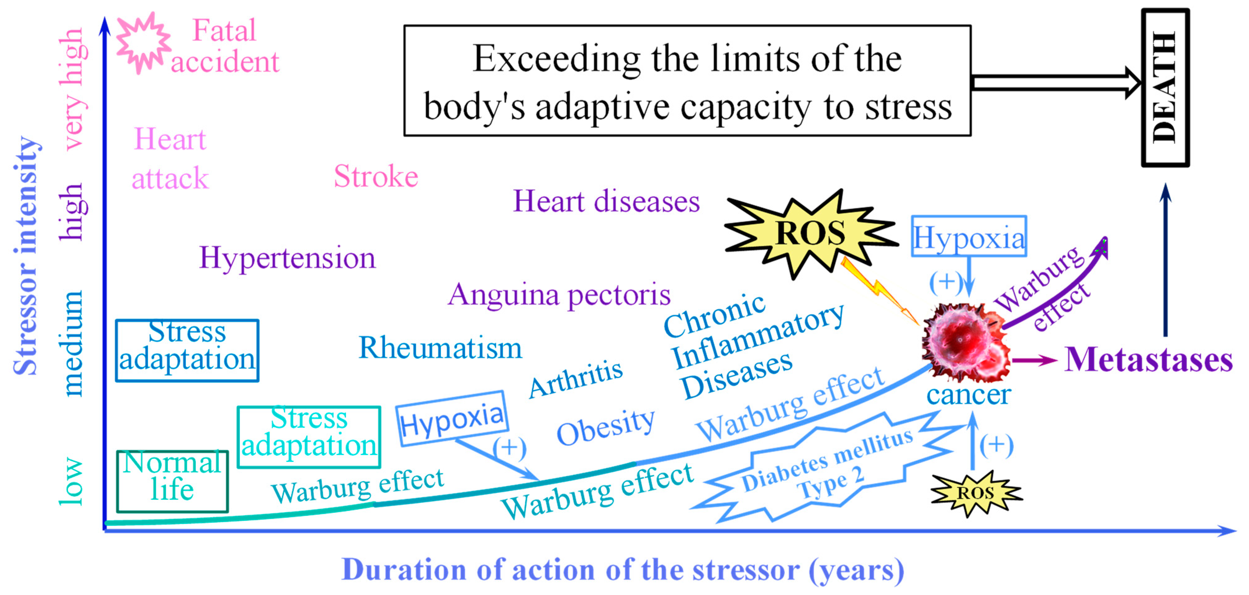
3. The Non-Specific Stress

Several studies suggest that stress may influence oncogenic development, and data from subhuman experiments have shown that aggressive offenses can enhance or suppress oncogenicity [63,64]. Thus, psychological factors may affect the risk and progression of tumour proliferation [65]. Increased tumour progression is evident following acute exposure to severe stress, and the impact of aggressive stimuli varies according to previous distress history and social living conditions [66]. In addition, prolonged depression can be associated with an increased risk of cancer [67]. Moreover, providing psychosocial support may help reduce depression, angst, and hurt, and can prolong the survival period with cancer. The relationship between sadness and cancer evolution assumes dysregulation of the hypothalamic-pituitary-adrenal alignment, particularly daytime changes in melatonin and cortisol. In general, depression affects the immune system, which can affect cancer control [68]. People with psychiatric disorders are no more susceptible than the general population to developing cancer, but they are more inclined to die from this disease [69]. All these findings suggest that there may be a close relationship between other forms of stress, mainly related to human personality.
Stress induces or worsens cardiovascular diseases, non-alcoholic fatty liver disease, depression, neurodegenerative disease, and cancer through peripheral inflammation as well as neuroinflammation [70]. Stress endangers central microglia and astrocytes, blood vessels, and immune system. It has been suggested that inflammation may be the common pathway for stress-related diseases, which may act as a contributing factor to disease progression or may occur very early during the disease development [70].

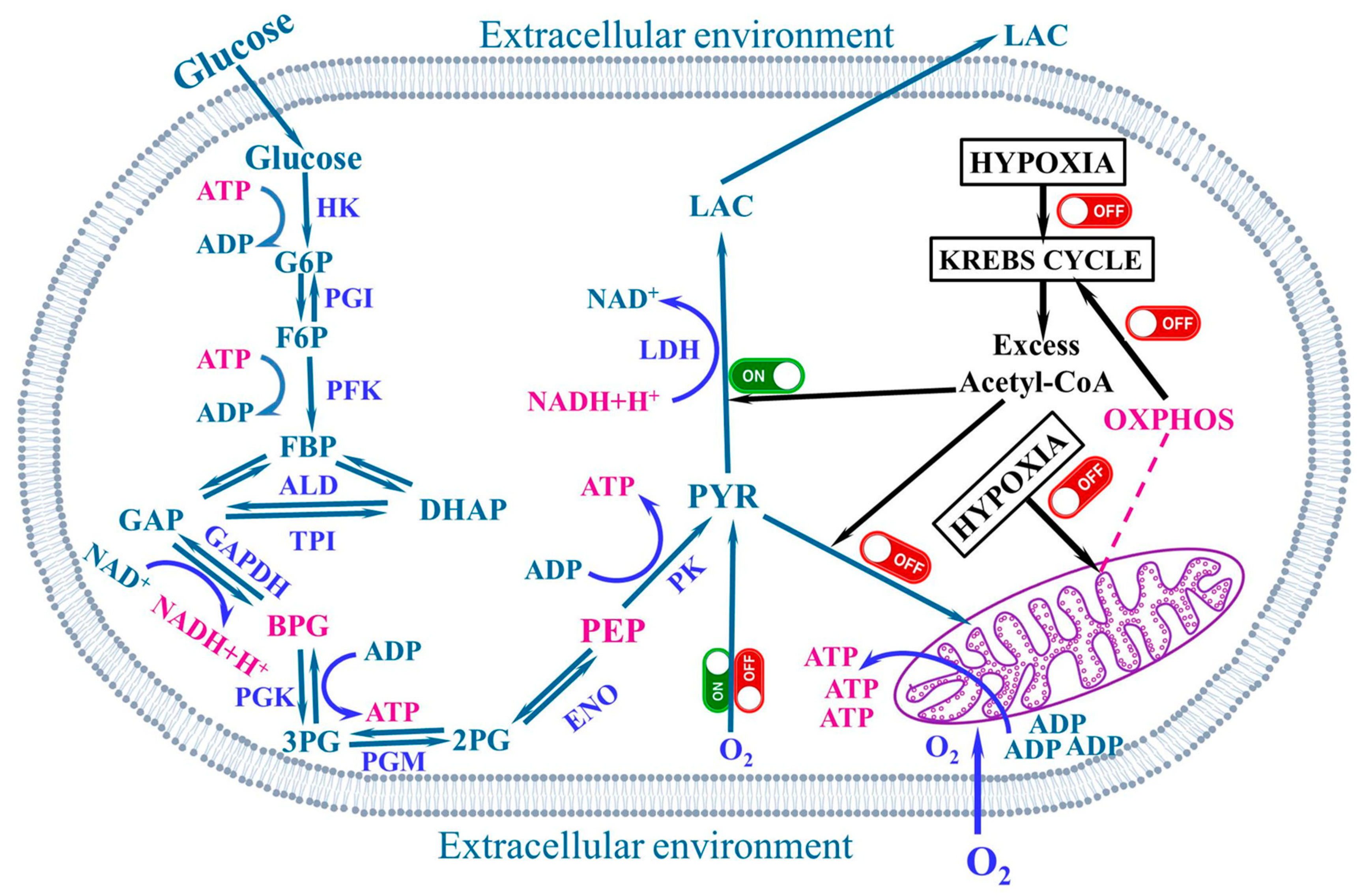
4. The Warburg Effect

A century ago, Otto Warburg and his colleagues observed that growing ascites cells converted most of their glucose to lactate, even under O2-rich surroundings [71]. He thought that such altered metabolism was specific to cancer cells, and that it arises from mitochondrial deficiencies that inhibit their capability to efficiently oxidize glucose to CO2. He concluded that the existence of dysfunctional mitochondria is one of the causes of cancer [72]. However, injured mitochondria have been shown not to affect aerobic glycolysis in most tumour cells; mitochondria in cancer cells are not damaged, but simply dysfunctional [73]. The metabolism of the cancer cell is altered and involves augmented glucose uptake and glucose fermentation to lactate [74]. This condition is known as the Warburg effect or aerobic glycolysis and is observed even in the presence of fully functional mitochondria, or even in the presence of oxygen [71,75]. Since respiration can maintain tumour viability, it was thought that these cells can be killed by depriving tumour cells of energy, so both glucose and oxygen should be removed [76]. However, Herbert Crabtree reported the heterogeneity of glycolysis in various tumours. Therefore, variable intensity of respiration in tumours was discovered [77]. Crabtree established that there is also variability in fermentation, probably due to environmental or genetic influences.
Nevertheless, Warburg proposed later that the origin of aerobic glycolysis is dysfunctional mitochondria [78]. Yet, Efraim Racker showed that tumours have respiratory capability. He advanced his own hypothesis about the Warburg effect by studying intracellular pH imbalances that disrupt the ATPase activity [79]. It has also been observed that aerobic glycolysis can be controlled by growth factor signalling. However, the identification of genes with a potential role in oncogenesis led to the conclusion that aberrant growth factor regulation could be the initial event in tumorigenesis [80,81]. Nonetheless, WE is necessary for tumour growth [3,4]. Therefore, targeting both aerobic glycolysis and mitochondrial metabolism might be required in cancer therapy [82,83,84]. Nevertheless, the functions of the Warburg effect have remained controversial for a long time.
The Warburg effect confers direct tumour cell signalling functions [85,86,87,88]. Thus, a direct contributory role of glycolytic metabolism in stimulating carcinogenesis through this signal transduction affecting other cellular processes is suggested. It has been thought that aerobic glycolysis may offer some advantage as it provides a favourable tumour microenvironment for cancer cell multiplication [1,89,90]. Under certain conditions, the Warburg effect could be the choice of an energy metabolism based on high glucose consumption.
Most tumour mitochondria are functional and are therefore able to perform oxidative phosphorylation. Nevertheless, mitochondrial metabolism in proliferating cells seems to be directed to macromolecular syntheses. Warburg and his colleagues did not consider such a possibility [1]. However, some authors consider that the Warburg effect is an initial event in carcinogenesis, being a direct result of an oncogenic mutation, which occurs before abnormal cell multiplication, and also in benign and early-stage lesions [91,92].
The Warburg effect was extensively investigated from multiple points of view [93]. Thus, metabolic alteration was understood as a necessity for rapid multiplication. It instantly generates energy in the form of ATP molecules, supports the biosynthesis of macromolecules and maintains the redox state of cells. Processes such as pH modification of tumour microenvironment, the stabilization of hypoxia-inducible factor (HIF), some mutation of tumour suppressor genes, and dysfunctions of mitochondria have been discussed. In addition, selective targeting by miRNA, altered glutamine metabolism and post-translational modifications were also investigated. These authors considered that a holistic understanding is needed to discover novel metabolism-based therapeutic strategies to hinder the Warburg effect and cancer advancement. Other authors found that the Warburg effect stimulates cancer metastasis and changes the tumour microenvironment; it may play a role in promoting angiogenesis, formation of cancer-associated fibroblasts, immune suppression, and drug resistance [94]. High uptake of glucose by cancer cells reduces considerably its accessibility in the tumour microenvironment, which results in a low-glucose extracellular environment and disturbs the activity of immune cells [74]. Moreover, tumour cells release high amounts of lactate, which induces an increase in the acidity of the microenvironment. Lactate can be utilized by some non-tumour cells in the liver to produce glucose with high energy consumption [95]. An acidic tumour microenvironment stimulates local invasion, and then metastasis and diminishes the anti-tumour action of immune cells [96,97].
In cancer cells, respiratory function decreases, and an increase in glycolysis proportional to the increase in the growth rate is observed [98]. However, decreased cellular respiration is not obligatory for an increased rate of cell proliferation.
Myc and HIF-1 activate the Warburg effect in reaction to growth factors and hypoxia. It is an important metabolic and energetic process that meets the requirements for fast gene replication [99]. Paradoxically, cancer appears to be a normal physiological phenomenon that follows precise rules, but it is also a degeneration, a dysregulation caused by a multitude of factors: lifestyle, diet, carcinogens, etc.

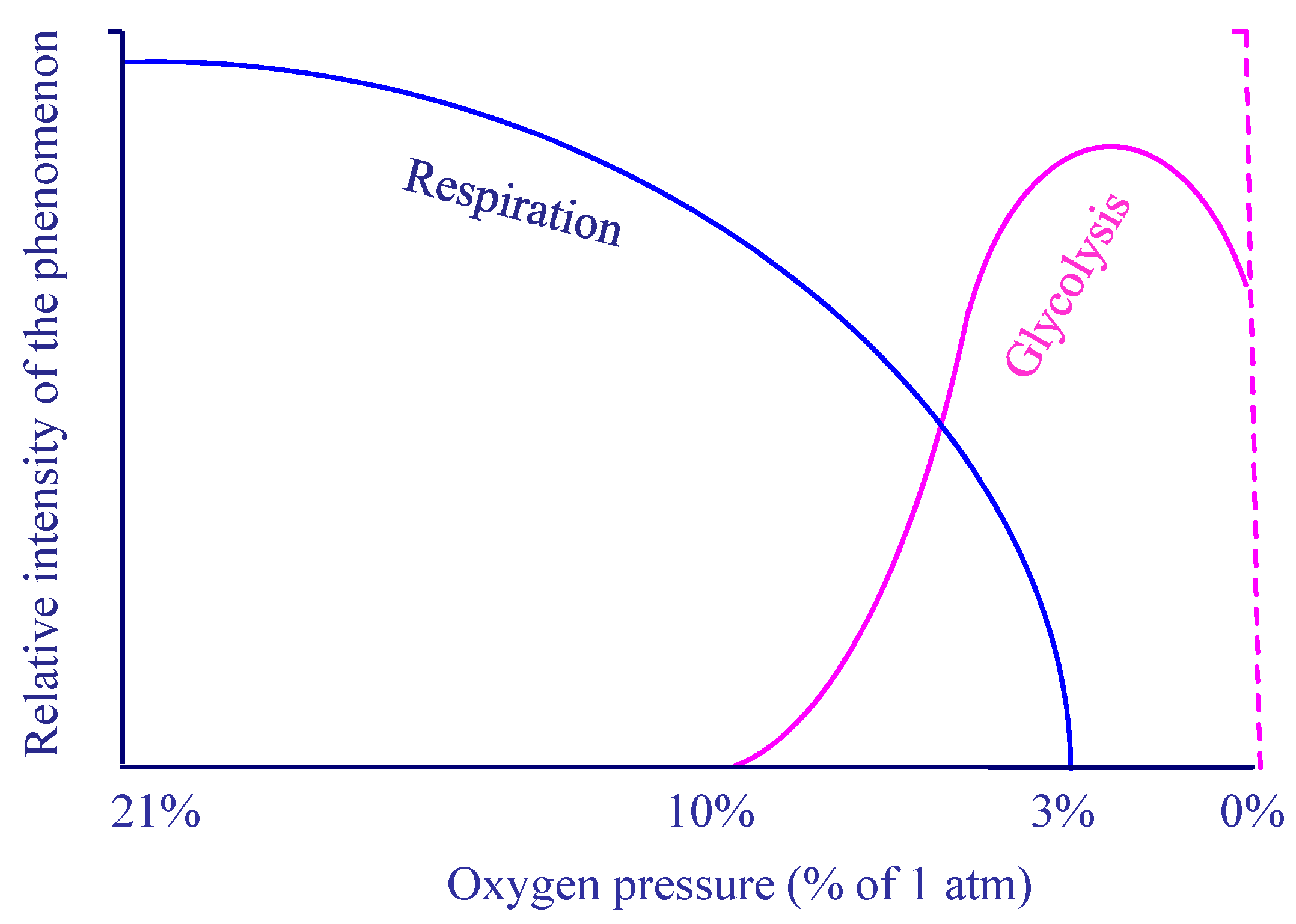
5. The Aerobic Glycolysis

Glycolysis is a primitive metabolic pathway that is essential for rapid multiplication of cancer cells, tissue regeneration, but also for growth of bacteria and viruses [99]. Aerobic glycolysis, which occurs not only in cancer cells, can be defined as an exaggerated increase in glucose consumption compared to oxygen supply, even when oxygen levels and delivery in the blood are sufficient to meet demand [100]. Therefore, this type of glycolysis is uneconomical and ATP generation is very low compared to the ATP produced by respiration [101,102]. Nevertheless, the amount of ATP synthesized in a given period of time is similar in both forms of glucose metabolism [103]. Therefore, the reason why the cancer cell uses aerobic glycolysis should be investigated and a suitable explanation should be found for this inherent difference in kinetics. A very simple explanation would be that there is a precise ratio between the concentration of ADP and ATP. Thus, decreasing the concentration of ADP will cause a phosphorylation reaction of ADP in order to keep the ratio of adenosine-di and triphosphate as constant as possible. It has been hypothesized that cells with higher glucose consumption, albeit with lower efficiency in ATP production, may have an advantage when competing for common and restricted energy resources [104,105]. On the contrary, we think differently: low oxygen concentration can lead to glycolysis [106]. Thus, it has been shown that when the cellular environment is altered to greatly increase ATP requirements, aerobic glycolysis increases rapidly, and oxidative phosphorylation remains constant [107]. In such cases, the aerobic glycolysis is considered an adaptive process to sustain the conditions for the biosynthesis of macromolecules and other compounds required by the uncontrolled multiplication. Therefore, increased glucose uptake is a carbon source for syntheses required to sustain tumoral growth [72,108,109,110]. The aerobic glycolysis is also required to support the rapid generation of ATP required to sustain chemical synthesis. However, the ATP requirement for cell growth and division is much lower than required, and ATP demand may never reach threshold values during cancer cell growth [111]. Similar mechanisms are also observed in other cell types linked to a rapid demand for ATP are also present in tumour cells. Thus, fast ATP synthesis based on creatine kinases in muscle is manifest in most tumour cells [74]. Compounds resulting in glycolysis are required for nucleotide, lipid, and protein synthesis [112,113,114,115]. Proliferating cells have a greater need for NADPH or NADH [116]. Increased synthesis of the reducing equivalents implies a higher utilization of glucose, which is then employed in the biosynthesis of lipids, amino acids and other biomolecules [1]. It was considered that the role of aerobic glycolysis is to regenerate NAD+ in the reaction of NADH+H+ with pyruvate, which produces lactate, the final product of aerobic glycolysis [117,118]. Lactate is not just a by-product of glycolysis, but has an important role in tumour metabolism, as identified by the Warburg effect studies [119]. Lactate plays a major role in cancer cell proliferation, but is also involved in inflammation, neural excitation, and many other biological processes.
NADH is generated in the reaction catalysed by glyceraldehyde phosphate dehydrogenase and is oxidized to NAD+, thus keeping glycolysis active. Glycolysis enables 3-phosphoglycerate to convert to serine for the production of NADPH and nucleotides [120]. NADPH homeostasis is regulated by several metabolic enzymes that undergo adaptive changes in cancer cells [121]. It is thought that modulating NADPH homeostasis in cancerous cells could be an effective strategy to eliminate them.
Aerobic glycolysis maintains a fertile environment that supports rapid biosynthesis to sustain multiplication and proliferation [122]. Cancer cells use aerobic glycolysis for energy metabolism, and a method to deprive malignant cells of glucose would prevent these cells from surviving and induce apoptosis in several types of cancer, which could be the basis of a potential treatment [123]. Cancer cells use glycolysis as an energy source although oxygen is present, this changed metabolism may provide a selective benefit for survival and growth, consistent with the Warburg effect. In addition, several molecules, such as NADPH, HIF, PKM2, and others, are important for the reproduction of cancer cells in the abnormal hypoxic medium.
It was also suggested that aerobic glycolysis is a pathway to support biosynthesis [124,125]. Although ATP production is inefficient, it can come at the cost of maintaining anabolic pathways, such as those involved in nucleotide and lipid metabolism. There may also be a limited number of mitochondria; thus, the necessary energy and biomass beyond mitochondrial capacity must be produced from aerobic glycolysis [126,127,128]. Therefore, aerobic glycolysis is considered to support biomass production when ATP production is limited. There is an apparent correlation between aerobic glycolysis and cell proliferation. The demand for NADPH is higher than the ATP requirement for biosynthesis. However, in aerobic glycolysis, the carbon atoms are not sequestered but are liberated extracellularly as lactate [2,111]. Acidosis can be beneficial for cancer cells; protons, H+, secreted by tumour cells may be liberated into the environment and modify the tumoral–stroma interface, permitting increased invasion [129]. Tumour-derived lactate has also been shown to contribute to tissue-associated M2 macrophage (TAM) polarization [130].
Glucose availability seems to be a result of intensive competition between resulted tumours and tumour-infiltrating lymphocytes (TILs) [131,132,133]. Intense aerobic glycolysis limits the availability of glucose to tumour-infiltrating lymphocytes, which need abundant glucose for their physiological functions [134,135,136]. Consequently, evidence is sought that inhibition of aerobic glycolysis in the tumour would allow increased glucose supply to TILs, thereby stimulating their function to eradicate tumour cells [137,138]. All these observations may suggest that malignant cells are in contact with cells in the immune system to sustain pro-tumour immunity [139,140,141,142,143].
Lactic acid plays a key role as it is capable of translocating through cell membranes, contributing to the cell-pH state, as well as influencing the complex immune response due to acidosis of the tumour microenvironment [99]. Even working brain tissues partly oxidize glucose and produce some lactic acid [100,144,145,146,147]. Therefore, aerobic glycolysis occurs normally when cells are stressed. Aerobic glycolysis occurs in astrocytes, where the Crabtree effect coincides with the Warburg effect. At the same time, neurons use both glucose and lactate, and there is a balance between glycolysis and respiration. This leads to the activation of Warburg and Crabtree effects in brain tissue, resulting in a high degree of aerobic glycolysis, indicating stimulation of astrocytes to generate neuronal ATP.

6. Copper and Cancer

Copper (Cu) is involved in numerous cellular processes, which include mitochondrial respiration, anti-oxidative defences, redox signalling, autophagy, kinase signalling, and regulation of protein quality [148]. Specific abnormalities of copper metabolism appear to have clinical potential as prognostic and predictive biomarkers [149]. Cu2+ ions are also capable of binding to growth factors, cell signalling proteins, or even structural proteins [150]. These ions can regulate the activity of several proteins; thus, many signalling metabolic reactions are dependent on copper. Mitochondria also play an essential role in copper homeostasis, which is important for mitochondrial physiology [151]. Cu is a component of cytochrome c oxidase, which is present in the respiratory chain in mitochondria [152]. Therefore, Cu is involved in energy production via oxidative phosphorylation [153]. In addition, copper is present in cell lysosomes, and some metallo-reductases maintain it in the Cu(I) form because lysosomes are an oxidative environment [154,155]. Such reductases are mainly located in the intracellular vesicles [156,157]. They are involved in the regulation of cell proliferation and apoptosis.
Glutathione (GSH) and metallothioneins (MTs), which are cysteine-rich cytoplasmic proteins, are greatly engaged in intracellular storage of excess copper [158]. GSH is implicated both in numerous mechanisms of metabolism and in the transfer and removal of metal ions, including copper ions, as Cu(I)–GSH complexes [159,160]. These complexes are thought to be related to the exchangeable pool of cytosolic copper [161,162].
Changes in Cu levels or in the Cu: Zn ratios have been observed in several forms of cancer [163,164]. Nevertheless, the Cu: Zn ratio changes with aging, inflammation, nutritional status, and OS. Increased copper concentration is accompanied by diminished levels of zinc in bladder cancer [165] and other forms of cancer [166,167,168,169]. However, certain authors testified that there are decreased copper levels in colorectal and breast cancers [170,171].
Increased levels of copper have been reported in tumour areas [172,173]. Copper is involved in proliferation and angiogenesis, two phenomena seen in tumorigenesis and cancer development. In addition, specific copper accumulation was reported in cancer cells themselves [174]. Thus, high levels of Cu were reported within the tumoral cells of breast cancer [175]. It is likely that copper ions induce the formation of secondary tumours by activating some enzymes implicated in cell multiplication [176]. Moreover, increases in serum copper in cancer pathology were sometimes correlated with cancer stage. In addition, in the case of patients who are resistant to chemotherapy, increased levels of serum copper have been measured [176]. Data on different types of cancer on this subject are contradictory. We suggest that a link between copper level and pH may exist. The isotopic 63Cu/65Cu ratio in the serum of tumour patients also seems to be altered [74], with increasing levels of the lighter isotope. These changes could be due to increased glycolysis and lactate formation. Furthermore, the Cu isotopic ratio could be used as an early diagnostic biomarker for cancer [176].
Some copper-related proteins, such as ATP7B and Ctr1, have been found to increase in breast cancer [177]. Dysregulation of several proteins involved in copper metabolism has an influence on cell migration and metastasis formation. Thus, Atox1 protein is elevated in several malign tissues [178,179]. It may also promote inflammatory neovascularization by acting putatively as a transcription factor and as a copper chaperone [180].
Cu-dependent LOX metalloenzymes play a significant function in tumour metastasis [181]. Thus, cancer cells produce LOX protein to promote collagen cross-linking and fibronectin biosynthesis. However, the pathway by which copper ions are delivered to copper-dependent LOX metalloenzymes is still unclear. ATP7A/B protein is used to limit copper toxicity and up-regulates cancerogenic enzymes, such as LOX and LOX-like proteins [182,183].

7. Cancer and Lifestyle

Only 5–10% of cancers are thought to be caused by inherited genetic defects. Numerous cancers are not inherited and are caused by various agents (environmental factors, physical factors, and hormones) [184]. Environmental factors encompass lifestyle (nutrition and overweight, over-smoking, over-drinking, stress, physical inactivity); physical factors (environmental pollutants, virus, bacteria and parasitic infections, ionizing and nonionizing radiation); as well as socio-economic and attitudinal factors. Therefore, most cancers have multiple possible concurring causes.
A healthy lifestyle includes a healthy diet, weight control, physical exercise, reducing alcohol drinking, and smoking avoidance [185]. About 25–30% of total cancer deaths are caused by tobacco, 30–35% are diet-related, approximately 15–20% are caused by infections, and the rest are attributable to other agents, such as radiation, stress, environmental pollutants, etc. In spite of medical progress, cancer incidence is expected to increase substantially in the near future [186]. It is also thought that all these carcinogens associated with lifestyle factors and all chemopreventive agents are connected with the long-term inflammation. Chronic inflammation is strongly associated with the tumorigenic trajectory, as evidenced by multiple findings [187]. Carcinogens activate while chemopreventive agents suppress NF-κB activation, which is a mediator of inflammation.
All living beings are constantly under stress, which is the nonspecific response of the body to any demand made upon it [188]. Unlike eustress or adaptive stress, distress affects immune responses, generally by exerting a suppressive effect. The stress-induced increases in tumour size are most probably a consequence of immunosuppression [189,190]. Some other authors showed that psychological stress is weakly associated with increased mortality from colon cancer [191]. However, chronic stress is associated with neuroendocrine abnormalities that can up-regulate inflammation and down-regulate protective immunity. Thus, the affected immune cells may not effectively control cancer cells and act as stromal cells, communicating with the tumour microenvironment and circulating cancer cells to promote tumour growth mechanisms, invasiveness, extravasation into the circulation and metastasis [192].
The response to physical and social stress involves a complex reaction at the cellular and molecular level [193]. NF-κB plays a key role in the cellular response to stress. Thus, stress up-regulates some genes, such as transcriptional genes that control cell growth, chromatin structure, cell cycle activation, and enzymes involved in nucleic acid and protein biosynthesis. Under stress, cell cycle inhibitors, the NF-κB inhibitor, apoptosis-related genes, antiproliferative cytokines, and Apo J are down-regulated. NF-κB is activated in response to many inflammatory factors such as carcinogens, chemotherapeutic agents, cytokines, hormones, mitogens, viral products, eukaryotic parasites, endotoxin, fatty acids, metals, radiation, hypoxia, and psychological, ROS, and chemical stresses [194]. Drugs that prevent cancer or inflammation have been proven to suppress NF-κB up-regulation. Curcumin and other polyphenols inhibit NF-κB, p53 pathways and potentiate Nrf2 activation [195].
Protein p53, which is a universal sensor of genotoxic stress, coordinates the cellular response to various genotoxic stimuli, determining cell death or survival [196]. ROS also appear to be involved in p53 signalling, being effective activators of p53 function. Some chemotherapeutic agents activate p53 due to their involvement in ROS production. However, ROS, generated following p53 activation, play a role in mediating apoptosis [196].
The role of transcription factors nuclear factor erythroid 2–related factor 2 (Nrf2) and nuclear factor-κB (NF-κB) related to OS was also investigated [197]. Thus, in response to OS, the transcription factor Nrf2 up-regulates the expression of antioxidants and detoxifying enzymes involved in antioxidant protection, being considered as the master regulator of redox homeostasis. The activation of the transcription factor NF-κB leads to the production of proinflammatory cytokines and chemokines, prostaglandins, free radicals such as NO and superoxide anions, and ultimately leads to chronic inflammation [198,199,200,201].
A healthy lifestyle has been associated with a substantial reduction in the overall risk of developing liver cancer [202]. Lifestyle improvements to combat cancer have long been recommended; however, there has been a renewed appreciation of their importance and relevance given the growing number of cancer survivors seeking alternative options for prevention and secondary cure [203]. Tumour survivors often face drug toxicity, also being at risk of cancer recurrence, a second primary cancer and high cause of mortality [204,205,206,207].
Most cancer survivors live with higher risks of complications and relapses, lower quality of life and reduced life expectation. There is an immediate need to improve cancer survivorship by improving lifestyle beyond clinical interventions. The association between a sedentary lifestyle and worsened survival after cancer was also noticed [208,209]. Physical exercises may be positively correlated with the control of tumour biology through specific effects on intrinsic tumour factors, such as Warburg-type high glycolytic metabolism [210]. Tumour metabolism can be selectively influenced by single exercise as well as by regularly applied exercise, depending on the intensity, duration, frequency, and mode of exercise. High intensity anaerobic exercise has been shown to inhibit glycolysis, and some animal studies have shown that the effects on tumour growth may be stronger compared to moderate intensity aerobic exercise. Of course, early detection and treatment can result in growing prevalence of survivors of cancer.
Stress-prone personalities, unfavourable coping styles and negative emotional responses, and poor quality of life were related to higher cancer incidence, poorer cancer survival, and higher cancer mortality [13]. Site-specific analyses indicate that PSF are associated with a higher incidence of lung cancer and poorer survival in patients with breast, lung, head and neck, hepatobiliary, and lymphoid or hematopoietic cancers. These analyses suggest that stress-related PSF have an adverse effect on cancer incidence and survival, although there is evidence of publication bias and results should be interpreted with caution. Some clinical studies have shown that psychological and/or pharmacological inhibition of excessive adrenergic and/or inflammatory stress signalling, especially in conjunction with cancer treatments, would improve prognosis [211]. There are some critical phases of cancer progression that are more sensitive to stress. Therefore, there is a need to focus on more vulnerable populations using individualised pharmacological and psychosocial approaches [212]. Addressing psychosocial stressors also raises the issue of distinguishing between human and laboratory animal cancers [211].

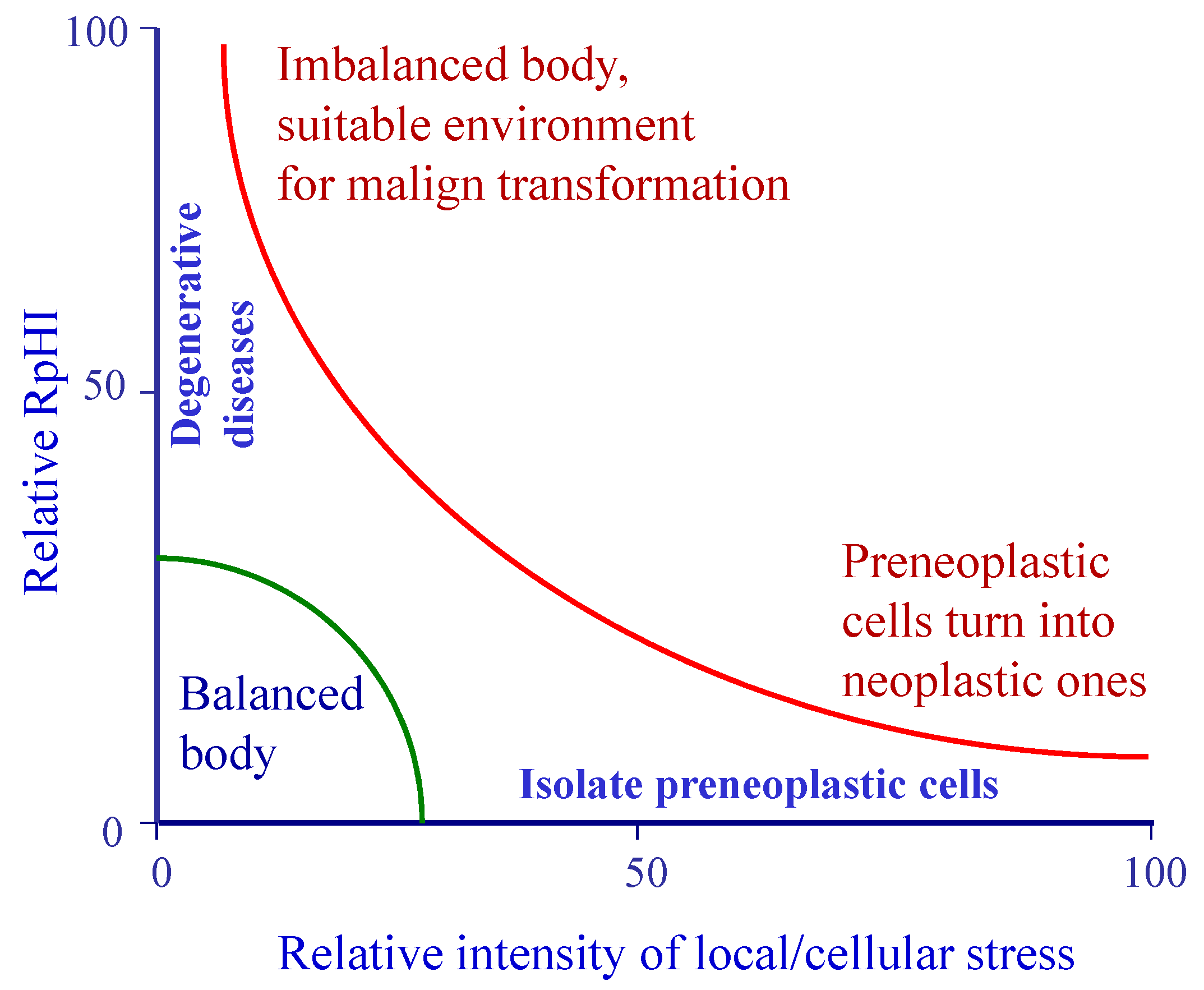
8. Respiration and pH Imbalance

In numerous pathologies, including cancer, an impaired respiration can be observed, along with a change in pH, due to a multitude of stress factors. In order to suggestively explain the imbalance between respiration and glycolysis, which leads to pH alteration, or more precisely, the imbalance of both respiration and pH of body fluids, the so-called respiratory and pH imbalance (RpHI) has been introduced [106]. There are striking similarities between the metabolic profiles of cancer cells and those of rapidly multiplying normal cells, such as aerobic glycolysis and increased biosynthesis [2]. The role of aerobic glycolysis in malignant growth should be elucidated, including whether there is metabolic reprogramming that may be related to chronically sustained proliferation [213]. However, altered metabolism is a hallmark of cancer, and metabolic reprogramming in cancer cells is seen in the main pathways of central carbon metabolism [214]. Different cancers are characterised by an intra-tumour hypoxia resulting from deregulated cell proliferation [215]. Tumour hypoxia is associated with poor prognosis and resistance to therapy [216]. Physiological responses triggered by hypoxia can impact all critical aspects of cancer progression, including immortalization, transformation, differentiation, genetic instability, angiogenesis, metabolic adaptation, autocrine growth factor signalling, invasion, metastasis, and resistance to treatment. Hypoxia-inducible factors (HIF) are key oxygen sensing factors that mediate the response to low oxygen pressure [217]. These transcription factors regulate cellular adaptation to hypoxia and protect cells by reacting acutely and inducing the production of endogenous metabolites and proteins to promptly regulate metabolic pathways. Therefore, hypoxia itself could be the trigger for the induction of aerobic glycolysis without any mitochondrial damage [218,219]. HIF1α activates via Activin/nodal signalling, and its increased expression redirects ATP production from oxidative phosphorylation to glycolysis. In addition, it has been reported that HIF1α-dependent expression of BNIP3 (a member of the apoptotic Bcl-2 proteins) promotes mitophagy to control ROS production and ROS-induced cell death [220].
Hypoxia, which is closely related to glycolysis, is common during carcinogenesis; it is associated with functional and structural modifications in proliferating cells [221]. Hypoxia also underlies the energetic processes of various activities in brain-like alertness, sensory processing, cognition, and physiological conditions. Its specific functions performed in cells are still less understood [100]. Aerobic glycolysis is characterized by excessive glucose utilization relative to oxygen consumption, even when oxygen levels and availability are adequate. Propranolol blocks aerobic glycolysis, including adrenal release of epinephrine, brain signalling through the vagus nerve, and an enhanced liberation of norepinephrine in the locus coeruleus. Sugar utilization is stimulated by norepinephrine and not oxygen consumption.
Glycolysis stoichiometry does not allow both biomass production and lactate generation, and NAD+ regeneration by lactate alone is not possible. It is therefore hard to see how the Warburg effect can directly stimulate biosynthesis. In general, cells allocate half of their genes to synthesize proteins engaged in glycolytic processes [208,222]. However, the cellular biosynthesis programs require lower amounts of protein. Therefore, the cost to produce proteins for aerobic glycolysis may be higher than the cost of producing proteins needed for biosynthesis. There is evidence that mitochondrial functions run concurrently with the Warburg effect, and thus, during aerobic glycolysis, mitochondrial activity is not impeded. It is thus unclear whether the Warburg effect functions to facilitate the various biosynthetic pathways. However, increased consumption of glucose to liberate lactate lowers the pH in the microenvironment [96].
In principle, RpHI can be regarded as a physiological reaction against any stress agents, and if the stressors are strong, an oxygen crisis within the body occurs, the busiest cells divide faster and faster, producing first preneoplastic cells and, in time, malignant tumours. The tumours acquired in this way, as well as their fermentation products, may disturb the normal functions of most cells and tissues in the body [223].
Since there are many forms of cancer depending on the organ or organs affected, the prognosis of the disease, and its stages, there are many forms of RpHI. Thus, a carcinogen can cause irritation of a tissue, followed by chronic inflammation and malignancy, while slow debilitation can lead to degenerative disease and, ultimately, to cancer. One can also speak of an increased concentration of glucose in cells and body fluids which can ferment in the presence of insufficient amounts of oxygen in the tissue although the patient’s breathing may appear normal. Thus, epinephrine (adrenaline) secretion during stress may explain the increased flow of glucose into certain active tissues, and higher concentrations in relation to oxygen intake may lead to fermentation and lactic acid formation. Therefore, a full description of RpHI requires further work.
Thus, under hypoxic conditions, overstressed cells, which receive less oxygen than necessary, undergo anaerobic fermentation to produce adenosine triphosphate (ATP). This process is associated with excessive multiplication and, ultimately, tumour growth. Indeed, severe hypoxia due to profoundly low arterial O2 content (hypoxemia) results in hypercapnic and metabolic acidosis, developed together with extensive lactate generation, with pH decreasing to under 6.8 [224]. Because hypoxia is dependent on the magnitude and duration of action of the causative factors, human and animal organisms can only compensate for hypoxia if the causative agents stop acting for a long time. Normally, various stress agents, such as physical and chemical stressors, viruses, other infectious agents, hormones in excess, but also, long-lasting anxiety, emotions, conflict states, etc., are able to affect the body as a whole [225,226,227,228,229,230].
Confronted with the external environment, the living organisms have several co-ordinate physiological processes to keep their internal states of equilibrium [231]. The living bodies react against aggression using metabolic energy obtained by the oxidation of organic substances, including organic acids in the mitochondria [232]. Under normal conditions, when the supply of oxygen is sufficient, cells can carry out aerobic respiration [233,234]. Hence, when busy, cells produce the energy they need from the food stores, including the organic acids they possess. During the Krebs cycle, carbon dioxide, of a weaker acid type, is released, normally outward [235,236]. Since stronger organic acids, such as succinic, malic, 2-ketoglutaric, and oxalylacetic acids, are replaced by carbon dioxide; the cell milieu tends to be more alkaline, i.e., just blood and urine [237]. The tendency of blood alkalization entails retention of carbon dioxide so that the pH of blood should alter as little as possible. The retention of carbon dioxide in blood does not allow the oxygen to shift at a normal rate in lungs [238,239]. As a consequence, oxygen pressure in blood and tissues decreases although it stays normal overall. However, oxygen may reach quite a low level without affecting cell breathing in any drastic way. Unfortunately, the busiest cells in the body need more oxygen. As oxygen partial pressure decreases lower, these cells receive less oxygen than they need, and fermentative processes develop alongside with breathing.
In fact, hypoxia is the natural environment in which DNA auto-replication and transcription take place in vivo in all eukaryotes [240,241,242,243]. Nuclear division unfolds anaerobically using the energy produced by glycolysis [244,245,246]. Consequently, the cells forced to manifest themselves in rather anaerobic conditions will divide more intensely [247]. Aerobic breathing, which provides cells with a great amount of energy, creates the necessary conditions for the existence of fine structures of the cells, and the specific functions run unimpaired [248]. Lack of oxygen, even partial, causes rupture of these structures, leading to the gradual disappearance of specific functions of cells as well as contact inhibition. At the same time, lack of oxygen entails cell-division to a greater extent than necessary for the tissue in question. It follows the first stage of the RpHI, which affects cell respiration and division. Nevertheless, the organism possesses buffer systems, lung ventilation, and kidney mechanisms to control the concentration of hydrogen ions within the cellular milieu (Figure 1) [2,249]. Alveolar ventilation is responsible for carbon dioxide elimination [250]. Mild acidosis occurs primarily when alveolar airflow is reduced or if CO2 generation is elevated. However, the organism has several compensatory systems to minimize a decrease in pH. For example, non-oxygenated haemoglobin easily buffers the blood environment to prevent significant pH alterations. Normally, carbon dioxide, a stimulant of respiration, induces an increase in minute ventilation to normalize the pH by eliminating increased quantities of CO2. Unfortunately, this effect is mitigated when CO2 concentrations remain elevated for more than a few hours. The kidneys are also capable of controlling both the blood pH and some other blood parameters. However, this process is slow and lasts for several hours or days. In fact, renal compensation begins in 6–12 h, but maximal compensation occurs in 3–5 days. The kidneys enhance the expulsion of protons, predominantly as ammonia. If the stress agents act continuously, the blood will become slightly more alkaline than usual, and the blood oxygen concentration will be lower than normal. Getting less oxygen than they need will lead to anaerobic fermentation in the overstrained cells. The overstrained cells also cause a lower content of NADH+H+ and NADPH+H+ a higher content of NAD+ and NADP+. Therefore, a decrease of the oxidation reduction potential will occur as well. The quantity of sulfhydryl groups in the blood and tissues also decreases. A marked decrease in succinic dehydrogenase and slight increase in cytochrome oxidase levels could be found, suggesting the alteration of the Krebs cycle. For this reason, a cell with excessive fermentation will not reach an upper energetic state if neighbouring cells and blood do not interfere with its metabolism. The second stage of the RpHI is reached when lactic acid is produced due to hypoxic conditions. In this case, the CO2 concentration may decrease; however, part of the produced carbon dioxide is not removed because the cell content may remain slightly alkaline in spite of the lactate production. Again, the kidneys should control the hydrogen concentration in blood, releasing acidic species, such as ammonium ions or phosphates, into urine [251]. The process is complicated by the existence of lactic acid in blood which decreases blood pH, while intracellular pH of overstrained cells is increased. However, if the stress agents act continuously, the neighbour cells involved in curing or rebalancing the overworked cells will also work hard while being overstrained and deprived of oxygen. There follows a third stage of the RpHI in which a real state of illness (infections, viruses) occurs. A very special balance between the two types of cells (attacked cells and neighbour ones) is established. The blood pH value is a little altered. If the stress agent is very strong, it can be lethal to the organism, and such a case does not reach the cancer stage. It is the case of microorganism- or virus-induced diseases, which, if untreated, can have a bad prognosis. On the contrary, a long-standing action by the stress agents may cause a slow shift in this imbalance even if the action is mild, resulting in a stepwise decrease of blood pH value. Long-standing infectious pathologies can thus lead to an RpHI, which creates the conditions for either the transformation of normal cells into preneoplastic cells, followed by the preneoplastic to neoplastic pathway, or the multiplication of malignant cells. It is well-known that only bodies with a significant amount of morbidity may become cancerous. Most cells become glycolytic, while fewer remain normal but overstressed. This pathway leads to the fourth stage of the RpHI, which is that of tumour formation.
Respiration and glycolysis are two independent biochemical processes that may occur simultaneously in the living cell (Figure 2). Given an oxygen concentration of 10 per cent or more in the surrounding atmosphere, respiration occurs in the living cell. Given a concentration below 3% of oxygen, fermentation (glycolysis) will occur. Both processes occur in the range from 3% to 10% oxygen (Figure 2). Growth and lifespan of human diploid cell strains at oxygen levels below 20% is increased, and an enhancement of around 25% in the lifetime of both cell types has been achieved by long-term cultivation under 10% oxygen [252]. It is well known that there is extremely low O2 content in growing tumours [253].
Indeed, if mitochondrial respiration in tumour cells were down-regulated, the accumulation of substrates from the Krebs cycle could also serve as a signal to stimulate glycolysis [254].
Whenever a cell within the metabolic system (muscle, liver, kidney, lung, etc.) uses oxygen at a faster rate than can be provided by the circulatory system, the cell begins to function anaerobically, reducing the pyruvate to lactate instead of oxidizing it further, as would happen if oxygen supplies were adequate [255,256,257]. Moreover, excess glucose can be glycolytically converted to lactic acid if the glucose: oxygen ratio increases. Lactate thus accumulates in that cell, diffuses through the bloodstream, and eventually reaches the liver, where it is re-oxidised to pyruvate and converted to glucose via the gluconeogenic pathway [258].
In fact, prokaryotic cells produce energy for their needs by glycolysis in the absence of oxygen. Glucose is thus metabolized into lactate or ethyl alcohol, depending on the cell type. In the cytoplasm of eukaryotic cells, including human cells, glycolysis takes place with the release of pyruvic acid or lactic acid [259,260]. These acids enter the mitochondria where they are degraded to CO2 and H2O, with the formation of reduced forms of NADH+H+ and FADH2. In the respiratory chain, the hydroxyl-rich compounds are oxidized and the chemical energy they contain is liberated and stored as ATP molecules. If there is insufficient oxygen, then the respiratory chain is blocked and NADH+H+ and FADH2 are no longer oxidized. Under these conditions, no new quantities of reduced NADH+H+ and FADH2 are formed; lactic and pyruvic acids accumulate in cells or are excreted extracellularly. This is a very simple mechanism for switching from respiration to glycolysis [258,261,262,263].
Figure 3 better suggests the molecular and biochemical pathways linking respiration and glycolytic processes to oxygen supply and hypoxia. The human cell is made up of the nucleus, which plays a role in the division and transmission of genetic information, the cell membrane, through which oxygen, glucose and other nutrients flow, and the cell organelles. Among the latter are mitochondria, which have an essential role in energy production in the form of adenosine triphosphate (ATP) molecules, oxidative degradation of fatty acids, metabolism of pyruvic acid and acetyl-Coenzyme A from fatty acids with the formation of NADH+H+ and FADH2 molecules. These reduced forms of nicotinamide dinucleotide (NAD+) and flavin adenine dinucleotide (FAD) compounds are involved in cellular energetics and multiple biochemical syntheses and constitute the fuel from which ATP is formed in the so-called electron transport chain (ETC). Glycolysis takes place in the cell cytoplasm and not in the mitochondria.
Glucose is degraded via the glycolytic pathway to pyruvate, which is either used as acetyl-CoA in mitochondria or released into the cytoplasm as lactate (Figure 3). Most of biochemical reactions are reversible. Several enzymes catalyse the conversion of glucose to pyruvic acid (PYR) with the release of two molecules of ATP. Pyruvic acid then reacts with Coenzyme A to form acetyl-CoA, which is broken down in the Krebs cycle with the release of CO2, while its hydrogen atoms reduce NAD+ and FAD to NADH+H+ and FADH2. These energy-rich molecules are used in the electron transport chain to form ATP in the presence of molecular oxygen. However, if oxygen is insufficient, the reduced NADH+H+ and FADH2 species cannot be used in ETC to generate the energy-rich molecules of ATP. The Krebs cycle is thus blocked, and acetyl-CoA is no longer needed. As a result, the pyruvic acid formed in the glycolytic process is no longer required in the mitochondria and is reduced to lactic acid by the existing NADH+H+. In this way, glycolysis occurs in the presence of insufficient oxygen levels to provide the required ATP, but glucose consumption is greatly increased for the same ATP concentrations required by the cells. Furthermore, the increase in NADH+H+, produced in the Krebs cycle due to hypoxia, inactivates phosphatase and tensin homolog (PTEN), which is encoded by the PTEN gene [264]. PTEN is involved in the regulation of the cell cycle, preventing cells from growing and dividing too rapidly. Thus, a close link between the Warburg effect and metabolic alterations in cancer cells has been found; it may gain a survival advantage and withstand therapeutic agents. The microenvironment of solid tumours is characterised by hypoxia, high lactate levels, extra-cellular acidosis, and depletion of glucose and glutamine [220]. Nevertheless, hypoxia might be responsible for the autophagy induction in tumour cells via HIF1α. NRF2 promotes HIFα activation, the metabolic switch, and colony formation [265]. ROS-induced NRF2 activates HIFα and drives the metabolic switch toward glycolytic energy production. However, further research is needed as such phenomena may be secondary to all physiological reactions.
However, if the glucose level is high enough, fermentative processes may occur due to the presence of glycolytic enzymes. Therefore, the so-called RpHI appears to be more complex and may comprise several biochemical pathways, which are interdependent. Therefore, this review suggests that aerobic glycolysis and malignant transformation could be controlled, at least in their early stages.

9. Discussion

Stress factors influence the body neurochemically, hormonally, and immunologically, and these factors have an impact on the carcinogenic process, suggesting a relationship between them and stress-induced changes in tumour growth [266]. Social stress influences tumour growth [267]. Reactive oxygen species (ROS) are also stressors that play important roles in a variety of normal biochemical functions and abnormal pathological processes [268]. Thus, ROS can induce cellular aging and cell death [269]. Conversely, an increase in ROS is related to an abnormal growth of cancer cells and indicates a disturbance of redox homeostasis, either due to an increase in ROS output or a decrease in ROS scavenging activity [270]. When ROS increases to a certain critical threshold that is incompatible with cell survival, ROS can cause a cytotoxic action, leading to cancer cell death and reduced cancer proliferation. Nevertheless, under intrinsic oxidative stress, most cancer cells adapt well to such stress and develop enhanced endogenous antioxidant ability.
Thus, low levels of ROS can increase the ability of cells to cope with stress, while an increase in ROS can cause damage to normal cells that can be killed or transformed into cancer cells. Furthermore, an exaggerated increase in ROS concentration can cause apoptosis of normal cells, but not cancer cells. Only very high amounts of ROS work as anti-tumorigenic agents [271]. Adequate levels of ROS are essential as excess ROS damages cellular membranes and nucleic acids [272]. Inadequate levels of ROS disrupt signalling mechanisms, which are useful for cell growth-like inactivating phosphatases and tensin homologues as well as tyrosine phosphatases. The Warburg effect can alter the redox potential of mitochondria, leading to ROS formation [273].
Hypoxia can induce enzymatic breakdown of cellular constituents into simple subunits, a phenomenon capable of sustaining glycolysis to maintain cellular ATP production [220].
It has been hypothesized that the primary reason for cachexia is elevated acidity of body tissues, which leads to increased and non-specific proteolysis of cell proteins. Hence, moderate hypoxia may be tightly linked to lactic acid formation throughout the body, not just around the cancer cells [274]. Indeed, hypoxia promotes acidosis by shifting from oxidative phosphorylation to glycolytic metabolism [275]. Inhibition of mitochondrial respiration induces increased NADH+H+ concentration, which can subsequently inactivate PTEN (phosphatase and tensin homologue) through a redox modification mechanism [264]. Cachexia can be a progressive body wasting disorder marked by loss of adipose tissue and skeletal muscle tissue in cancer, infection, acquired immunodeficiency status, and heart congestion [276,277]. Sarcopenia judged by skeletal muscle mass volume is a prognostic marker in some cancer patients [278,279]. It is related to ageing, but can also be caused by poor nutritional status, and inflammatory, endocrine, and malignant diseases [280]. The relationship between cancer and sarcopenia is well-recognized. Numerous inflammatory agents that facilitate tumour progression are also associated with cancer cachexia, pain, weakness, and poor survival.
Inflammation is a critical component of tumour progression [281]. The role of inflammation in the pathogenesis of various diseases has also been examined [282]. Inflammatory responses may occur acutely following traumatic tissue injury or infection, or may be induced chronically by malignant cells, degenerative alterations or tissue ischemia due to oxygen deprivation [283]. Many cancers arise from areas of infection, chronic irritation and inflammation. Inflammatory cells clearly participate in the neoplastic process, promoting proliferation, survival, and migration. Furthermore, tumour cells take up innate immune system signalling molecules, such as selectins, chemokines, and their receptors for invasion, migration, and metastasis. Therefore, an anti-inflammatory therapeutic approach can be considered in cancer.
DNA damage mediated by chronic inflammation increases cytokine expression or ROS release contributes to type 2 diabetes, heart disease, various cancers, and stroke [282]. The release of proinflammatory cytokines, such as TNF-α and IL-1, modulates innate immune cells that release inflammatory mediators, chemokines, interferons, recruited neutrophils, and adhesion molecules. Figure 4 shows a suggestive scheme illustrating the relationship between stressors, chronic inflammation, the Warburg effect, and various medical conditions. TNF-α stimulates COX-2 expression and nitric oxide synthesis by activating NF-κB [284,285]. COX-2 catalyses the synthesis of inflammatory prostaglandins (PGs) from arachidonic acid, which in turn causes chronic inflammation [286].
Disturbed copper homeostasis is seen in many types of cancer. It may be related to increases or decreases in protein status. Moreover, copper consumption by growing cancer cells increases. Both proteins involved in copper metabolism and copper-containing proteins are exposed to multiple dysregulations, which results in higher carcinogenicity. Oxidative stress, aerobic glycolysis, hypoxia, etc., create conditions for increased acidity around cancer cells and body fluids and increased intracellular pH. Copper ions react with phosphate ions with the formation of insoluble copper phosphate, resulting in decreased copper concentration in some biological fluids. Excess copper, relative to the amount of phosphate available, can react with lactic acid produced by glycolysis. The result would be increased copper ion concentration in other media. In addition, as the pH of a solution increases, copper ions increasingly bind to proteins and peptides to form complexes. This may explain the measurements of variable copper concentrations in patients with malignant tumours.
However, a correct understanding of biochemical and physiological processes that manifest in a cancer pathology is only possible if our conception of living organisms is greatly improved. Eugen Macovschi advanced a so-called biostructural theory on cancerogenesis [287,288]. In addition, older hypotheses and theories, all based on the molecular outlook on living systems, should be replaced by others that are more adequate to understanding living phenomena [289]. For example, carcinogens in the environment, acting on living tissues, cause partial, sometimes reversible, breakdown of the biostructure described by Eugen Macovschi [290]. They cause cellular hypoxia which induces alteration of the state of the biostructure, and thus, of living matter. The whole body is affected, and malignant cells are in fact normal cells that receive less oxygen than necessary and undergo glycolysis, dividing more than necessary. Therefore, carcinogens that come into contact with living tissue must be removed at all costs. These carcinogens cannot be destroyed or removed by the body alone despite its exhausting efforts. The main cause of cancer would thus be a so-called respiration and pH imbalance, and not gene mutations, as the molecular theory claims.
Current molecular medicine relies on physico-chemical laws to investigate biological phenomena associated with cancer, which are thought to occur only at the molecular level of living organisms/Only chemical reactions take place there [223]. However, a structural-phenomenological outlook seems to be more appropriate to illustrate the observed aspects of living organisms and the relationships between the biological levels and soul (psychostructural level), mind (noesistructural level) or between mind and consciousness. In other words, cancer can be conceived as a phenomenon which occurs on both biological (biostructure) and physico-chemical structures, while cancer aetiology seems to be related to specific breakdown of the biostructure of the whole organism. The cancer cell biostructure is found under an altered, abnormal state and the investigation of this state is essential to understand carcinogenesis. In addition, a mathematical hypothesis of networks of multidimensional hierarchic evolution with various ranks was advanced, and the self-organization of living was analysed in the frame of Macovschi’s biostructural conception [291,292]. The network’s complexity varies horizontally, within the same level, and vertically, from the lower to the upper level.
The Warburg effect appears to be a normal physiological process due to the low oxygen supply relative to the needs of rapidly multiplying cells. Nerve cells and circulatory system cells are exceptions to this rule. Their overload usually leads to their destruction as opposed to excessive multiplication. Malignant cells adapt to the partially anaerobic environment, undergo protein degradation processes, and are unable to behave normally in the presence of oxygen. The phenomenon of excessive multiplication is similar to wound healing. The cells involved are too overworked to cope with the stress of the external environment, receive too little oxygen in relation to their increased needs, multiply excessively, and then the body eliminates the unnecessary cells. In the case of cancer cells, the body is no longer able to destroy cells that have multiplied in excess of the needs of the tissue in question.
Pathways leading to increased glycolysis can also cause inhibition of mitochondrial activity [293]. HIF-1 is thought to stimulate essential glycolysis pathways, but also regulates genes that control angiogenesis, cell survival, and invasion. However, high levels of HIF-1 are observed in some tumours, even in the presence of oxygen. Therefore, not only hypoxia, but also other factors (e.g., hormones and growth factors) could induce stabilisation of HIF-1 expression [294].
The cell’s response to hypoxia is also controlled by HIF-1, which activates expression of specific genes involved in angiogenesis, glucose uptake, glycolysis, growth factor signalling, apoptosis, invasion, and metastasis [295]. Hypoxia can induce enzymatic breakdown of cellular constituents into simple subunits, a phenomenon capable of sustaining glycolysis to maintain cellular ATP production [220]. Thus, HIF-1 not only stimulates glucose influx and utilization in tumour cells, but also stabilizes mitochondria through various mechanisms. Stimulation of mitochondrial activity would cause cellular energy metabolism to return to the phenotype characteristic of non-malignant cells and would also promote ROS production by mitochondria, leading to apoptotic cell death of tumour cells [218,296]. We believe, however, that hypoxia causes the breakdown of the triplet states of the biologically active molecules in ECT responsible for the electromagnetic transfer of energy from NADH and FADH2 to ATP [297]. This hypothesis is also supported by the fact that hypoxia greatly reduces the number of mitochondria in the body’s cells [298,299].
The anti-cancer effect of many conventional treatments, such as ionising radiation, etoposide, and arsenic trioxide, is based on the stimulation of ROS production [218,300]. According to the General Adaptation Syndrome (GAS) described by Hans Selye, there are different stages of stress, namely (i) alarm, (ii) resistance, and (iii) exhaustion [189]. The psychological factors can also play a significant role in the stress process. Prolonged stressors can cause psychosomatic disorders, depending on their intensity and duration. Under the action of stressors, at any stage, a person can die. However, we believe that there may be a fourth, cancerous stage in which the body fluids become more acidic due to fermentative processes.
Studies during the past decade suggest that the WE is more closely related to alterations in signalling pathways that govern glucose uptake than to mitochondrial defects [93]. Although glycolysis is indeed greatly increased in cancer cells, mitochondrial respiration continues to function normally at rates proportional to oxygen uptake. There is, instead, an up-regulation of glycolysis, not a switch from OXPHOS to glycolysis [301].

10. Concluding Remarks

A substantial body of research links stressors, including psychosocial factors, to the Warburg effect and increased cancer incidence. The literature findings reviewed here support the idea of a close relationship between oxidative stress, or other forms of stress, hypoxia, aerobic glycolysis, and carcinogenesis. Currently, there are all the prerequisites for a correct understanding of the aetiology of the various forms of cancer and their development and treatment. However, given the multitude of cancer-causing factors as well as different forms of cancer, it is difficult to design a unified theory of oncogenesis. In addition, theories based solely on chemical reactions appear to be unable to provide a satisfactory explanation for the relationship between stress and disease. A nature-of-life approach is possible, which takes into account the whole biological phenomenology of disease and not just its molecular aspects. In this brief review, we have sought general information, leaving aside some particular aspects of several forms of cancer, although these too would have better completed the picture of this disease. From the few findings reviewed here, a picture emerges in which the importance of physiological and biochemical aspects is highlighted. The role of hypoxia and the way in which it occurs and manifests itself has thus been highlighted in greater depth, although it is more likely to be a matter of oxygen insufficiency of overstimulated cells. It was highlighted that respiration and glycolysis are two biochemical processes that can occur simultaneously in living cells, and aerobic glycolysis also takes place under normal physiological conditions. The Warburg effect can thus be considered a normal physiological process due to the low oxygen supply in relation to the needs of the overworked cells. The recently advanced physiological mechanism may better explain the Warburg effect than the older theories on cancer occurrence. Starting from molecular biology and medicine, supramolecular theories could be developed, followed by improved biostructural and structural-phenomenological concepts of the disease state to fully understand and design revolutionary therapies for different forms of cancer.

Funding

The present research has not received any external support.

Data Availability Statement

Not applicable.

Acknowledgments

The authors greatly appreciate the constructive suggestions of the reviewers, which have very much contributed to the improvement of this review. In addition, GD greatfully addresses the Editorial Team who greatly improved the style and English language of this work.

Conflicts of Interest

I declare that there is no conflict of interest associated with this review.

References

  1. Ward, P.S.; Thompson, C.B. Metabolic reprogramming: A cancer hallmark even Warburg did not anticipate. Cancer Cell 2012, 21, 297–308. [Google Scholar] [CrossRef] [PubMed] [Green Version]
  2. Keibler, M.A.; Wasylenko, T.M.; Kelleher, J.K.; Iliopoulos, O.; Vander Heiden, M.G.; Stephanopoulos, G. Metabolic requirements for cancer cell proliferation. Cancer Metab. 2016, 4, 16. [Google Scholar] [CrossRef] [PubMed] [Green Version]
  3. Fantin, V.R.; St-Pierre, J.; Leder, P. Attenuation of LDH-A expression uncovers a link between glycolysis, mitochondrial physiology, and tumor maintenance. Cancer Cell 2006, 9, 425–434. [Google Scholar] [CrossRef] [PubMed] [Green Version]
  4. Shim, H.; Chun, Y.S.; Lewis, B.C.; Dang, C.V. A unique glucose-dependent apoptotic pathway induced by c366 Myc. Proc. Nat. Acad. Sci. USA 1998, 95, 1511–1516. [Google Scholar] [CrossRef] [Green Version]
  5. Warburg, O.H. The Prime Cause and Prevention of Cancer; K. Triltsch: Würtzburg, Germany, 1969; pp. 6–16. [Google Scholar]
  6. Bose, S.; Le, A. Glucose metabolism in cancer. Adv. Exp. Med. Biol. 2018, 1063, 3–12. [Google Scholar] [PubMed]
  7. Weiss, F.; Lauffenburger, D.; Friedl, P. Towards targeting of shared mechanisms of cancer metastasis and therapy resistance. Nat. Rev. Cancer 2022, 22, 157–173. [Google Scholar] [CrossRef]
  8. Hayes, J.D.; Dinkova-Kostova, A.T.; Tew, K.D. Oxidative stress in cancer. Cancer Cell 2020, 38, 167–197. [Google Scholar] [CrossRef]
  9. Galadari, S.; Rahman, A.; Pallichankandy, S.; Thayyullathil, F. Reactive oxygen species and cancer paradox: To promote or to suppress? Free Radical Biol. Med. 2017, 104, 144–164. [Google Scholar] [CrossRef] [PubMed]
  10. Kirtonia, A.; Sethi, G.; Garg, M. The multifaceted role of reactive oxygen species in tumorigenesis. Cell. Mol. Life Sci. 2020, 77, 4459–4483. [Google Scholar] [CrossRef]
  11. Zhang, J.; Duan, D.; Song, Z.L.; Liu, T.; Hou, Y.; Fang, J. Small molecules regulating reactive oxygen species homeostasis for cancer therapy. Med. Res. Rev. 2021, 41, 342–394. [Google Scholar] [CrossRef]
  12. Nakamura, H.; Takada, K. Reactive oxygen species in cancer: Current findings and future directions. Cancer Sci. 2021, 112, 3945–3952. [Google Scholar] [CrossRef] [PubMed]
  13. Chida, Y.; Hamer, M.; Wardle, J.; Steptoe, A. Do stress-related psychosocial factors contribute to cancer incidence and survival? Nat. Clin. Pract. Oncol. 2008, 5, 466–475. [Google Scholar] [CrossRef] [PubMed]
  14. Kim, H.S.; Kim, Y.J.; Seo, Y.R. An overview of carcinogenic heavy metal: Molecular toxicity mechanism and prevention. J. Cancer Prev. 2015, 20, 232–240. [Google Scholar] [CrossRef] [PubMed]
  15. Romaniuk, A.; Lyndin, M.; Sikora, V.; Lyndina, Y.; Romaniuk, S.; Sikora, K. Heavy metals effect on breast cancer progression. J. Occup. Med. Toxicol. 2017, 12, 32. [Google Scholar] [CrossRef] [PubMed] [Green Version]
  16. Sell, S. Stem cell origin of cancer and differentiation therapy. Crit. Rev. Oncol. Hemat. 2004, 51, 1–28. [Google Scholar] [CrossRef] [PubMed]
  17. Sharma, A.; Blériot, C.; Currenti, J.; Ginhoux, F.; Ginhoux, F. Oncofetal reprogramming in tumour development and progression. Nat. Rev. Cancer 2022, 22, 593–602. [Google Scholar] [CrossRef]
  18. Lesuffleur, T.; Barbat, A.; Dussaulx, E.; Zweibaum, A. Growth adaptation to methotrexate of HT-29 human colon carcinoma cells is associated with their ability to differentiate into columnar absorptive and mucus-secreting cells. Cancer Res. 1990, 50, 6334–6343. [Google Scholar] [PubMed]
  19. Nieto, M.A. Epithelial plasticity: A common theme in embryonic and cancer cells. Science 2013, 342, 1234850. [Google Scholar] [CrossRef] [Green Version]
  20. Sell, S. (Ed.) Cancer Markers: Diagnostic and Developmental Significance; Springer Science & Business Media: Berlin/Heidelberg, Germany, 2012; Volume 1, pp. 6–18. [Google Scholar] [CrossRef]
  21. Eales, K.; Hollinshead, K.; Tennant, D. Hypoxia and metabolic adaptation of cancer cells. Oncogenesis 2016, 5, e190. [Google Scholar] [CrossRef] [Green Version]
  22. Della Rocca, Y.; Fonticoli, L.; Rajan, T.S.; Trubiani, O.; Caputi, S.; Diomede, F.; Pizzicannella, J.; Guya Diletta Marconi, G.D. Hypoxia: Molecular pathophysiological mechanisms in human diseases. J. Physiol. Biochem. 2022, 78, 739–752. [Google Scholar] [CrossRef]
  23. Georgakilas, A.G. Oxidative stress, DNA damage and repair in carcinogenesis: Have we established a connection? Cancer Lett. 2012, 327, 3–4. [Google Scholar] [CrossRef]
  24. Kitao, H.; Iimori, M.; Kataoka, Y.; Wakasa, T.; Tokunaga, E.; Saeki, H.; Oki, E.; Maehara, Y. DNA replication stress and cancer chemotherapy. Cancer Sci. 2018, 109, 264–271. [Google Scholar] [CrossRef] [Green Version]
  25. Sosa, V.; Moliné, T.; Somoza, R.; Paciucci, R.; Kondoh, H.; Leonart, M.E. Oxidative stress and cancer: An overview. Ageing Res. Rev. 2013, 12, 376–390. [Google Scholar] [CrossRef] [PubMed]
  26. Bylund, J.; Brown, K.L.; Movitz, C.; Dahlgren, C.; Karlsson, A. Intracellular generation of superoxide by the phagocyte NADPH oxidase: How, where, and what for? Free Radic. Biol. Med. 2010, 49, 1834–1845. [Google Scholar] [CrossRef] [PubMed]
  27. Agarwal, A.; Banerjee, A.; Banerjee, U.C. Xanthine oxidoreductase: A journey from purine metabolism to cardiovascular excitation-contraction coupling. Crit. Rev. Biotechnol. 2011, 31, 264–280. [Google Scholar] [CrossRef]
  28. Zhang, Q.; Zou, P.; Zhan, H.; Zhang, M.; Zhang, L.; Ge, R.S.; Huang, Y. Dihydrolipoamide dehydrogenase and cAMP are associated with cadmium-mediated Leydig cell damage. Toxicol. Lett. 2011, 205, 183–189. [Google Scholar] [CrossRef]
  29. Mittler, R.; Vanderauwera, S.; Suzuki, N.; Miller, G.A.D.; Tognetti, V.B.; Vandepoele, K.; Gollery, M.; Shulaev, V.; Van Breusegem, F. ROS signalling: The new wave? Trends Plant Sci. 2011, 16, 300–309. [Google Scholar] [CrossRef]
  30. Sies, H. Oxidative stress: A concept in redox biology and medicine. Redox Biol. 2015, 4, 180–183. [Google Scholar] [CrossRef] [Green Version]
  31. Reczek, C.R.; Birsoy, K.; Kong, H.; Martínez-Reyes, I.; Wang, T.; Gao, P.; Sabatini, D.M.; Chandel, N.S. A CRISPR screen identifies a pathway required for paraquat-induced cell death. Nat. Chem. Biol. 2017, 13, 1274–1279. [Google Scholar] [CrossRef] [Green Version]
  32. Wilson, J.N.; Pierce, J.D.; Clancy, R.L. Reactive oxygen species in acute respiratory distress syndrome. Heart Lung 2001, 30, 370–375. [Google Scholar] [CrossRef] [PubMed]
  33. Dugan, L.L.; Quick, K.L. Reactive oxygen species and aging: Evolving questions. Sci. Aging Knowl. Environ. 2005, 26, pe20. [Google Scholar] [CrossRef]
  34. Brown, N.S.; Bicknell, R. Hypoxia and oxidative stress in breast cancer. Oxidative stress: Its effects on the growth, metastatic potential and response to therapy of breast cancer. Breast Cancer Res. 2001, 3, 323–327. [Google Scholar] [CrossRef] [PubMed] [Green Version]
  35. Sumi, D.; Shinkai, Y.; Kumagai, Y. Signal transduction pathways and transcription factors triggered by arsenic trioxide in leukemia cells. Toxicol. Appl. Pharmacol. 2010, 244, 385–392. [Google Scholar] [CrossRef] [PubMed]
  36. Gukovskaya, A.S.; Pandol, S.J. Cell death pathways in pancreatitis and pancreatic cancer. Pancreatology 2004, 4, 567–586. [Google Scholar] [CrossRef]
  37. Chan, D.W.; Liu, V.W.; Tsao, G.S.; Yao, K.M.; Furukawa, T.; Chan, K.K.; Ngan, H.Y. Loss of MKP3 mediated by oxidative stress enhances tumorigenicity and chemoresistance of ovarian cancer cells. Carcinogenesis 2008, 29, 1742–1750. [Google Scholar] [CrossRef] [Green Version]
  38. Dodson, M.; Castro-Portuguez, R.; Zhang, D.D. NRF2 plays a critical role in mitigating lipid peroxidation and ferroptosis. Redox Biol. 2019, 23, 101107. [Google Scholar] [CrossRef]
  39. Redza-Dutordoir, M.; Averill-Bates, D.A. Activation of apoptosis signalling pathways by reactive oxygen species. Biochim. Biophys. Acta 2016, 1863, 2977–2992. [Google Scholar] [CrossRef] [PubMed]
  40. Halliwell, B.; Gutteridge, J.M.C. Free Radicals in Biology and Medicine, 3rd ed.; Oxford University Press: Oxford, UK, 2015; pp. 30–65. [Google Scholar]
  41. Breitzig, M.; Bhimineni, C.; Lockey, R.; Kolliputi, N. 4-Hydroxy-2-nonenal: A critical target in oxidative stress? Am. J. Physiol. Cell Physiol. 2016, 311, C537–C543. [Google Scholar] [CrossRef] [Green Version]
  42. Murphy, M.P. How mitochondria produce reactive oxygen species. Biochem. J. 2009, 417, 1–13. [Google Scholar] [CrossRef] [Green Version]
  43. Brown, D.I.; Griendling, K.K. Nox proteins in signal transduction. Free Radic. Biol. Med. 2009, 47, 1239–1253. [Google Scholar] [CrossRef] [Green Version]
  44. Radi, R. Oxygen radicals, nitric oxide, and peroxynitrite: Redox pathways in molecular medicine. Proc. Natl. Acad. Sci. USA 2018, 115, 5839–5848. [Google Scholar] [CrossRef] [PubMed] [Green Version]
  45. Gamcsik, M.P.; Kasibhatla, M.S.; Teeter, S.D.; Colvin, O.M. Glutathione levels in human tumors. Biomarkers 2012, 17, 671–691. [Google Scholar] [CrossRef] [Green Version]
  46. Sheng, Y.; Abreu, I.A.; Cabelli, D.E.; Maroney, M.J.; Miller, A.F.; Teixeira, M.; Valentine, J.S. Superoxide dismutases and superoxide reductases. Chem. Rev. 2014, 114, 3854–3918. [Google Scholar] [CrossRef] [PubMed]
  47. Gill, J.G.; Piskounova, E.; Morrison, S.J. Cancer, oxidative stress, and metastasis. Cold Spring Harb. Symp. Quant. Biol. 2016, 81, 163–175. [Google Scholar] [CrossRef] [Green Version]
  48. Kirkman, H.N.; Gaetani, G.F. Mammalian catalase: A venerable enzyme with new mysteries. Trends Biochem. Sci. 2007, 32, 44–50. [Google Scholar] [CrossRef] [PubMed]
  49. Cairns, R.A.; Harris, I.S.; Mak, T.W. Regulation of cancer cell metabolism. Nat. Rev. Cancer 2011, 11, 85–95. [Google Scholar] [CrossRef] [Green Version]
  50. Cheung, E.C.; Vousden, K.H. The role of ROS in tumour development and progression. Nat. Rev. Cancer 2022, 22, 280–297. [Google Scholar] [CrossRef]
  51. Sasaki, Y. Does oxidative stress participate in the development of hepatocellular carcinoma. J. Gastroenterol. 2006, 41, 1135–1148. [Google Scholar] [CrossRef]
  52. Tripathy, B.C.; Oelmüller, R. Reactive oxygen species generation and signaling in plants. Plant Signal. Behav. 2012, 7, 1621–1633. [Google Scholar] [CrossRef] [Green Version]
  53. Overmyer, K.; Brosché, M.; Kangasjärvi, J. Reactive oxygen species and hormonal control of cell death. Trends Plant Sci. 2003, 8, 335–342. [Google Scholar] [CrossRef]
  54. Wang, Z.; Li, Z.; Ye, Y.; Xie, L.; Li, W. Oxidative stress and liver cancer: Etiology and therapeutic targets. Oxid. Med. Cell. Longev. 2016, 2016, 7891574. [Google Scholar] [CrossRef] [PubMed] [Green Version]
  55. Murakami, T.; Kim, T.; Nakamura, H. Hepatitis, cirrhosis, and hepatoma. J. Magn. Reson. Imaging 1998, 8, 346–358. [Google Scholar] [CrossRef]
  56. Hussain, S.P.; Hofseth, L.J.; Harris, C.C. Radical causes of cancer. Nat. Rev. Cancer 2003, 3, 276–285. [Google Scholar] [CrossRef] [PubMed]
  57. Hothorn, T.; Lausen, B.; Benner, A.; Radespiel-Tröger, M. Bagging survival trees. Stat. Med. 2004, 23, 77–91. [Google Scholar] [CrossRef]
  58. Georgakilas, A.G.; Mosley, W.G.; Georgakila, S.; Ziech, D.; Panayiotidis, M.I. Viral-induced human carcinogenesis: An oxidative stress perspective. Mol. BioSystems 2010, 6, 1162–1172. [Google Scholar] [CrossRef] [PubMed]
  59. Liou, G.Y.; Storz, P. Reactive oxygen species in cancer. Free Radical. Res. 2010, 44, 479–496. [Google Scholar] [CrossRef] [Green Version]
  60. Saikolappan, S.; Kumar, B.; Shishodia, G.; Koul, S.; Koul, H.K. Reactive oxygen species and cancer: A complex interaction. Cancer Lett. 2019, 452, 132–143. [Google Scholar] [CrossRef]
  61. Lazarus, E.; Bays, H.E. Cancer and obesity: An obesity medicine association (OMA) clinical practice statement (CPS) 2022. Obes. Pillars 2022, 3, 100026. [Google Scholar] [CrossRef]
  62. Wilcock, D.J.; Badrock, A.P.; Wong, C.W.; Owen, R.; Guerin, M.; Southam, A.D.; Johnston, H.; Telfer, B.A.; Fullwood, P.; Watson, J.; et al. Oxidative stress from DGAT1 oncoprotein inhibition in melanoma suppresses tumor growth when ROS defenses are also breached. Cell Rep. 2022, 39, 110995. [Google Scholar] [CrossRef]
  63. Rakoczy, K.; Szlasa, W.; Sauer, N.; Saczko, J.; Kulbacka, J. Molecular relation between biological stress and carcinogenesis. Mol. Biol. Rep. 2022, 49, 9929–9945. [Google Scholar] [CrossRef]
  64. Dalton, S.O.; Boesen, E.H.; Ross, L.; Schapiro, I.R.; Johansen, C. Mind and cancer: Do psychological factors cause cancer? Eur. J. Cancer 2002, 38, 1313–1323. [Google Scholar] [CrossRef]
  65. Edelman, S.; Kidman, A.D. Mind and cancer: Is there a relationship?—A review of evidence. Aust. Psychol. 1997, 32, 79–85. [Google Scholar] [CrossRef]
  66. Tosevski, D.L.; Milovancevic, M.P. Stressful life events and physical health. Curr. Opin. Psychiatr. 2006, 19, 184–189. [Google Scholar] [CrossRef] [PubMed]
  67. Spiegel, D.; Giese-Davis, J. Depression and cancer: Mechanisms and disease progression. Biol. Psychiatr. 2003, 54, 269–282. [Google Scholar] [CrossRef]
  68. Tubbs, J.D.; Ding, J.; Baum, L.; Sham, P.C. Immune dysregulation in depression: Evidence from genome-wide association. Brain Behav. Immun. Health 2020, 7, 100108. [Google Scholar] [CrossRef]
  69. Kisely, S.; Crowe, E.; Lawrence, D. Cancer-related mortality in people with mental illness. JAMA Psychiatr. 2013, 70, 209–217. [Google Scholar] [CrossRef] [Green Version]
  70. Liu, Y.Z.; Wang, Y.X.; Jiang, C.L. Inflammation: The common pathway of stress-related diseases. Front. Hum. Neurosci. 2017, 11, 316. [Google Scholar] [CrossRef] [PubMed] [Green Version]
  71. Warburg, O.; Posener, K.; Negelein, E. Ueber den stoffwechsel der tumoren. Biochem. Z. 1924, 152, 319–344. [Google Scholar]
  72. Koppenol, W.H.; Bounds, P.L.; Dang, C.V. Otto Warburg’s contributions to current concepts of cancer metabolism. Nat. Rev. Cancer 2011, 11, 325–337. [Google Scholar] [CrossRef]
  73. Kim, A. Mitochondria in cancer energy metabolism: Culprits or bystanders? Toxicol. Res. 2015, 31, 323–330. [Google Scholar] [CrossRef] [Green Version]
  74. Liberti, M.V.; Locasale, J.W. The Warburg effect: How does it benefit cancer cells. Trends Biochem. Sci. 2016, 41, 211–218. [Google Scholar] [CrossRef] [PubMed] [Green Version]
  75. Warburg, O. The metabolism of carcinoma cells. J. Cancer Res. 1925, 9, 148–163. [Google Scholar] [CrossRef] [Green Version]
  76. Warburg, O.; Wind, F.; Negelein, E. The metabolism of tumors in the body. J. Gen. Physiol. 1927, 8, 519–530. [Google Scholar] [CrossRef] [Green Version]
  77. Crabtree, H.G. Observations on the carbohydrate metabolism of tumours. Biochem. J. 1929, 23, 536–545. [Google Scholar] [CrossRef] [PubMed]
  78. Warburg, O. On the origin of cancer cells. Science 1956, 123, 309–314. [Google Scholar] [CrossRef]
  79. Racker, E. Bioenergetics and the problem of tumor growth: An understanding of the mechanism of the generation and control of biological energy may shed light on the problem of tumor growth. Am. Sci. 1972, 60, 56–63. [Google Scholar] [PubMed]
  80. Flier, J.S.; Mueckler, M.M.; Usher, P.; Lodish, H.F. Elevated levels of glucose transport and transporter messenger RNA are induced by ras or src oncogenes. Science 1987, 235, 1492–1495. [Google Scholar] [CrossRef]
  81. Birnbaum, M.J.; Haspel, H.C.; Rosen, O.M. Transformation of rat fibroblasts by FSV rapidly increases glucose transporter gene transcription. Science 1987, 235, 1495–1498. [Google Scholar] [CrossRef]
  82. Dang, C.V.; Hamaker, M.; Sun, P.; Le, A.; Gao, P. Therapeutic targeting of cancer cell metabolism. J. Mol. Med. 2011, 89, 205–212. [Google Scholar] [CrossRef] [Green Version]
  83. Weinberg, S.E.; Chandel, N.S. Targeting mitochondria metabolism for cancer therapy. Nat. Chem. Biol. 2015, 11, 9–15. [Google Scholar] [CrossRef] [Green Version]
  84. Reinfeld, B.I.; Rathmell, W.K.; Kim, T.K.; Rathmell, J.C. The therapeutic implications of immunosuppressive tumor aerobic glycolysis. Cell. Mol. Immunol. 2022, 19, 46–58. [Google Scholar] [CrossRef]
  85. Wellen, K.E.; Thompson, C.B. A two-way street: Reciprocal regulation of metabolism and signalling. Nat. Rev. Mol. Cell Biol. 2012, 13, 270–276. [Google Scholar] [CrossRef]
  86. Wellen, K.E.; Thompson, C.B. Cellular metabolic stress: Considering how cells respond to nutrient excess. Mol. Cell 2010, 40, 323–332. [Google Scholar] [CrossRef] [PubMed] [Green Version]
  87. Vadivalagan, C.; Krishnan, A.; Chen, S.J.; Hseu, Y.C.; Muthu, S.; Dhar, R.; Tambuwala, M.M. The Warburg effect in osteoporosis: Cellular signaling and epigenetic regulation of energy metabolic events to targeting the osteocalcin for phenotypic alteration. Cell. Signall. 2022, 100, 110488. [Google Scholar] [CrossRef]
  88. Hamanaka, R.B.; Chandel, N.S. Warburg effect and redox balance. Science 2011, 334, 1219–1220. [Google Scholar] [CrossRef] [PubMed]
  89. Justus, C.R.; Sanderlin, E.J.; Yang, L.V. Molecular connections between cancer cell metabolism and the tumor microenvironment. Int. J. Mol. Sci. 2015, 16, 11055–11086. [Google Scholar] [CrossRef] [Green Version]
  90. Martínez-Reyes, I.; Chandel, N.S. Cancer metabolism: Looking forward. Nat. Rev. Cancer 2021, 21, 669–680. [Google Scholar] [CrossRef] [PubMed]
  91. Ying, H.; Kimmelman, A.C.; Lyssiotis, C.A.; Hua, S.; Chu, G.C.; Fletcher-Sananikone, E.; Locasale, J.W.; Son, J.; Zhang, H.; Coloff, J.L.; et al. Oncogenic Kras maintains pancreatic tumors through regulation of anabolic glucose metabolism. Cell 2012, 149, 656–670. [Google Scholar] [CrossRef] [PubMed] [Green Version]
  92. Shain, A.H.; Yeh, I.; Kovalyshyn, I.; Sriharan, A.; Talevich, E.; Gagnon, A.; Dummer, R.; North, J.; Pincus, L.; Ruben, B.; et al. The genetic evolution of melanoma from precursor lesions. N. Engl. J. Med. 2015, 20, 1926–1936. [Google Scholar] [CrossRef]
  93. Iheagwam, F.N.; Iheagwam, O.T.; Odiba, J.K.; Ogunlana, O.O.; Chinedu, S.N. Cancer and glucose metabolism: A review on Warburg mechanisms. Trop. J. Nat. Prod. Res. 2022, 6, 661–667. [Google Scholar]
  94. Zhong, X.; He, X.; Wang, Y.; Hu, Z.; Huang, H.; Zhao, S.; Wei, P.; Li, D. Warburg effect in colorectal cancer: The emerging roles in tumor microenvironment and therapeutic implications. J. Hematol. Oncol. 2022, 15, 160. [Google Scholar] [CrossRef] [PubMed]
  95. Ippolito, L.; Morandi, A.; Giannoni, E.; Chiarugi, P. Lactate: A metabolic driver in the tumour landscape. Trends Biochem. Sci. 2019, 44, 153–166. [Google Scholar] [CrossRef] [PubMed]
  96. Estrella, V.; Chen, T.; Lloyd, M.; Wojtkowiak, J.; Cornnell, H.; Ibrahim-Hashim, A.; Bailey, K.; Balagurunathan, Y.; Rothberg, J.M.; Sloane, B.F.; et al. Acidity generated by the tumor microenvironment drives local invasion. Cancer Res. 2013, 73, 1524–1535. [Google Scholar] [CrossRef] [Green Version]
  97. Bergers, G.; Fendt, S. The metabolism of cancer cells during metastasis. Nat. Rev. Cancer 2021, 21, 162–180. [Google Scholar] [CrossRef]
  98. Pinto, M.M.; Paumard, P.; Bouchez, C.; Ransac, S.; Duvezin-Caubet, S.; Mazat, J.P.; Rigoulet, M.; Devin, A. The Warburg effect and mitochondrial oxidative phosphorylation: Friends or foes. BBA-Bioenergetics 2023, 1864, 148931. [Google Scholar] [CrossRef]
  99. Pouysségur, J.; Marchiq, I.; Parks, S.K.; Durivault, J.; Ždralević, M.; Vucetic, M. ‘Warburg effect’ controls tumor growth, bacterial, viral infections and immunity-genetic deconstruction and therapeutic perspectives. Semin. Cancer Biol. 2022, 86, 334–346. [Google Scholar] [CrossRef]
  100. Dienel, G.A.; Cruz, N.F. Aerobic glycolysis during brain activation: Adrenergic regulation and influence of norepinephrine on astrocytic metabolism. J. Neurochem. 2016, 138, 14–52. [Google Scholar] [CrossRef] [Green Version]
  101. Vander Heiden, M.G.; Cantley, L.C.; Thompson, C.B. Understanding the Warburg effect: The metabolic requirements of cell proliferation. Science 2009, 324, 1029–1033. [Google Scholar] [CrossRef] [PubMed] [Green Version]
  102. Locasale, J.W.; Cantley, L.C. Metabolic flux and the regulation of mammalian cell growth. Cell Metab. 2011, 14, 443–451. [Google Scholar] [CrossRef] [PubMed] [Green Version]
  103. Shestov, A.A.; Liu, X.; Ser, Z.; Cluntun, A.A.; Hung, Y.P.; Huang, L.; Dongsung Kim, D.; Le, A.; Yellen, G.; Albeck, J.G.; et al. Quantitative determinants of aerobic glycolysis identify flux through the enzyme GAPDH as a limiting step. eLife 2014, 3, e03342. [Google Scholar] [CrossRef]
  104. Pfeiffer, T.; Schuster, S.; Bonhoeffer, S. Cooperation and competition in the evolution of ATP-producing pathways. Science 2001, 292, 504–507. [Google Scholar] [CrossRef] [PubMed] [Green Version]
  105. Slavov, N.; Budnik, B.A.; Schwab, D.; Airoldi, E.M.; van Oudenaarden, A. Constant growth rate can be supported by decreasing energy flux and increasing aerobic glycolysis. Cell Rep. 2014, 7, 705–714. [Google Scholar] [CrossRef] [PubMed] [Green Version]
  106. Drochioiu, G. The influence of respiratory and pH imbalance in cancer development. Int. J. Biochem. Res. Rev. 2014, 4, 386–409. [Google Scholar] [CrossRef]
  107. Epstein, T.; Xu, L.; Gillies, R.J.; Gatenby, R.A. Separation of metabolic supply and demand: Aerobic glycolysis as a normal physiological response to fluctuating energetic demands in the membrane. Cancer Metab. 2014, 2, 7. [Google Scholar] [CrossRef] [Green Version]
  108. Levine, A.J.; Puzio-Kuter, A.M. The control of the metabolic switch in cancers by oncogenes and tumor suppressor genes. Science 2010, 330, 1340–1344. [Google Scholar] [CrossRef] [PubMed] [Green Version]
  109. DeBerardinis, R.J.; Lum, J.J.; Hatzivassiliou, G.; Thompson, C.B. The biology of cancer: Metabolic reprogramming fuels cell growth and proliferation. Cell Metab. 2008, 7, 11–20. [Google Scholar] [CrossRef] [PubMed] [Green Version]
  110. Boroughs, L.K.; DeBerardinis, R.J. Metabolic pathways promoting cancer cell survival and growth. Nat. Cell Biol. 2015, 17, 351–359. [Google Scholar] [CrossRef] [Green Version]
  111. Lunt, S.Y.; Vander Heiden, M.G. Aerobic glycolysis: Meeting the metabolic requirements of cell proliferation. Ann. Rev. Cell Develop. Biol. 2011, 27, 441–464. [Google Scholar] [CrossRef] [Green Version]
  112. Zhao, X.; Fu, J.; Du, J.; Xu, W. The role of D-3-phosphoglycerate dehydrogenase in cancer. Int. J. Biol. Sci. 2020, 16, 1495. [Google Scholar] [CrossRef]
  113. Amelio, I.; Cutruzzolá, F.; Antonov, A.; Agostini, M.; Melino, G. Serine and glycine metabolism in cancer. Trends Biochem. Sci. 2014, 39, 191–198. [Google Scholar] [CrossRef]
  114. Mullarky, E.; Xu, J.; Robin, A.D.; Huggins, D.J.; Jennings, A.; Noguchi, N.; Olland, A.; Lakshminarasimhan, D.; Miller, M.; Tomita, D.; et al. Inhibition of 3-phosphoglycerate dehydrogenase (PHGDH) by indole amides abrogates de novo serine synthesis in cancer cells. Bioorg. Med. Chem. Lett. 2019, 29, 2503–2510. [Google Scholar] [CrossRef] [PubMed]
  115. Yang, M.; Vousden, K.H. Serine and one-carbon metabolism in cancer. Nat. Rev. Cancer 2016, 16, 650–662. [Google Scholar] [CrossRef] [PubMed]
  116. Mullen, A.R.; Hu, Z.; Shi, X.; Jiang, L.; Boroughs, L.K.; Kovacs, Z.; Boriack, R.; Rakheja, D.; Sullivan, L.B.; Linehan, W.M.; et al. Oxidation of alpha-ketoglutarate is required for reductive carboxylation in cancer cells with mitochondrial defects. Cell Rep. 2014, 7, 1679–1690. [Google Scholar] [CrossRef] [Green Version]
  117. Manoj, K.M.; Nirusimhan, V.; Parashar, A.; Edward, J.; Gideon, D.A. Murburn precepts for lactic-acidosis, Cori cycle, and Warburg effect: Interactive dynamics of dehydrogenases, protons, and oxygen. J. Cell. Physiol. 2022, 237, 1902–1922. [Google Scholar] [CrossRef] [PubMed]
  118. Claps, G.; Faouzi, S.; Quidville, V.; Chehade, F.; Shen, S.; Vagner, S.; Robert, C. The multiple roles of LDH in cancer. Nat. Rev. Clin. Oncol. 2022, 19, 749–762. [Google Scholar] [CrossRef]
  119. Li, X.; Yang, Y.; Zhang, B.; Lin, X.; Fu, X.; An, Y.; Zou, Y.; Wang, J.X.; Wang, Z.; Yu, T. Lactate metabolism in human health and disease. Signal Transduct. Target. Ther. 2022, 7, 305. [Google Scholar] [CrossRef]
  120. Ahn, W.S.; Dong, W.; Zhang, Z.; Cantor, J.R.; Sabatini, D.M.; Iliopoulos, O.; Stephanopoulos, G. Glyceraldehyde 3-phosphate dehydrogenase modulates nonoxidative pentose phosphate pathway to provide anabolic precursors in hypoxic tumor cells. AIChE J. 2018, 64, 4289–4296. [Google Scholar] [CrossRef]
  121. Ju, H.Q.; Lin, J.F.; Tian, T.; Xie, D.; Xu, R.H. NADPH homeostasis in cancer: Functions, mechanisms and therapeutic implications. Signal Transduct. Target. Ther. 2020, 5, 231. [Google Scholar] [CrossRef]
  122. DeBerardinis, R.J.; Chandel, N.S. We need to talk about the Warburg effect. Nat. Metab. 2020, 2, 127–129. [Google Scholar] [CrossRef] [Green Version]
  123. Zam, W.; Ahmed, I.; Yousef, H. The Warburg effect on cancer cells survival: The role of sugar starvation in cancer therapy. Curr. Rev. Clin. Experim. Pharmacol. 2021, 16, 30–38. [Google Scholar]
  124. Hermsen, R.; Okano, H.; You, C.; Werner, N.; Hwa, T. A growth-rate composition formula for the growth of E. coli on co-utilized carbon substrates. Mol. Syst. Biol. 2015, 11, 801. [Google Scholar] [CrossRef]
  125. Hui, S.; Silverman, J.M.; Chen, S.S.; Erickson, D.W.; Basan, M.; Wang, J.; Hwa, T.; Williamson, J.R. Quantitative proteomic analysis reveals a simple strategy of global resource allocation in bacteria. Mol. Syst. Biol. 2015, 11, 784. [Google Scholar] [CrossRef]
  126. Molenaar, D.; Van Berlo, R.; De Ridder, D.; Teusink, B. Shifts in growth strategies reflect tradeoffs in cellular economics. Mol. Syst. Biol. 2009, 5, 323. [Google Scholar] [CrossRef] [PubMed]
  127. Vazquez, A.; Liu, J.; Zhou, Y.; Oltvai, Z.N. Catabolic efficiency of aerobic glycolysis: The Warburg effect revisited. BMC Syst. Biol. 2010, 4, 58. [Google Scholar] [CrossRef] [Green Version]
  128. Shlomi, T.; Benyamini, T.; Gottlieb, E.; Sharan, R.; Ruppin, E. Genome-scale metabolic modeling elucidates the role of proliferative adaptation in causing the Warburg effect. PLoS Comput. Biol. 2011, 7, e1002018. [Google Scholar] [CrossRef] [PubMed] [Green Version]
  129. Gatenby, R.A.; Gawlinski, E.T. A reaction-diffusion model of cancer invasion. Cancer Res. 1996, 56, 5745–5753. [Google Scholar]
  130. Colegio, O.R.; Chu, N.Q.; Szabo, A.L.; Chu, T.; Rhebergen, A.M.; Jairam, V.; Cyrus, N.; Brokowski, C.E.; Eisenbarth, S.C.; Phillips, G.M.; et al. Functional polarization of tumour-associated macrophages by tumour-derived lactic acid. Nature 2014, 513, 559–563. [Google Scholar] [CrossRef] [PubMed] [Green Version]
  131. Van Acker, H.H.; Ma, S.; Scolaro, T.; Kaech, S.M.; Mazzone, M. How metabolism bridles cytotoxic CD8+ T cells through epigenetic modifications. Trends Immunol. 2021, 42, 401–417. [Google Scholar] [CrossRef]
  132. Zhang, L.; Romero, P. Metabolic control of CD8+ T cell fate decisions and antitumor immunity. Trends Mol. Med. 2018, 24, 30–48. [Google Scholar] [CrossRef]
  133. Lee, N.; Zakka, L.R.; Mihm, M.C., Jr.; Schatton, T. Tumour-infiltrating lymphocytes in melanoma prognosis and cancer immunotherapy. Pathology 2016, 48, 177–187. [Google Scholar] [CrossRef]
  134. Gemta, L.F.; Siska, P.J.; Nelson, M.E.; Gao, X.; Liu, X.; Locasale, J.W.; Yagita, H.; Slingluff, C.L., Jr.; Hoehn, K.L.; Rathmell, J.C.; et al. Impaired enolase 1 glycolytic activity restrains effector functions of tumor-infiltrating CD8+ T cells. Sci. Immunol. 2019, 4, eaap9520. [Google Scholar] [CrossRef] [PubMed]
  135. Siska, P.J.; Beckermann, K.E.; Mason, F.M.; Andrejeva, G.; Greenplate, A.R.; Sendor, A.B.; Chiang, Y.C.J.; Corona, A.L.; Gemta, L.F.; Vincent, B.G.; et al. Mitochondrial dysregulation and glycolytic insufficiency functionally impair CD8 T cells infiltrating human renal cell carcinoma. JCI Insight 2017, 2, e93411. [Google Scholar] [CrossRef] [PubMed]
  136. Beckermann, K.E.; Hongo, R.; Ye, X.; Young, K.; Carbonell, K.; Healey, D.C.C.; Siska, P.J.; Barone, S.; Roe, C.E.; Smith, C.C.; et al. CD28 costimulation drives tumor-infiltrating T cell glycolysis to promote inflammation. JCI Insight 2020, 5, e138729. [Google Scholar] [CrossRef]
  137. Beezhold, K.; Byersdorfer, C.A. Targeting immuno-metabolism to improve anti-cancer therapies. Cancer Lett. 2018, 414, 127–135. [Google Scholar] [CrossRef]
  138. Cascone, T.; McKenzie, J.A.; Mbofung, R.M.; Punt, S.; Wang, Z.; Xu, C.; Williams, L.J.; Wang, Z.; Bristow, C.A.; Carugo, A.; et al. Increased tumor glycolysis characterizes immune resistance to adoptive T cell therapy. Cell Metab. 2018, 27, 977–987. [Google Scholar] [CrossRef] [PubMed]
  139. Palucka, K.; Ueno, H.; Fay, J.; Banchereau, J. Dendritic cells and immunity against cancer. J. Intern. Med. 2011, 269, 64–73. [Google Scholar] [CrossRef] [Green Version]
  140. Nagarsheth, N.; Wicha, M.S.; Zou, W. Chemokines in the cancer microenvironment and their relevance in cancer immunotherapy. Nat. Rev. Immunol. 2017, 17, 559–572. [Google Scholar] [CrossRef] [Green Version]
  141. Zou, W.; Restifo, N.P. TH17 cells in tumour immunity and immunotherapy. Nat. Rev. Immunol. 2010, 10, 248–256. [Google Scholar] [CrossRef] [Green Version]
  142. Shi, Y.; Du, L.; Lin, L.; Wang, Y. Tumour-associated mesenchymal stem/stromal cells: Emerging therapeutic targets. Nat. Rev. Drug Discov. 2017, 16, 35–52. [Google Scholar] [CrossRef]
  143. Kalluri, R. The biology and function of fibroblasts in cancer. Nat. Rev. Cancer 2016, 16, 582–598. [Google Scholar] [CrossRef]
  144. Chih, C.P.; Lipton, P.; Roberts, E.L., Jr. Do active cerebral neurons really use lactate rather than glucose. Trends Neurosci. 2001, 24, 573–578. [Google Scholar] [CrossRef] [PubMed]
  145. Dienel, G.A. Lack of appropriate stoichiometry: Strong evidence against an energetically important astrocyte–neuron lactate shuttle in brain. J. Neurosci. Res. 2017, 95, 2103–2125. [Google Scholar] [CrossRef] [Green Version]
  146. Dienel, G.A. Brain lactate metabolism: The discoveries and the controversies. J. Cerebr. Blood Flow Metab. 2012, 32, 1107–1138. [Google Scholar] [CrossRef] [PubMed] [Green Version]
  147. Díaz-García, C.M.; Yellen, G. Neurons rely on glucose rather than astrocytic lactate during stimulation. J. Neurosci. Res. 2019, 97, 883–889. [Google Scholar] [CrossRef] [Green Version]
  148. Ge, E.J.; Bush, A.I.; Casini, A.; Cobine, P.A.; Cross, J.R.; DeNicola, G.M.; Dou, Q.P.; Franz, K.J.; Gohil, V.M.; Gupta, S.; et al. Connecting copper and cancer: From transition metal signalling to metalloplasia. Nat. Rev. Cancer 2022, 22, 102–113. [Google Scholar] [CrossRef] [PubMed]
  149. Lelièvre, P.; Sancey, L.; Coll, J.L.; Deniaud, A.; Busser, B. The multifaceted roles of copper in cancer: A trace metal element with dysregulated metabolism, but also a target or a bullet for therapy. Cancers 2020, 12, 3594. [Google Scholar] [CrossRef]
  150. Grubman, A.; White, A.R. Copper as a key regulator of cell signalling pathways. Expert Rev. Mol. Med. 2014, 16, e11. [Google Scholar] [CrossRef] [PubMed]
  151. Baker, Z.N.; Cobine, P.A.; Leary, S.C. The mitochondrion: A central architect of copper homeostasis. Met. Integr. Biometal Sci. 2017, 9, 1501–1512. [Google Scholar] [CrossRef]
  152. Timón-Gómez, A.; Nývltová, E.; Abriata, L.A.; Vila, A.J.; Hosler, J.; Barrientos, A. Mitochondrial cytochrome c oxidase biogenesis: Recent developments. Semin. Cell Dev. Biol. 2018, 76, 163–178. [Google Scholar] [CrossRef]
  153. Zischka, H.; Einer, C. Mitochondrial copper homeostasis and its derailment in Wilson disease. Int. J. Biochem. Cell Biol. 2018, 102, 71–75. [Google Scholar] [CrossRef]
  154. Polishchuk, E.V.; Polishchuk, R.S. The emerging role of lysosomes in copper homeostasis. Met. Integr. Biometal Sci. 2016, 8, 853–862. [Google Scholar] [CrossRef] [PubMed]
  155. Polishchuk, E.V.; Concilli, M.; Iacobacci, S.; Chesi, G.; Pastore, N.; Piccolo, P.; Paladino, S.; Baldantoni, D.; van IJzendoorn, S.C.D.; Chan, J.; et al. Wilson disease protein ATP7B utilizes lysosomal exocytosis to maintain copper homeostasis. Dev. Cell 2014, 29, 686–700. [Google Scholar] [CrossRef] [PubMed] [Green Version]
  156. Ohgami, R.S.; Campagna, D.R.; McDonald, A.; Fleming, M.D. The Steap proteins are metalloreductases. Blood 2006, 108, 1388–1394. [Google Scholar] [CrossRef] [PubMed]
  157. Gomes, I.M.; Maia, C.J.; Santos, C.R. STEAP Proteins: From structure to applications in cancer therapy. Mol. Cancer Res. 2012, 10, 573–587. [Google Scholar] [CrossRef] [PubMed] [Green Version]
  158. Si, M.; Lang, J. The roles of metallothioneins in carcinogenesis. J. Hematol. Oncol. 2018, 11, 107. [Google Scholar] [CrossRef]
  159. Aquilano, K.; Baldelli, S.; Ciriolo, M.R. Glutathione: New roles in redox signaling for an old antioxidant. Front. Pharmacol. 2014, 5, 196. [Google Scholar] [CrossRef] [Green Version]
  160. Kardos, J.; Héja, L.; Simon, Á.; Jablonkai, I.; Kovács, R.; Jemnitz, K. Copper signalling: Causes and consequences. Cell Commun. Signal. 2018, 16, 71. [Google Scholar] [CrossRef] [Green Version]
  161. Morgan, M.T.; Bourassa, D.; Harankhedkar, S.; McCallum, A.M.; Zlatic, S.A.; Calvo, J.S.; Meloni, G.; Faundez, V.; Fahrni, C.J. Ratiometric two-photon microscopy reveals attomolar copper buffering in normal and Menkes mutant cells. Proc. Natl. Acad. Sci. USA 2019, 116, 12167–12172. [Google Scholar] [CrossRef] [Green Version]
  162. Maryon, E.B.; Molloy, S.A.; Kaplan, J.H. Cellular glutathione plays a key role in copper uptake mediated by human copper transporter 1. Am. J. Physiol. Cell Physiol. 2013, 304, C768–C779. [Google Scholar] [CrossRef] [Green Version]
  163. Guo, C.H.; Chen, P.C.; Yeh, M.S.; Hsiung, D.Y.; Wang, C.L. Cu/Zn ratios are associated with nutritional status, oxidative stress, inflammation, and immune abnormalities in patients on peritoneal dialysis. Clin. Biochem. 2011, 44, 275–280. [Google Scholar] [CrossRef]
  164. Mezzetti, A.; Pierdomenico, S.D.; Costantini, F.; Romano, F.; De Cesare, D.; Cuccurullo, F.; Imbastaro, T.; Riario-Sforza, G.; Di Giacomo, F.; Zuliani, G. Copper/zinc ratio and systemic oxidant load: Effect of aging and aging-related degenerative diseases. Free Radic. Biol. Med. 1998, 25, 676–681. [Google Scholar] [CrossRef] [PubMed]
  165. Mao, S.; Huang, S. Zinc and copper levels in bladder cancer: A systematic review and meta-analysis. Biol. Trace Elem. Res. 2013, 153, 5–10. [Google Scholar] [CrossRef] [PubMed]
  166. Saleh, S.A.K.; Adly, H.M.; Abdelkhaliq, A.A.; Nassir, A.M. Serum levels of selenium, zinc, copper, manganese, and iron in prostate cancer patients. Curr. Urol. 2020, 14, 44–49. [Google Scholar] [CrossRef]
  167. Yücel, I.; Arpaci, F.; Özet, A.; Döner, B.; Karayilanoglu, T.; Sayar, A.; Berk, Ö. Serum copper and zinc levels and copper/zinc ratio in patients with breast cancer. Biol. Trace Elem. Res. 1994, 40, 31–38. [Google Scholar] [CrossRef]
  168. Khoshdel, Z.; Naghibalhossaini, F.; Abdollahi, K.; Shojaei, S.; Moradi, M.; Malekzadeh, M. Serum copper and zinc levels among Iranian colorectal cancer patients. Biol. Trace Elem. Res. 2016, 170, 294–299. [Google Scholar] [CrossRef]
  169. Cunzhi, H.; Jiexian, J.; Xianwen, Z.; Jingang, G.; Shumin, Z.; Lili, D. Serum and tissue levels of six trace elements and copper/zinc ratio in patients with cervical cancer and uterine myoma. Biol. Trace Elem. Res. 2003, 94, 113–122. [Google Scholar] [CrossRef]
  170. Kucharzewski, M.; Braziewicz, J.; Majewska, U.; Gózdz, S. Selenium, copper, and zinc concentrations in intestinal cancer tissue and in colon and rectum polyps. Biol. Trace Elem. Res. 2003, 92, 1–10. [Google Scholar] [CrossRef]
  171. Jouybari, L.; Kiani, F.; Islami, F.; Sanagoo, A.; Sayehmiri, F.; Hosnedlova, B.; Do¸sa, M.D.; Kizek, R.; Chirumbolo, S.; Bjørklund, G. Copper concentrations in breast cancer: A systematic review and meta-analysis. Curr. Med. Chem. 2019, 26, 6373–6383. [Google Scholar] [CrossRef]
  172. Lavilla, I.; Costas, M.; Miguel, P.S.; Millos, J.; Bendicho, C. Elemental fingerprinting of tumorous and adjacent non-tumorous tissues from patients with colorectal cancer using ICP-MS, ICP-OES and chemometric analysis. BioMetals 2009, 22, 863–875. [Google Scholar] [CrossRef] [PubMed]
  173. Yoshida, D.; Ikeda, Y.; Nakazawa, S. Quantitative analysis of copper, zinc and copper/zinc ratio in selected human brain tumors. J. Neurooncol. 1993, 16, 109–115. [Google Scholar] [CrossRef]
  174. Callejón-Leblic, B.; Gómez-Ariza, J.L.; Pereira-Vega, A.; García-Barrera, T. Metal dyshomeostasis based biomarkers of lung cancer using human biofluids. Metallomics 2018, 10, 1444–1451. [Google Scholar] [CrossRef] [PubMed] [Green Version]
  175. Blockhuys, S.; Malmberg, P.; Wittung-Stafshede, P. Copper distribution in breast cancer cells detected by time-of-flight secondary ion mass spectrometry with delayed extraction methodology. Biointerphases 2018, 13, E412. [Google Scholar] [CrossRef]
  176. Majumder, S.; Chatterjee, S.; Pal, S.; Biswas, J.; Efferth, T.; Choudhuri, S.K. The role of copper in drug-resistant murine and human tumors. BioMetals 2009, 22, 377–384. [Google Scholar] [CrossRef]
  177. Blockhuys, S.; Celauro, E.; Hildesjö, C.; Feizi, A.; Stål, O.; Fierro-González, J.C.; Wittung-Stafshede, P. Defining the human copper proteome and analysis of its expression variation in cancers. Metallomics 2017, 9, 112–123. [Google Scholar] [CrossRef] [PubMed] [Green Version]
  178. Jana, A.; Das, A.; Krett, N.L.; Guzman, G.; Thomas, A.; Mancinelli, G.; Bauer, J.; Ushio-Fukai, M.; Fukai, T.; Jung, B. Nuclear translocation of Atox1 potentiates activin A-induced cell migration and colony formation in colon cancer. PLoS ONE 2020, 15, e0227916. [Google Scholar] [CrossRef] [PubMed] [Green Version]
  179. Kim, Y.J.; Bond, G.J.; Tsang, T.; Posimo, J.M.; Busino, L.; Brady, D.C. Copper chaperone ATOX1 is required for MAPK signaling and growth in BRAF mutation-positive melanoma. Metallomics 2019, 11, 1430–1440. [Google Scholar] [CrossRef] [Green Version]
  180. Chen, G.F.; Sudhahar, V.; Youn, S.W.; Das, A.; Cho, J.; Kamiya, T.; Urao, N.; McKinney, R.D.; Surenkhuu, B.; Hamakubo, T. Copper Transport Protein Antioxidant-1 Promotes Inflammatory Neovascularization via Chaperone and Transcription Factor Function. Sci. Rep. 2015, 5, 14780. [Google Scholar] [CrossRef] [Green Version]
  181. Xiao, Q.; Ge, G. Lysyl Oxidase, Extracellular matrix remodeling and cancer metastasis. Cancer Microenviron. 2012, 5, 261–273. [Google Scholar] [CrossRef] [Green Version]
  182. Petruzzelli, R.; Polishchuk, R.S. Activity and trafficking of copper-transporting ATPases in tumor development and defense against platinum-based drugs. Cells 2019, 8, 1080. [Google Scholar] [CrossRef] [Green Version]
  183. Katano, K.; Kondo, A.; Safaei, R.; Holzer, A.; Samimi, G.; Mishima, M.; Kuo, Y.M.; Rochdi, M.; Howell, S.B. Acquisition of resistance to cisplatin is accompanied by changes in the cellular pharmacology of copper. Cancer Res. 2002, 62, 6559–6565. [Google Scholar]
  184. Fymat, A.L. Genetics, epigenetics and cancer. Cancer Ther. Oncol. Int. J. 2017, 4, 555634. [Google Scholar] [CrossRef]
  185. Khan, N.; Afaq, F.; Mukhtar, H. Lifestyle as risk factor for cancer: Evidence from human studies. Cancer Lett. 2010, 293, 133–143. [Google Scholar] [CrossRef] [PubMed] [Green Version]
  186. Katzke, V.A.; Kaaks, R.; Kühn, T. Lifestyle and cancer risk. Cancer J. 2015, 21, 104–110. [Google Scholar] [CrossRef] [PubMed] [Green Version]
  187. Anand, P.; Kunnumakara, A.B.; Sundaram, C.; Harikumar, K.B.; Tharakan, S.T.; Lai, O.S.; Sung, B.; Aggarwal, B.B. Cancer is a preventable disease that requires major lifestyle changes. Pharm. Res. 2008, 25, 2097–2116. [Google Scholar] [CrossRef]
  188. Selye, H. Stress in Health and Disease; Butterworth-Heinemann: Oxford, UK, 2013; pp. 442–450, 869–872. [Google Scholar]
  189. Dhabhar, F.S. Enhancing versus suppressive effects of stress on immune function: Implications for immunoprotection versus immunopathology. Allergy Asthma Clin. Immunol. 2008, 4, 2. [Google Scholar] [CrossRef] [Green Version]
  190. Wu, Y.; Zhou, L.; Zhang, X.; Yang, X.; Niedermann, G.; Xue, J. Psychological distress and eustress in cancer and cancer treatment: Advances and perspectives. Sci. Adv. 2022, 8, eabq7982. [Google Scholar] [CrossRef]
  191. Kojima, M.; Wakai, K.; Tokudome, S.; Tamakoshi, K.; Toyoshima, H.; Watanabe, Y.; Hayakawa, N.; Suzuki, K.; Hashimoto, S.; Kawado, M.; et al. Perceived psychologic stress and colorectal cancer mortality: Findings from the Japan Collaborative Cohort Study. Psychosom. Med. 2005, 67, 72–77. [Google Scholar] [CrossRef]
  192. Kitamura, T.; Qian, B.Z.; Pollard, J.W. Immune cell promotion of metastasis. Nat. Rev. Immunol. 2015, 15, 73–86. [Google Scholar] [CrossRef] [Green Version]
  193. Feodorova, Y.N.; Sarafian, V.S. Psychological stress, cellular and molecular mechanisms. Folia Med. 2012, 54, 5–13. [Google Scholar] [CrossRef] [Green Version]
  194. Aggarwal, B.B.; Sethi, G.; Nair, A.; Ichikawa, H. Nuclear factor-κB: A holy grail in cancer prevention and therapy. Curr. Sign. Transd. Therap. 2006, 1, 25–52. [Google Scholar] [CrossRef] [Green Version]
  195. Shen, C.; Wang, J.; Feng, M.; Peng, J.; Du, X.; Chu, H.; Chen, X. The mitochondrial-derived peptide MOTS-c attenuates oxidative stress injury and the inflammatory response of H9c2 cells through the Nrf2/ARE and NF-κB pathways. Cardiovasc. Eng. Techn. 2022, 13, 651–661. [Google Scholar] [CrossRef] [PubMed]
  196. Martindale, J.L.; Holbrook, N.J. Cellular response to oxidative stress: Signaling for suicide and survival. J. Cell. Physiol. 2002, 192, 1–15. [Google Scholar] [CrossRef] [PubMed]
  197. Jazvinšćak Jembrek, M.; Oršolić, N.; Mandić, L.; Sadžak, A.; Šegota, S. Anti-oxidative, anti-inflammatory and anti-apoptotic effects of flavonols: Targeting Nrf2, NF-κB and p53 pathways in neurodegeneration. Antioxidants 2021, 10, 1628. [Google Scholar] [CrossRef]
  198. Liu, T.; Zhang, L.; Joo, D.; Sun, S.C. NF-κB signaling in inflammation. Signal Transduct. Target. Ther. 2017, 2, 17023. [Google Scholar] [CrossRef] [PubMed] [Green Version]
  199. Ginwala, R.; Bhavsar, R.; Chigbu, D.I.; Jain, P.; Khan, Z.K. Potential role of flavonoids in treating chronic inflammatory diseases with a special focus on the anti-inflammatory activity of apigenin. Antioxidants 2019, 8, 35. [Google Scholar] [CrossRef] [Green Version]
  200. Yang, H.; Zhang, W.; Pan, H.; Feldser, H.G.; Lainez, E.; Miller, C.; Leung, S.; Zhong, Z.; Zhao, H.; Sweitzer, S. SIRT1 activators suppress inflammatory responses through promotion of p65 deacetylation and inhibition of NF-κB activity. PLoS ONE 2012, 7, e46364. [Google Scholar] [CrossRef] [Green Version]
  201. Tian, W.; Heo, S.; Kim, D.-W.; Kim, I.-S.; Ahn, D.; Tae, H.-J.; Kim, M.-K.; Park, B.-Y. Ethanol extract of Maclura tricuspidata fruit protects SH-SY5Y neuroblastoma cells against H2O2-induced oxidative damage via inhibiting MAPK and NF-κB signaling. Int. J. Mol. Sci. 2021, 22, 6946. [Google Scholar] [CrossRef]
  202. Song, C.; Lv, J.; Yu, C.; Zhu, M.; Yu, C.; Guo, Y.; Yang, L.; Chen, Y.; Chen, Z.; Jiang, T.; et al. Adherence to Healthy Lifestyle and Liver cancer in Chinese: A prospective cohort study of 0.5 million people. Br. J. Cancer 2022, 126, 815–821. [Google Scholar] [CrossRef]
  203. Rivers, D. Lifestyle interventions for cancer survivors. Nat. Rev. Cancer 2022, 22, 130. [Google Scholar] [CrossRef]
  204. Argiris, A.; Brockstein, B.E.; Haraf, D.J.; Stenson, K.M.; Mittal, B.B.; Kies, M.S.; Rosen, F.R.; Jovanovic, B.; Rosen, F.R.; Jovanovic, B.; et al. Competing causes of death and second primary tumors in patients with locoregionally advanced head and neck cancer treated with chemoradiotherapy. Clin. Cancer Res. 2004, 10, 1956–1962. [Google Scholar] [CrossRef] [Green Version]
  205. Cao, C.; Friedenreich, C.M.; Yang, L. Concerns remain regarding the association of sitting time and physical activity with cancer survivorship-reply. JAMA Oncol. 2022, 8, 945–946. [Google Scholar] [CrossRef] [PubMed]
  206. Cathcart-Rake, E.J.; Ruddy, K.J. Evidence-based guidance for breast cancer survivorship. Hematol. Oncol. Clinics 2023, 37, 225–243. [Google Scholar] [CrossRef] [PubMed]
  207. Maresso, K.C.; Basen-Engquist, K.; Hawk, E. Cancer epidemiology, prevention, and survivorship. In Perioperative Care of the Cancer Patient; Elsevier: Amsterdam, The Netherlands, 2023; pp. 3–14. [Google Scholar]
  208. Kousar, K.; Ahmad, T.; Naseer, F.; Kakar, S.; Anjum, S. Immune landscape and immunotherapy options in cervical carcinoma. Cancers 2022, 14, 4458. [Google Scholar] [CrossRef]
  209. Emery, J.; Butow, P.; Lai-Kwon, J.; Nekhlyudov, L.; Rynderman, M.; Jefford, M. Management of common clinical problems experienced by survivors of cancer. Lancet 2022, 399, 1537–1550. [Google Scholar] [CrossRef]
  210. Hofmann, P. Cancer and exercise: Warburg hypothesis, tumour metabolism and high-intensity anaerobic exercise. Sports 2018, 6, 10. [Google Scholar] [CrossRef] [Green Version]
  211. Notari, L.; Kirton, R.; Mills, D.S. Psycho-behavioural changes in dogs treated with corticosteroids: A clinical behaviour perspective. Animals 2022, 12, 592. [Google Scholar] [CrossRef]
  212. Eckerling, A.; Ricon-Becker, I.; Sorski, L.; Sandbank, E.; Ben-Eliyahu, S. Stress and cancer: Mechanisms, significance and future directions. Nat. Rev. Cancer 2021, 21, 767–785. [Google Scholar] [CrossRef]
  213. Hanahan, D.; Weinberg, R.A. Hallmarks of cancer: The next generation. Cell 2011, 144, 646–674. [Google Scholar] [CrossRef] [PubMed] [Green Version]
  214. Seth Nanda, C.; Venkateswaran, S.V.; Patani, N.; Yuneva, M. Defining a metabolic landscape of tumours: Genome meets metabolism. Br. J. Cancer 2020, 122, 136–149. [Google Scholar] [CrossRef]
  215. Jögi, A. Tumour hypoxia and the hypoxia-inducible transcription factors: Key players in cancer progression and metastasis. In Tumor Cell Metabolism; Springer: Vienna, Austria, 2015; pp. 65–98. [Google Scholar] [CrossRef]
  216. Brown, J.M.; Wilson, W.R. Exploiting tumour hypoxia in cancer treatment. Nat. Rev. Cancer 2004, 4, 437–447. [Google Scholar] [CrossRef]
  217. Kumar, H.; Choi, D.K. Hypoxia inducible factor pathway and physiological adaptation: A cell survival pathway. Mediat. Inflamm. 2015, 2015, 584758. [Google Scholar] [CrossRef] [PubMed] [Green Version]
  218. Gogvadze, V.; Orrenius, S.; Zhivotovsky, B. Mitochondria in cancer cells: What is so special about them. Trends Cell Biol. 2008, 18, 165–173. [Google Scholar] [CrossRef] [PubMed]
  219. Denko, N.C. Hypoxia, HIF1 and glucose metabolism in the solid tumour. Nat. Rev. Cancer 2008, 8, 705–713. [Google Scholar] [CrossRef] [PubMed]
  220. Rodolfo, C.; Di Bartolomeo, S.; Cecconi, F. Autophagy in stem and progenitor cells. Cell. Mol. Life Sci. 2016, 73, 475–496. [Google Scholar] [CrossRef]
  221. Buzalewicz, I.; Mrozowska, M.; Kmiecik, A.; Kulus, M.; Haczkiewicz-Leśniak, K.; Dzięgiel, P.; Podhorska-Okołów, M.; Zadka, Ł. Quantitative phase imaging detecting the hypoxia-induced patterns in healthy and neoplastic human colonic epithelial cells. Cells 2022, 11, 3599. [Google Scholar] [CrossRef]
  222. Madhukar, N.S.; Warmoes, M.O.; Locasale, J.W. Organization of enzyme concentration across the metabolic network in cancer cells. PLoS ONE 2015, 10, e0117131. [Google Scholar] [CrossRef]
  223. Drochioiu, G. Biological systems: A structural-phenomenological approach. In Cybernetics and Systems, Proceedings of the Nineteenth Meeting on Cybernetics and Systems Research, Vienna, Austria, 25–28 March 2008; Trappl, R., Ed.; Austrian Society for Cybernetic Studies: Vienna, Austria, 2008; Volume 1, pp. 203–208. [Google Scholar]
  224. Swenson, E.R. The many acid–base manifestations and consequences of hypoxia. Curr. Opin. Physiol. 2019, 7, 72–81. [Google Scholar] [CrossRef]
  225. Chrousos, G.P. Stress and disorders of the stress system. Nat. Rev. Endocrinol. 2009, 5, 374–381. [Google Scholar] [CrossRef]
  226. Aldwin, C.M. Stress, Coping, and Development: An Integrative Perspective; Guilford Press: New York, NY, USA, 2007; pp. 37–54. [Google Scholar]
  227. Lovallo, W.R. Stress and Health: Biological and Psychological Interactions, 2nd ed.; Sage: Thousand Oaks, CA, USA; London, UK; New Delhi, India, 1997; pp. 11–22. [Google Scholar]
  228. Stranks, J. (Ed.) Stress at Work; Burlington: Burlington, NJ, USA, 2005; pp. 4–21, 41–44. [Google Scholar]
  229. Bendelow, G. Health, Emotion and the Body; Bendelow, G., Ed.; Polity Press: Cambridge, UK; Malden, MA, USA, 2009; pp. 80–105. [Google Scholar]
  230. Spiegel, D. Cancer. In Encyclopedia of Stress; Fink, G., Ed.; Academic Press: Cambridge, MA, USA, 2000; Volume 1, pp. 368–375. [Google Scholar]
  231. Kraemer, W.J.; Fleck, S.J.; Deschenes, M.R. Exercise Physiology: Integrating Theory and Application; Lippincott Williams & Wilkins: Philadelphia, PA, USA, 2011; pp. 27–66,103–134, 167–196. [Google Scholar]
  232. Valavanidis, A.; Vlachogianni, T.; Fiotakis, K.; Loridas, S. Pulmonary oxidative stress, inflammation and cancer: Respirable particulate matter, fibrous dusts and ozone as major causes of lung carcinogenesis through reactive oxygen species mechanisms. Int. J. Environ. Res. Public Health 2013, 10, 3886–3907. [Google Scholar] [CrossRef]
  233. Taylor, C.T. Mitochondria and cellular oxygen sensing in the HIF pathway. Biochem. J. 2008, 409, 19–26. [Google Scholar] [CrossRef] [Green Version]
  234. Erecińska, M.; Silver, I.A. Tissue oxygen tension and brain sensitivity to hypoxia. Res. Physiol. 2001, 128, 263–276. [Google Scholar] [CrossRef] [PubMed]
  235. King, A.; Selak, M.A.; Gottlieb, E. Succinate dehydrogenase and fumarate hydratase: Linking mitochondrial dysfunction and cancer. Oncogene 2006, 25, 4675–4682. [Google Scholar] [CrossRef] [PubMed] [Green Version]
  236. Wiklund, L. Carbon dioxide formation and elimination in man. Upsala J. Med. Sci. 1996, 101, 35–68. [Google Scholar] [CrossRef] [PubMed] [Green Version]
  237. Williamson, J.R.; Jákob, A.; Scholz, R. Energy cost of gluconeogenesis in rat liver. Metabolism 1971, 20, 13–26. [Google Scholar] [CrossRef]
  238. Robin, E.D. Abnormalities of acid-base regulation in chronic pulmonary disease, with special reference to hypercapnia and extracellular alkalosis. N. Engl. J. Med. 1963, 268, 917–922. [Google Scholar] [CrossRef]
  239. West, J.B. Pulmonary Physiology and Pathophysiology: An Integrated, Case-Based Approach, 2nd ed.; Wolters Kluwer Health/Lippincott Williams & Wilkins: Baltimore, MD, USA; Philadelphia, PA, USA, 2007; pp. 15–24. [Google Scholar]
  240. Das, M.; Bouchey, D.M.; Moore, M.J.; Hopkins, D.C.; Nemenoff, R.A.; Stenmark, K.R. Hypoxiainduced proliferative response of vascular adventitial fibroblasts is dependent on G protein-mediated activation of mitogen-activated protein kinases. J. Biol. Chem. 2001, 276, 15631–15640. [Google Scholar] [CrossRef] [Green Version]
  241. Hall-Stoodley, L.; Costerton, J.W.; Stoodley, P. Bacterial biofilms: From the natural environment to infectious diseases. Nat. Rev. Microbiol. 2004, 2, 95–108. [Google Scholar] [CrossRef]
  242. Boutilier, R.G. Mechanisms of cell survival in hypoxia and hypothermia. J. Exp. Biol. 2001, 204, 3171–3181. [Google Scholar] [CrossRef]
  243. Bergers, G.; Benjamin, L.E. Angiogenesis: Tumorigenesis and the angiogenic switch. Nat. Rev. Cancer 2003, 3, 401–410. [Google Scholar] [CrossRef] [PubMed]
  244. Martinou, J.C.; Green, D.R. Breaking the mitochondrial barrier. Nat. Rev. Mol. Cell Biol. 2001, 2, 63–67. [Google Scholar] [CrossRef]
  245. Morowitz, H.; Smith, E. Energy flow and the organization of life. Complexity 2007, 13, 51–59. [Google Scholar] [CrossRef]
  246. Embley, T.M.; Martin, W. Eukaryotic evolution, changes and challenges. Nature 2006, 440, 623–630. [Google Scholar] [CrossRef] [PubMed] [Green Version]
  247. Schmitt, C.A. Cellular senescence and cancer treatment. BBA-Rev. Cancer 2007, 1775, 5–20. [Google Scholar] [CrossRef] [PubMed]
  248. Nyakas, C.; Buwald, B.; Luiten, P.G.M. Hypoxia and brain development. Prog. Neurobiol. 1996, 49, 25–27. [Google Scholar] [CrossRef] [PubMed]
  249. Hall, J.E.; Hall, M.E. Guyton and Hall Textbook of Medical Physiology e-Book; Elsevier Health Sciences: Amsterdam, The Netherlands, 2020. [Google Scholar]
  250. Reglin, B.; Secomb, T.W.; Pries, A.R. Structural adaptation of microvessel diameters in response to metabolic stimuli: Where are the oxygen sensors? Am. J. Physiol. 2009, 297, H2206–H2219. [Google Scholar] [CrossRef] [Green Version]
  251. Ravikumar, N.P.; Pao, A.C.; Raphael, K.L. Acid-mediated kidney injury across the spectrum of metabolic acidosis. Adv. Chronic Kidney Dis. 2022, 29, 406–415. [Google Scholar] [CrossRef]
  252. Fais, S. Proton pump inhibitor-induced tumour cell death by inhibition of a detoxification mechanism. J. Intern. Med. 2010, 267, 515–525. [Google Scholar] [CrossRef]
  253. Bongaerts, G.P.A.; Van Halteren, H.K.; Verhagen, C.A.M.; Wagener, D.T. Cancer cachexia demonstrates the energetic impact of gluconeogenesis in human metabolism. Med. Hypotheses 2006, 67, 1213–1222. [Google Scholar] [CrossRef]
  254. Gottlieb, E.; Tomlinson, I.P. Mitochondrial tumour suppressors: A genetic and biochemical update. Nat. Rev. Cancer 2005, 5, 857–866. [Google Scholar] [CrossRef]
  255. Brooks, G.A. The science and translation of lactate shuttle theory. Cell Metab. 2018, 27, 757–785. [Google Scholar] [CrossRef] [Green Version]
  256. Van Hall, G. Lactate kinetics in human tissues at rest and during exercise. Acta Physiol. 2010, 199, 499–508. [Google Scholar] [CrossRef] [PubMed]
  257. Poole, D.C.; Rossiter, H.B.; Brooks, G.A.; Gladden, L.B. The anaerobic threshold: 50+ years of controversy. J. Physiol. 2021, 599, 737–767. [Google Scholar] [CrossRef] [PubMed]
  258. Rosenthal, M.D.; Glew, R.H. Medical biochemistry: Human Metabolism in Health and Disease; John Wiley & Sons: Hoboken, NJ, USA, 2011. [Google Scholar]
  259. Lodish, H.; Berk, A.; Zipursky, S.L.; Matsudaira, P.; Baltimore, D.; Darnell, J. Oxidation of glucose and fatty acids to CO2. In Molecular Cell Biology, 4th ed.; W.H. Freeman & Comp.: New York, NY, USA, 2000. [Google Scholar]
  260. Freeman, W.H.; Gray, L.R.; Tompkins, S.C.; Taylor, E.B. Regulation of pyruvate metabolism and human disease. Cell. Mol. Life Sci. 2014, 71, 2577–2604. [Google Scholar]
  261. Fadaka, A.; Ajiboye, B.; Ojo, O.; Adewale, O.; Olayide, I.; Emuowhochere, R. Biology of glucose metabolization in cancer cells. J. Oncol. Sci. 2017, 3, 45–51. [Google Scholar] [CrossRef]
  262. Dashty, M. A quick look at biochemistry: Carbohydrate metabolism. Clin. Biochem. 2013, 46, 1339–1352. [Google Scholar] [CrossRef]
  263. Dienel, G.A. Brain glucose metabolism: Integration of energetics with function. Physiol. Rev. 2019, 99, 949–1045. [Google Scholar] [CrossRef]
  264. Pelicano, H.; Xu, R.H.; Du, M.; Feng, L.; Sasaki, R.; Carew, J.S.; Hu, Y.; Ramdas, L.; Hu, L.; Keating, M.J.; et al. Mitochondrial respiration defects in cancer cells cause activation of Akt survival pathway through a redox-mediated mechanism. J. Cell Biol. 2006, 175, 913–923. [Google Scholar] [CrossRef] [Green Version]
  265. Hawkins, K.E.; Joy, S.; Delhove, J.M.K.M.; Kotiadis, V.N.; Fernandez, E.; Fitzpatrick, L.M.; Whiteford, J.R.; King, P.J.; Bolanos, J.P.; Duchen, M.R.; et al. NRF2 orchestrates the metabolic shift during induced pluripotent stem cell reprogramming. Cell Rep. 2016, 14, 1883–1891. [Google Scholar] [CrossRef] [Green Version]
  266. Singh, A.; Singh, U.P. Role of stress in initiation and progression of cancer: An overview. Indian Exp. Soc. Sci. Human. 2018, 12, 54–59. [Google Scholar]
  267. Sklar, L.S.; Anisman, H. Social stress influences tumor growth. Psychosom. Med. 1980, 42, 347–365. [Google Scholar] [CrossRef]
  268. Pelicano, H.; Carney, D.; Huang, P. ROS stress in cancer cells and therapeutic implications. Drug Resist. Update 2004, 7, 97–110. [Google Scholar] [CrossRef] [PubMed]
  269. Nakamura, T.; Naguro, I.; Ichijo, H. Iron homeostasis and iron-regulated ROS in cell death, senescence and human diseases. BBA-Gen. Subj. 2019, 1863, 1398–1409. [Google Scholar] [CrossRef] [PubMed]
  270. Trachootham, D.; Alexandre, J.; Huang, P. Targeting cancer cells by ROS-mediated mechanisms: A radical therapeutic approach. Nat. Rev. Drug Discov. 2009, 8, 579–591. [Google Scholar] [CrossRef] [PubMed]
  271. Jin Shin, K.; Jin Lee, Y.; Ryoul Yang, Y.; Park, S.; Suh, P.G.; Yung Follo, M.; Cocco, L.; Ho Ryu, S.; Ho Ryu, S. Molecular mechanisms underlying psychological stress and cancer. Curr. Pharm. Des. 2016, 22, 2389–2402. [Google Scholar] [CrossRef] [PubMed]
  272. Sena, L.A.; Chandel, N.S. Physiological roles of mitochondrial reactive oxygen species. Mol. Cell 2012, 48, 158–167. [Google Scholar] [CrossRef] [Green Version]
  273. Yu, W.; Tu, Y.; Long, Z.; Liu, J.; Kong, D.; Peng, J.; Wu, H.; Zheng, G.; Zhao, J.; Chen, Y.; et al. Reactive oxygen species bridge the gap between chronic inflammation and tumor development. Oxid. Med. Cell. Longev. 2022, 2022, 2606928. [Google Scholar] [CrossRef] [PubMed]
  274. Drochioiu, G. Chronic metabolic acidosis may be the cause of cachexia: Body fluid pH correction may be an effective therapy. Med. Hypotheses 2008, 70, 1167–1173. [Google Scholar] [CrossRef]
  275. Chiche, J.; Brahimi-Horn, M.C.; Pouysségur, J. Tumour hypoxia induces a metabolic shift causing acidosis: A common feature in cancer. J. Cell. Mol. Med. 2010, 14, 771–794. [Google Scholar] [CrossRef] [Green Version]
  276. MacDonald, N. Cancer cachexia and targeting chronic inflammation: A unified approach to cancer treatment and palliative/supportive care. J. Support. Oncol. 2007, 5, 157–162. [Google Scholar]
  277. Melstrom, L.G.; Melstrom, K.A., Jr.; Ding, X.Z.; Adrian, T.E. Mechanisms of skeletal muscle degradation and its therapy in cancer cachexia. Histol. Histopathol. 2007, 22, 805–814. [Google Scholar]
  278. Horie, K.; Matsuda, T.; Yamashita, K.; Hasegawa, H.; Utsumi, M.; Urakawa, N.; Kanajia, S.; Oshikiria, T.; Kakeji, Y. Sarcopenia assessed by skeletal muscle mass volume is a prognostic factor for oncological outcomes of rectal cancer patients undergoing neoadjuvant chemoradiotherapy followed by surgery. Eur. J. Surg. Oncol. 2022, 48, 850–856. [Google Scholar] [CrossRef] [PubMed]
  279. Cruz-Jentoft, A.J. European working group on sarcopenia in older people: Sarcopenia: European consensus on definition and diagnosis. Report of the European working group on sarcopenia in older people. Age Ageing 2010, 39, 412–423. [Google Scholar] [CrossRef] [PubMed] [Green Version]
  280. Fearon, K.; Strasser, F.; Anker, S.D.; Bosaeus, I.; Bruera, E.; Fainsinger, R.L.; Jatoi, A.; Loprinzi, C.; MacDonald, N.; Mantovani, G.; et al. Definition and classification of cancer cachexia: An international consensus. Lancet Oncol. 2011, 12, 489–495. [Google Scholar] [CrossRef] [PubMed]
  281. Coussens, L.M.; Werb, Z. Inflammation and cancer. Nature 2002, 420, 860–867. [Google Scholar] [CrossRef]
  282. Kaur, B.; Singh, P. Inflammation: Biochemistry, cellular targets, anti-inflammatory agents and challenges with special emphasis on cyclooxygenase-2. Bioorg. Chem. 2022, 121, 105663. [Google Scholar] [CrossRef] [PubMed]
  283. Rittler, P.; Jauch, K.W.; Hartl, W. Metabolische Unterschiede zwischen Anorexie, Katabolie und Kachexie. Akt. Ernähr. Med. 2007, 32, 93–98. [Google Scholar] [CrossRef]
  284. Kany, S.; Vollrath, J.T.; Relja, B. Cytokines in inflammatory disease. Int. J. Mol. Sci. 2019, 20, 6008. [Google Scholar] [CrossRef] [Green Version]
  285. Hayden, M.S.; Ghosh, S. Regulation of NF-κB by TNF family cytokines. Semin. Immunol. 2014, 26, 253–266. [Google Scholar] [CrossRef] [Green Version]
  286. Wang, B.; Wu, L.; Chen, J.; Dong, L.; Chen, C.; Wen, Z.; Hu, J.; Fleming, I.; Wang, D.W. Metabolism pathways of arachidonic acids: Mechanisms and potential therapeutic targets. Signal Transduct. Target. Ther. 2021, 6, 94. [Google Scholar] [CrossRef]
  287. Murariu, M.; Drochioiu, G. Biostructural theory of the living systems. BioSystems 2012, 109, 126–132. [Google Scholar] [CrossRef]
  288. Macovschi, E. The biostructural theory of cancerogenesis. Rev. Roum. Biochim. 1984, 21, 3–11. [Google Scholar]
  289. Drochioiu, G.; Oniscu, C.; Gradinaru, R.; Murariu, M. The biostructural theory versus the chemiosmotic theory. Rom. Biotechnol. Lett. 2004, 9, 1579–1586. [Google Scholar]
  290. Drochioiu, G.; Murariu, M. The relationship between the carcinogens in the environment, the biostructure of the living organisms and cancer. Sci. Permits Work. Paper 2001, S1574-0331, 4. [Google Scholar]
  291. Baiculescu, S.; Acalugaritei, G. (ANs) Networks within the Context of Macovschi’s Biostructural Theory (MBt). In Proceedings of the 12th International WOSC Congress 4th Workshop of IIGSS, Pitssburg, PA, USA, 24–26 March 2002. [Google Scholar]
  292. Baiculescu, S. Spaces and ideas. In Propedeutics of the Essay “Space of Experience” (Prolegómena); Scholars’ Press: Chisinau, Moldova, 2022; pp. 2–30. [Google Scholar]
  293. Yang, M.; Darwish, T.; Larraufie, P.; Rimmington, D.; Cimino, I.; Goldspink, D.A.; Jenkins, B.; Koulman, A.; Brighton, C.A.; Ma, M.; et al. Inhibition of mitochondrial function by metformin increases glucose uptake, glycolysis and GDF-15 release from intestinal cells. Sci. Rep. 2021, 11, 2529. [Google Scholar] [CrossRef]
  294. Hägg, M.; Wennström, S. Activation of hypoxia-induced transcription in normoxia. Exp. Cell Res. 2005, 306, 180–191. [Google Scholar] [CrossRef] [Green Version]
  295. Pelicano, H.; Martin, D.S.; Xu, R.A.; Huang, P. Glycolysis inhibition for anticancer treatment. Oncogene 2006, 25, 4633–4646. [Google Scholar] [CrossRef] [Green Version]
  296. Haugrud, A.B.; Zhuang, Y.; Coppock, J.D.; Miskimins, W.K. Dichloroacetate enhances apoptotic cell death via oxidative damage and attenuates lactate production in metformin-treated breast cancer cells. Breast Cancer Res. Treat. 2014, 147, 539–550. [Google Scholar] [CrossRef] [PubMed] [Green Version]
  297. Drochioiu, G. The role of bacteriorhodopsin in light harvesting and ATP production by Halobacterium salinarum cells. Int. Multidiscip. Sci. GeoConf. SGEM 2022, 22, 137–144. [Google Scholar]
  298. Hoppeler, H.; Vogt, M.; Weibel, E.R.; Flück, M. Response of skeletal muscle mitochondria to hypoxia. Exp. Physiol. 2003, 88, 109–119. [Google Scholar] [CrossRef] [PubMed]
  299. Tohme, S.; Yazdani, H.O.; Liu, Y.; Loughran, P.; van der Windt, D.J.; Huang, H.; Yazdani, H.O.; Liu, Y.; Loughran, P.; van der Windt, D.J.; et al. Hypoxia mediates mitochondrial biogenesis in hepatocellular carcinoma to promote tumor growth through HMGB1 and TLR9 interaction. Hepatology 2017, 66, 182–197. [Google Scholar] [CrossRef] [Green Version]
  300. Bhujade, A.; Gupta, G.; Talmale, S.; Das, S.K.; Patil, M.B. Induction of apoptosis in A431 skin cancer cells by Cissus quadrangularis Linn stem extract by altering Bax–Bcl-2 ratio, release of cytochrome c from mitochondria and PARP cleavage. Food Funct. 2013, 4, 338–346. [Google Scholar] [CrossRef] [PubMed]
  301. Chen, X.; Qian, Y.; Wu, S. The Warburg effect: Evolving interpretations of an established concept. Free Radical Biol. Med. 2015, 79, 253–263. [Google Scholar] [CrossRef] [PubMed] [Green Version]
Figure 1. Possible role of respiration and pH imbalance (RpHI) in cancer progression. Relative RpHI means both the relative intensity and duration of RpHI within the body, whereas local/cellular stress may have different intensities or is variably long standing [106].
Stresses 03 00036 g001
Figure 2. Schematic presentation of the relative intensities of cellular respiration and glycolysis as a function of oxygen partial pressure [103].
Stresses 03 00036 g002
Figure 3. Schematic presentation of the biochemical process of spontaneous transition from ATP formation by respiration (OXPHOS) to ATP production by glycolysis, depending on oxygen availability. Here: G6P-glucose-6-phosphate, F6P-fructose-6-phosphate, FBP-fructose-1,6-bisphosphate, GAP-glcyceraldehyde-3-phosphate, DHAP-dihydroxyacetone phosphate, BPG-1,3 bisphosphoglycerate, 3PG-3-phosphoglycerate, 2PG-2-phosphoglycerate, PEP-phosphoenolpyruvate, PYR—pyruvate, Lac—lactate, HK—hexokinase, PGI—phosphoglucoisomerase, PFK—phosphofructokinase, ALD—aldolase, TPI—triosephosphoisomerase, GAPDH—glyceraldehyde-phosphate dehydrogenase, PGK—phosphoglycerate kinase, PGM—phosphoglycerate mutase, ENO—enolase, PK—pyruvate kinase, LDH—lactate dehydrogenase, O2—molecular oxygen, ADP—adenosine diphosphate, ATP—adenosine 5′-triphosphate, OXPHOS—oxidative phosphorylation, PDH—pyruvate dehydrogenase.
Stresses 03 00036 g003
Figure 4. Suggestive presentation of the relationship between stressors, chronic inflammation, the Warburg effect and various medical conditions such as arthritis, obesity, myocardial infarction, stroke, cancers and others. Stressors include heavy metals, psychosocial stressors, carcinogenic chemicals, asbestos, viruses, bacteria, etc.
Stresses 03 00036 g004
Disclaimer/Publisher’s Note: The statements, opinions and data contained in all publications are solely those of the individual author(s) and contributor(s) and not of MDPI and/or the editor(s). MDPI and/or the editor(s) disclaim responsibility for any injury to people or property resulting from any ideas, methods, instructions or products referred to in the content.

