1. Introduction
Cross-education, also known as the cross-training effect, is a phenomenon whereby improved motor output of the untrained contralateral limb occurs following unilateral training of the ipsilateral limb [
1,
2] and has been observed in healthy and pathological populations [
3]. Alterations in motor output appear to be specific to the homologous muscles within the contralateral limb, suggesting that the observed enhanced activity (i.e., muscle strength and/or motor skills) can be explained by either structural adaptations (i.e., muscle hypertrophy) or functional adaptations (neurological adaptations) [
3,
4,
5]. Theoretical predictions posit that repetitive muscle contractions provide the necessary mechanical stimuli for the synthesis of contractile proteins, which are responsible for muscle hypertrophy and subsequent improvements in muscle strength [
6]. Functional nervous system modification arises from muscle activation that promotes central adaptations originating within the brain rather than peripheral changes in the muscle tissue [
7].
The fundamental interaction of structural and functional adaptations plays a crucial role in our understanding of how cross-education potentially impacts postural control in both healthy and pathological populations. Current evidence of cross-education and its neural underpinnings largely derives from upper limb motor tasks. A notable gap remains regarding practical applications to lower limb function and how to apply the findings to optimize performance in rehabilitation programs, sports training, or clinical interventions.
Previous investigations of cross-education address both acute and chronic approaches, examining factors like fatigue and training, respectively [
3,
5,
8]. The acute effects of fatigue are of particular interest for the current investigation as understanding the impacts of the transfer of skills and strength between limbs has implications for rehabilitation and sports performance [
1,
3,
9]. Muscle fatigue—defined as an acute impairment in the ability to produce maximum force, regardless of whether or not the task itself can still be performed successfully [
10]—affects the mechanical muscle characteristics and the proprioceptive system, which is essential for maintaining balance [
3,
8,
11]. Fatigue can be attributed to both peripheral and central mechanisms. Peripheral fatigue is often caused by changes at the neuromuscular junction, leading to a decrease in muscular force. Central fatigue is caused by a reduction in voluntary activation independent of skeletal muscle contractions [
9,
12].
The combination of central and peripheral fatigue results from neural changes that transfer from higher central nervous system levels to muscle fibers, leading to decreased motor unit recruitment and force production as a protective mechanism against excessive effort [
9]. Fatigue has pronounced effects on balance. Specifically, muscle fatigue leads to altered proprioceptive feedback, which impairs the ability to execute effective compensatory movements for maintaining posture [
13]. The degree of fatigue varies according to various factors, such as the nature of the exercise task and exercise type [
14]. Such physical fatigue also compromises sensorimotor coordination, leading to decreased stability in double-leg or single-leg postural stances [
15,
16]. This decline in postural control has been shown to elevate the risk of musculoskeletal injuries as it necessitates altered motor patterns for maintaining posture [
17]. Thus, understanding the mechanisms of muscular fatigue is crucial for assessing the impact on performance, injury risk, and postural control mechanisms.
Postural control and balance are essential for maintaining upright standing and performing daily activities. This control is often assessed by measuring parameters like the displacement of the center of mass, center of foot pressure, and body segment activity [
18,
19,
20]. Typically, decreased postural control manifests as increased center of pressure displacements and greater sway velocity [
5]. One common method to evaluate balance is the single-leg stance as it inherently challenges postural stability by narrowing the base of support. Additionally, increased postural challenge occurs with modifications in surface compliance (i.e., hard and soft surfaces) and has the potential means to elucidate the neural adaptations associated with cross-over fatigue within the lower limbs.
Previous research has demonstrated that proximal muscles around the hip and knee contribute more significantly to maintaining upright stance than distal muscles [
21,
22]. Fatigue in these proximal muscles has been associated with increased medio-lateral sway, whereas fatigue in the ankle primarily affects antero-posterior sway [
11]. However, contrasting findings exist when comparing dominant and non-dominant legs. Limb dominance appears to be context-dependent and influenced by factors such as sports preferences, repeated motor tasks, and body mass distribution [
23]. While the dominant leg is typically associated with one-sided tasks (i.e., kicking), lower limb dominance is more task-dependent during weight-bearing, locomotion, or dynamic tasks.
Therefore, the purpose of this study was to examine the cross-over influence of lower limb fatigue on postural control. Based on previous evidence [
13,
24], it was hypothesized that fatiguing the dominant limb would result in altered postural control while standing on the non-fatigued limb and that increasing the level of postural difficulty (i.e., foam surface) would further reveal the effects of cross-over fatigue. In addition, this study aimed to enhance our understanding between fatigue, cross-education, and postural control. This knowledge can inform therapeutic strategies, rehabilitation programs, and training techniques aimed at improving postural abilities, preventing new injuries, and optimizing sport performance in various populations, including patients and athletes.
2. Experiment 1 Materials and Methods
2.1. Participants
Fifteen healthy young adults (age = 20.5 ± 1.0 years old, height = 172.2 ± 9.0 cm, weight = 68.4 ± 12.20 kg), eight males and seven females, participated in this study. Recruitment of participants included informational flyers posted around the university and through word-of-mouth. Inclusion criteria consisted of no lower extremity injuries within the past year, normal or corrected-to-normal vision, no known balance impairment, and no neuromuscular disorder. Participants provided English-language informed consent before participating in the study, with all procedures approved by the Institutional Review Board (IRB) and constructed in accordance with the Declaration of Helsinki.
To determine an adequate sample size, an a priori power analysis was run using an effect size based on the existing literature. The previous findings indicated an effect size (f) of 0.62 for cross-over fatigue influence on center-of-pressure variables. Using an alpha error probability (α err prob) of 0.05 and a desired statistical power level of 90%, it was determined that a minimum of eight participants were needed. With the sample collected, a 95% statistical power was achieved.
2.2. Experimental Protocol
Participants completed the Waterloo Footedness Questionnaire to determine dominant leg. Pre- and post-fatigue balance tasks were performed on a force plate (OR6-7, AMTI, Watertown, MA, USA) with a sampling frequency of 100 Hz. Postural sway data were collected with a force place that recorded forces and moment data. Data obtained from the force plate were used to compute center of pressure (COP) trajectories in the anterior–posterior and medial–lateral directions during each balance task. All trials were executed without shoes and consisted of single-leg standing with variations between the right and left leg, use of vision (eyes open and closed), and surface types (stable and unstable). Participants performed two trials for each experimental condition, ensuring repeatable data collection. The order of tasks was randomized, and each task was performed for 30 s with approximately 30 s of rest between tasks. During the balance tasks, participants were instructed to maintain an upright stance with hands on hips, to hold the non-weight-bearing leg relaxed in a 90-degree knee flexion position, and to maintain a forward gaze at a target located approximately one meter in front of them at eye level. Failed attempts were considered by the placement of the non-weight-bearing leg on the ground or losing contact with the force plate. Trials were repeated up to two times following failed attempts.
Following the pre-fatigue balance tasks, individuals completed the fatiguing exercise on the dominant leg. The fatiguing protocol included four bouts of single-leg squats for one minute with 30 s of rest given between each bout. A wooden box (height: 50 cm) was used to perform the single-leg squats. Standing in an elevated position allowed participants to relax the non-exercising leg along the side of the box during the fatiguing exercise. Participants were verbally encouraged to perform the exercise continuously for one minute with the correct form. Following the fatiguing exercise, the participants reported a level of fatigue on a scale from 1–10 and then immediately began the post-fatigue balance tasks in the same manner as the pre-fatigue session.
2.3. Data Processing
Data were exported to Matlab (Mathworks, v. 2019b), where custom-written code was used to compute COP measurements. To ensure the participant was standing firmly on the force plate during balance tasks, the first three and last two seconds were cropped from the collected data. Analysis of the COP trajectories consisted of linear and nonlinear dependent variables. To evaluate the magnitude of postural sway, the COP trajectories were analyzed for the standard deviation in the medial–lateral (SDML) and anterior–posterior directions (SDAP) with each trial. The non-linear fractal analysis of detrended fluctuation of analysis (DFA) was used on the medial–lateral (dfaML) and anterior–posterior trajectories (dfaAP) to index the temporal structure of each COP trajectory.
The use of DFA is an analytical technique which is commonly used in time series analyses for understanding complex data patterns and temporal correlations [
25]. The methodology of DFA has found significant utility in investigating postural stability and control, with a specific focus on center of pressure (COP) signals during periods of static standing. DFA functions by assessing the fractal-like autocorrelation properties with a time series signal and provides a proxy metric of the overall system complexity and provides insight into the complex interactions of underlying mechanisms involved in postural control and stability [
25,
26].
The outcome variable of DFA is the
alpha scaling exponent, which is computed in the following steps: (a) the time series is centered to a zero mean and integrated—
Forceln and then detrended—
ForceDe, (b) the time series is divided into a number of time scales with an equal number of data points to compute the root mean square (i.e., difference between the integrated and detrended signal).
Recent evidence recommends removal of very short time scales and the use of even spacing within the DFA plot in order to increase the reliability of the α values [
27,
28]. Thus, a range of time scales (ts) from 10 to n/4 were used, where n was the number of data points in the time series. For fractal signals, the power-law function, corresponding to the slope (i.e., linear fit) of the plot F(ts) against ts in log–log coordinates, ranges between 0 and 2.0. Notably, the alpha value is often benchmarked against the theoretically anticipated 1/f noise and holds substantial functional relevance as an indicator of system complexity. The fractal exponent that characterizes the signal can reflect Gaussian noise and contains persistent long-range correlation α values between 0.5 and 1. For alpha values of 1.5, Brownian motion (regular, predictable) exists within the time series, while values nearing 1.0 are considered 1/
f noise.
2.4. Statistical Analysis
For statistical analysis, data from the eyes closed condition were removed due to participants’ inability to maintain balance for the entire 30 s duration of the task. In order to examine the effects of cross-over fatigue, we examined the postural sway metrics of the non-dominant leg using a univariate ANOVA with the fixed factors of phase (pre- and post-fatigue) and surface type (hard surface and foam surface). Dominant leg balance performance was also evaluated using the same approach to address the impact of the fatiguing task on postural sway. All analyses were conducted in SPSS software (IBM SPSS Statistics for Windows, version 29. (2023). IBM Corp., Armonk, NY, USA) and the significance level was set at p < 0.05.
4. Experiment 2 Materials and Methods
4.1. Participants
Twenty individuals (20.95 ± 2.58 years., 178 ± 0.9 cm, 78.95 ± 10.29 kg, three females, and seventeen males) between 18 and 35 years of age were recruited to take part in the study. Individuals were excluded if they reported having one or more of the following: known balance, visual, or neuromuscular disorder or impairment; ankle, knee, or hip injury (such as a sprain) within the last two years; a history of lower extremity surgery of any sort. Recruitment occurred within our local community including on and off campus sites through word-of-mouth and email. All procedures were approved by the local Institutional Review Board (IRB) prior to the start of data collection. All participants signed an approved consent form prior to participation in the study and participated on a voluntary basis.
4.2. Experimental Protocol
Participants performed the pre- and post-fatigue balance tasks while standing barefooted on a force plate under the conditions of right and left leg standing and on a stable and unstable (foam) surface. Instructions provided to the participants included: to maintain an upright stance with their hands on their hips, to keep the non-weight-bearing leg relaxed in a 90-degree knee flexion position, and to maintain a forward gaze at a target located approximately two meters in front of them. Any failed attempts were defined as either placing the non-weight-bearing leg on the ground or losing contact with the force plate. Trials were repeated up to two times following any failed attempts.
Fatigue was induced within the calf muscles by participants performing a set of calf raises on their dominant leg until exhaustion. A 60-s rest period was then provided where participants were seated in a chair. Then, another exhaustive set of calf raises was performed on the dominant leg. Immediately after this set, performance was reassessed on the force plate, but only for the stable surface conditions for both legs. This cycle of calf raises to exhaustion followed by performance measurement continued. After another exhaustive calf raise session on the dominant leg, the final performance was gauged on the force plate for unstable surface conditions with both legs. The sequential approach aimed to thoroughly fatigue the calf muscles on the dominant leg and subsequently study the effects on postural control and stability. After completing the fatiguing task, participants underwent post-fatigue balance tasks following an identical order as the pre-fatigue balance task described earlier.
4.3. Data Processing
Data processing for Experiment 2 closely followed the methodology outlined in Experiment 1. Data were transferred to Matlab (Mathworks, v. 2019b) and COP measurements were computed using a previously established custom code. Data from the initial three seconds and final two seconds were omitted to ensure accuracy. As before, the COP signals were processed using a low-pass, second-order Butterworth filter with a 10 Hz cut-off frequency. Computation of linear COP dependent variables, such as SDML, SDAP, dfaML, and dfaAP, remained consistent with the procedures from Experiment 1.
4.4. Statistical Analysis
The statistical approach for the data within Experiment 2 consisted of separate analyses conducted on the dominant and non-dominant leg. Within univariate ANOVAs, the dependent variables of the COP trajectories were examined with the fixed factors of phase (pre- and post-fatigue states) and surface type (stable and unstable). SPSS software (IBM SPSS Statistics for Windows, version 29, 2023, IBM Corp., Armonk, NY, USA) was used and the statistical significance threshold was again set at p < 0.05.
6. Discussion
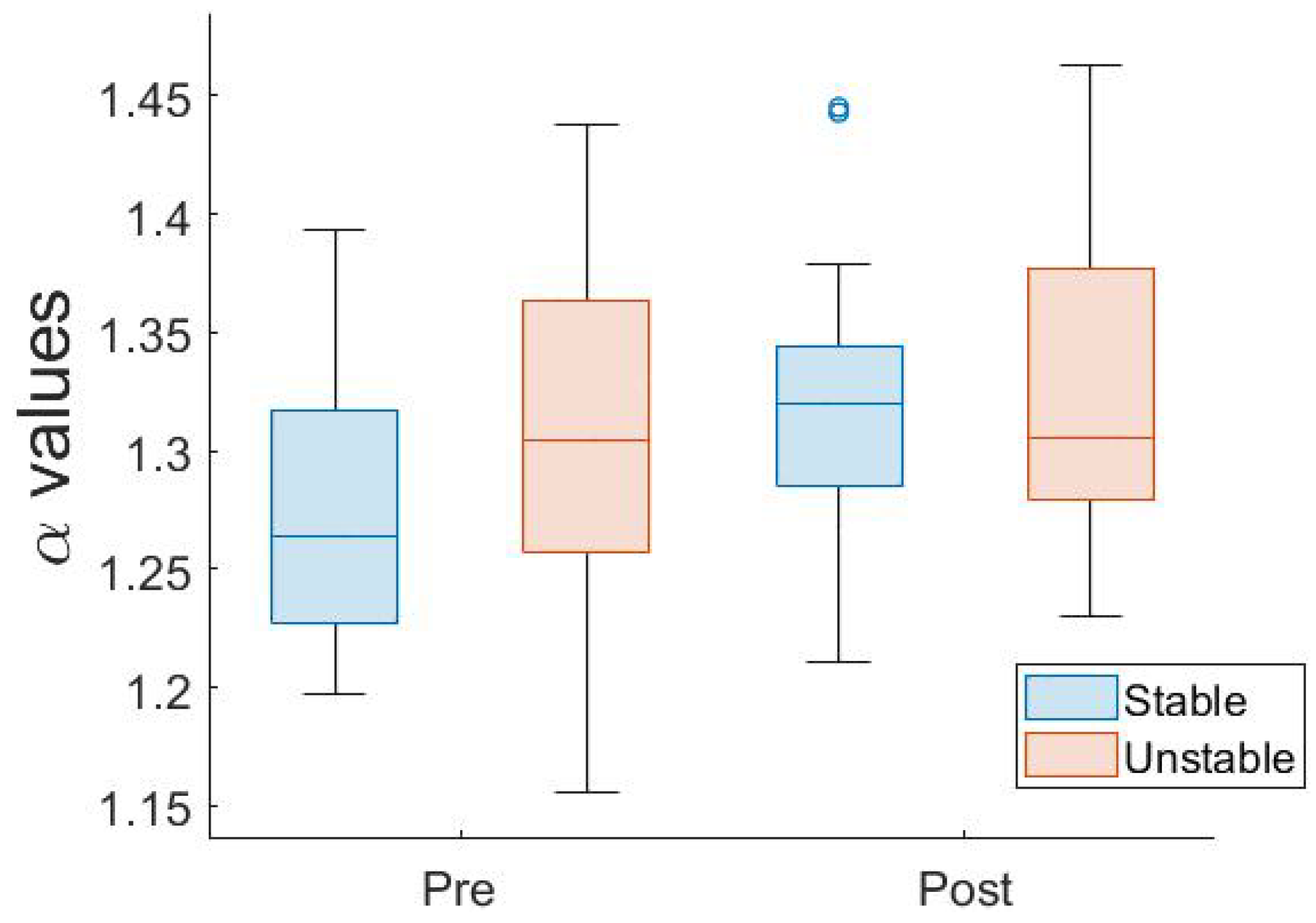
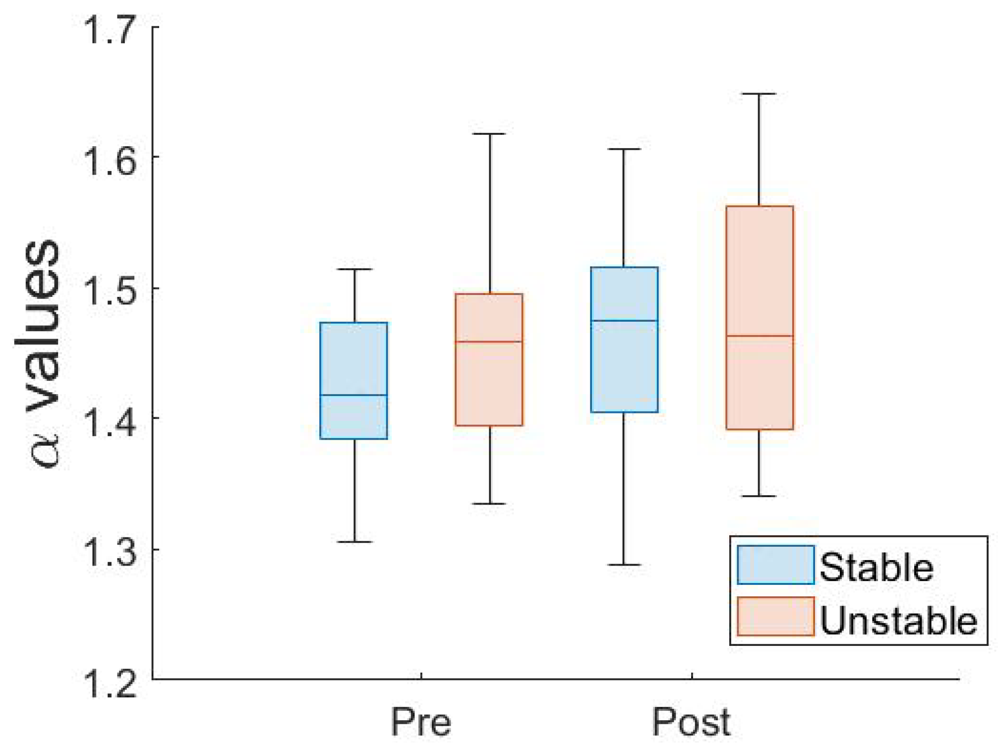
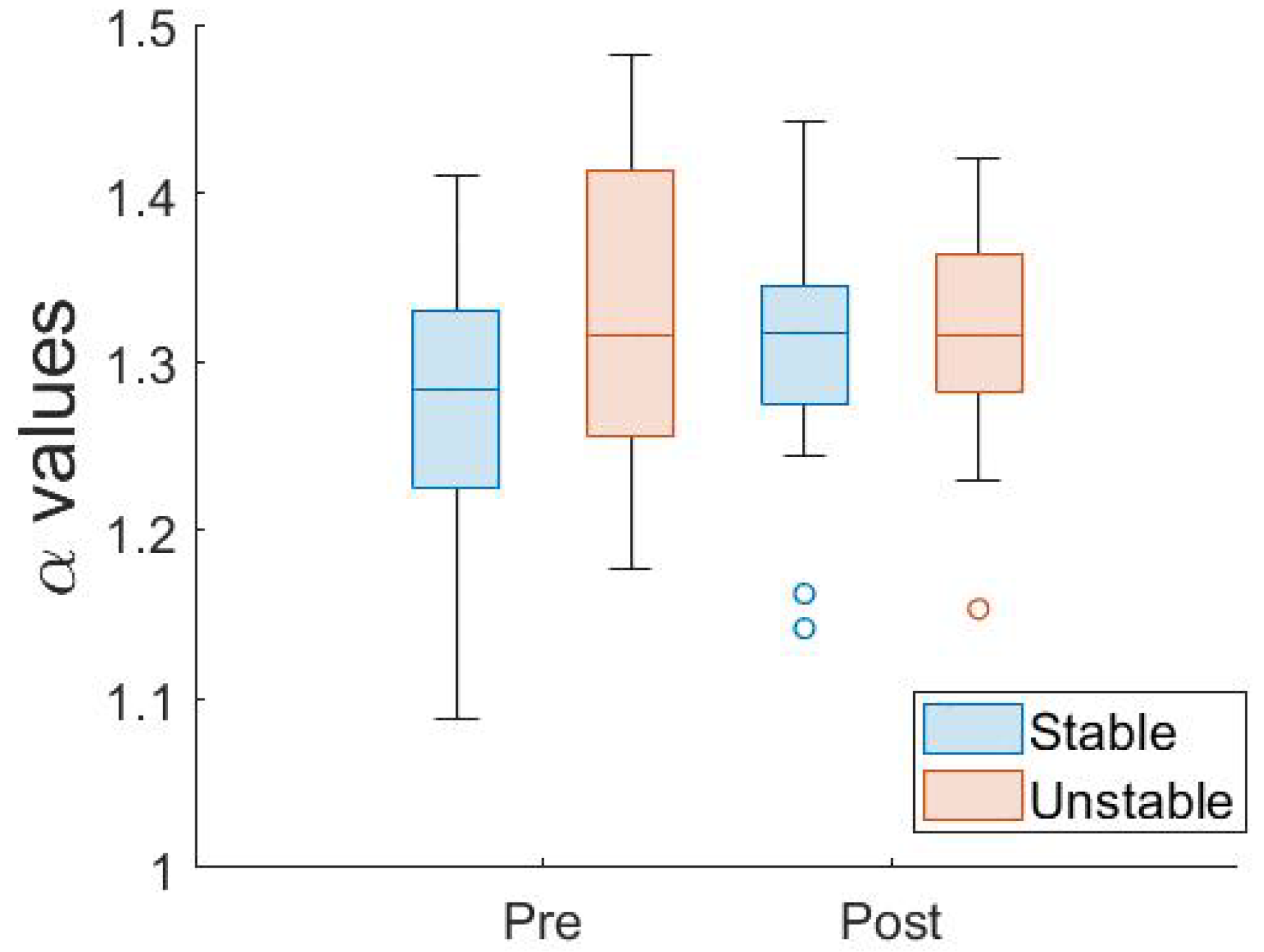
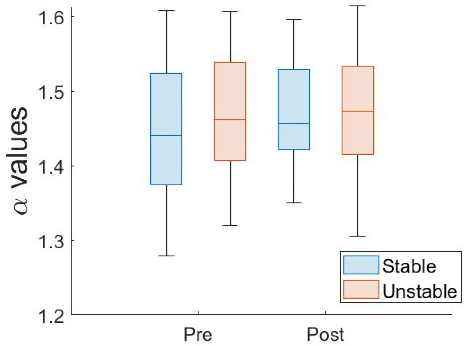
Our two-experiment approach examined the cross-education influence of lower limb fatigue on postural control by inducing fatigue in a proximal and distal muscle group. Based on the results, our hypotheses were not supported in that the completion of the fatiguing exercise task did not significantly alter the postural sway of the non-fatigued leg. However, for the dominant limb, fatigue showed significant differences in postural sway metrics; specifically, phase had a significant effect on both dfaML and dfaAP and the surface influenced dfaML. For the dominant limb in Experiment 2, similar significant differences in postural sway were also observed following the fatiguing exercise. While our results did not fully confirm the expected outcomes, the findings provide valuable insights into understanding the types of fatigue that produce meaningful cross-education effects for postural control.
The current study assessed the immediate impact of fatiguing exercises on the postural control of the contralateral leg. In contrast to evidence from previous studies that underscore the significant influence of fatigue on the stability [
12,
23,
28], our results did not illustrate a significant deterioration in the postural sway of the non-fatigue leg. For instance, Son [
29] found that fatigue of the ipsilateral plantar flexor muscle led to immediate alterations in postural control of the contralateral leg. Similarly, Paillard et al. [
24] demonstrated that both voluntary contractions (VOL) and electrical stimulation (ES) of the quadriceps muscle led to disturbances in postural sway of the contralateral limb, with more pronounced alterations after VOL-induced fatigue. The discrepancies in findings relative to our study could be attributed to several factors related to the fatigue protocol components. In a previous work [
24], two different fatigue protocols (VOL and ES) were examined that induce fatigue in the dominant leg knee extensor muscles and were shown to have a significant influence on the postural control of the contralateral limb. Such findings suggest that the specific muscles targeted for fatigue and the method of inducing fatigue can critically determine the cross-over effects on the contralateral limb. While Arora et al. [
13] conceptualized fatigue as the muscle’s decreased force-producing capability and measured it through the reduction in MVC, Paillard et al. [
24] assessed fatigue using two distinct methods: voluntary contractions and electrical stimulation, leading to differing physiological and biomechanical implications. The contrasting results emphasize the importance of considering the interaction of factors like the primary muscle, specific fatiguing exercises, and methods of fatigue induction when investigating cross-over fatigue effects on postural control.
The present study did not illustrate a significant effect of unilateral fatigue for either the ML or AP direction. Paillard et al. [
24] showed that cross-over fatigue was more pronounced in the AP direction than the ML direction. Specifically, all frequency bands of the AP direction showed changes post-fatigue, while only the first frequency band (0.05 to 0.5 Hz) in the ML direction increased. Lower frequency bands have been linked to sensory feedback mechanisms and may experience greater influence than feedforward mechanisms. The change in contralateral postural control within the sagittal plane might be linked to the role of the quadriceps femoris that stabilizes the body in that specific plane. The discrepancy in findings between Paillard et al. [
24] and the current study may be attributed to differences in the fatigue protocols. Overall, the intensity and duration of the fatiguing exercises emerge as potent modulators of the observed differences in postural control and the impact of cross-over fatigue [
24].
Several considerations arise around the components of different fatigue protocols. Specifically, the alignment between fatiguing exercises and muscles used in postural control might play a crucial role [
8]. For example, after localized muscle fatigue in the lower back and ankle, younger individuals displayed significant alterations in their postural sway [
30], suggesting that the post-fatigue state appeared to increasingly rely on other sensory systems [
30]. Additionally, the effects of general and local fatigue can alter both sensory inputs and motor outputs [
8].
A variety of aspects of cross-education require further investigation to better understand the neural mechanisms of cross-education. One potential interpretation relates to how unilateral motor activity can activate excitatory pathways interconnecting the ipsilateral and contralateral primary motor cortex, a process known as motor irradiation [
13,
31]. However, during unilateral contractions, there can also be active inhibition of the non-targeted muscle group, as postulated by the theory of default bilateral interaction [
32]. In the current study, it is plausible that these opposing effects—the excitatory and inhibitory mechanisms—counteracted each other. This might explain why no significant changes were observed in the postural sway parameters of the non-fatigued limb. Examining the underlying mechanisms of cross-over fatigue is crucial for future research.
In the original hypothesis, we predicted that the surface type would significantly impact postural sway of the non-fatigued leg; however, the findings revealed similar sway magnitude variability in both the medial–lateral (SDML) and anterior–posterior (SDAP) directions between stable or unstable balance conditions. While not statistically significant, greater COP variability was observed for unstable surfaces, suggesting that these posed a higher degree of postural challenge, but these conditions requires further investigation. Similarly, Hatton et al. [
33] found that textured surfaces introduce a degree of instability but failed to significantly alter postural sway in young adults during static standing. These findings indicates that postural control might be influenced differently by the nature and intensity of the specific surface characteristics.
Understanding how the duration of the fatigue protocols impacts the potential of cross-over fatigue on postural control requires further consideration. Experiment 1 targeted proximal muscles and consisted of four sets of one-minute-duration single-leg squats interspersed with 30 s rest intervals, while Experiment 2 fatigued a distal muscle group using a single-leg standing calf raise performed until exhaustion. Paillard et al. [
24] observed that exercises inducing only central fatigue without peripheral fatigue resulted in disturbances in contralateral single-leg stance postural control. In contrast, shorter duration exercises, like the approach used by Arora and colleagues [
13], that included a protocol of 15 contractions for five minutes at 30% MVC, did not exhibit significant alterations in muscle strength, EMG activity, or postural control of the non-exercised leg. These findings indicate that longer durations of the fatiguing task appear to produce more pronounced cross-over effects on the contralateral limb. The inconsistent results might be attributed to the nature of fatigue elicited in that central fatigue appears to negatively affect contralateral monopedal postural control and peripheral fatigue alone may not be sufficient to produce such disturbances predicted by cross-education theories. In light of these variable findings, it is evident that the duration of the fatiguing task plays a pivotal role in determining the extent of its cross-over effects, especially when the exercise induces central fatigue.
In addition to factors like fatigue duration and intensity, discrepancies in the neural and muscular mechanisms activated during the fatigue and balance tasks may have contributed to the lack of observed cross-over effects. In particular, the contralateral motor commands activated in the brain and spinal pathways during a squat task might differ from those triggered during balance stances [
34]. Hortobágyi et al. [
34] highlighted that voluntary contractions of a muscle in one limb can modulate motor cortical and segmental output in the contralateral limb, suggesting that the specific neural interactions during the fatiguing task can play a crucial role in determining cross-over effects. Furthermore, cross-over effects typically manifest when the fatiguing task and the balance assessment engage homologous contralateral muscle groups. Therefore, ensuring that the muscle groups targeted during fatigue align closely with those active during balance tasks is pivotal for observing cross-over effects [
3]. The squatting task used in Experiment 1 may have lacked the specificity of muscle contractions similar to those activated during the unilateral postural control stance. Thus, the fatiguing task in the current study may have produced larger effects of general fatigue, as compared to an isometric maximum voluntary contraction test, by involving multiple joints of the body, thereby lacking a specific muscle group relating to unilateral postural control stances [
5].
Compensation strategies can occur during postural control stances and muscles likely contribute to altered individual strategies used following fatigue [
24]. Proximal muscle fatigue of the hip and knee can result in greater deficits of unilateral stances compared to ankle muscle fatigue [
16,
24]. The squat task was performed in the AP plane while the balance tasks were performed using muscles in the AP and ML direction. Because the squatting task emphasized fatigue in the AP direction, balance compensation may have occurred with ML postural control muscles [
8,
35]. When transitioning to single-leg balance tasks, which require stabilization in both the AP and medial–lateral (ML) directions, fatigued muscles in the frontal plane could potentially compromise stability. This likely prompted a compensatory mechanism, where the ML postural control muscles assumed a more pronounced role to counterbalance the diminished support from the fatigued AP muscles. Compensations through coactivation of lower limb muscles may be contributing to variable results, with some individuals displaying both increased and decreased postural sway after exercise [
35].
Future research resulting from our study is anticipated to significantly expand the understanding of fatigue-induced changes in postural control, with a particular focus on three key areas. First, there is a vital need to explore these changes across a broader spectrum of the population. By incorporating a more diverse demographic in future studies, including varied age groups and a balanced gender representation, we can gain a more comprehensive understanding of how fatigue impacts postural control across different segments of the population. This approach is critical for generalizing findings and understanding population-specific variations. Secondly, an in-depth examination of the long-term effects of different fatigue protocols in clinical settings is warranted. This research is particularly crucial in the context of rehabilitation for individuals with neuromuscular disorders. Understanding the prolonged impact of these fatigue protocols can aid in developing more effective treatment and management strategies, adapted to the specific needs of patients in rehabilitation settings. Finally, there is a compelling need to delve deeper into the neurophysiological mechanisms that mediate the relationship between fatigue, balance, and muscle coordination. Investigating these underlying mechanisms can unravel the complex physiological processes influenced by fatigue. These future research directions, grounded in our study’s findings, open up new frontiers for exploration and have the potential to make significant contributions to the fields of rehabilitation and neuromuscular research.


