Article

Toxocara canis and Toxocara cati in Stray Dogs and Cats in Bangkok, Thailand: Molecular Prevalence and Risk Factors

by
Pornkamol Phoosangwalthong
1,2,3,
Nam Hung Luong
3,
Jutamas Wongwigkan
1,2,3,
Ketsarin Kamyingkird
3,
Jumnongjit Phasuk
3,
Khampee Pattanatanang
3,
Nipa Thammasonthijarern
3,
Chanya Kengradomkij
3,
Wissanuwat Chimnoi
3,
Peter Odermatt
4,5 and
Tawin Inpankaew
1,2,3,*
1
Center for Agricultural Biotechnology, Kamphaeng Saen Campus, Kasetsart University, Nakhon Pathom 73140, Thailand
2
Center of Excellence on Agricultural Biotechnology: (AG-BIO/MHESI), Bangkok 10900, Thailand
3
Department of Parasitology, Faculty of Veterinary Medicine, Kasetsart University, Bangkok 10900, Thailand
4
Department of Epidemiology and Public Health, Swiss Tropical and Public Health Institute, 4123 Allschwil, Switzerland
5
University of Basel, 4001 Basel, Switzerland
*
Author to whom correspondence should be addressed.
Parasitologia 2022, 2(2), 88-94; https://doi.org/10.3390/parasitologia2020009
Submission received: 3 March 2022 / Revised: 25 April 2022 / Accepted: 26 April 2022 / Published: 28 April 2022

Abstract

:
Toxocara canis and Toxocara cati are known as common roundworm in dogs and cats, respectively. The objective of the current study was to determine the molecular prevalence and risk factors associated with T. canis and T. cati infections in stray dogs and cats, respectively, in Bangkok, Thailand. In total, 1000 fecal samples (from 500 dogs and 500 cats) were collected from stray dogs and cats residing in Bangkok temples. DNA was extracted and screened for T. canis and T. cati using polymerase chain reaction (PCR) with the internal transcribed spacer (ITS) region. The overall prevalence of T. canis in the stray dogs was 5.4% (27/500) and 0.6% (3/500) for T. cati in the stray cats. Toxocara canis infections were detected in stray dogs from 11 out of 48 districts (22.9%), with Bang Khen district having the highest proportion of infected dogs, whereas T. cati infections were only detected in the stray cats in Lat Krabang district. These results indicated that stray animals residing in Bangkok temples pose a considerable public health risk due to zoonotic parasites, especially T. canis.

1. Introduction

Toxocara canis and Toxocara cati are some of the most common parasitic roundworms that cause functional intestinal disorders in dogs and cats worldwide [1]. T. canis and T. cati are in the group causing larva migrans syndrome (LMS), visceral larva migrans (VLM), ocular larva migrans (OLM), neural larva migrans (NLM), and covert infection [2]. Following ingestion of the embryonated eggs by the host, the larvae hatch in the small intestine. The larvae then migrate through the intestinal mucosa to the liver and lungs, where they mature into adult worms in the small intestine [3]. Humans, who act as paratenic hosts, can become infected with these parasites through infected eggs from contaminated soil or water, unwashed hands, raw vegetables, or the ingestion of larvae in the undercooked or raw and infected organ or muscle tissues of other paratenic hosts [4].
In research on the coprological investigation of fecal samples, the identification of helminth eggs as Toxocara spp. is usually recorded more than the classification of the particular species [1]. Even though the differences between Toxocara spp. are clinically and epidemiologically important, the measurement of the egg dimensions does not allow for the differentiation between Toxocara spp. within the genus due to similar egg sizes [5]. A polymerase chain reaction (PCR)-based molecular approach has been demonstrated in some studies and could provide reliable markers to accurately identify Toxocara spp. [6,7]. The eggs of Toxocara spp. are in the host feces and can be identified accurately using PCR with specific primers.
In Thailand, the percentage of stray dogs and cats increases each year [8]. According to the Bureau of Disease Control and Veterinary Services [9] (2016), 10.3% of dogs and 15.7% of cats are not owned. Although stray dogs and cats are potential reservoirs for zoonotic infections in humans, little data exist on the prevalence of animal infections. There have been few studies in Bangkok or other provinces [8,10,11,12]. Stray or neglected dogs and cats are often in poor health with several parasitic diseases and they are viewed as one of the most important sources of zoonosis. Environmental contamination with helminth eggs or protozoa (oo) cysts has been known to be associated with the prevalence of intestinal parasitic infections in animal populations [13]. The objective of the current study was to determine the molecular prevalence and risk factors associated with T. canis and T. cati in stray dogs and cats residing in Bangkok temples using a molecular technique.

2. Results

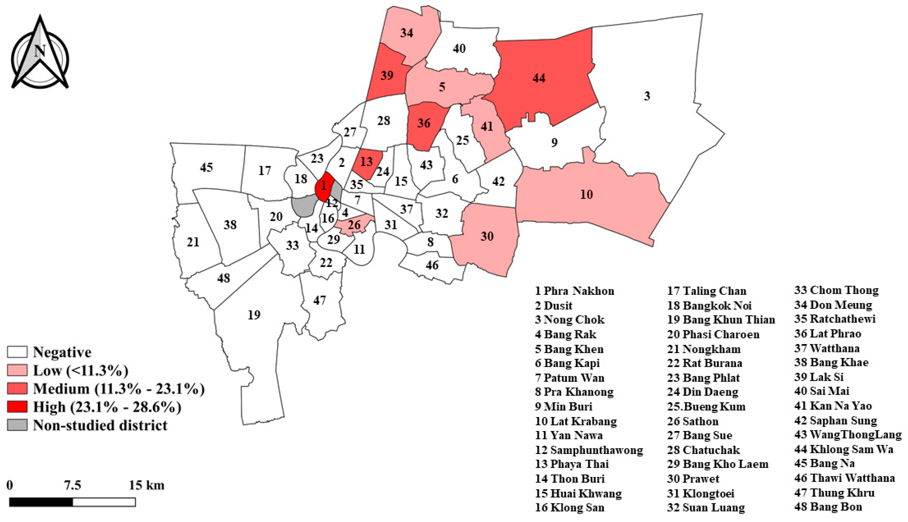
2.1. Prevalence of Toxocara canis in Stray Dogs

The results of the partial ITS region from the PCR-based amplification indicated that the prevalence of T. canis infection was 5.4% (n = 27) in the 500 stray dogs. Moreover, T. canis infections were detected in temple dogs in 11 out of the 48 districts (22.9%; Figure 1), with Bang Khen district having the highest proportion of T. canis-infected dogs (6/27, 22.2%) followed by Lad Prao (5/27, 18.5%) and Khlong Sam Wa districts (4/27, 14.8%), as shown in Figure 1.

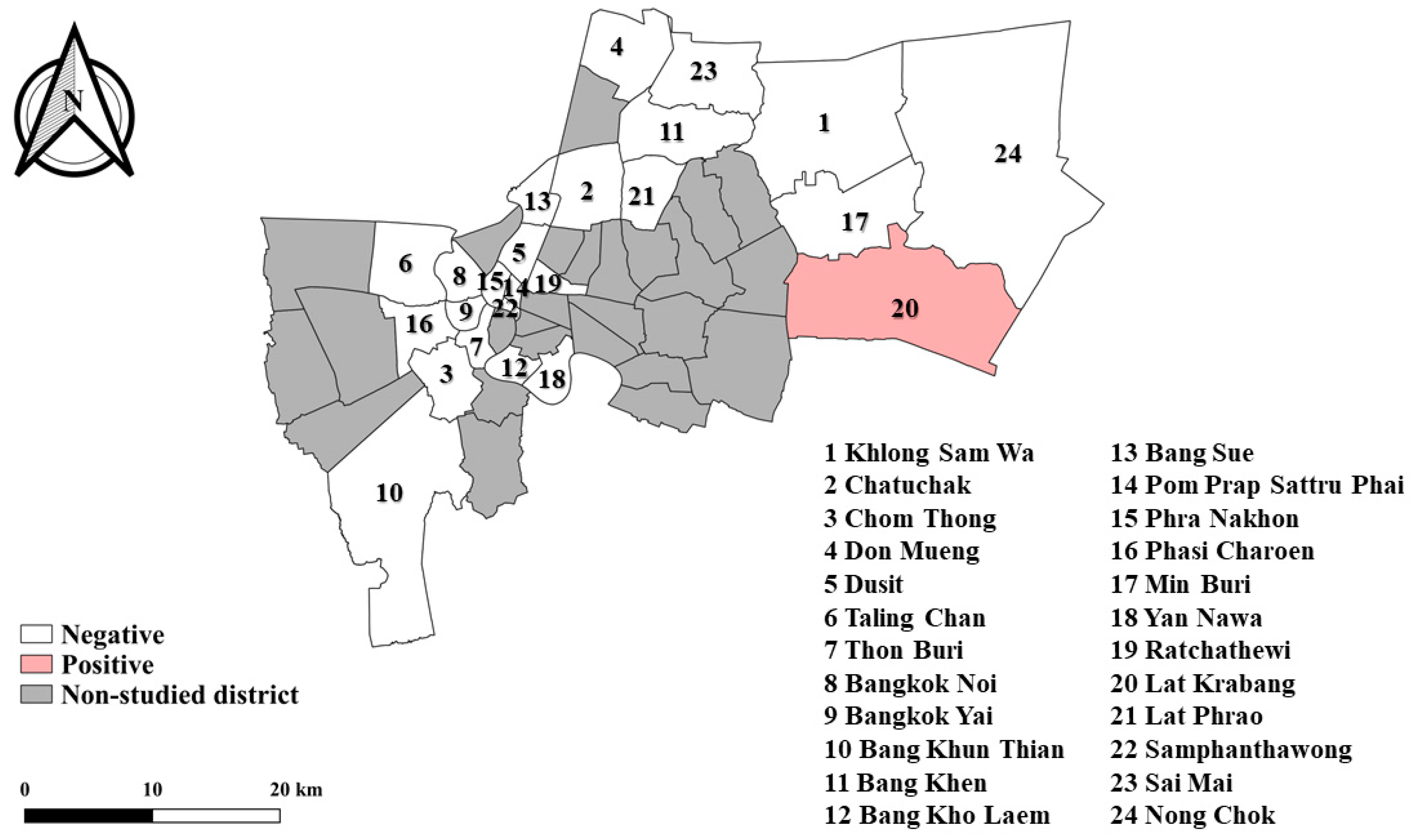
2.2. Prevalence of Toxocara cati in Stray Cats

The prevalence of T. cati infection was 0.6% (n = 3) among the 500 stray cats. All the T. cati infections were detected in the stray cats from Lat Krabang district (3/3, 100%), as shown in Figure 2.

2.3. Risk Factors Associated with Toxocara canis and Toxocara cati Infections

T. canis infections were found more frequently in free-roaming, young, dewormed, male dogs from suburban areas compared to the corresponding comparison group. However, none of the observed infection rates increased and none of the increased OR were statistically significant. T. cati-positive cats were also of younger age (1–5 years), with no statistically significant association with potential risk factors.

3. Discussion

Based on other studies, the prevalence of Toxocara spp. in stray dogs and cats in European countries was 1.4–30.5% and 8–76%, respectively [14]. The prevalence of T. canis in dogs has previously been reported as higher than in the current study in Thailand, with 37.7% in Vietnam [15], 32.0% in Poland [16], and 11.4% in Brazil [17]. The prevalence of T. cati in cats has not been widely reported, but it has been as high as 47.8% in Vietnam [15], 16.7% in Brazil [17], and 17.8% in China [18]. The prevalence of toxocariasis in cats is high in comparison to dogs and may be caused by a reduced resistance to reinfection in adult cats, whereas other possible explanations could be the transmission route of the tranmammary in young cats and the differences in environment, food supply, and eating behavior [19,20]. Previous research in Thailand also found that the most common Ascarid nematode infections were T. canis in dogs and T. cati in cats in Bangkok and other provinces [8,11,12,21]. The current study identified the prevalence of T. canis at 5.4% as rather low and at about half of the global prevalence (10.6–11.7%) and estimated prevalence in Southeast Asia (6.8–18.2%) in 2020 [22,23]. However, the variations between the districts are considerable as some districts had a prevalence exceeding 20%, which is a potential infection risk if infected stray dogs near temples leave their feces on the ground where the eggs and protozoan cysts can stay viable for a long time, posing a serious threat to public health [24].
Although previous studies found an association between Toxocara infection rates and risk factors such as age, sex, and location, in this study no such association could be established. This may be due to the low infection rates observed although the sampling pool size selection was based on previous studies, the relatively limited number of data points as a result of the low infection rates (and the subsequent insignificant differences among the groups defined by the risk factor variables) could lead to a low amount of data from our study. However, it is interesting to note that in our study the dewormed dogs had a statistically significant increased risk for T. canis infection (OR 2.2, 95% CI 1.0–4.9). This seems to be counterintuitive; however, it could be due to the fact that T. canis-infected dogs appear sick and hence have a higher chance of being dewormed by their owners compared to healthy, uninfected dogs.
It is commonly accepted that there is no relationship between the gender of dogs and cats and the Toxocara spp. infection status [14]. In our study, we observed that there was no difference between the gender groups.
The infection rates in puppies and kittens are also higher than in adult dogs and cats, and notably, the susceptibility of young animals to the infection and transmission of toxocariasis through infected mother’s milk can lead to serious infections [25]. This factor was supported by the current results, with the percentage of infected dogs aged under 1 year and over 5 years being higher than that of infected adult dogs (aged 1–5 years).
A weakness of our study was that for many stray animals the data on some of the risk factors were difficult to collect, e.g., the presence of previous deworming. Hence, some misclassification could have occurred which could have contributed to the lack of statistically significant differences. Even though PCRs can detect certain parasites with high sensitivity and specificity, they are notably time-consuming and do not provide quantitative results. Furthermore, the low prevalence of Toxocara in stray animals might be due to the fact that endogenous controls were not included in this study. It is vital to use endogenous controls to demonstrate that the negative reactions are genuine since sometimes PCRs can be inhibited. To overcome this, it is recommended to use endogenous controls in order to verify the negative results in further studies.
Despite the absence of statistically significant differences between the risk groups, our results provided an initial insight into the infection prevalence of Toxocara spp. in stray dogs and cats in Bangkok, Thailand. Further studies should broaden the research parameters by comparing different animal species or different regions to provide updated information on roundworms and more broadly on endoparasites in Thailand. Such information can be used to raise public awareness and increase knowledge about zoonotic parasites. In addition, a characterization of these parasites in humans and animals living in a shared environment is needed to determine the transmission between humans and animals.

4. Materials and Methods

4.1. Ethical Approval

This study was approved by the Kasetsart University Institutional Animal Care and Use Committee under the Ethical Review Board of the Office of National Research Council of Thailand (NRCT) with approval No. ACKU60-VET-006.

4.2. Study Period, Sample Collection, and Study Areas

Samples were collected from June to December 2015. In total, 1000 fecal samples (500 dogs and 500 cats) were collected from stray dogs in 91 temples in 48 districts and from stray cats from 43 temples in 24 districts in Bangkok. A fecal sample was individually collected from the rectum of dogs and cats, placed into a plastic bag, and maintained in cool conditions (4 °C) during transportation within 4–6 h to the Department of Parasitology, Faculty of Veterinary Medicine, Kasetsart University, Bangkok, and screened for T. canis in dogs and T. cati in cats using molecular techniques. Moreover, for further information about each animal, the monks or animal caretakers were interviewed for a questionnaire related to the animal’s environmental and hygiene conditions. Information was recorded on sex (male or female), age (<1 year, 1–5 years, and >5 years), free-roaming status (yes or no), history of veterinary attention (yes or no), and deworming status (yes or no).

4.3. DNA Extraction and Molecular Detection of Toxocara Species

All collected fecal samples were extracted using an E.Z.N.A® Stool DNA kit (Omega Bio-tek Inc.; Norcross, GA, USA) according to the manufacturers’ instructions with minor modifications. In general, Toxocara eggs have a thick eggshell that must be broken in order to obtain genomic DNA. If the procedure is insufficient, DNA cannot be obtained, resulting in a false-negative result. Therefore, samples were digested overnight at 56 °C (minimum of 16 h) with proteinase K digestion as recommended to successfully break unembryonated Toxocara eggs so that gDNA can be detected by PCR [26]. All gDNA samples were measured for quality at 260/280 nm and quantity at 260/230 nm using a BioSpectrometer (Eppendorf AG, Hamburg, Germany). The DNA was stored at −20 °C until further analysis. Diagnostic PCR was performed as previously described [26]. The internal transcribed spacer (ITS) regions were used to amplify 400 bp of Toxocara using the primers Tcan1 and NC2 for T. canis in dogs and the primers Tcat1 and NC2 for T. cati in cats. Each PCR reaction carried out with positive (DNA extracted from T. canis and T. cati eggs) and negative controls (nuclease-free water). All PCR products were run on a 1% agarose gel containing SYBR SafeGel Stain (Invitrogen; Waltham, MA, USA) and visualized using a UV transilluminator (Clare Chemical Research; Dolores, CO, USA).

4.4. Statistical Analysis

Data were evaluated using version 3.6.1 of the R software package [27]. Descriptive statistics were calculated on infection and risk factors. Univariate analysis was performed to determine the association between the infection status with Toxocara spp. (outcome) and risk factors including sex, age, and the animal’s environmental and hygiene conditions. Therefore, the odds ratio (OR) and 95% confidence interval (95% CI) were calculated. Significance was tested at p ≤ 0.05.

5. Conclusions

The current study identified the molecular prevalence of T. canis in dogs as being at 5.4% and of T. cati in cats as being at 0.6%. These results showed that stray dogs and cats residing in Bangkok temples acted as a potential source of roundworm infection. Despite the low prevalence of these parasites, they could still pose a low-to-moderate threat to public health, especially T. cati. Therefore, proper control and preventive measures against these parasites must be established including the use of disinfectants in contaminated areas where animals have defecated.

Author Contributions

Conceptualization, T.I.; methodology, P.P., N.H.L. and J.W.; data curation, P.P., W.C., K.K., J.P., N.T. and C.K.; writing—original draft preparation, P.P. and N.H.L.; investigation, P.P., K.K., J.P., K.P., N.T., P.O. and T.I.; writing—review and editing, T.I. and P.O.; supervision, T.I. and P.O.; funding acquisition, T.I. All authors have read and agreed to the published version of the manuscript.

Funding

This work was financially supported by the Office of the Ministry of Higher Education, Science, Research and Innovation; and the Thailand Science Research and Innovation through the Kasetsart University Reinventing University Program 2021 with grant no. CASAF-04. The research was supported by the Center of Excellence on Agricultural Biotechnology, Office of the Permanent Secretary, Ministry of Higher Education, Science, Research and Innovation. (AG-BIO/MHESI) with grant no. AG-BIO/60-004-002.

Institutional Review Board Statement

The study was conducted according to the guidelines of the Declaration of Helsinki and approved by the Animal Ethics Committee of Kasetsart University, Bangkok, Thailand (approval ID ACKU60-VET006).

Informed Consent Statement

Not applicable.

Data Availability Statement

The data presented in this study are available upon request from the corresponding author.

Acknowledgments

The authors would like to thank the staff and graduate students from the Department of Parasitology, Faculty of Veterinary Medicine, Kasetsart University, Bangkok, Thailand, and Pacharathon Simking and Sinsamuth Saengow from the Division of Animal Health Science, Faculty of Agricultural Technology, Rajamangala University of Technology Thanyaburi, Pathum Thani, Thailand, who helped during sample collection.

Conflicts of Interest

The authors declare that they have no personal conflicting or competing financial interest that could impact the work reported in this study.

References

  1. Okulewicz, A.; Perec-Matysiak, A.; Buńkowska, K.; Hildebrand, J. Toxocara canis, Toxocara cati and Toxascaris leonina in wild and domestic carnivores. Helminthologia 2012, 49, 3–10. [Google Scholar] [CrossRef] [Green Version]
  2. Joy, A.T.; Chris, O.I.; Godwin, N.C. Toxocariasis and Public Health: An Epidemiological Review. Glob. J. Infect. Dis. Clin. Res. 2017, 3, 28–39. [Google Scholar]
  3. Bruschi, F.; Dupouy-Camet, J. Helminth Infections and Their Impact on Global Public Health; Springer: Vienna, Austria, 2014. [Google Scholar]
  4. Wolfe, A.; Wright, I. Human toxocariasis and direct contact with dogs. Vet. Rec. 2003, 152, 419–422. [Google Scholar] [CrossRef]
  5. Uga, S.; Matsuo, J.; Kimura, D.; Rai, S.; Koshino, Y.; Igarashi, K. Differentiation of Toxocara canis and T. cati eggs by light and scanning electron microscopy. Vet. Parasitol. 2000, 92, 287–294. [Google Scholar] [CrossRef]
  6. Lee, A.C.; Schantz, P.M.; Kazacos, K.R.; Montgomery, S.P.; Bowman, D.D. Epidemiologic and zoonotic aspects of ascarid infections in dogs and cats. Trends Parasitol. 2010, 26, 155–161. [Google Scholar] [CrossRef] [PubMed]
  7. Oguz, B.; Ozdal, N.; Deger, M.S. Genetic analysis of Toxocara spp. in stray cats and dogs in Van province, Eastern Turkey. J. Vet. Res. 2018, 62, 291–295. [Google Scholar] [CrossRef] [Green Version]
  8. Rojekittikhun, W.; Chaisiri, K.; Mahittikorn, A.; Pubampen, S.; Sa-Nguankiat, S.; Kusolsuk, T.; Maipanich, W.; Udonsom, R.; Mori, H. Gastrointestinal parasites of dogs and cats in a refuge in Nakhon Nayok, Thailand. Southeast Asian J. Trop. Med. Public Health 2014, 45, 31–39. [Google Scholar]
  9. Bureau of Disease Control and Veterinary Services, Department of Livestock Development, Ministry of Agriculture and Cooperatives. Survey of Dog and Cat Populations in the Year 2016; Services BoDCaV: Bangkok, Thailand, 2016. (In Thai)
  10. Kaewthamasorn, M.; Niwetpathomwat, A.; Assarasakorn, S.; Wongsamee, S.; Tiawsirisup, S. A Surveillance of Canine Gastrointestinal Parasites in Fecal Samples from Public Areas of Bangkok, Thailand. J. Anim. Vet. Adv. 2006, 5, 1209–1213. [Google Scholar]
  11. Inpankaew, T.; Traub, R.; Thompson, R.C.; Sukthana, Y. Canine parasitic zoonoses in Bangkok temples. Southeast Asian J. Trop. Med. Public Health 2007, 38, 247–255. [Google Scholar]
  12. Phasuk, N.; Punsawad, C. Seroprevalence of Toxocara canis infection and associated risk factors among primary schoolchildren in rural Southern Thailand. Trop. Med. Health 2020, 48, 23. [Google Scholar] [CrossRef]
  13. Overgaauw, P.A.; van Knapen, F. Veterinary and public health aspects of Toxocara spp. Vet. Parasitol. 2013, 193, 398–403. [Google Scholar] [CrossRef] [PubMed] [Green Version]
  14. Baneth, G.; Thamsborg, S.M.; Otranto, D.; Guillot, J.; Blaga, R.; Deplazes, P. Major Parasitic Zoonoses Associated with Dogs and Cats in Europe. J. Comp. Pathol. 2016, 155 (Suppl. S1), S54–S74. [Google Scholar] [CrossRef] [PubMed] [Green Version]
  15. Anh, N.T.; Thuy, D.T.; Hoan, D.H.; Hop, N.T.; Dung, D.T. Levels of Toxocara infections in dogs and cats from urban Vietnam together with associated risk factors for transmission. J. Helminthol. 2016, 90, 508–510. [Google Scholar] [CrossRef] [PubMed]
  16. Luty, T. Prevalence of species of Toxocara in dogs, cats and red foxes from the Poznan region. Pol. J Helminthol. 2001, 75, 153–156. [Google Scholar]
  17. Dantas-Torres, F. Toxocara prevalence in dogs and cats in Brazil. In Advances in Parasitology; Bowman, D.D., Ed.; Academic Press: Cambridge, MA, USA, 2020; Chapter 33; pp. 715–741. [Google Scholar]
  18. Yang, Y.; Liang, H. Prevalence and Risk Factors of Intestinal Parasites in Cats from China. Biomed. Res. Int. 2015, 2015, 967238. [Google Scholar] [CrossRef] [PubMed]
  19. Swerczek, T.W.; Nielsen, S.W.; Helmboldt, C.F. Transmammary passage of Toxocara cati in the cat. Am. J. Vet. Res. 1971, 32, 89–92. [Google Scholar]
  20. Coati, N.; Schnieder, T.; Epe, C. Vertical transmission of Toxocara cati Schrank 1788 (Anisakidae) in the cat. Parasitol. Res. 2004, 92, 142–146. [Google Scholar] [CrossRef]
  21. Jittapalapong, S.; Inparnkaew, T.; Pinyopanuwat, N.; Kengradomkij, C.; Sangvaranond, A.; Wongnakphet, S. Gastrointestinal parasites of stray cats in Bangkok metropolitan areas Thailand. Agric. Nat. Resour. 2007, 41, 69–73. [Google Scholar]
  22. Rostami, A.; Sepidarkish, M.; Ma, G.; Wang, T.; Ebrahimi, M.; Fakhri, Y.; Mirjalali, H.; Hofmann, A.; Macpherson, C.N.L.; Hotez, P.J.; et al. Global prevalence of Toxocara infection in cats. In Advances in Parasitology; Bowman, D.D., Ed.; Academic Press: Cambridge, MA, USA, 2020; Chapter 30; pp. 615–639. [Google Scholar]
  23. Rostami, A.; Riahi, S.M.; Hofmann, A.; Ma, G.; Wang, T.; Behniafar, H.; Taghipour, A.; Fakhri, Y.; Spotin, A.; Chang, B.C.; et al. Global prevalence of Toxocara infection in dogs. Adv. Parasitol. 2020, 109, 561–583. [Google Scholar]
  24. Pinyopanuwat, N.; Kengradomkij, C.; Kamyingkird, K.; Chimnoi, W.; Suraruangchai, D.; Inpankaew, T. Stray animals (dogs and cats) as sources of soil-transmitted parasite eggs/cysts in temple grounds of Bangkok Metropolitan, Thailand. J. Trop. Med. Parasitol. 2018, 41, 15–20. [Google Scholar]
  25. Zibaei, M.; Sadjjadi, S.M. Trend of toxocariasis in Iran: A review on human and animal dimensions. Iran. J. Vet. Res. 2017, 18, 233–242. [Google Scholar] [PubMed]
  26. Borecka, A.; Gawor, J. Modification of gDNA extraction from soil for PCR designed for the routine examination of soil samples contaminated with Toxocara spp. eggs. J. Helminthol. 2008, 82, 119–122. [Google Scholar] [CrossRef] [PubMed]
  27. R Core Team. R: A Language and Environment for Statistical Computing; R Foundation for Statistical Computing: Vienna, Austria, 2020; Available online: https://www.R-project.org/ (accessed on 28 February 2022).
Figure 1. Map of Bangkok with Toxocara canis positive dogs (in red) and negative dogs (in white) by district and non-studied district (in gray).
Parasitologia 02 00009 g001
Figure 2. Map of Bangkok with Toxocara cati positive cats (in red) and negative cats (in white) by district and non-studied district (in gray).
Parasitologia 02 00009 g002
Publisher’s Note: MDPI stays neutral with regard to jurisdictional claims in published maps and institutional affiliations.

