
Haemorrhagic Artefacts Produced by Ant Activity on Human Cadavers in the Early Post-Mortem Period


Abstract
:1. Introduction
2. Materials and Methods
3. Results and Discussion
3.1. Droplet Pattern (Minute Haemorrhages with Droplet-Shaped Pattern)
3.2. Stripe Pattern
3.3. Pool Pattern
3.4. Mixed Pattern

| Date and Season | Place of Discovery | Sex Age | Stage of Decomposition and Thanatological Signs: RM = Rigor Mortis 1 LM = Livor Mortis 2 AM = Algor Mortis 3 | Last Time Seen Alive | Cause of Death | Position of the Body | Presence of Clothing | Anatomical Region of and Type of Artefact Caused by Ants (HA 4, DH 5) | Bloodstain Pattern Observed | Case # |
|---|---|---|---|---|---|---|---|---|---|---|
| 03.11.2015 Dry season (summer) | Body found indoors, at a house with good hygiene standards | M 30 | Fresh RM = full LM = legs AM = cooler than normal body temperature | 5 h before the police was informed | Suicide by hanging | Partial hanging, from the ceiling fan of the hall area of house | Yes | HA = lower limbs (Figure 6) | Mixed, stripe and droplet | 1 |
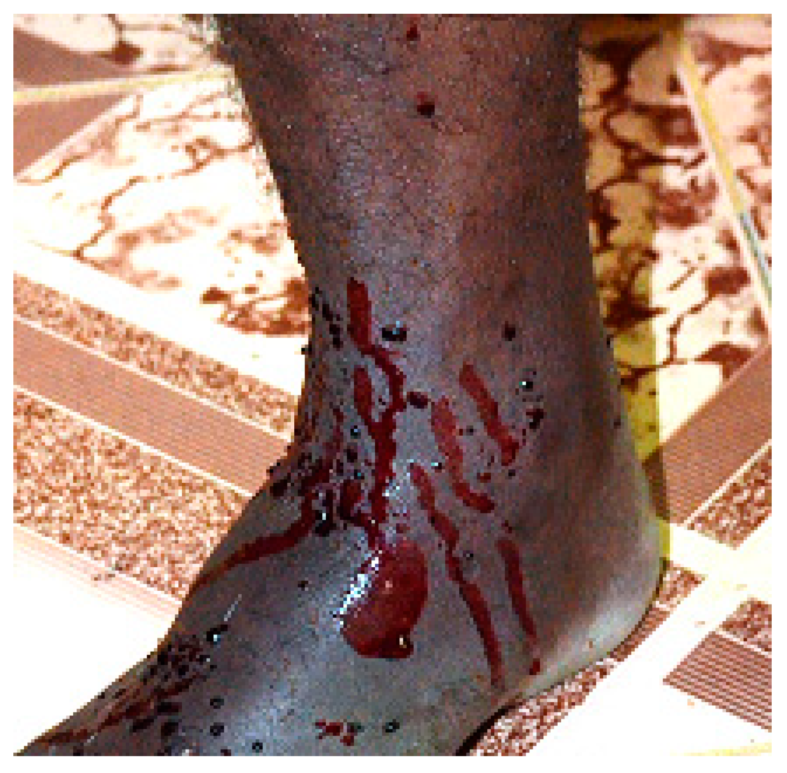
| HA = feet (Figure 3a) | Droplet | |||||||||
| Presence of black ants on eyes, with no HA/DH associated | nil | |||||||||
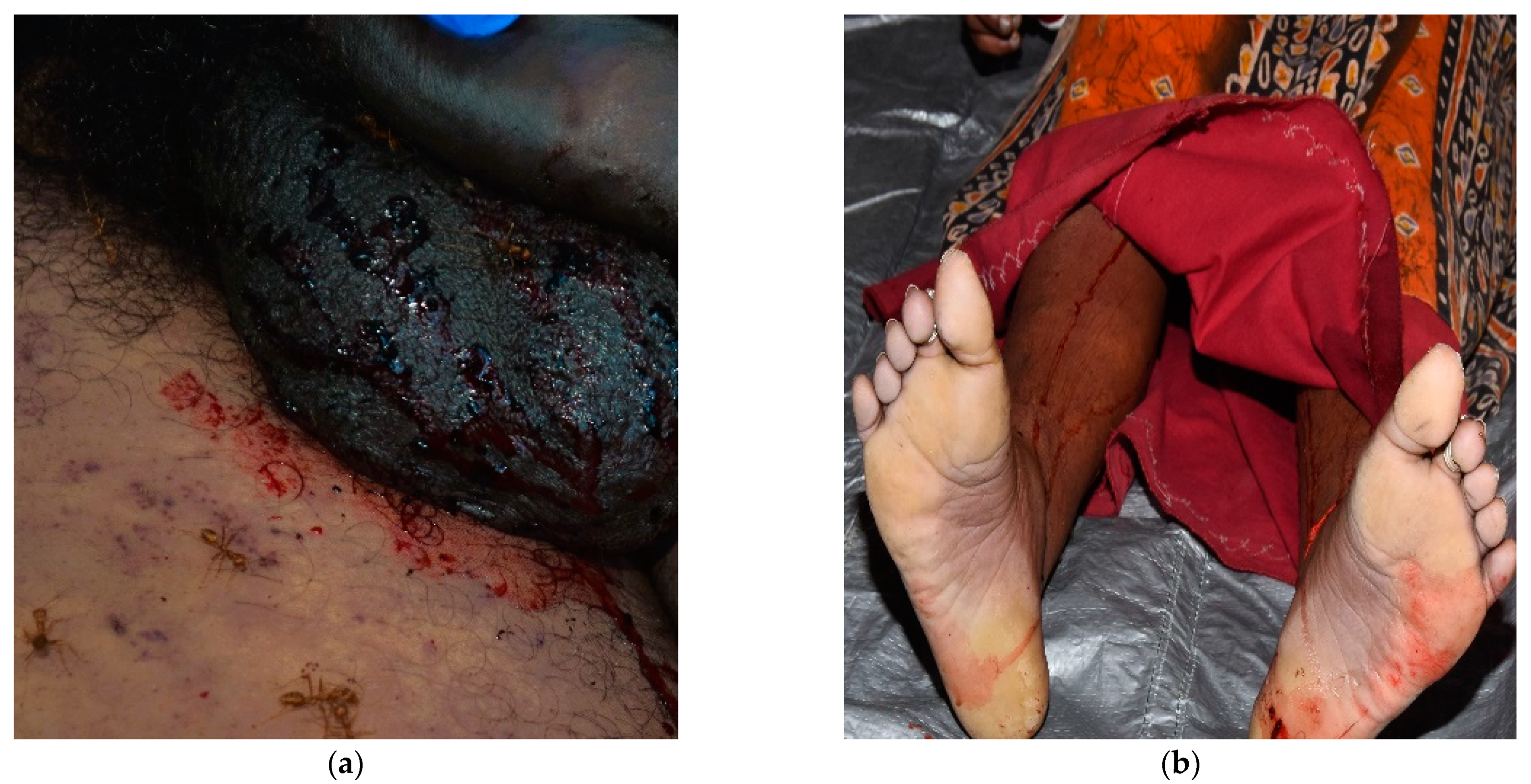
| 15.03.2016 Dry season (summer) | Body found outdoors, in an area near bushes alongside a road | M 45 | Fresh RM = full LM = back AM = cooler than normal body temperature | Last seen the night before; body found in the morning | Natural death in known history of chronic alcoholism. Alcohol test positive | Supine on the ground of a road side, surrounded by dry leaves | Yes | HA = middle and lower back | Mixed | 2 |
| DH = right arm and hand | Multiple small abrasions | |||||||||
| 19.03.2016 Dry season (summer) | Body found outdoors, in a forest area | F 16 | Fresh RM = full LM = tongue AM = cooler than normal body temperature | 1 day before | Suicide by hanging | Supine on ground of a forest area, surrounded by dry leaves | Yes | HA = tongue (Figure 8a) | Pool | 3 |
| DH = area of the face and neck | Multiple small abrasions | |||||||||
| 11.06.2016 Rainy season | Body found indoors, location in moderate hygiene standards | M 44 | Fresh RM = full LM = back AM = cooler than normal body temperature | Last seen the night before; body found in the morning | Natural death in known history of chronic alcoholism | Supine on the house floor, in an apartment located at the first floor of a building | Yes (trousers only) | HA = internal corner of the right eye (Figure 8b) | Pool | 4 |
| 29.10.2016 Dry season (summer) | Body found outdoors, in a forest area | M 26 | Fresh RM = not yet developed LM = back AM = cooler than normal body temperature | 5 h before | Suicide by hanging | Full hanging from a tree in an open forest area with low tree density | Yes | HA = tongue (Figure 2) | Droplet | 5 |
| 12.11.2016 Dry season (summer) | Body found indoors, at a house with moderate hygiene standards | F 65 | Fresh RM = full LM = hands and feet AM = cooler than normal body temperature | Last seen the night before; body found in the morning | Suicide by hanging | Partial hanging, from wooden ceiling beam of bathroom area of a house | Yes | HA = lower lip | Mixed | 6 |
| DH = upper lip area | Multiple small abrasions | |||||||||
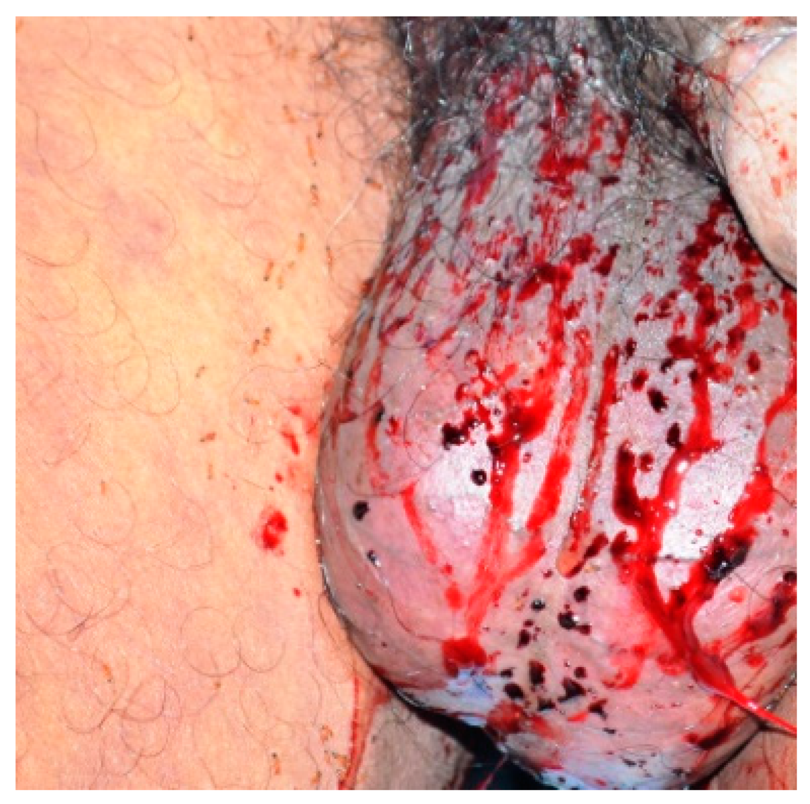
| 25.04.2017 Dry season (summer) | Body found indoors, at a temporary house with moderate hygiene standards | M 63 | Fresh RM = full LM = tongue, hands, and feet AM = cooler than normal body temperature | 4 h before | Suicide by hanging | Partial hanging, from ceiling wooden beam of a temporary house (shed made of tin) | Yes | HA = genital area (scrotum, Figure 4); | Mixed, stripe and droplet | 7 |
| HA = gluteal region and proximal medial thighs (Figure 3b) | Mixed, stripe and droplet | |||||||||
| 07.05.2018 Rainy season | Body found outdoors, in a forest area | M 83 | Fresh RM = in development LM = tongue, hands, and legs AM = cooler than normal body temperature | 4 h before | Suicide by hanging | Partial hanging, from a tree in forest area | Yes | HA = genital area (especially scrotum) (Figure 7a) | Stripe | 8 |
| HA = tongue (Figure 8c) | Pool | |||||||||
| 18.03.2020 Dry season (summer) | Body found indoors, at a house with moderate hygiene standards | F 34 | Fresh RM = full LM = palm of hands AM = cooler than normal body temperature | Last seen the night before; body found in the morning | Suicide by hanging | Partial hanging, from a wooden beam of a house/shed constructed between farms | Yes | HA = lower limbs (Figure 7b) | Stripe | 9 |
| DH = neck and upper back area | Multiple small abrasions | |||||||||
| 30.09.2021 Rainy season | Body found outdoors, in a forest area | M 44 | Fresh RM = in development LM = palm of hands and soles of feet AM = cooler than normal body temperature | 6 h before | Suicide by hanging | Full hanging, from a tree in a forest area | Yes | HA = genital area and lower limbs (Figure 5) | Stripe | 10 |
| DH = area of the face and neck | Multiple small abrasions |
4. Conclusions
Author Contributions
Funding
Institutional Review Board Statement
Informed Consent Statement
Data Availability Statement
Acknowledgments
Conflicts of Interest
References
- Haglund, W.D.; Sorg, M. Advances in Forensic Taphonomy (Method, Theory and Archaeological Perspectives); CRC Press: Boca Raton, FL, USA, 2002. [Google Scholar]
- Byrd, J.H.; Tomberlin, J.K. Insects of forensic importance. In Forensic Entomology—The Utility of Arthropods in Legal Investigation, 3rd ed.; Byrd, J.H., Tomberlin, J.K., Eds.; CRC Press: Boca Raton, FL, USA, 2020; pp. 15–62. [Google Scholar]
- Benecke, M. Forensic Entomology: Arthropods and Corpses. In Forensic Pathology Reviews; Tsokos, M., Ed.; Humana Press: Totowa, NJ, USA, 2004; Volume 2, pp. 207–240. [Google Scholar]
- Amendt, J.; Campobasso, C.P.; Gaudry, E.; Reiter, C.; LeBlanc, H.N.; Hall, M.J. Best practice in forensic entomology-Standards and guidelines. Int. J. Leg. Med. 2007, 121, 90–104. [Google Scholar] [CrossRef]
- Wells, J.D. A forensic entomological analysis can yield an estimate of postmortem interval, and not just a minimum postmortem interval: An explanation and illustration using a case. J. Forensic Sci. 2019, 64, 634–637. [Google Scholar] [CrossRef]
- Chophi, R.; Sharma, S.; Sharma, S.; Singh, R. Forensic entomotoxicology: Current concepts, trends and challenges. J. Forensic Leg. Med. 2019, 67, 28–36. [Google Scholar] [PubMed]
- Rivers, D.; Geiman, T. Insect artifacts are more than just altered bloodstains. Insects 2017, 8, 37. [Google Scholar] [CrossRef]
- Viero, A.; Montisci, M.; Pelletti, G.; Vanin, S. Crime scene and body alterations caused by arthropods: Implications in death investigation. Int. J. Leg. Med. 2019, 133, 307–316. [Google Scholar]
- Magni, P.A.; Massimelli, M.; Messina, R.; Mazzucco, P.; Di Luise, E. Entomologia Forense. In Gli Insetti Nelle Indagini Giudiziarie e Medico-Legali; Ed. Minerva Medica: Turin, Italy, 2008. [Google Scholar]
- Heo, C.C.; Mohamad, A.R.; Rosli, H.; Nurul Ashikin, A.; Chen, C.D.; John, J.; Hiromu, K.; Baharudin, O. Ants (Hymenoptera: Formicidae) associated with pig carcasses in Malaysia. Trop. Biomed. 2009, 26, 106–109. [Google Scholar]
- Andrade-Silva, J.; Pereira, E.K.C.; Silva, O.; Santos, C.L.C.; Rabêlo, J.M.M. Ants (Hymenoptera: Formicidae) associated with pig carcasses in an urban area. Sociobiology 2015, 62, 527–532. [Google Scholar] [CrossRef]
- Nooten, S.S.; Chan, K.H.; Schultheiss, P.; Bogar, T.A.; Guénard, B. Ant body size mediates functional performance and species interactions in carrion decomposer communities. Funct. Ecol. 2022, 36, 1279–1291. [Google Scholar] [CrossRef]
- Haden-Pinneri, K.; Sanchez, L. The postmortem picnic. In Proceedings of the American Academy for Forensic Sciences (AAFS), San Antonio, TX, USA, 12–16 February 2007. [Google Scholar]
- Singh, S.; Abdullah, N.A.B.; Carbaugh, J.; Heo, C.C. Ants associated with a rat carcass: Its implications in forensic entomology with special emphasis on Carebara diversa (Hymenoptera: Formicidae). Int. J. Trop. Insect Sci. 2020, 40, 703–706. [Google Scholar] [CrossRef]
- Mendonça, R.; Santos-Prezoto, H.H.; Prezoto, F. Actions of the fire ant Solenopsis saevissima (Smith) (Hymenoptera: Formicidae) on a big-eared opossum carcass. Fla. Entomol. 2019, 102, 435–437. [Google Scholar] [CrossRef]
- Maciel, T.T.; Castro, M.M.; Barbosa, B.C.; Fernandes, E.F.; Prezoto, H.H.S.; Prezoto, F. Foraging behavior of fire ant Solenopsis saevissima (Smith) (Hymenoptera: Formicidae) in Felis catus Linnaeus (Carnivora: Felidae) carcass. Sociobiology 2015, 62, 610–612. [Google Scholar] [CrossRef]
- Maciel, T.T.; Barbosa, B.C.; Prezoto, H.H.S.; Prezoto, F. Record of foraging of ants (Hymenoptera, Formicidae) in vertebrate carcasses. Acta Sci. 2016, 38, 491–494. [Google Scholar] [CrossRef]
- Gotwald, W.H. Comparative morphological studies of the ants, with particular reference to the mouthparts (Hymenoptera: Formicidae). Mem. Cornell. Univ. Agric. Exper. Stat. 1969, 408, 1–150. [Google Scholar]
- Philip, S.; Barton, P.S.; Archer, M.S.; Quaggiotto, M.M.; Wallman, J.F. Invertebrate succession in natural terrestrial environments. In Forensic Entomology—The Utility of Arthropods in Legal Investigation, 3rd ed.; Byrd, J.H., Tomberlin, J.K., Eds.; CRC Press: Boca Raton, FL, USA, 2020; pp. 141–154. [Google Scholar]
- Campobasso, C.P.; Marchetti, D.; Introna, F.; Colonna, M.F. Post-mortem artifacts made by ants and the effect of ant activity on decompositional rates. Am. J. Forensic Med. Pathol. 2009, 30, 84–87. [Google Scholar] [CrossRef] [PubMed]
- Byard, R.W.; Heath, K.J. Patterned postmortem ant abrasions outlining clothing and body position after death. J. Forensic Legal. Med. 2014, 26, 10–13. [Google Scholar] [CrossRef]
- Jayaprakash, P.T. Postmortem skin erosions caused by ants and their significance in crime reconstruction. J. For. Ident. 2006, 56, 972–999. [Google Scholar]
- James, S.H.; Kish, P.E.; Sutton, T.P. Principles of Bloodstain Pattern Analysis: Theory and Practice; CRC Press: Boca Raton, FL, USA, 2005. [Google Scholar]
- Peschel, O.; Kunz, S.N.; Rothschild, M.A.; Mützel, E. Blood Stain Pattern Analysis. Forensic Sci. Med. Pathol. 2011, 7, 257–270. [Google Scholar] [CrossRef]
- Saukko, P.J.; Knight, B. Knight’s Forensic Pathology, 4th ed.; CRC Press: Boca Raton, FL, USA, 2016. [Google Scholar]
- Obando Brito, T.; Elzubair, A.; Sales Araújo, L.; de Souza Camargo, S.A.; Pereira Souza, J.L.; Almeida, L.H. Characterization of the mandible Atta Laevigata and the bioinspiration for the development of a biomimetic surgical clamp. Mat. Res. 2017, 20, 1525–1533. [Google Scholar] [CrossRef]
- Anonymous. An Integrated Environmentally Sound Development Strategy for the Andaman and Nicobar Islands; Government of India: New Delhi, India, 1986. [Google Scholar]
- AA.VV. Crime in India; Ministry of Home Affairs: New Delhi, India, 2021. Available online: https://ncrb.gov.in/en/Crime-in-India-2021 (accessed on 30 June 2023).
- AA.VV. Crime in India; Ministry of Home Affairs: New Delhi, India, 2018. Available online: https://ncrb.gov.in/en/Crime-in-India-2018 (accessed on 30 June 2023).
- Bharti, H.; Guénard, B.; Bharti, M.; Economo, E.P. An updated checklist of the ants of India with their specific distributions in Indian states (Hymenoptera, Formicidae). ZooKeys 2016, 551, 1–83. [Google Scholar]
- Mohanraj, P.; Ali, M.; Veenakumari, K. Formicidae of the Andaman and Nicobar Islands (Indian Ocean: Bay of Bengal). J. Insect. Sci. 2010, 10, 172. [Google Scholar] [CrossRef]
- McVean, D.N. Report on Land Use in the Andaman and Nicobar Islands; United Nations Environment Programme: Morges, Switzerland, 1976; Available online: https://wedocs.unep.org/handle/20.500.11822/29905 (accessed on 30 June 2023).
- Chhotani, O.B.; Marti, P.K. Contribution to the knowledge of Formicidae of the Andaman islands. Newsl. Zool. Surv. India 1977, 3, 17–20. [Google Scholar]
- Tiwari, R.N.; Jonathan, J.K. A new species of Liomyrmex Mayr from Andaman islands (Hymenoptera: Formicidae). Rec. Zool. Surv. India 1986, 83, 87–90. [Google Scholar] [CrossRef]
- Tiwari, R.N.; Jonathan, J.K. A new species of Metapone Forel from Nicobar islands (Hymenoptera: Formicidae: Myrmicinae). Rec. Zool. Surv. India 1986, 83, 149–153. [Google Scholar] [CrossRef]
- Agavekar, G.; Garcia, F.H.; Economo, E.P. Taxonomic overview of the hyperdiverse ant genus Tetramorium Mayr (Hymenoptera, Formicidae) in India with descriptions and X-ray microtomography of two new species from the Andaman Islands. PeerJ 2017, 5, e3800. [Google Scholar] [CrossRef] [PubMed]
- Adam, C.D. Fundamental studies of bloodstain formation and characteristics. Forensic Sci. Int. 2012, 219, 76–87. [Google Scholar] [CrossRef]
- Pal, S.K.; Rana, A.; Sharma, A.; Kaushik, N.; Sehgal, A. A forensic analysis of ligature material in suicidal hangings. Int. J. Forensic Sci. 2018, 3, 000138. [Google Scholar]
- Bambaradeniya, T.B.; Magni, P.A.; Dadour, I.R. A Summary of concepts, procedures and techniques used by forensic entomologists and proxies. Insects 2023, 14, 536. [Google Scholar] [CrossRef]
- van de Goot, F.R.W. The chronological dating of injury. In Essentials of Autopsy Practice; Rutty, G.N., Ed.; Springer: London, UK, 2008. [Google Scholar]
- Vinay, J.; Harish, S.; Mangala, G.S.R.; Hugar, B.S. A study on postmortem wound dating by gross and histopathological examination of abrasions. Am. J. Forensic Med. Pathol. 2017, 38, 167–173. [Google Scholar] [CrossRef]
- Esposito-Fava, A.; Marchand, E.; Gauchotte, G. Skin injuries in forensic histopathology: A descriptive study. Forensic Sci. Med. Pathol. 2023. [Google Scholar] [CrossRef]


Disclaimer/Publisher’s Note: The statements, opinions and data contained in all publications are solely those of the individual author(s) and contributor(s) and not of MDPI and/or the editor(s). MDPI and/or the editor(s) disclaim responsibility for any injury to people or property resulting from any ideas, methods, instructions or products referred to in the content. |
© 2023 by the authors. Licensee MDPI, Basel, Switzerland. This article is an open access article distributed under the terms and conditions of the Creative Commons Attribution (CC BY) license (https://creativecommons.org/licenses/by/4.0/).
Share and Cite
Kumar, Y.; Guareschi, E.E.; Bharti, H.; Magni, P.A. Haemorrhagic Artefacts Produced by Ant Activity on Human Cadavers in the Early Post-Mortem Period. Forensic Sci. 2023, 3, 506-520. https://doi.org/10.3390/forensicsci3030035
Kumar Y, Guareschi EE, Bharti H, Magni PA. Haemorrhagic Artefacts Produced by Ant Activity on Human Cadavers in the Early Post-Mortem Period. Forensic Sciences. 2023; 3(3):506-520. https://doi.org/10.3390/forensicsci3030035
Chicago/Turabian StyleKumar, Yogesh, Edda E. Guareschi, Himender Bharti, and Paola A. Magni. 2023. "Haemorrhagic Artefacts Produced by Ant Activity on Human Cadavers in the Early Post-Mortem Period" Forensic Sciences 3, no. 3: 506-520. https://doi.org/10.3390/forensicsci3030035