


Mind Your Decompositional Assumptions


International Forensic Research & Consulting, P.O. Box 250411, 50520 Cologne, Germany
*
Author to whom correspondence should be addressed.
Forensic Sci. 2022, 2(4), 725-740; https://doi.org/10.3390/forensicsci2040054
Submission received: 21 September 2022
/
Revised: 7 October 2022
/
Accepted: 27 October 2022
/
Published: 3 November 2022
Abstract
:Decompositional processes depend on temperature, humidity, and light, as well as on animals that can access the body. In low-level cases, suicides, or cases of self-neglect especially, these factors are not always taken into consideration because a full investigation may not have taken place. Misinterpretations of environmental factors can put crime scene investigators on the wrong track, and natural causes of missing tissue may be wrongly interpreted as violence or wounds. Here, we give some examples for the influence of invertebrate land animals on corpses.
1. Introduction
1.1. Invertebrates in General
Land-based invertebrates may cause tissue defects (missing tissue) on deceased persons. Such wound artifacts, if they remain unnoticed, may cause misinterpretations and could be challenging during forensic case investigations [1,2,3,4,5,6].
Invertebrate animals such as insects use corpses as a food source or a breeding site, or they may predatorily feed on other corpse colonizers. Few reports address the importance of postmortem tissue changes caused by insects, even though the wounds may appear so similar to an injury that they could be misinterpreted as such [9,10,11] (Figure 1).
To clarify the origin of such defects, an investigation of the place where the corpse was found and its surroundings is necessary since biologically important information may easily be lost in the dissecting room, given that ants, beetles, etc., may no longer be identified as having (possibly misleadingly) caused the loss of tissue and possible wounds.
Here, we present some examples for injuries caused postmortem by animals; these cases were or would have been challenging during case investigations.
1.2. Ants
Feeding defects of ants may look very similar to skin abrasions [12,13], strangulation marks, or (electrical) burns, and may therefore be misidentified as being vital injuries [8,12,13,14,15,16,17].
The red imported fire ant Solenopsis invicta Buren 1972 originally comes from South America and feeds on human tissue and human skin [18]. This species leaves wound-like defects that appear similar to burns (Figure 2). As a result of global trade and global warming, the red imported fire ant is becoming increasingly common at latitudes with formerly lower temperatures, i.e., uninhabitable environments for the ants. “Burn patterns” on corpses found in open land and in residences should be interpreted with reservations, and ants at the scene considered to be evidence should be documented and collected for identification purposes.
In one of our cases, the finding of ants in the face and on a suspects’ boot and the blouse of the deceased also led to the conviction of the suspect, a priest who had killed his wife. Lasius foliginosus (Latreille, 1798), the jet black ant, was found in the immediate vicinity of the crime scene, which was just a few meters away from the nest. Animals of this species were collected and were seen on the body in pictures of the crime scene, where they had caused a loss of tissue by feeding from the skin. A coincidental meeting of the boot and the nest (or other nests of the same ant species) was extremely unlikely as the ant species in question does not travel far away from the nest [17].
1.3. Beetles
Some beetle species may also cause circular holes that can be confused with gunshot wounds [21,22] (Figure 4 and Figure 5).
The occurrence of beetles and beetle larvae is often associated with later stages of decomposition [23]. Rove and dermestid beetles may, however, predatorily feed on fly maggots (rove) or actual corpse tissue (dermestids) earlier in succession (Figure 6 and Figure 7). In one of our cases, the skin defects caused by beetle feeding did resemble stabbing injuries (Figure 8).
This observation has been confirmed by Caballero and Leon-Cortes in 2014 [24]. Several beetle species may occur during the entire decomposition process, even at unexpected times, due to climate change.
1.4. Maggots and Adult Blow Flies
Fly larvae cause tissue loss which may be misinterpreted as pre-mortem or postmortem injury patterns (Figure 9 and Figure 10). In 1988, Pollak and Reiter [9] reported a case of a highly decomposed body where such a loss of tissue due to maggots was incorrectly interpreted as being shot wounds in the left orbital roof.
Differentiation between Bloodstains and Fly Artefacts
Presumed blood spatter arising from an acceleration of blood at a crime scene may have been caused by flies (Figure 11). DNA is not always an effective means of help here, as flies can also absorb genuine blood from the crime scene and then distribute it (through vomiting, excrement, etc.).
Even though fly artifacts may be differentiated from blood spatter patterns from humans by DNA analysis [27,28] and also by the following steps [26], we recommend:
A documentation of the fly activity (and presence of dead flies) at the possible crime scene: if (dead) flies are found then fly artifacts may also be present.
A range of stains/possible spatters: fly activity is frequent in the vicinity of light sources, on light-colored walls, windows, and mirrors. A comparison of stains near the corpse with those that are to be found far apart from the body is therefore recommended. Adult dead flies may be found in rooms other than where the corpse is found.
A comparison of the pattern at the scene with known/experimental fly artifact patterns.
An identification of presumed human blood stains that appear in a point-like or teardrop-like form (and could potentially be used for reconstruction) and an exclusion of the following stains:
- Traces with a tail–body relationship greater than 1 (Figure 11);
- Traces with a spermatozoa or tadpole-like structure;
- Traces that are similar to a spermatozoid and do not end in a small point;
- all traces without a distinguishable body and tail;
- all traces with a wavelike or uneven structure;
The absence of known characteristics of human blood stains in finger-like shapes around the edges.
An attempt to cover blood stains on the floor with paper, particularly those on the ground, so that they are preserved as well as possible, and not destroyed by investigators, emergency personnel, etc.
Good close-up photographs using a high-resolution camera and a scale on every picture.
1.5. Snails
Slugs alter the tissue of freshly dead or decomposing corpses (Figure 12).
They produce (smaller) tissue defects by scraping with their radula tongues at soft tissue. Due to penetration, and therefore the process of stretching these defects by continued rasping and the properties of the tissue itself, the abrasions become larger; occasionally, they become actual holes (Figure 13). The shape of the defects varies. We repeatedly observed slugs on piglets’ tongues (Figure 14). Misinterpretations regarding possible trauma may easily occur.
1.6. Wasps
There are only a few reports on wasps found on corpses. A study from Brazil by Simoes et al. [29] reported wasps on dead pigs and the influence of wasps on the speed of decomposition. Certain species of wasp may prefer certain stages of decomposition.
Wasps can feed on eggs of blow flies and thus wrongly reset the “developmental clock” on the postmortem interval. Their feeding activity caused traces on the body’s tissue similar to skin abrasions or scratch marks (Figure 15).
Due to their feeding activity, wasps also sometimes expose bones under thinner areas of the skin (Figure 16). These skin defects caused by feeding must be differentiated from traces that are caused by violence.
Barbosa et al. [30] present postmortem injuries to pig carcasses caused by the South American wasp species Agelaia fulvofasciata (Deg. 1773). These wasps prefer to feed from the lips but also on the snout and the anus. At the same time, they feed predatorily on fly maggots of a fly species that is used to calculate the colonization interval of corpses. If corpse colonizers are eaten by predators, then this may influence the calculations by wrongly “resetting” the decompositional starting point.
In our field trainings for students in a garden in Cologne, Germany, whole chunks of meat the size of a human head were consumed by local wasps (Vespula vulgaris (Linnaeus, 1758)) within a few days.
2. Environmental Influences
Decompositional processes depend, to a large extent, on environmental factors, including temperature, humidity, light, rainfall, soil, location, and the condition of the corpse et cetera [31,32,33,34].
Access for corpse-visiting insects to dead bodies is also dependent on the environment. It is therefore not only the location where the corpse is found that must be considered (forest, town, field edge, buried, sunny, in shadow, indoors, and open land [25]), but also the animals that are present there (insects, vertebrates (such as pets) [3,4,5,6,35,36], wild animals [37], birds [38], etc.). The animal’s life habits in turn play an important role in the later interpretation of possible feeding defects and in differentiating them from genuine injury patterns.
Naturally, caution has to be taken in cases in which the body may have been moved after insect colonization has started. The range of animals found on a body may not only depend on the environment in which the body was found, but also possible former environments with a probable different range of insects (or other animals).
The following cases illustrate the importance of light and temperature in the insect colonization of corpses in indoor areas.
2.1. Case 1: Influence of Clothing and Mummification
On 11 July 2017, the dead body of a 39 year old man was found in his apartment in a small district town in the eastern Ruhr area in Germany. The man was last seen on 24 June 2017. He suffered from a personality disorder, as well as drug and alcohol addiction. The body was found near the town center. There was urban greenery vegetation in the street; yet, there were no parks or larger green areas near to where the body was found.
The apartment was untidy; the body was lying on the floor, clothed in sweatpants and a pullover. Greasy brown deposits on the front left outer edge of the sofa and on the upper edge of a bucket for excrement and vomit alongside the sofa to the left were visible (Figure 17). The bed sheets in the bed room also exhibited similar brown deposits.
The body was in an advanced state of decomposition, and the skin on the trunk and back, as well as the extremities, did show some red–green to grey–green color. The epidermis of the stomach area had detached in strips in places. Much of the epidermis of the trunk was detached.
While the front side of the upper body was dried out, the back section appeared damp and putrid.
There were numerous maggots and fly pupae found on both the body and in the clothing of the deceased, and numerous egg clutches were present too.
Tissue defects had been caused by feeding blow fly larvae on the left lower side of the thorax, the flank area, and above the side area of the right eyebrow. Maggots were found on and in parts of the corpse, as well as in the perineum and anus.
The left arm was covered in clothing that otherwise appears to have been pulled up to the neck; the right arm and the entire upper body were uncovered up to the neck by the clothing.
In contrast to the rest of the body, hands and feet were clearly mummified. In particular, the hands at the wrist were strongly distinct from the lighter skin of the upper arm (Figure 18).
The clothing may have been pulled up by the emergency physician so that the clothing had formerly protected the area of the body underneath against desiccation.
Sun was shining from both the left side as well as from above onto the body (Figure 18). The lower side of the foot that was facing the left window was mildly dried out; the head was turned in the direction of the upper window and also exhibited dryness (partial mummification) in the forehead, nose, and eye area.
The fact that clothing protects against desiccation but also against decomposition was shown in experiments using piglets. Partially clothed piglets were lain out in open land. The head often decomposed considerably more quickly than the rest of the body which was largely covered by the clothing (Figure 19 and Figure 20). The higher attractiveness of the face to deposit eggs, and the stronger maggot feeding activity in this area on the one hand, as well as the protective function of the clothing on the other, was evident [36]. Overall, decomposition may happen in unevenly distributed patterns that can only be understood from the situation at the original scene, but sometimes not on the autopsy table.
2.2. Case 2: Influence of Light
In August 2001, the partly mummified body of a man was found in his bed in a large city in the Rhineland area of Germany [39]. The eye sockets had been emptied by fly larvae feeding from the tissue. The lips and the left area of his hips also exhibited signs of maggot feeding activity. The corpse was separated from the rest of the large room that had no fully closed separate room units due to a room partition wall. A lamp was attached on the right side of the bed, with the light bulb pointing away from the bed.
The corpse showed an uneven (differential) colonization by blow fly maggots of the eye socket facing the light source, showing that juvenile stages of blow flies tend to avoid light (Figure 21). They initially fed on the eye (socket) that faced away from the light, and then changed to the eye socket that was turned towards the light as a result of a food shortage. This finding could have led to a misinterpretation of the colonization sequence and the wrong assumption of an injury to the eye before death. As maggots prefer injuries compared to natural body openings to lay their eggs (and for adult colonization), it sounds plausible but would have been still wrong to assume that, for example, a puncture site in or near the eye may have led to unusual maggot colonization. (The deceased worked in a nearby hospital as a medical doctor; during this time, no injuries were seen.)
3. Conclusions
When a corpse is discovered, the overall “biological” picture from and at the scene should be considered and documented by good photographs with a scale.
The contribution of flies in the creation of what may, at first glance, be taken to be a blood stain spatter, in particular indoors, should be borne in mind.
If a partial decomposition pattern or a pattern of different signs of decomposition are observed on a body, then the micro-climate underneath the clothing should also be considered. The clothing can partially protect the tissue from light, rain, sunshine, etc., and hence lead to extreme differences in the decomposition process [40,41]. Snails and ants are also of relevance, as well as cockroaches [12,13].
Environmental influences may be checked experimentally.
4. Impact Statement
This communication illustrates the importance of taking environmental factors and animals at crime scenes into account so that misinterpretations and wrong assumptions can be avoided in forensic case work.
Author Contributions
writing—original draft preparation and editing, K.B. (Kristina Baumjohann); writing—review and editing, M.B. (Mark Benecke); case work case 1—M.B., correspondence with crime scene unit in case 1—K.B.; supervision—M.B.; conceptualization—K.B. All authors have read and agreed to the published version of the manuscript.
Funding
This research received no external funding.
Institutional Review Board Statement
Not applicable.
Informed Consent Statement
Informed consent was not required because these cases were judicial autopsies.
Data Availability Statement
Not applicable.
Conflicts of Interest
The authors declare no conflict of interest.
References
- Haglund, W.D. Contribution of rodents to postmortem artifacts of bone and soft tissue. J. Forensic Sci. 1992, 37, 1459–1465. [Google Scholar] [CrossRef] [PubMed]
- Patel, F. Artefact in forensic medicine: Postmortem rodent activity. J. Forensic Sci. 1994, 39, 257–260. [Google Scholar] [CrossRef]
- Ropohl, D.; Scheithauer, R.; Pollak, S. Postmortem injuries inflicted by domestic golden hamster: Morphological aspects and evidence by DNA typing. Forensic Sci. Int. 1995, 72, 81–90. [Google Scholar] [CrossRef]
- Steadman, D.W.; Worne, H. Canine scavenging of human remains in an indoor setting. Forensic Sci. Int. 2007, 173, 78–82. [Google Scholar] [CrossRef] [PubMed]
- Tsokos, M.; Schulz, F. Indoor postmortem animal interference by carnivores and rodents: Report of two cases and review of the literature. Int. J. Leg. Med. 1999, 112, 115–119. [Google Scholar] [CrossRef]
- Willey, P.; Snyder, L.M. Canid modification of human remains: Implications for time-since-death estimations. J. Forensic Sci. 1989, 34, 894–901. [Google Scholar] [CrossRef]
- Asamura, H.; Takayanagi, K.; Ota, M. Unusual characteristic patterns of postmortem injuries. J. Forensic Sci. 2004, 49, 592–594. [Google Scholar] [CrossRef]
- Campobasso, C.P.; Marchetti, D.; Introna, F.; Colonna, M.F. Postmortem artifacts made by ants and the effect of ant activity on decompositional rates. Am. J. Forensic Med. Pathol. 2009, 30, 84–87. [Google Scholar] [CrossRef]
- Pollak, S.; Reiter, C. Vortäuschung von Schussverletzungen durch postmortalen Madenfraß. Arch. Kriminol. 1988, 181, 146–154. [Google Scholar]
- Haskell, N.H.; Hall, R.D.; Cervenka, V.J.; Clark, M.A. On the body: Insects’ life stage presence, their postmortem artifacts. In Forensic Taphonomy: The Postmortem Fate of Human Remains, 1st ed.; Haglund, W.D., Sorg, M.H., Eds.; CRC Press: Boca Raton, FL, USA, 1997; pp. 415–448. [Google Scholar]
- Ortloff, A.; Albornoz, S.; Romero, M.; Vivallo, G. Skin artefacts due to post-mortem damage caused by Notiothauma reedi: A insect of forensic importance in forest communities of Chile. Egypt. J. Forensic Sci. 2016, 6, 411–415. [Google Scholar] [CrossRef] [Green Version]
- Weimann, W.; Prokop, O. Atlas der Gerichtlichen Medizin, 1st ed.; VEB Verlag Volk und Gesundheit: Berlin, Germany, 1963; pp. 36–44. [Google Scholar]
- Benecke, M. A brief survey of the history of forensic entomology. Acta Biol. Benr. 2008, 14, 15–38. [Google Scholar]
- Von Hofmann, E.R. Lehrbuch der Gerichtlichen Medizin, 6th ed.; Urban & Schwarzenberg: Vienna, Austria; Leipzig, Germany, 1893; pp. 359–819. [Google Scholar]
- Dietz, G. Gerichtliche Medizin für Juristen, Kriminalisten, Studierende der Rechtswissenschaften und Medizin, 5th ed.; Johann Ambrosius Barth: Leipzig, Germany, 1970; p. 36. [Google Scholar]
- Bonacci, T.; Benecke, M.; Scapoli, C.; Vercillo, V.; Pezzi, M. Severe post mortem damages by ants on a human corpse. Rom. J. Leg. Med. 2019, 27, 269–271. [Google Scholar] [CrossRef]
- Benecke, M.; Seifert, B. Forensische Entomologie am Beispiel eines Tötungsdeliktes. Eine kombinierte Spuren- und Liegezeitanalyse. Arch. Kriminol. 1999, 204, 52–60. [Google Scholar] [PubMed]
- Byrd, J.H.; Castner, J.L. Forensic Entomology. The Utility of Arthropods in Legal Investigations, 2nd ed.; CRC Press: Boca Raton, FL, USA, 2009. [Google Scholar]
- Payne, J.A.; King, E.W. Arthropod succession and decomposition of buried pigs. Nature 1968, 219, 1180–1181. [Google Scholar] [CrossRef]
- Payne, J.A.; King, E.W. Hymenoptera associated with pig carrion. Proc. Entomol. Soc. Wash. 1971, 73, 132–141. [Google Scholar]
- Baumjohann, K.; Benecke, M.; Rothschild, M. Schussverletzungen oder Käferfraß. Rechtsmedizin 2014, 24, 114–117. [Google Scholar] [CrossRef]
- Benecke, M. Leichenerscheinungen und Todeszeitbestimmung—Besiedlung durch Gliedertiere. In Handbuch Gerichtliche Medizin, 1st ed.; Brinkmann, B., Madea, B., Eds.; Springer: Berlin/Heidelberg, Germany; New York, NY, USA; Tokyo, Japan, 2003; pp. 170–187. [Google Scholar]
- Kulshrestha, P.; Satpathy, D.K. Use of beetles in forensic entomology. Forensic Sci. Int. 2001, 120, 15–17. [Google Scholar] [CrossRef]
- Caballero, U.; Leon-Cortes, J.L. Beetle succession and diversity between clothed sun-exposed and shaded pig carrion in a tropical dry forest landscape in Southern Mexico. Forensic Sci. Int. 2014, 245, 143–150. [Google Scholar] [CrossRef]
- Fiedler, A.; Halbach, M.; Sinclair, B.; Benecke, M. What is the edge of a forest? A Diversity Analysis of Adult Diptera Found on Decomposing Piglets inside and on the Edge of a Western German Woodland inspired by a Courtroom Question. Entomol. Heute 2008, 20, 173–191. [Google Scholar]
- Benecke, M.; Barksdale, L. Distinction of bloodstain patterns from fly artifacts. Forensic Sci. Int. 2003, 137, 152–159. [Google Scholar] [CrossRef]
- Rivers, D.B.; Cavanagh, G.; Greisman, V.; McGregor, A.; Brogan, R.; Schoeffield, A. Immunoassay detection of fly artifacts produced by several species of necrophagous flies following feeding on human blood. Forensic Sci. Int. Synerg. 2019, 1, 1–10. [Google Scholar] [CrossRef] [PubMed]
- Bini, C.; Giorgetti, A.; Iuvaro, A.; Giovannini, E.; Gianfreda, D.; Pelletti, G.; Pelotti, S. A DNA-based method for distinction of fly artifacts from human bloodstains. Int. J. Leg. Med. 2021, 135, 2155–2161. [Google Scholar] [CrossRef]
- Simoes, M.H.; Guedes, A.O.; Thyssen, P.J.; Souza-Silva, M. Ecological and Forensic Implications of Social Wasps on Pig Carcass Degradation in Brazilian Savannah. Bangladesh J. Fertil. Steril. 2013, 2, 286–293. [Google Scholar] [CrossRef] [Green Version]
- Barbosa, R.R.; Carrico, C.; Souto, R.N.P.; Andena, S.R.; Urufahy-Rodrigues, A.; Queiroz, M.M.C. Record of postmortem injuries caused by the Neotropical social wasp Agelaia fulvofasciata (Degeer) (Hymenoptera, Vespidae) on pig carcasses in the Eastern Amazon region: Implications in forensic taphonomy. Rev. Bras. Entomol. 2015, 59, 257–259. [Google Scholar] [CrossRef]
- Mann, R.W.; Bass, W.M.; Meadows, L. Time Since Death and Decomposition of the Human Body: Variables and Observations in Case and Experimental Field Studies. J. Forensic Sci. 1990, 35, 103–111. [Google Scholar] [CrossRef] [PubMed] [Green Version]
- Aturaliya, S.; Lukasewyz, A. Experimental forensic and bioanthropological aspects of soft tissue taphonomy: 1. Factors influencing postmortem tissue desiccation rate. J. Forensic Sci. 1999, 44, 893–896. [Google Scholar] [CrossRef]
- Dautartas, A.M. The Effect of Various Coverings on the rate of Human Decomposition. Master Thesis, University of Tennessee, Knoxville, TN, USA, August 2009. [Google Scholar]
- Dent, B.B.; Forbes, S.L.; Stuart, B.H. Review of human decomposition processes in soil. Environ. Geol. 2004, 45, 576–585. [Google Scholar] [CrossRef]
- Rossi, M.L.; Shahrom, A.W.; Chapman, R.C.; Vanezis, P. Postmortem injuries by indoor pets. Am. J. Forensic Med. Pathol. 1994, 15, 105–109. [Google Scholar] [CrossRef]
- Rothschild, M.A.; Schneider, V. On the temporal onset of postmortem animal scavenging. Motivation of the animal. Forensic Sci. Int. 1997, 89, 57–64. [Google Scholar] [CrossRef]
- Jones, A.L. Animal Scavengers as Agents of Decomposition: The Postmortem Succession of Louisiana wildlife. Master Thesis, Louisiana State University and Agricultural and Mechanical College, Baton Rouge, LA, USA, August 2011. [Google Scholar]
- Spradley, M.K.; Hamilton, M.D.; Giordano, A. Spatial patterning of vulture scavenged human remains. Forensic Sci. Int. 2012, 219, 57–63. [Google Scholar] [CrossRef]
- Benecke, M. Rein einseitiges Auftreten von Schmeißfliegenmaden im Gesicht einer Faulleiche. Arch. Kriminol. 2001, 208, 182–185. [Google Scholar] [PubMed]
- Kelly, J.A.; van der Linde, T.C.; Anderson, G.S. The influence of clothing and wrapping on carcass decomposition and arthropod succession during the warmer seasons in central South Africa. J. Forensic Sci. 2009, 54, 1105–1112. [Google Scholar] [CrossRef] [PubMed]
- Matuszewski, S.; Fratczak, K.; Konwerski, S.; Bajerlein, D.; Szpila, K.; Jarmusz, M.; Szafałowicz, M.; Grzywacz, A.; Madra, A. Effect of body mass and clothing on carrion entomofauna. Int. J. Leg. Med. 2016, 130, 221–232. [Google Scholar] [CrossRef] [PubMed]
Figure 1.
Postmortem tissue changes caused by insects or by an external force impact before or after death.
Figure 1.
Postmortem tissue changes caused by insects or by an external force impact before or after death.

Figure 2.
Post-mortem skin lesions caused by the red imported fire ant may appear similar to burns or chemical burn, further information in [18].
Figure 2.
Post-mortem skin lesions caused by the red imported fire ant may appear similar to burns or chemical burn, further information in [18].

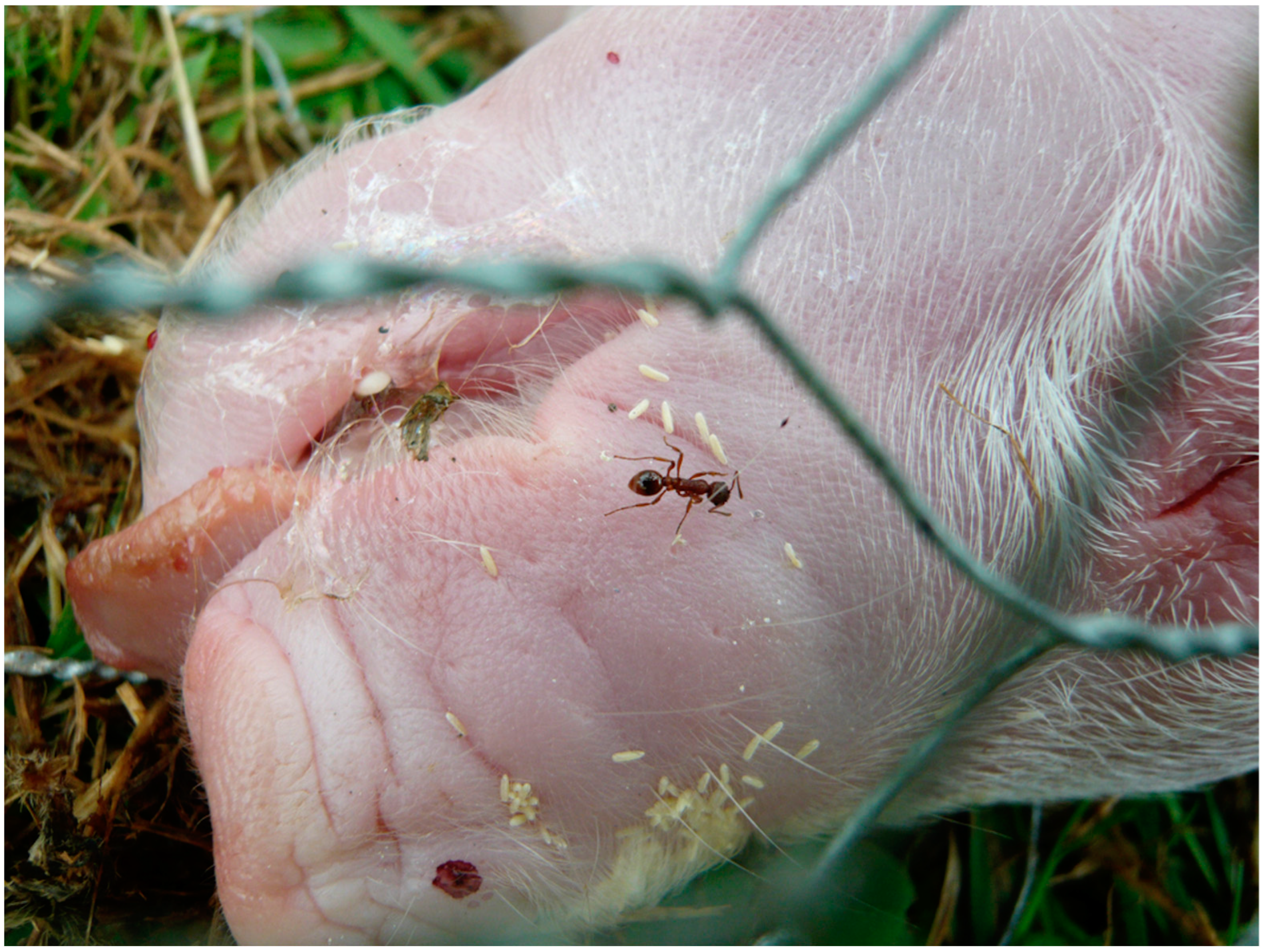
Figure 3.
Ant on a piglet’s cadaver.

Figure 4.
Hole in the shoulder region of a human body produced by beetles.

Figure 5.
Hole produced by dung beetles (MB observed the development of this hole). Possible confusion with close-range superimposed shot wound, further information in [22].
Figure 5.
Hole produced by dung beetles (MB observed the development of this hole). Possible confusion with close-range superimposed shot wound, further information in [22].

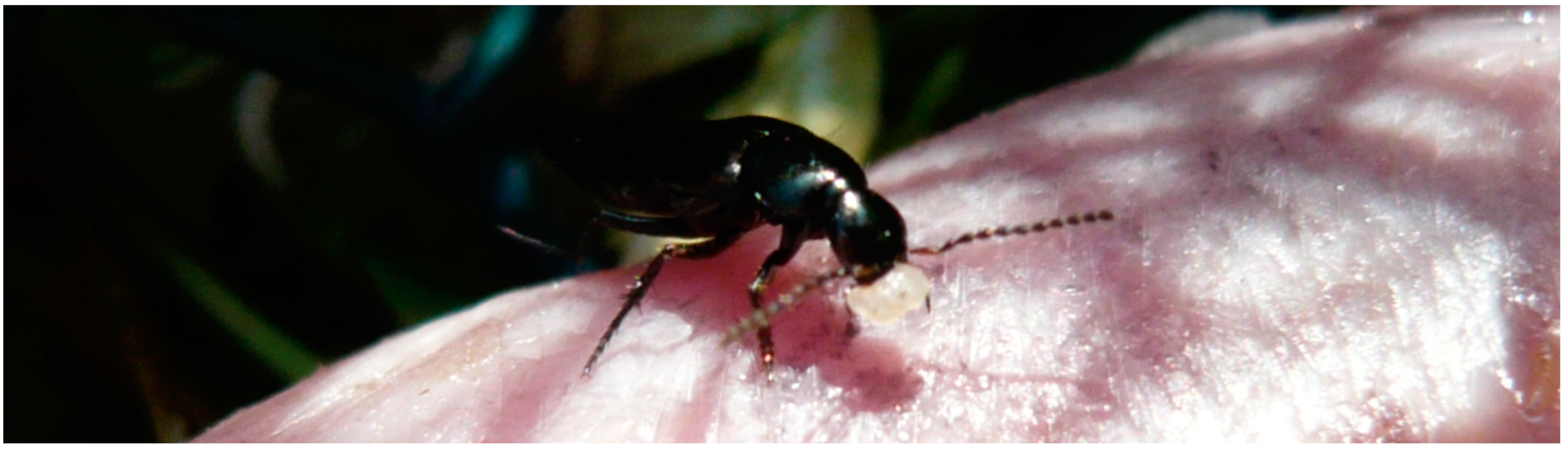
Figure 6.
Rove beetle predatorily feeding on fly maggots and “swiping”, with some feeding on a piglet’s cadaver.
Figure 6.
Rove beetle predatorily feeding on fly maggots and “swiping”, with some feeding on a piglet’s cadaver.

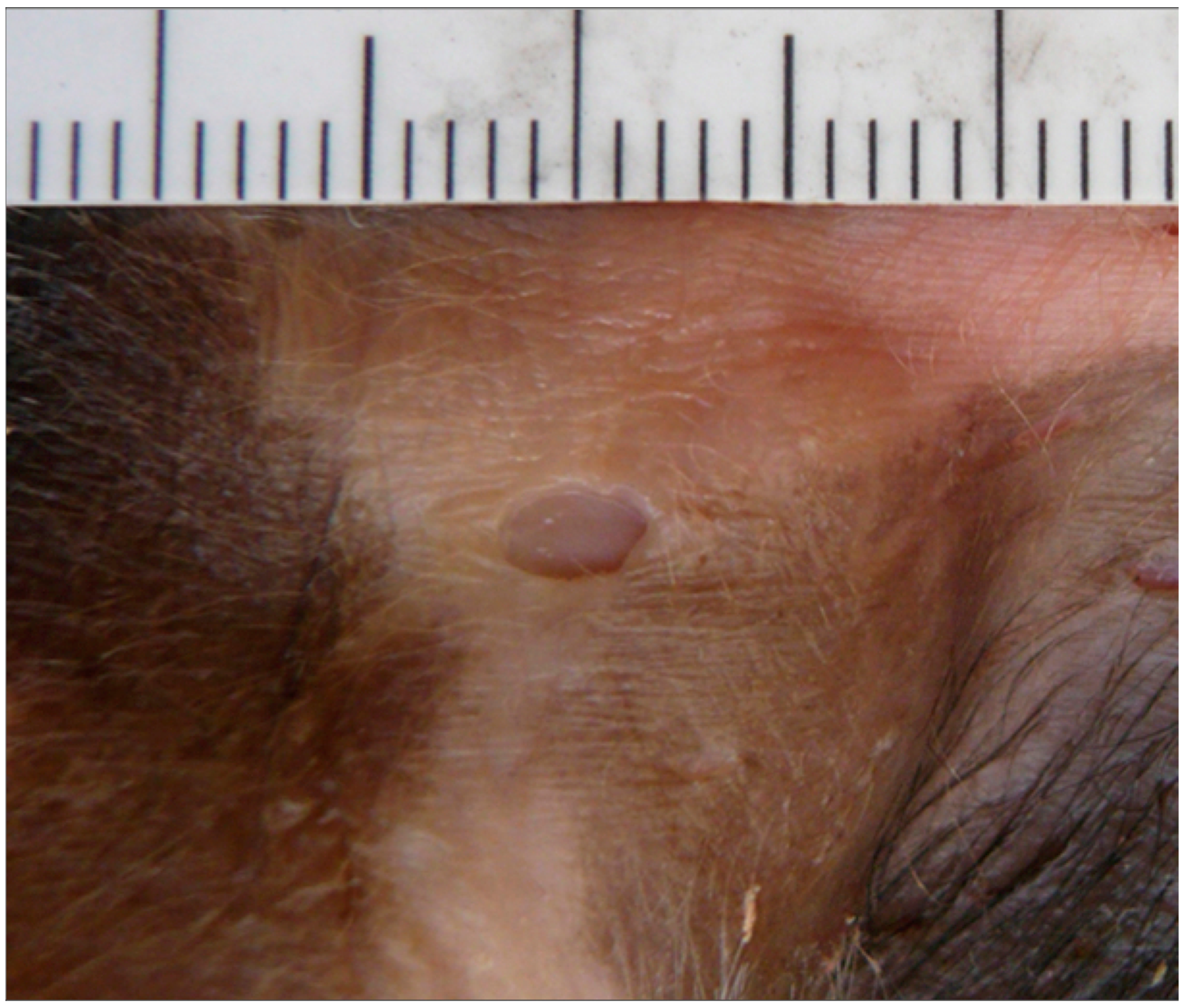
Figure 7.
Flat abrasion caused by feeding from dermestid beetles on a fresh piglet cadaver.

Figure 8.
Experimental wound reconstruction from Figure 7. A stabbing wound with the left tool resembles Figure 7.

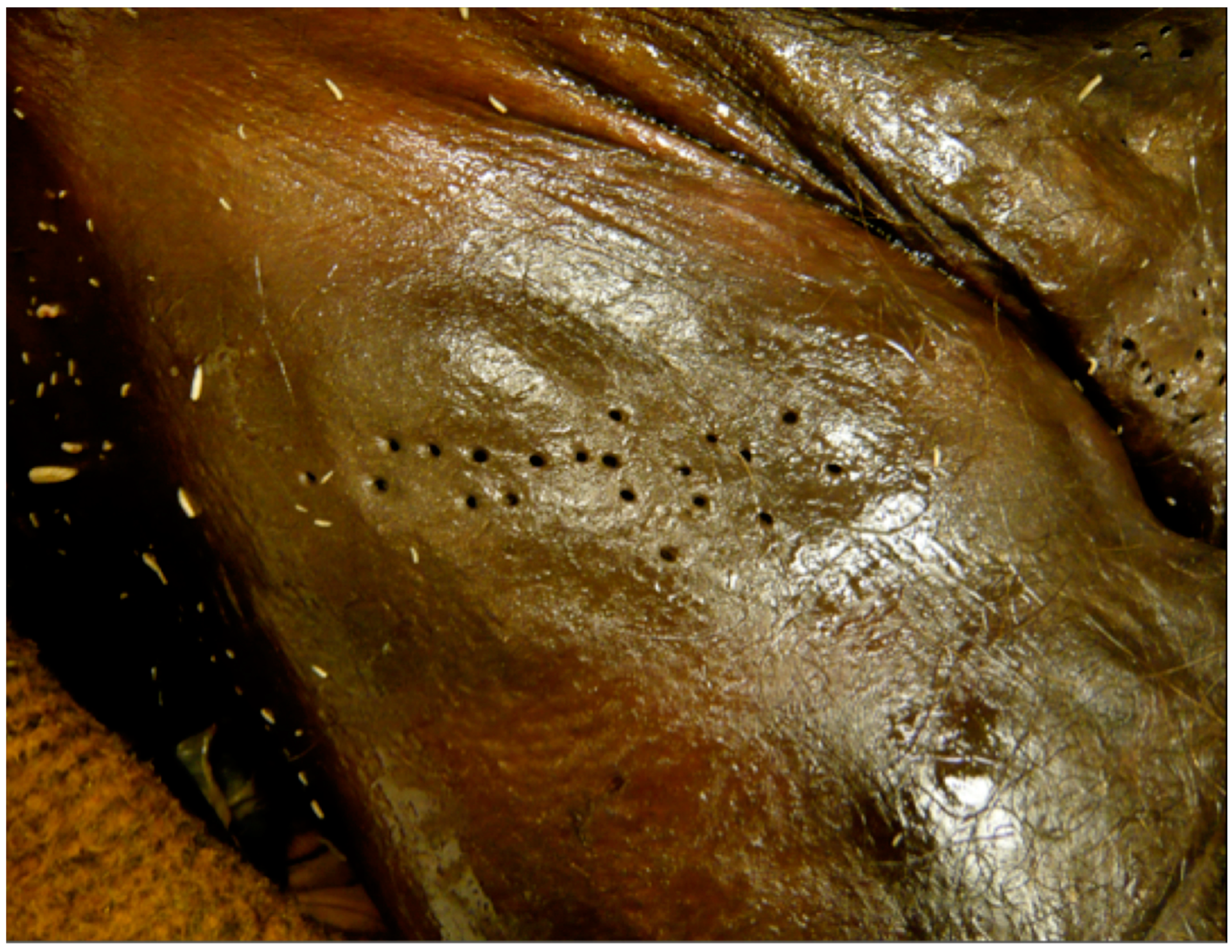
Figure 9.
Hole-like skin defects caused by maggots resembling scatter-gun holes.

Figure 10.
Another example for alleged “shotgun holes”. Trace caused by insects on the Anthroplogical Research Facility (“Body Farm”), Tennessee, USA.
Figure 10.
Another example for alleged “shotgun holes”. Trace caused by insects on the Anthroplogical Research Facility (“Body Farm”), Tennessee, USA.

Figure 11.
Homicide with more than ten stab wounds (top) (source: our case) vs. defecation/spitting traces by flies (bottom) [26]. Artefacts by insects may resemble blood stains; their body-length-to-tail ratio is often larger than 1. Gray bar = 1 cm.
Figure 11.
Homicide with more than ten stab wounds (top) (source: our case) vs. defecation/spitting traces by flies (bottom) [26]. Artefacts by insects may resemble blood stains; their body-length-to-tail ratio is often larger than 1. Gray bar = 1 cm.

Figure 12.
Preference for cadaver tissue by snails, revealing a possible source of change in postmortem interval (colonization time) calculations using the size and age of blow fly eggs. Additionally, rasping on dried-out areas/lines on the neck. Danger of confusion with injuries caused by assault against the head/neck, especially with scratch marks.

Figure 13.
Snails eating dead tissue and thereby causing holes which may resemble wounds. Left: snail feeding on pig’s tongue; right: hole in pig’s tongue produced by the same snail.
Figure 13.
Snails eating dead tissue and thereby causing holes which may resemble wounds. Left: snail feeding on pig’s tongue; right: hole in pig’s tongue produced by the same snail.

Figure 14.
Feeding defects by snails may have unusual shapes and can occur on different body parts. Left: snail feeding on right foot leg of piglet; right: wound-like tissue defect (also note the presence of blow fly eggs).
Figure 14.
Feeding defects by snails may have unusual shapes and can occur on different body parts. Left: snail feeding on right foot leg of piglet; right: wound-like tissue defect (also note the presence of blow fly eggs).

Figure 15.
By feeding on eggs of blow flies, wasps wrongly reset the “developmental clock” on the post-mortem interval. They also create skin defects similar to skin abrasions or scratch marks.
Figure 15.
By feeding on eggs of blow flies, wasps wrongly reset the “developmental clock” on the post-mortem interval. They also create skin defects similar to skin abrasions or scratch marks.

Figure 16.
Skin defects caused by wasps vary strongly. Left: the edges of these feeding traces are often irregular and “frayed”. Bones (right) can appear as if they have been gnawed off neatly.
Figure 16.
Skin defects caused by wasps vary strongly. Left: the edges of these feeding traces are often irregular and “frayed”. Bones (right) can appear as if they have been gnawed off neatly.

Figure 17.
Brownish greasy spots on the left forward outer edge of the sofa as well as on the edge of the bucket on the left side of the sofa.
Figure 17.
Brownish greasy spots on the left forward outer edge of the sofa as well as on the edge of the bucket on the left side of the sofa.

Figure 18.
Finding situation of the body. Arrows mark the direction of light coming through the window in the apartment of the deceased (arrowhead = window). Distinct dried-out areas of the face, hands, and feet.
Figure 18.
Finding situation of the body. Arrows mark the direction of light coming through the window in the apartment of the deceased (arrowhead = window). Distinct dried-out areas of the face, hands, and feet.

Figure 19.
Differential decomposition: clear differences in the decomposition process of a piglet between head and clothed lower body in open land (forest). (Experiments were conducted by Kristina Baumjohann, unpublished.)
Figure 19.
Differential decomposition: clear differences in the decomposition process of a piglet between head and clothed lower body in open land (forest). (Experiments were conducted by Kristina Baumjohann, unpublished.)

Figure 20.
Visible differences in the decomposition process of a piglet between clothed and unclothed body areas in open land (forest). Top: Original situation; bottom left: shirt removed to show differential state of decomposition: the area that was formerly clothed does not show maggot activity; bottom right: shirt liftet for inspection: no maggot acitivy under the shirt. Note that this pattern may be reversed under different weather or climate conditions. (Experiments were conducted by Kristina Baumjohann, unpublished.)

Figure 21.
Unilateral colonization by blow fly maggots of the eye socket due to light conditions, not to injury; further information in [39].
Figure 21.
Unilateral colonization by blow fly maggots of the eye socket due to light conditions, not to injury; further information in [39].

Publisher’s Note: MDPI stays neutral with regard to jurisdictional claims in published maps and institutional affiliations. |
© 2022 by the authors. Licensee MDPI, Basel, Switzerland. This article is an open access article distributed under the terms and conditions of the Creative Commons Attribution (CC BY) license (https://creativecommons.org/licenses/by/4.0/).
Share and Cite
MDPI and ACS Style
Baumjohann, K.; Benecke, M. Mind Your Decompositional Assumptions. Forensic Sci. 2022, 2, 725-740. https://doi.org/10.3390/forensicsci2040054
AMA Style
Baumjohann K, Benecke M. Mind Your Decompositional Assumptions. Forensic Sciences. 2022; 2(4):725-740. https://doi.org/10.3390/forensicsci2040054
Chicago/Turabian StyleBaumjohann, Kristina, and Mark Benecke. 2022. "Mind Your Decompositional Assumptions" Forensic Sciences 2, no. 4: 725-740. https://doi.org/10.3390/forensicsci2040054