Article

Cytotoxic Effects of Indonesian Betel Quid Components on Oral Keratinocytes and Fibroblasts

by
Elizabeth Fitriana Sari
1,2,*,
Ali I. Mohammed
3,
Antonio Celentano
3,
Michael John McCullough
3 and
Nicola Cirillo
3,*
1
Dentistry and Oral Health Discipline, Department of Rural Clinical Science, La Trobe Rural Health School, Bendigo, VIC 3552, Australia
2
Faculty of Dentistry, Universitas Padjadjaran, Bandung 45363, Indonesia
3
Melbourne Dental School, The University of Melbourne, Carlton, VIC 3053, Australia
*
Authors to whom correspondence should be addressed.
BioChem 2023, 3(4), 153-169; https://doi.org/10.3390/biochem3040011
Submission received: 14 June 2023 / Revised: 23 July 2023 / Accepted: 5 October 2023 / Published: 23 October 2023

Abstract

:
A betel quid (BQ) chewing habit has been strongly associated with the development of several oral mucosal diseases. In order to investigate whether individual components of BQ mixtures have distinct physio-pathological effects on oral mucosal cells, we examined the impact of areca nut (AN), Piper betle leaf (Leaf), Piper betle stem inflorescence (SI), areca husk (Husk) and the complete BQ mixture on the growth of oral keratinocytes (OKF-6) and primary oral fibroblasts (MMF-1). Based on their known chemical properties, we selected BQ samples from Banda Aceh (BA) and West Papua (WP) regions for our in vitro study. We used a fluorescein diacetate assay (FDA) to assess the cell viability of BQ components on OKF-6 and MMF-1 cells. The cytotoxic effect of WP-AN on the OKF-6 cell line was observed at a concentration of 100 μg/mL, resulting in a 50% reduction in cell viability (IC50) after a 2-day incubation. Similarly, BA-AN exhibited cytotoxic effect, although at a higher concentration (500 μg/mL). WP-SI also displayed cytotoxic effects at a concentration of 500 μg/mL following 2 days of incubation. In contrast, Leaf, BQ mixture and husk extracts did not show any cytotoxic effects even after 3 days of incubation. No cytotoxic effects were observed at any concentration of BQ components when exposed to MMF-1 cells. Regarding cell proliferation, MMF-1 cells exposed to BA-AN and WP-AN showed increased growth on day 1, followed by decreased growth on day 2, in a dose- and time-dependent manner. Overall, our study indicates that BQ components induce distinctive cytotoxic effects on stromal and epithelial cells from the oral cavity.

1. Introduction

Several epidemiological and case control studies in the Indian subcontinent, South-East Asia and South Pacific islands have reported a strong correlation of betel quid (BQ) chewing habit with oral cancer and oral potentially malignant disorders (OPMDs) [1,2,3,4,5,6]. In particular, BQ chewing is associated with an increased risk of oral submucous fibrosis (OSMF) [1,2], an insidious disease that originates in the stromal fibroblasts [3,6]. The pathophysiology of these BQ-induced mucosal changes is not well understood, and the data on potential BQ toxicity are heterogenous due to differences in methods of BQ extraction as well as the cytotoxicity assays utilised [1,7].
The ingredients of BQ differ in different countries, but the major components being used are areca nut (AN), betel leaf or betel stem inflorescence (SI) and slaked lime. Tobacco and other spices are optional and reflect local chewing practices [8,9,10]. Approximately 600 million individuals worldwide are estimated to be AN or BQ chewers [8,9].
Our previous studies on Indonesian BQ components from four different regions has successfully profiled the metabolomic and antioxidant properties and highlighted the presence of chemicals and metabolites that are potentially harmful, but also of molecules that may be protective [11,12]. In the present study, cytotoxicity assays were used to investigate the pathobiological effect of BQ components on human oral mucosal cells. We selected BQ components from BA and WP regions as these had the lowest and the highest level of antioxidants, polyphenols, and arecoline, respectively. Furthermore, our pilot clinical data suggest that BA and WP are among the regions with the lowest and highest prevalence of oral submucous fibrosis (OSMF), respectively [13]. Hence, in this study we aimed to investigate whether the cytotoxic effect of BQ on oral mucosal cells correlated with its clinical-molecular profile, with the hypothesis that distinct BQ ingredients harbour different physio-pathological effects [14].
AN is the main component of BQ and is considered to be the major aetiologic factor of BQ-related oral cancer [4,7]. AN extract (ANE) contains mainly tannin and areca alkaloids, such as arecoline, arecaidine, guvacine, and guvacoline, which are cytotoxic but can also work as potential carcinogens. These substances are released from BQ components during the period of chewing BQ and with time can dramatically alter the normal oral mucosa and induce pathological changes typical of OSMF [2]. Previous studies have reported that ANE, arecoline and BQ extracts were cytotoxic to cultured oral keratinocytes and fibroblasts [2,15,16,17,18,19], and this is partially in contrast with clinical data that imply a pro-carcinogenic effect of BQ mixtures. Surprisingly, the potential cytotoxic effects of individual BQ components have not been investigated so far. It is also important to differentiate between keratinocytes and fibroblasts as these cell types have different functions in the pathogenesis of OSMF and oral cancer. Hence, in the current study our objective was to evaluate the thresholds for acute cytotoxicity of different Indonesian BQ formulations in oral keratinocytes (OKF-6) and primary oral fibroblasts (MMF-1). This assessment aims to provide guidance for the creation of appropriate in vitro models for BQ-related disease. Furthermore, gaining a comprehensive insight into the impact of individual BQ components could prove vital in shaping targeted preventive and chemo-preventive approaches to mitigate the occurrence of OSMF and BQ-related oral cancer.

2. Material and Methods

2.1. Preparation of Betel Quid Aqueous Extracts

Extraction of dried areca nut, betel leaf, betel stem and areca husk was performed following published methods with modifications [20]. Seeds and husks of areca nut were manually separated. All samples were freeze-dried for 72 h using an FD3 Freeze Drier (Dynavac Engineering, Cannington, WA, Australia) and finely ground using an electronic grinder (Multigrinder II, model EM0405, Sunbeam, Auckland, New Zealand). Then, 5 g of each sample powder was weighed into a 50 mL centrifuge tube prefilled with nitrogen gas (99.99% Grade 4.0, Coregas, Yennora, NSW, Australia), with 50 mL of methanol then added as the extraction solution. Extractions were carried out at 20 °C for 48 h under shaking at 150 rpm (Incubator shaker ZWYR-240, LABWIT Scientific, Melbourne, VIC, Australia). After the extraction, the samples were centrifuged at 6500 rpm for 10 min at 20 °C. The supernatants were transferred to a clean tube and evaporated at 40 °C to remove the solvent entirely using a Hei-VAO Value rotary evaporator (Heidolp Instruments GmbH & Co.KG, Schwabach, Germany), then redissolved in 10 mL of LC-grade methanol. The final extract was flushed with nitrogen gas and sealed with parafilm to avoid oxidation, and stored at 4 °C in darkness until analysis. The extract stock solution was filtered using a 30 mm × 0.45 µm nylon syringe filter (Thermo Fisher Scientific, Scoresby, VIC, Australia) to gain a clear solution before further analyses.
The extract concentrations were established based on a study conducted by Van Wyk et al. Cells were exposed to AN and Leaf extract concentrations of 100, 250 and 500 μg/mL [19]. The determination of BQ mixture concentrations was informed by a preliminary study that indicated cellular changes upon exposure to BQ mixture extracts at 1 mg/mL, 15 mg/mL and a doubled concentration of 30 mg/mL. The determination of husk concentrations was carried out following a similar approach, wherein husk extracts induced cellular changes at 1 mg/mL, 22.5 mg/mL and a doubled concentration of 45 mg/mL.

2.2. Cells and Culture Conditions

The primary fibroblast (MMF-1) cell line used in this study was derived from early passages (<12) of normal human oral fibroblasts isolated from the buccal mucosa of a healthy 57-year-old patient. The ethical approval for the collection of the material from which these cells were obtained was granted by the Human Research Ethics Committee (HREC, number 1340716) for the study titled “The alteration of the oral glucocorticoid system in oral squamous cell carcinoma primary cultures compared with normal oral keratinocytes”.
These primary fibroblasts were cultured in a growth medium composed of Dulbecco’s modified Eagle’s medium (DMEM, Merk, cat #D5796, Sigma-Aldrich, Castle Hill, NSW, Australia) with 2 mM L-glutamine, supplemented with 10% (v/v) fetal bovine serum (FBS), 100 IU/mL penicillin and 100 μg/mL streptomycin. The cultures were maintained at 37 °C in a humidified atmosphere with 5% CO2.
Immortalized normal human oral keratinocytes’ (OKF-6/TERT) use in this experiment was established by Dickson et al. [21]. These oral keratinocytes were isolated from the floor of the mouth of a 57-year-old male patient. The OKF-6 cell line was kindly supplied by the Oral Health Cooperative Research Centre (OHCRC) at the University of Melbourne in Australia. Prior to experimentation, the OKF-6 cells underwent karyotype analysis at the Cytogenetics Laboratory of the Victorian Clinical Genetic Service in the Royal Children’s Hospital in Parkville, Victoria [21].
OKF-6 cells were cultured in standard cell suspending medium (CSM), containing GIBCO Keratinocyte Serum-Free Medium (GIBCO KSFM, #17005-042, Thermo Fisher Scientific, Mulgrave, VIC, Australia) supplemented with 25 μg/mL pituitary bovine extract (PBE), 0.2 ng/mL epidermal growth factor (EGF), 100 IU/mL penicillin and 100 μg/mL streptomycin, and containing 10% (v/v) new calf serum (NCS). Keratinocytes were maintained in a humidified incubator with 5% CO2 at 37 °C.

2.3. Treatments

Sample solutions for treating cells were areca nut, leaf, husk and BQ mixtures from West Papua and Banda Aceh. Those BQs from two regions were, respectively selected from our previous studies based on the highest and lowest extracts of areca nut, BQ mixture, husk, leaf and stem inflorescence (SI) that contained polyphenols and alkaloids [11,12]. Samples were prepared in the cell culture media containing 1% fetal bovine serum (FBS) for primary fibroblasts (MMF1) and 1% containing new calf serum (NCS) for OKF-6. The extract concentrations for the cytotoxicity assay were varied: for areca nut and leaf/stem, they were made at 100 μg/mL, 250 μg/mL and 500 μg/mL; for the BQ mixture, they were made at 1 mg/mL, 22.5 mg/mL and 45 mg/mL; and for areca husk, they were made at 1 mg/mL, 15 mg/mL and 30 mg/mL. These concentrations were determined based on the initial range of findings from previous studies and a preliminary study we conducted.

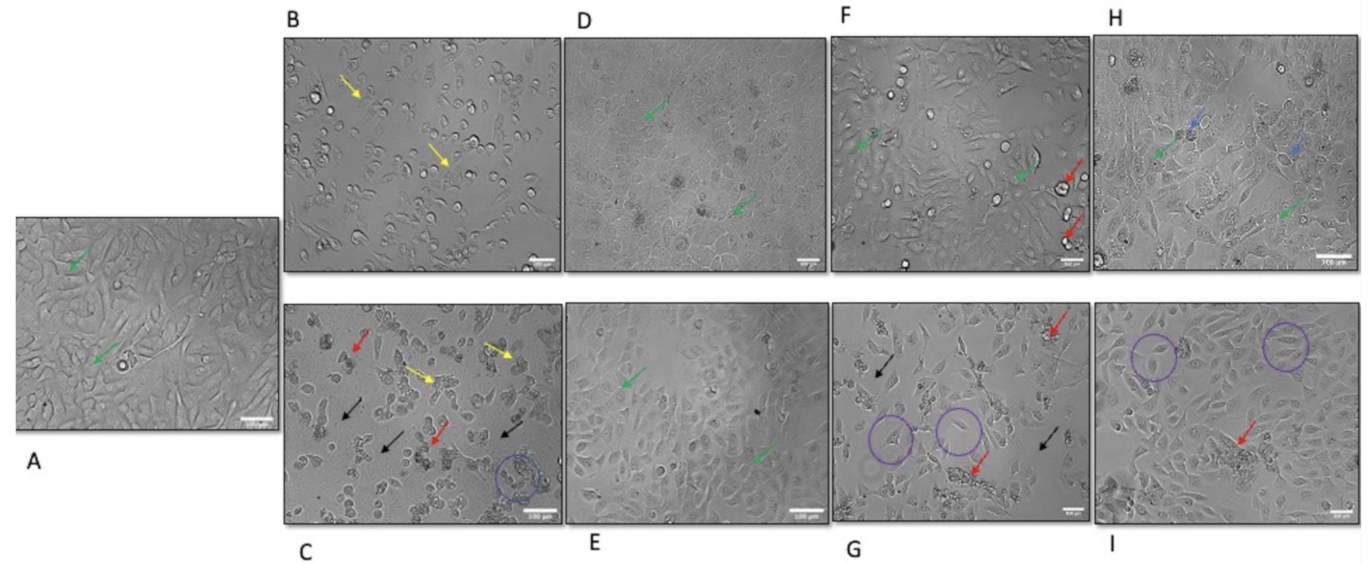
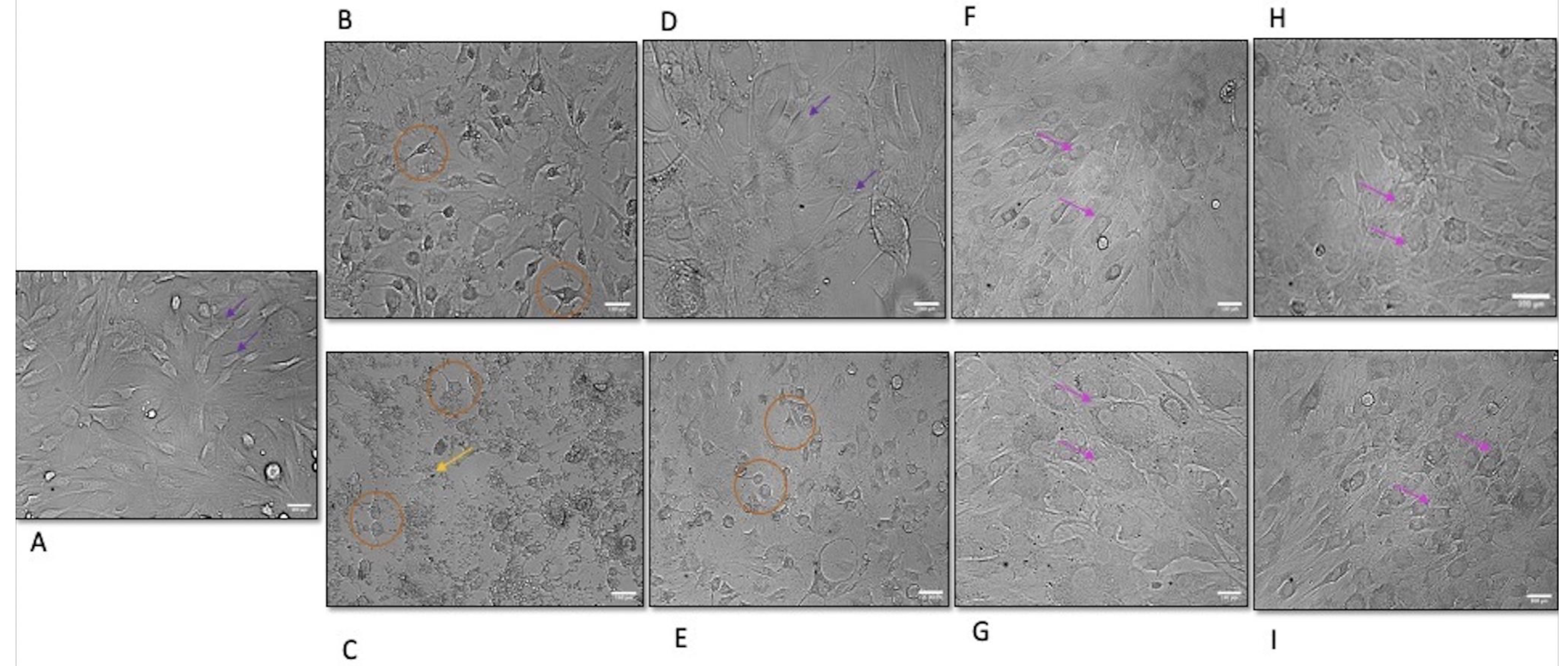
2.4. Morphological Evaluation

Morphological evaluation was performed using an inverted microscope (FLoid™ Cell Imaging Station, Life Technologies, Mulgrave, VIC, Australia). The viable cells after exposure to arecoline were taken up via fluorescein diacetate (FDA), stained and exhibited green or white light, while non-viable cells remained dark.

2.5. Viability Assay

The assay was carried out according to the procedure reported by Gonzalez-Reyes et al. with some modifications [22,23]. Briefly, 96-well Costar® microplates (Sigma-Aldrich, Castle Hill, NSW, Australia #CLS3603-48EA) were used and plated with 100 µL at 2 × 10⁴ cells/mL of OKF-6 or 1.35 × 10⁴ cells/mL of MMF1 cell suspension. The cell viability of seeded cells was at least 90% when determined with trypan blue stain. A humidified incubator (Thermo Scientific, Forma direct heat, CO2 incubator) was used to incubate cells, and this was performed at 37 °C in 5% CO2. After 24 h of seeding, plates were removed from the incubator and washed with 100 µL/well PBS, and the cells were treated with 100 µL/well BQ concentrations as previously prepared. Three wells/plate were used as controls. Control wells contained untreated cells that were incubated with 100 µL culture medium containing Dulbecco’s modified Eagle’s medium (DMEM) with 2 mM L-glutamine, supplemented with 10% (v/v) FBS, 100 IU/mL penicillin and 100 μg/mL streptomycin. Blank wells (without cells) contained 100 µL culture medium only. After treating the cells with various BQ concentrations, they were incubated in a 37 °C/5% CO2 incubator for 1 day. After 1 day, control media or test compounds were removed, the cells were rinsed with 100 µL PBS and, subsequently, 100 µL of FDA solution prepared in serum-free medium was added to each well. The plates were incubated at 37 °C/5% CO2 for 40 min; then, FDA fluorescence was measured via spectrometry at wavelengths of 485/20 nm and 528/20 nm, respectively, for excitation and emission, in a Synergy HTX Multi-Mode Reader (Bio-Tek, Shoreline, WA, USA). The FDA-stained cells were viewed through an inverted microscope (Floid cell imaging station, Life Technologies) with fluorescence/green or white light as required to see whether the cells were correctly stained. The steps were repeated after treatment with BQ for 2 days and 3 days of incubation. To validate the result of spectrometry readings in the Synergy HTX Multi-Mode Reader, one BQ cytotoxic concentration (IC50) was selected with its time point (1 time point over 3 days of incubation). Cells were seeded in a 6-well plate in triplicate for 24 h of incubation to attach and, after 24 h, the cells were treated with BQ at a cytotoxic concentration (IC50) according to previous spectrometry finding. After exposing the cells to BQ components at the found IC50 and incubating them for the IC50 time point of incubation (whether the IC50 happened on day 1, day 2 or day 3 of incubation), cells were counted with an automated cell mincounter (Bio-Rad, South Granville, NSW, Australia) and stained with trypan blue to count the viable cells. If the results of IC50 were consistently similar to the spectrometry readings, then the IC50 read using spectrometry was considered to be correct.

2.6. Statistical Analysis

Statistical analysis was conducted using MS Excel, Minitab Express© version 1.5.1. 2017 and R commander. The significant difference was tested using one-way ANOVA, with the expected p-value < 0.05 and a 95% CI. Tukey’s HSD post hoc test was used to find significant differences between internal groups. The cell cytotoxicity (IC50) was measured as a 50% inhibition of cell viability.

3. Results

With few exceptions, exposure of oral mucosal cells to BQ components from BA and WP regions for over 3 days influenced cell growth in a dose- and time-dependent manner (Table 1 and Table 2). Notably, all BQ components significantly reduced cell viability, albeit to a variable extent.

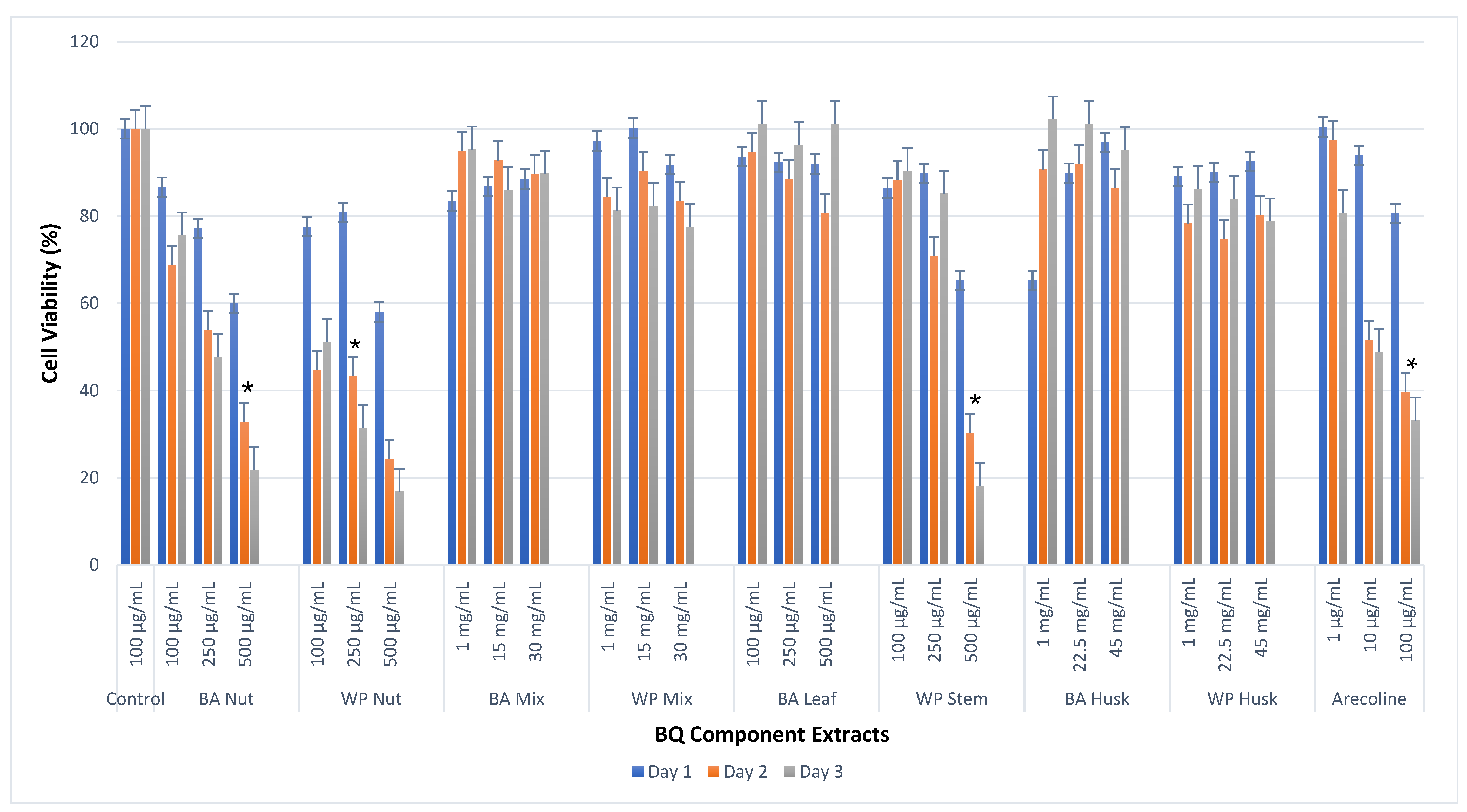
3.1. Cell Viability Effects of BQ Extracts on OKF-6

BA-AN and WP-ANE both inhibited OKF-6 growth in a dose- and time-dependent fashion, with the exception of 100 μg/mL at 2 and 3 days. The IC50 of WP-ANE for OKF-6 was 100 μg/mL (2 days), while ANE from the BA region exhibited cytotoxicity at 500 μg/mL (2 days).
BA-BQ mixture and WP-BQ mixture (1, 15 and 30 mg/mL) induced mild cytotoxicity during 3 days of incubation. In pilot experiments, BA-BQ mixture and WP-BQ mixture (100, 250, 500 and 1000 μg/mL) induced cell proliferation; hence, we raised the concentrations to 1, 15 and 30 mg/mL to clearly observe the cytotoxic effect of BQ mixture on OKF-6.
The cytotoxic effect of WP-SI was significant at 500 μg/mL (2 days), whereas BA leaf did not induce a reduction in OKF-6 cell viability at any concentration used (100, 250 and 500 μg/mL), except with 500 μg/mL on Day 2.
Oral keratinocytes incubated with husk extract from WP, but not from BA, exhibited a significant difference in cell viability at all concentrations assessed (1, 22.5 and 45 mg/mL) after 48 and 72 h of incubation. The IC50 of arecoline was reached at 100 μg/mL after 2 days of incubation. The reduction in cell viability induced by arecoline was dose and time dependent, as shown in Table 1 and Figure 1.

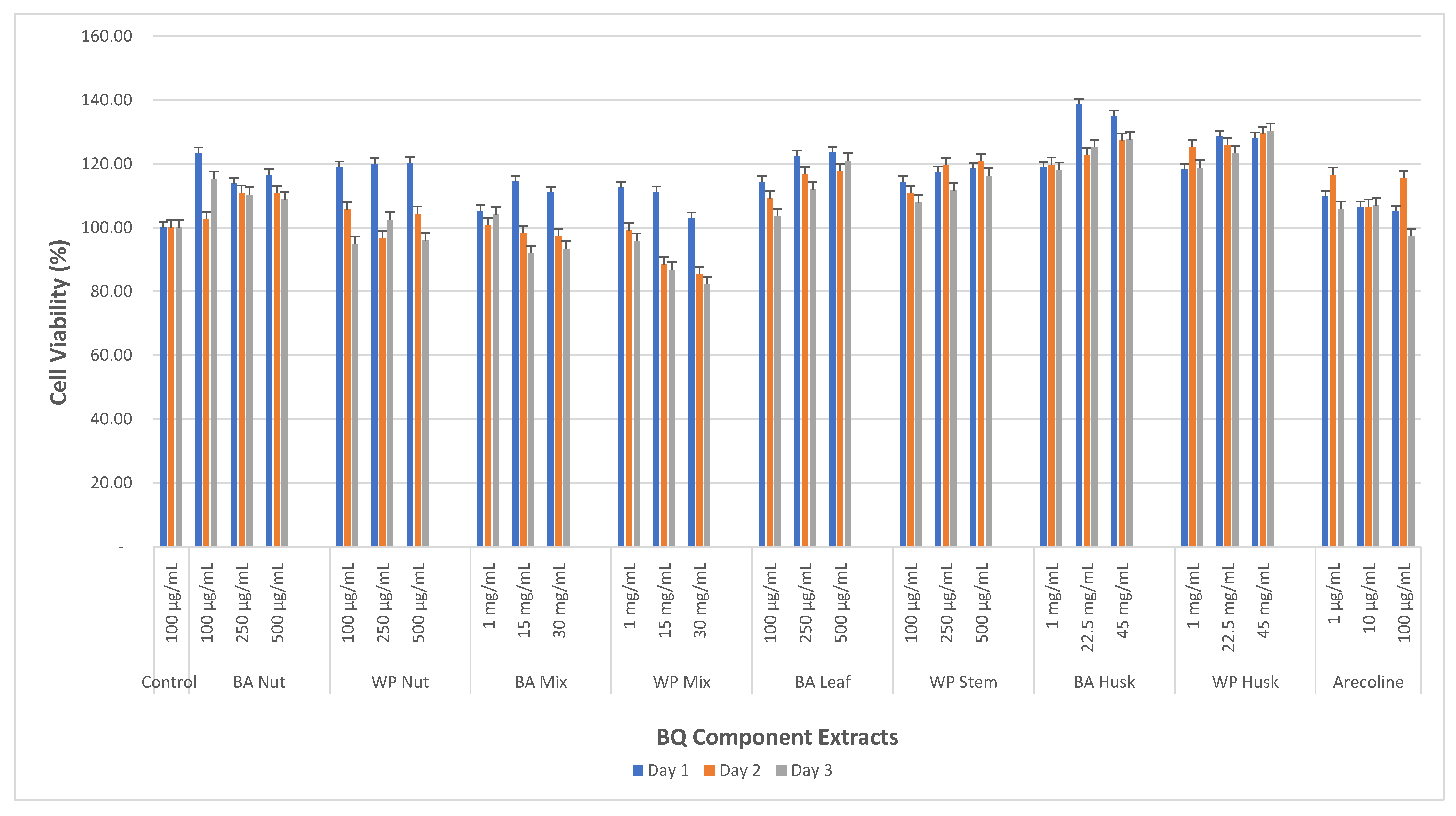
3.2. Cell Viability Effects of BQ Extracts on MMF-1

None of the BQ components from any region exhibited cytotoxicity to MMF-1 at the concentration used for up to 3 days and, importantly, arecoline failed to induce cytotoxic effects at all concentrations used (1, 10 and 100 μg/mL) for up to 3 days of incubation (Table 2). Notably, all BQ components induced proliferation of oral primary fibroblasts, as shown by the increased cell viability compared to untreated controls. This cell proliferation only occurred in the first day of incubation, and the number of viable cells declined consistently thereafter (Table 2 and Figure 2).

3.3. Morphological Alteration of OKF-6 Following Exposure to BQ Components from BA and WP Regions

Morphological alterations in cultured OKF-6 following exposure to BQ components were observed. Untreated normal OKF-6 showed ovoid, rounded or polygonal appearances with well-defined intercellular spaces (Figure 3A). Exposure of OKF-6 to 500 μg/mL ANE from BA for 1 day induced loss of intercellular space and cell shrinkage (Figure 3B). Exposure of OKF-6 to 500 μg/mL of ANE from WP for 1 day showed substantial loss of intercellular spaces, cells were rounded and shrunk, the architecture of nuclei and nucleoli was not clearly demarcated, and some debris and dead cells were present, along with evidence of intercellular vacuolization (Figure 3C). Thus, both WP-AN and BA-AN caused morphological change consistent with cellular degeneration and death (Figure 3).
Cells exposed to BA-BQ mixture extract (30 mg/mL) for 1 day showed polygonal appearance, and intercellular spaces were sharply defined (Figure 3D). Exposure of OKF-6 to 30 mg/mL of BQ mixture extract from WP over 1 day of incubation showed well-defined intercellular spaces, no loss of intercellular spaces and a polygonal shape of cells (Figure 3E). Keratinocytes exposed to 500 μg/mL of BA leaf extract for 1 day displayed both polygonal and elongated appearance, and some dead cells were also present (Figure 3F). Exposure to 500 μg/mL of SI extract for 1 day induced loss of intercellular spaces, architectural changes including fibrastoid and shell-like appearance, and dead cells (Figure 3G). Exposure to BA husk extract (45 mg/mL) showed polygonal and rounded cell appearances, intercellular spaces were intact, some dead cells were also present, and cells’ architecture changed (Figure 3H). Exposure of OKF-6 to 45 mg/mL of WP husk extract for 1 day showed well-defined enlargement of intercellular spaces. Cytologically, oral keratinocytes were polygonal with evidence of shell-like appearance, and vacuolization was evident (Figure 3I).

3.4. Morphological Alteration of MMF-1 Following Exposure to BQ Components from BA and WP Regions

Morphological alteration of oral primary fibroblasts exposed to BQ components was evident and various changes in cell characteristics were observed. The normal untreated MMF-1 (control) showed a spindle shape with well-defined intercellular spaces (Figure 4A). MMF-1 was exposed to 500 μg/mL of ANE from BA for 1 day and showed stellate form and unclear intercellular spaces (Figure 4B). The morphological change of MMF-1 exposed to 500 μg/mL of WP-ANE for 1 day showed stellate form with debris, and dead cells were present, while less intercellular space was observed (Figure 4C).
Exposure of MMF-1 to 30 mg/mL of BA-BQ mixture for 1 day showed cell architectural changes (Figure 4D). Exposure of MMF-1 to 30 mg/mL of WP-BQ mixture for 1 day showed a stellate cell form and intercellular spaces (Figure 4E). Exposure of MMF-1 to 500 μg/mL BA leaf extract for 1 day showed epithelioid formations (Figure 4F). Exposure of MMF-1 to 500 μg/mL SI extract for 1 day showed epithelioid cell forms, and no intercellular space loss was observed (Figure 4G). Exposure of MMF-1 to 45 mg/mL of husk extract from BA over 3 days of incubation showed epithelioid forms with well-defined intercellular spaces (Figure 4H). Exposure of MMF-1 to 45 mg/mL of husk extract from WP showed cell architecture change into epithelioid forms and no loss of intercellular spaces (Figure 4I).

4. Discussion

In the present study, we show that BQ ingredients not only induce distinct functional and morphological effects in oral mucosal cells but, also, that these effects are mostly time, dose, and cell type dependent. Cell viability as measured by FDA assay was performed to investigate the basic pathobiological effect of BQ components on oral keratinocyte (OKF-6) and oral fibroblast (MMF-1) cells. Whilst individual ingredients of BQ were cytotoxic at the concentration used, whole BQ mixtures induced cell proliferation and reduced cell viability only at much higher levels. BQ mixtures externed distinct effects of keratinocytes and fibroblasts, surprisingly, the same chemical components induced transient cell proliferation in oral fibroblasts. The different cytotoxicity seen between BA-AN and WP-AN may be due to the different maturity of AN. BA-AN is a ripe mature AN, while WP-AN is an unripe type. Our previous study [11,12], that was consistent with several studies, reported that unripe AN contains more polyphenols as an antimutagenic rather than ripe AN [8]. WP-AN had antioxidants activities (TPC, FRAP, DPPH scavenging activities) almost 2-fold higher than BA-AN, and contains more polyphenols quantification as shown in Table 1 and Table 2. However, the excessive amount of phenolics could lead to cytotoxic instead of protective effects [24]. Furthermore, we also showed that arecoline quantification by High pressure liquid chromatography (HPLC) was higher in WP-AN compared to BA-AN, 250 mg/mL ANE from WP, contain 6.92 ± 1.64 μg/mL, while 250 mg/mL ANE from BA contains 6.10 ± 0.99 μg/mL [11,12]. Arecoline is believed to be the major alkaloid identified in ANE and has been shown to exhibit cytotoxicity on epithelial cells in cancer cells line [17,18,25]. Previous studies have shown that a concentration of 100 μg/mL of an aqueous ANE contains 10 μg/mL of arecoline [19,26]. A further study reported that ANE exhibited cytotoxicity to cultured gingival keratinocytes (C3H10T/12) at concentrations higher than 320 mg/ mL, as shown by a decrease in colony numbers [2]. These previously reported cytotoxic effects of AN in these studies are consistent with our current results. The variation of IC50 of ANE may have occurred due to different methods of extraction used, maturity of AN, and cytotoxicity assays used by researchers [8,19,27]. Unlike ANE, no cytotoxic effect, as measured by 50% decreased cell viability, was induced by BQ Mixture from both regions at any concentration used over 3 days of incubation (Table 1). This can be due to the fact that BQ Mixture not only contains AN but also leaf or SI, as well as slaked lime which could counterbalance the effect of arecoline. Consistently, our previous phytochemical study showed that arecoline was not detected in betel leaf and SI samples. This finding is in line with other studies showing that betel leaf and SI mostly contains polyphenols without any alkaloids [8,28,29]. Polyphenols contained in the leaf and SI might act as chemo-preventive compounds to oral cancer [8,30]. In particular, hydroxychavicol could reduce oral cancer risk in people who chew BQ with tobacco [31].
Although slaked lime in the BQ Mixture could decrease the concentration of phenolics by extreme changes of pH as well as cause direct damage [32], long-term lime exposure to oral mucosa would be necessary to produce oral lesions. For example, previous study reported that only long-term exposure of Chinese hamster cheek pouch to slaked lime (calcium hydroxide) was able to create hyperplastic lesions in these animals. Further studies showed that repeated topical application of lime also could led to oral leukoplakia [7,33]. These data suggest that the small quantities of slaked lime in BQ would be able to induce cellular changes of the oral mucosa only if given repeatedly and for long term. In the present study, cells were exposed to slaked lime contained in BQ in a short-term over 3 days-time and in the presence of other potentially chemo-preventive compounds such as betel leaf or SI. This might explain why BQ Mixture showed no cytotoxic effect on OKF-6 over 3 days.
People living in BA region tend to chew BQ by wrapping all ingredients in leaf of piper betle, while in contrast people living in WP region pick a part of SI piper betle to be chewed together with other BQ components [34]. The results of the current study show that BA-Leaf resulted in growth stimulation of OKF-6. Our previous phytochemical study identified a number of polyphenols in betel leaf and SI [11,12]. Previous studies of betel leaf indicated that of hydroxy chavicol (69.46%), 4-chromanol (24%) and eugenol (4.86%), possess antioxidant, anti-inflammatory, anti-platelet and antithrombotic, antibacterial and antifungal activities [35].
In contrast, WP-SI showed cytotoxic effect at 500 μg/mL after incubation for 2 days. Even though WP-SI contains a range of polyphenols that could be protective to the cell [11,12], it also contains other toxic phytochemicals that could overcome the effect of polyphenols. Our previous study using GC-MS identified safrole in high amount in WP-SI sample [12] and several studies have shown safrole as a possible human carcinogen [8,29]. High concentration (15 mg/mL) of safrole has been found in SI [6]. Consequently, chewing BQ containing SI may expose the oral mucosa to safrole and, consistently, this compound has been shown to be present in concentrations of about 420 μM in saliva during BQ chewing [36]. Safrole is also classified as a hepatocarcinogen in mice and rats [36]. A previous study reported that betel SI extract at concentration 100–400 mg/mL is cytotoxic on cultured gingival keratinocytes [25]. These studies support the view that SI confers cytotoxic properties to BQ from WP.
The husk extract from both regions did not exhibit cytotoxic effects on OKF-6 at any concentration used over the 3 days of incubation, although these did reduce cell viability significantly to a different extent. Our previous study on arecoline quantification using HPLC detected low arecoline concentrations in husk from most regions, where WP-Husk had the highest arecoline amount and BA-Husk did not contain detectable arecoline [11]. Arecoline’s role in cell inhibition may be reflected in WP-Husk-induced inhibition OKF-6 growth by dose and time fashion, while BA-Husk showed very little inhibition of cell growth. Both husks contain several polyphenols, such as sinapic acid and 5-(hydroxymethyl) furfural [11], which could slow the detrimental effect of arecoline on OKF-6. The cytotoxic effect of husk containing arecoline could provide additional information to some countries such as Guam, China, and Taiwan that include husk with BQ chewing habit [8]. Furthermore, proper husk waste management need to be considered to avoid ongoing environmental pollution.
The increase of cell viability compared to control indicated that BA-AN and WP-AN induced cell proliferation of oral primary fibroblasts over 3 days of incubation. Harvey et al. also recorded increased human fibroblast proliferation with areca nut and showed cytotoxicity at 100 μg/mL in 5 days of incubation [16,19]. This result is consistent with our study that BA-AN and WP-AN showed increased number of oral fibroblasts. In our experiments, cell proliferation was transient and occurred primarily within the first 24 h of incubation to then decrease steadily during the following 2 days.
AN as the major ingredient of BQ Mixture is most likely to induce cell proliferation via the action of areca alkaloids, especially arecoline [16,19]. The increase of cell viability in oral fibroblasts incubated with BA and WP-BQ Mixture was seen on 1 day of incubation (Table 2). However, the number of viable cells started to decline within 2 days, in particular in WP-AN samples. The Mixture of BQ ingredients allows interaction between alkaloids [16,37] and polyphenols [7] possessed by nut and leaf/SI. The polyphenols in leaf/SI and short-term exposure of slaked lime to cells could lead to delayed cell inhibition [32] that is consistent with the results of the current study.
BA-Leaf and WP-SI did not exhibit cell cytotoxicity after 3 days of incubation with MMF-1. In contrast, cell proliferation occurred up to 3 days of incubation at all concentration used. This cell proliferation might be due to fact that leaf/SI contain polyphenols. Our phytochemistry study identified polyphenols in leaf and SI, such as sinapic acid and 5-(hydroxymethyl) furfural [11,12]. A previous study that reported areca nut carcinogenicity was suppressed by the betel leaf when injected to mice [38]. This areca alkaloids-antagonistic action by betel leaf has been attributed to the antioxidative nature of phenolics, particularly of appreciable quantities of hydroxychavicol. Antioxidative property of betel leaf has been reported to be much more either butylated hydroxy anisole or butylated hydroxytoluene [39,40].
BA-Husk and WP-Husk did not exhibit cytotoxic effect on MMF-1 up until 3 days of incubation. The appreciable numbers of identified polyphenols and small amount of arecoline in both husks [11,12] might slow the inhibition of cell growth and rather more likely to stimulate cell growth as shown by the increased cells number compared to control.
Arecoline exhibited cytotoxicity on OKF-6 at 100 μg/mL after incubation for 2 days. Arecoline suppressed growth of OKF-6 at 1 μg/mL, 10 μg/mL, and 100 μg/mL respectively by 80.75%, 48.79%, and 33.16% after incubation for 3 days. However, arecoline induced cell proliferation on MMF-1 at all concentrations used over 3 days of incubation. Several researchers reported that arecoline suppressed oral keratinocyte cell vitality at different ranges of concentration [2,7,17,18,25]. In contrast, there are well-documented reports of proliferation and collagen synthesis in human buccal mucosal fibroblasts induced by arecoline and arecaidine [16]. Our findings are consistent with the pathogenesis of OSMF, in which arecoline has a proliferative effect on fibroblast [41,42,43] and also has an anti-proliferative and/or cytotoxic effects on epithelial, which may have further implications on the oral mucosa and vascular function, respectively [41,44]. The effect of arecoline on fibroblasts, keratinocytes and endothelial cells contribute to the pathogenesis of OSMF [41,44].
Gross morphological changes were present in both OKF-6 and MMF-1 following BQ exposure. The morphological alteration differed widely and were dependent upon individual components and cell line tested.
It was observed that ANE exposure to OKF-6 led to cell shrinkage and death as well as intercellular space loss. Jeng et al. [25] reported morphological alterations after exposure to AN in the change of cell characterization, form, and size, consistent with our current study. The exposure of leaf, BQ Mixture, and husk extracts to OKF-6 caused less to no intercellular space’s loss.
When comparing exposure to AN from BA and WP region by the same dose and incubation time to OKF-6, WP-AN was associated with the presence of more dead cells and a higher loss of intercellular spaces. WP-Husk extract led to higher loss of intercellular spaces rather than BA-Husk extract and this is more likely due to higher arecoline concentration carried by WP-AN.
The normal spindle form of MMF-1 was altered to stellate form following exposure to BA-AN and WP-AN. However, the loss of intercellular spaces was more prominent in MMF-1 exposed by WP-AN compared to BA-AN. The oral primary fibroblast morphology alteration most likely occurred due to areca arecoline in the ANE, as also reported by Abhisek et al. [45].

5. Conclusions

Our present study indicated that extracts from both Banda Aceh and West Papua regions of areca had cytotoxic effects on OKF-6 over a 3-day incubation period. SI extracts also exhibited cytotoxicity towards OKF-6, albeit to a lesser extent. In contrast, the extract of the leaf, BQ mixture and husk did not demonstrate cytotoxic effects within the first 3 days of incubation. When exposed to fibroblast (MMF-1), the BQ components from both regions showed no cytotoxicity at any of the tested concentrations. BA and WP-ANE led to some MMF-1 proliferation after 1 day of incubation, followed by growth inhibition on the 2nd day of incubation in a dose- and time- dependent manners.
Morphological changes were observed in OKF-6 and MMF-1 cells following exposure to BQ. These changes included alterations in various cellular features, such as cell architecture, form, size, loss of cell demarcation, and the formation of intercellular spaces.
The phytochemicals findings from our previous study are consistent with the observed cytotoxic effect on both cultured fibroblast and keratinocytes. These results suggest that unripe areca nut, SI and husk from West Papua (WP) might contribute to the development of oral submucous fibrosis. It could be postulated that the consumption of ripe areca nuts, without the inclusion of either stem inflorescence of piper betle or areca husk, has potential to decrease the development of oral submucous fibrosis and ultimately reduce the morbidity and mortality associated with this horrific disease.

Author Contributions

Conceptualization, E.F.S., M.J.M. and N.C.; Methodology, E.F.S., M.J.M. and N.C.; Formal Analysis, E.F.S.; Investigation, E.F.S. and A.I.M.; Resources, E.F.S., M.J.M., N.C. and A.C.; Writing—Original Draft, E.F.S., M.J.M. and N.C.; Writing—Review and Editing, M.J.M., N.C. and A.C.; Visualization, E.F.S.; Supervision, M.J.M. and N.C.; Project Administration, E.F.S. and N.C.; Funding Acquisition, N.C. and E.F.S. All authors have read and agreed to the published version of the manuscript.

Funding

This study was funded by the Australian Dental Research Foundation Inc. (Grant ID: 109-2016) and the Indonesian Endowment Fund for Education (LPDP).

Institutional Review Board Statement

Not applicable.

Informed Consent Statement

Informed consent was obtained from all subjects involved in this study.

Data Availability Statement

The data presented in this study are available on request form the corresponding author.

Acknowledgments

This study was conducted at the Tissue Culture Laboratory of the Melbourne Dental School, the University of Melbourne.

Conflicts of Interest

The authors declare no conflict of interest. The funders had no role in the design of the study; in the collection, analyses or interpretation of data; in the writing of the manuscript; or in the decision to publish the results.

Abbreviations

AN = Areca nuts, ANE = Areca nut extract, BA = Banda Aceh, BA-AN = Banda Aceh areca nut, BQ = Betel quid, BQ Mixture = Betel quid mixture of areca nut + slaked lime + leaf, BA-Husk = Banda Aceh husk, BA-Leaf = Banda Aceh betel leaf, OSMF = Oral submucous fibrosis, SD = Standard deviation, SI = stem inflorescence, TPC = Total phenolic content, WP = West Papua, WP-AN = West Papua areca nut, WP-Husk = West Papua husk, WP-SI= West Papua betel stem inflorescence, μ = Micro, © = Copy right, mg = Milligram, mL = Millilitre, g = Gram.

References

  1. IARC. Monographs on the Evaluation of Carcinogenic Risk of Chemicals to Humans; IARC: Lyon, France, 1985; Volume 73.
  2. Yeh, C.-Y.; Chen, H.-M.; Chang, M.-C.; Kok, S.-H.; Lee, J.-J.; Chang, B.-E.; Jeng, P.-Y.; Chan, C.-P.; Jeng, J.-H. Cytotoxicity and transformation of C3H10T1/2 cells induced by areca nut components. J. Formos. Med. Assoc. 2016, 115, 108–112. [Google Scholar] [CrossRef] [PubMed]
  3. Hosein, M.; Mohiuddin, S.; Fatima, N. Association Between Grading of Oral Submucous Fibrosis With Frequency and Consumption of Areca Nut and Its Derivatives in a Wide Age Group: A Multi-centric Cross Sectional Study From Karachi, Pakistan. J. Cancer Prev. 2015, 20, 216–222. [Google Scholar] [CrossRef] [PubMed]
  4. Thomas, S.; Kearsley, J. Betel Quid and Oral Cancer: A Review. Eur. J. Cancer Oral Oncol. 1992, 298, 251–255. [Google Scholar] [CrossRef]
  5. Wang, M.; Xiao, C.; Ni, P.; Yu, J.J.; Wang, X.W.; Sun, H. Correlation of Betel Quid with Oral Cancer from 1998 to 2017: A Study Based on Bibliometric Analysis. Chin. Med. J. 2018, 131, 1975–1982. [Google Scholar] [CrossRef] [PubMed]
  6. Wollina, U.; Verma, S.B.; Ali, F.M.; Patil, K. Oral submucous fibrosis: An update. Clin. Cosmet. Investig. Dermatol. 2015, 8, 193–204. [Google Scholar] [CrossRef] [PubMed]
  7. Sen, S.; Talukder, G.; Sharma, A. Betel Cytotoxicity. J. Ethnopharmacol. 1989, 26, 217–247. [Google Scholar] [CrossRef]
  8. IARC. Betel-quid and Areca-nut Chewing and Some Areca-nut-derived Nitrosamines. In Monographs on the Evaluation of Carcinogenic Risks to Humans, 85th ed.; IARC Press: Lyon, France, 2004. [Google Scholar]
  9. Gupta, P.C.; Warnakulasuriya, S. Global epidemiology of areca nut usage. Addict. Biol. 2002, 7, 77–83. [Google Scholar] [CrossRef]
  10. Williams, S.; Malik, A.; Chowdhury, S.; Chauhan, S. Sociocultural aspects of areca nut use. Addict. Biol. 2002, 7, 147–154. [Google Scholar] [CrossRef]
  11. Sari, E.F.; Prayogo, G.P.; Loo, Y.T.; Zhang, P.; McCullough, M.J.; Cirillo, N. Distinct phenolic, alkaloid and antioxidant profile in betel quids from four regions of Indonesia. Sci. Rep. 2020, 10, 16254. [Google Scholar] [CrossRef]
  12. Zhang, P.; Sari, E.F.; McCullough, M.J.; Cirillo, N. Metabolomic Profile of Indonesian Betel Quids. Biomolecules 2022, 12, 1469. [Google Scholar] [CrossRef]
  13. Sari, E.F.; Johnson, N.W.; McCullough, M.J.; Cirillo, N. Global Burden of Disease Data for Indonesia. Lancet Glob. Health 2023, 11, e336. [Google Scholar] [CrossRef]
  14. Cirillo, N.; Duong, P.H.; Er, W.T.; Do, C.T.N.; De Silva, M.E.H.; Dong, Y.; Cheong, S.C.; Sari, E.F.; McCullough, M.J.; Zhang, P.; et al. Are There Betel Quid Mixtures Less Harmful than Others? A Scoping Review of the Association between Different Betel Quid Ingredients and the Risk of Oral Submucous Fibrosis. Biomolecules 2022, 12, 664. [Google Scholar] [CrossRef]
  15. Elias, S.T.; Diniz, J.; Rubens, S.S.; Almeida Alvarenga, N.; Simeoni, L.A.; Ferro, E.; Silva Guerra, E.N.; Motoyama, A.B. Cytotoxic effect of tobacco extract on human oral squamous cell carcinoma cell-line. Oral Oncol. 2010, 46, 869–873. [Google Scholar] [CrossRef]
  16. Harvey, W.; Scutt, A.; Meghji, S.; Canniff, J.P. Stimulation Of Human Buccal Mucosa Fibrobalst In Vitro by Betel Nut Alkaloids. Arch. Oral Biol. 1986, 31, 45–49. [Google Scholar] [CrossRef]
  17. Lee, P.-H.; Chang, M.-C.; Chang, W.-H.; Wang, T.-M.; Wang, Y.-J.; Hahn, L.-J.; Ho, Y.-S.; Lin, C.-Y.; Jeng, J.-H. Prolonged exposure to arecoline arrested human KB epithelial cell growth: Regulatory mechanisms of cell cycle and apoptosis. Toxicology 2006, 220, 81–89. [Google Scholar] [CrossRef] [PubMed]
  18. Tseng, S.-K.; Chang, M.-C.; Hsu, M.-L.; Su, C.-Y.; Chi, L.-Y.; Lan, W.-C.; Jeng, J.-H. Arecoline inhibits endothelial cell growth and migration and the attachment to mononuclear cells. J. Dent. Sci. 2014, 9, 258–264. [Google Scholar] [CrossRef]
  19. Van Wyk, C.W.; Olivier, A.; De Miranda, C.M.; Van der Bijl, P.; Grobler-Rabie, A.F. Observation on the effect of areca nut extracts on oral fibroblast proliferation. J. Oral Pathol. Med. 1994, 23, 145–148. [Google Scholar] [PubMed]
  20. Kammerer, D.; Claus, A.; Carle, R.; Schieber, A. Polyphenol Screening of Pomace from Red and White Grape Varieties (Vitis vinifera L.) by HPLC-DAD. J. Agric. Food Chem. 2004, 52, 4360–4367. [Google Scholar] [CrossRef]
  21. Dickson, M.A.; Hahn, W.C.; Ino, Y.; Ronfard, V.; Wu, J.Y.; Weinberg, R.A.; Rheinwald, J.G. Human Keratinocytes That Express hTERT and Also Bypass a p16INK4a-Enforced Mechanism That Limits Life Span Become Immortal yet Retain Normal Growth and Differentiation Characteristics. Mol. Cell. Biol. 2000, 20, 1436–1447. [Google Scholar] [CrossRef] [PubMed]
  22. Gonzales-Reyes, S.; Guzman-Beltran, S.; Medine-Campos, O.N.; Pedraza-Chaverri, J. Curcumin pretreatment induces Nrf2 and an antioxidant response and prevents hemin-induced toxicity in primary cultures of cerebellar granule neurons of rats. Oxid. Med. Cell. Longev. 2013, 2013, 801418. [Google Scholar] [CrossRef]
  23. Prime, S.S.; Nixon, S.V.; Crane, I.J.; Stone, A.; Matthews, J.B.; Maitland, N.J.; Remnant, L.; Powell, S.K.; Game, S.M.; Scully, C. The behaviour of human oral squamous cell carcinoma in cell culture. J. Pathol. 1990, 160, 259–269. [Google Scholar] [CrossRef]
  24. Petti, S.; Scully, C. Polyphenols, oral health and disease: A review. J. Dent. 2009, 37, 413–423. [Google Scholar] [CrossRef] [PubMed]
  25. Jeng, J.H.; Hahn, L.J.; Lin, B.R.; Hsieh, C.C.; Chan, C.P.; Chang, M.C. Effects of areca nut, inflorescence piper betle extracts and arecoline on cytotoxicity, total and unscheduled DNA synthesis in cultured gingival keratinocytes. Oral Pathol. Med. 1999, 28, 64–71. [Google Scholar] [CrossRef] [PubMed]
  26. Wary, K.K.; Sharan, R.N. Aqueous extract of betel-nut in North-East India induces DNA-stiand breaks and enhanc- es rate of cell proliferation in vitro. J. Cancer Rex. Ctin. Oncol. 1988, 114, 579–582. [Google Scholar] [CrossRef] [PubMed]
  27. Awang, M.N. Fate of betel nut chemical constituents following nut treatment prior lo chewing anJ its relation to oral precancerous and cancerous lesions. Dent. J. Malays. 1988, 10, 33–37. [Google Scholar]
  28. Hwang, L.S.; Wang, C.K.; Sheu, M.J.; Kao, L.S. Phenolic compounds of Piper betle flower as flavoring and neuronal activity modulating agents. In Phenolic Compounds in Food and their Effects on Health, Analysis, Occurrence and Chemistry. Food and Their Effects on Health; Ho, C.T., Lee, C.Y., Huang, M.T., Eds.; American Chemical Society: Washington, DC, USA, 1992; Volume 1. [Google Scholar]
  29. IARC. Monographs on the Evaluation of the Carcinogenic Risk of Chemicals to Humans; Some Naturally Occurring Substances; IARC: Lyon, France, 1976.
  30. Baldasquin-Caceres, B.; Gomez-Garcia, F.J.; Lopez-Jornet, P.; Castillo-Sanchez, J.; Vicente-Ortega, V. Chemopreventive potential of phenolic compounds in oral carcinogenesis. Arch. Oral Biol. 2014, 59, 1101–1107. [Google Scholar] [CrossRef]
  31. Amonkar, A.J.; Padma, P.R.; Bhide, S.V. Protective effect of hydroxychavicol, a phenolic component of betel leaf, against the tobacco-specific carcinogens. Mutat. Res. 1989, 210, 249–253. [Google Scholar] [CrossRef]
  32. Steven, J.; Thomas, R.M. Slaked lime and betel nut cancer in Papua New Guinea. Lancet 1992, 340, 577–578. [Google Scholar]
  33. Dunham, L.J.; Muir, C.S.; Hamner, J.E. Epithelial atypia in hamster cheek pouches treated repeatedly with calcium hydroxide. Br. J. Cancer 1966, 20, 586–593. [Google Scholar] [CrossRef]
  34. Sari, E.F.; McCullough, M.; Cirillo, N. Oral Pre-malignant and malignant lesion detection among Indonesian: The Prevalence and Risk Factors. Head Neck 2017, 39, E58. [Google Scholar]
  35. Muruganandam, L.; Krishna, A.; Reddy, J.; Nirmala, G.S. Optimization studies on extraction of phytocomponents from betel leaves. Resour.-Effic. Technol. 2017, 3, 385–393. [Google Scholar]
  36. Chen, C.-L.; Chi, C.-W.; Chang, K.-W.; Liu, T.-Y. Safrole-like DNA adducts in oral tissue from oral cancer patients with a betel quid chewing history. Carcinogenesis 1999, 20, 2331–2334. [Google Scholar] [CrossRef] [PubMed]
  37. Franke, A.A.; Biggs, L.; Yew, J.Y.; Lai, J.F. Areca alkaloids measured from buccal cells using DART-MS serve as accurate biomarkers for areca nut chewing. Drug Test. Anal. 2019, 11, 906–911. [Google Scholar] [CrossRef] [PubMed]
  38. Shivapurkar, N.M.; Ranadive, S.N.; Gothoskar, S.V.; Bhide, S.V.; Ranadive, K.J. Tumorigenic effect of aqueous and polyphenolic fractions of betel nut in Swiss strain mice. Indian J. Exp. Biol. 1980, 18, 1159–1161. [Google Scholar] [PubMed]
  39. Patek, R.S.; Rajorhia, G.S. Antioxidative role of curry (Murray koenigi) and betel (Piper betle) leaves in ghee. J. Food Sci. Technol. 1991, 16, 158–160. [Google Scholar]
  40. Sethi, S.C.; Aggarwal, J.S. Stabilization of edible fats by spices. Part II. A new antioxidant from betel leaf. J. Sci. Ind. Res. 1956, 15, 34–36. [Google Scholar]
  41. Arakeri, G.; Rai, K.K.; Hunasgi, S.; Merkx, M.A.W.; Gao, S.; Brennan, P.A. Oral submucous fibrosis: An update on current theories of pathogenesis. J. Oral Pathol. Med. 2017, 46, 406–412. [Google Scholar] [CrossRef]
  42. Reichart, P.A.; Van Wyk, C.W.; Becker, J.; Schuppan, D. Distribution of procollagen type III, collagen type IV and tenascin in oral submucous fibrosis. J. Oral Pathol. Med. 1994, 23, 394–398. [Google Scholar] [CrossRef]
  43. Angadi, P.V.; Rao, S.S. Areca nut in pathogenesis of oral submucous fibrosis: Revisited. Oral Maxillofac. Surg. 2011, 15, 1–9. [Google Scholar] [CrossRef]
  44. Ekanayaka, R.P.; Tilakaratne, W.M. Oral submucous fibrosis: Review on mechanisms of pathogenesis and malignant transformation. J. Carcinog. Mutagen. 2013, S5, 002. [Google Scholar] [CrossRef]
  45. Banerjee, A.; Kamath, V.V.; Kotrashetti, V.; Bhatt, K. Fibroblastic phenotype in oral submucous fibrosis—A cell culture analysis. Saudi J. Pathol. Microbiol. 2017, 2, 36–47. [Google Scholar]
Figure 1. Cell viability of betel quid (BQ) component extracts from Banda Aceh (BA) and West Papua (WP) regions, and arecoline, on OKF6 cells. BA-AN = Banda Aceh areca nut, WP-AN = West Papua areca nut, BA-Mix = Banda Aceh BQ mixture, WP-Mix = West Papua BQ mixture, WP-SI = West Papua stem inflorescence, BA-H = Banda Aceh husk, WP-H = West Papua husk. Asterisks (*) on top of the bars show cytotoxic mean values (IC50) at the earliest time.
Biochem 03 00011 g001
Figure 2. Cell viability of betel quid (BQ) components extracts from Banda Aceh (BA) and West Papua (WP) regions, and arecoline, on MMF-1 cells. BA-AN = Banda Aceh areca nut, WP-AN = West Papua areca nut, BA-Mix = Banda Aceh BQ mixture, WP-Mix = West Papua BQ mixture, WP-SI = West Papua stem inflorescence, BA-H = Banda Aceh husk, WP-H = West Papua husk.
Biochem 03 00011 g002
Figure 3. Evaluation of oral keratinocyte (OKF6) morphology after treatment with BQ extracts. OKF-6 morphology after 1 day of growth in DMEM culture medium (A), with addition of 500 μg/mL ANE from BA (B) or WP (C); with 30 mg/mL of BQ mixture from BA (D) or WP (E); with 500 μg/mL of leaf extract from BA (F) or SI from WP (G); with 45 mg/mL of husk extract from BA (H) or WP (I). Note: Polygonal cells are pointed out with green arrows, cells shrinkage is pointed out with yellow arrows, dead cells are pointed out with red arrows, vacuolization is pointed out with blue circles, shell-like cells are pointed out with purple circles, intercellular spaces are pointed out with black arrows and rounded cells are pointed out with blue arrows. DMEM = Dulbecco’s modified Eagle’s medium. ANE = areca nut extract, BA = Banda Aceh, WP = West Papua, SI = stem inflorescence, OKF-6 = immortalized normal human oral keratinocytes.
Biochem 03 00011 g003
Figure 4. Evaluation of oral fibroblast (MMF-1) morphology after treatment with BQ extracts. MMF-1 morphology after 1 day of growth in DMEM culture medium (A), with addition of 500 μg/mL ANE from BA (B) or WP (C). With 30 mg/mL BQ Mixture from BA (D) or SI from WP (E); with 500 μg/mL of leaf extract from BA (F) or WP (G); with 45 mg/mL of husk extract from BA (H) or WP (I). Note: Spindle forms are pointed out with purple arrows, stellate form cells are pointed out with brown circles, debris is pointed out with orange arrows, epithelioid forms are pointed out with pink arrows. DMEM = Dulbecco’s modified Eagle’s medium. ANE = areca nut extract, BA = Banda Aceh, WP = West Papua, SI = stem inflorescence, MMF-1 = primary fibroblast cell line.
Biochem 03 00011 g004
Table 1. Cell viability levels of OKF-6 after exposure to BQ components in extracts.
BQ ComponentsConcentration1-Day
Cell Viability (%) ± SD
2-Day
Cell Viability (%) ± SD
3-Day
Cell Viability (%) ± SD
p-Value
Control0100100100
BA-AN100 μg/mL86.6 ± 3.068.8 ± 4.275.6 ± 7.8<0.05
250 μg/mL77.1 ± 353.8 ± 2.047.6 ± 4.4
500 μg/mL59.9 ± 3.432.8 ± 1.421.8 ± 1.0
WP-AN100 μg/mL77.5 ± 5.844.6 ± 4.751.1 ± 2.3<0.05
250 μg/mL80.8 ± 3.643.3 ± 0.731.48 ± 2.1
500 μg/mL58 ± 4.624.4 ± 2.116.86 ± 0.5
BA-BQ Mixture1 mg/mL83.5 ± 6.995.0 ± 3.195.3 ± 7.7<0.05
15 mg/mL86.8 ± 4.692.8 ± 486.0 ± 8.4
30 mg/mL88.5 ± 1.84 89.6 ± 2.889.8 ±3.1
WP-BQ Mixture1 mg/mL97.2 ± 4.384.4 ± 381.3 ± 6.7 <0.05
15 mg/mL100.2 ± 2.590.3 ± 4.282.3 ± 5.2
30 mg/mL91.8 ± 383.4 ± 5.077.5 ± 1.8
BA-Leaf100 μg/mL93.6 ± 8.494.7 ± 5.7101.2 ± 4.3<0.05
250 μg/mL92.3 ± 2.688.6 ± 2.896.2 ± 4
500 μg/mL91.9 ± 6.380.7 ± 4.6101.1 ± 6.3
WP-SI100 μg/mL86.4 ± 5.388.33 ± 10.190.3 ± 2.1<0.05
250 μg/mL89.8 ± 3.470.7 ± 6.985.2 ± 4.1
500 μg/mL65.3 ± 1.430.3 ± 0.718.1 ± 1.3
BA-Husk1 mg/mL93.4 ± 5.890.7 ± 2.3102.2 ± 1.1<0.05
22.5 mg/mL89.8 ± 5.091.9 ± 7.6101.1 ± 4.6
45 mg/mL96.9 ± 2.786.4 ± 1.295.2 ± 5.8
WP-Husk1 mg/mL89.1 ± 6.878.3 ± 4.786.2 ± 3.6<0.05
22.5 mg/mL90 ± 4.974.8 ± 10.684 ± 3.4
45 mg/mL92.5 ± 2.780.2 ± 578.8 ± 5.2
Arecoline1 μg/mL100.5 ± 7.997.4 ± 0.4 80.8 ± 18.2<0.05
10 μg/mL93.9 ± 11.851.63 ± 148.8 ± 1.5
100 μg/mL80.6 ± 8.439.7 ± 5.533.2 ± 1.7
Cytotoxic mean values (IC50) at the earliest time and lowest concentration on OKF-6
Significantly different mean values (number of OKF-6) compared to control value
Not significantly different mean values (number of OKF-6) compared to control value
Tested using Tukey comparison to find significant differences between different concentrations and the control on each day of incubation. Those significantly different from the control value are marked in different colors.
Table 2. Cell viability levels of MMF-1 after exposure to BQ components in extracts.
BQ ComponentsConcentration1-Day
Cell Viability (%) ± SD
2-Day
Cell Viability (%) ± SD
3-Day
Cell Viability (%) ± SD
p-Value
Control0100100100
BA-AN100 μg/mL 123.4 ± 2.8102.8 ± 7.7115.2 ± 5.1<0.05
250 μg/mL 113.8 ± 4.1110.9 ± 0.8110.3 ± 5.6
500 μg/mL 116.6 ± 5.7110.9 ± 4.6108.9 ± 2.5
WP-AN100 μg/mL 119.0 ± 1.0105.7 ± 3.394.8 ±3.7<0.05
250 μg/mL120.0 ± 1.196.7 ± 13.9102.4 ± 2.4
500 μg/mL 120.4 ± 0.2104.4 ± 2.696 ± 3.3
BA-BQ Mixture 1 mg/mL 105.2 ± 5.2100.7 ± 5.6104.2 ± 3.9<0.05
15 mg/mL114.5 ± 1.398.4 ± 4.592 ± 3
30 mg/mL111.2 ± 0.997.4 ± 6.193.4± 3.0
WP-BQ Mixture1 mg/mL112.6 ± 2.799.1 ± 2.595.8 ± 6.6<0.05
15 mg/mL111.2 ± 0.588.4 ± 5.286.7 ± 0.7
30 mg/mL103 ± 2.685.5 ± 182.2 ± 2.9
BA-Leaf100 μg/mL114.4 ± 3.9109.2 ±3.7103.5 ± 6.6<0.05
250 μg/mL122.4 ± 8.8116.8 ± 1.6111.9 ± 5.2
500 μg/mL123.7 ± 6.5117.7 ± 1.2121 ± 3.7
WP-SI100 μg/mL114.4 ± 4.2107.8 ± 4.9110.9 ± 2.6<0.05
250 μg/mL117.4 ± 3.3111.6 ± 0.8119.7 ± 2.9
500 μg/mL118.5 ± 2.1116.2 ± 2.6120.8 ± 3.5
BA-Husk1 mg/mL118.9 ± 4.9118.1 ± 1.2119.8 ± 4.8<0.05
22.5 mg/mL138.7 ± 2.3125.2 ± 6.4122.8 ± 5.7
45 mg/mL135.0 ± 2.7127.6 ± 7.8127.3 ± 6.5
WP-Husk 1 mg/mL118.2 ± 3.4118.8 ± 2.6 125.4 ± 0.9<0.05
22.5 mg/mL128.5 ± 0.9123.3 ± 2.4125.9 ± 0.6
45 mg/mL128.1 ± 9.2130.3 ± 2.1129.5 ± 2.6
Arecoline 1 μg/mL109.8 ± 2.5116.6 ± 6.4105.8 ± 5.7<0.05
10 μg/mL107.4 ± 4.2106.5 ± 5.1106.9 ± 5.3
100 μg/mL105.9 ± 2.2115.5 ± 3.897.3 ± 5.9
Significantly higher cell proliferation
Significantly lower cell proliferation
Not significantly different
Tested using Tukey comparison to find significant differences between different concentrations and the control on each day of incubation. Those significantly different from the control value are marked in different colors.
Disclaimer/Publisher’s Note: The statements, opinions and data contained in all publications are solely those of the individual author(s) and contributor(s) and not of MDPI and/or the editor(s). MDPI and/or the editor(s) disclaim responsibility for any injury to people or property resulting from any ideas, methods, instructions or products referred to in the content.

