
Zirconia Implants: A Mapping Review

Preventive and Restorative Department, Arthur A. Dugoni School of Dentistry, University of the Pacific, San Francisco, CA 94103, USA
Oral 2024, 4(1), 9-22; https://doi.org/10.3390/oral4010002
Submission received: 13 November 2023
/
Revised: 22 December 2023
/
Accepted: 22 December 2023
/
Published: 25 December 2023
(This article belongs to the Special Issue Current Issues in Oral Health)
Abstract
:The advent of zirconia ceramics with excellent mechanical, biomechanical, and optical properties has made them attractive metal-free substitutes for titanium implants. Both animal and human studies have documented shortcomings with titanium implants. A mapping review of the current literature on three iterations of zirconia implant designs has been challenging due to heterogeneous success data and limited follow-up. Zirconia implants hold promise for a new generation of dental implants, but technical developments are needed for design and material enhancements that will need to be validated by long-term rigorous studies.
1. Introduction
When Branemark introduced osseointegration to North America at the Toronto Conference, in 1982, it was considered a quantum leap in the replacement of missing teeth with an implant that could mimic nature. With the continual optimization of macrogeometry and microgeometry of dental implants and clinical techniques, implant survival rates have reached 96.4% in a 10-year follow-up [1]. However, the prevalence of peri-implantitis and its intractability to treatment has called into question whether titanium is the “gold standard” for dental implants [2]. In fact, there has been a growing confluence of forces, catalyzed by a growing emphasis on esthetics and health concerns, that has promoted the use of metal-free alternatives such as zirconia that could potentially address biological, esthetic, and sensitivity concerns with titanium implants.
Accumulations of titanium particles have been detected in surrounding peri-implant tissues [3,4]. Moreover, when titanium was placed in contact with fluoride in the saliva, a corrosion process was demonstrated [5]. Sridhar et al. also found that bacterial biofilms potentiated oxidation on the surface of titanium implants in an acidic environment [6]. After the exposure of the implant in the oral cavity through a transmucosal abutment, a pellicle is derived from components of the saliva and host tissue. This acts as a substrate for predominantly Gram-positive cocci and rods (e.g., Actinomyces viscosus) and smaller proportions of Gram-negative bacteria (e.g., Porphyromonas gingivalis), forming a biofilm. Higher proportions of these colonies, especially the Gram-negative ones, characterize peri-implant disease. The longer implants were in situ; researchers found higher concentrations of corrosion products. There was also a synergistic effect of bacteria biofilm and cyclic occlusal loading (tribocorrosion) negatively affecting the surface of titanium dental implants. It has been reported that early formed biofilms have shown a higher accumulation of titanium implants compared with zirconia implants [7]. Furthermore, zirconia has been documented to have a lower surface energy and wettability than titanium [8,9].
Another limitation of titanium is its grey color, which is difficult to mask in patients with a thin gingival biotype even when implants are placed in the correct facial–lingual position, with at least 1.1 mm of remaining facial bone [10,11]. This has led to a widespread regimen of using tissue grafts to increase mucosal thickness [11]. While marginal bone loss has not been linked to mucosal thickness or even loading protocols in the short term, continual craniofacial growth has been shown to lead to the labialization of anterior implants over a lifetime [12,13,14]. This has resulted in soft and hard tissue recessions in the anterior sextant, as well as the potential for grey show-through in the soft tissues with titanium implants [15]. It is of note that zirconia implants with a 7-year follow-up have a 90% approval rating for esthetics [16]. Due to the growing cosmetic demand, which has increased by 12% per year largely because of the media, patients are becoming highly discriminating when there is even a slight discrepancy in the gingival color scalloping an implant restoration in the esthetic zone [17,18]. Using the Pink Esthetic Score (PES), the esthetic superiority of zirconia implants has been established and compared to titanium [19]. Even without soft tissue grafting, zirconia implants led to less discoloration of the mucosa than titanium implants [20].
Allergy to metals has also been implicated in autoimmunity and a putative link to neuroendocrinology [21,22,23]. Investigators have postulated that, in vivo, metal ions activate T-cells via cytokines, which affects the hypothalamus–pituitary–adrenal axis. While titanium allergy has been estimated at a low prevalence (0.6%), a significantly higher risk of an allergic reaction was found in patients with a predilection for a post-operative allergy-compatible response [24]. For these patients, an allergy test would be propitious to perform. Hypersensitivity reactions to zirconia, on the other hand, are rare [25].
2. Chemistry of Zirconia
Zirconium dioxide (ZrO2), a chemically inert material, is most often applied in dentistry as 3 mol% yittria-stabilized tetragonal zirconia polycrystal (3Y-TZP) to afford a flexural strength similar to titanium alloy while having comparable osteoconductivity [26,27]. This strength is achieved through a mechanism of phase transformation that toughens with an opportune 4% volume expansion to counteract crack propagation [28,29]. However, a legitimate concern about zirconia is its low-temperature degradation (LTD) in the presence of water, resulting in a slow transformation from the tetragonal phase to the monoclinic phase, causing degradation [30]. Zirconia has manifold applications in dentistry, due not only to its superior toughness and fatigue resistance but also its excellent wear resistance. These applications include the production of crowns, posts, fixed dental prostheses, complete arch fixed implant prostheses, and implant abutments, as well as dental implants.
3. Biocompatibility of Zirconia
When evaluating osseointegration, most studies report no significant differences in bone-to-implant contact and removal torque values between titanium and zirconia implants [31]. With soft-tissue integration, the proliferation of human gingival fibroblasts was significantly faster on smooth zirconia disks than on rough or smooth titanium disks [32]. It was concluded that the wettability of zirconia could enhance the adsorption of protein and distribution of fibroblasts [33]. Soft tissues surrounding healing abutments coated with zirconia rather than titanium demonstrated a lower expression of proinflammatory cytokines (tumor necrosis factor-alpha and interleukin-6) [34]. Finally, from a limited number of studies, peri-implantitis with zirconia implants has either not been observed or commonly reported [35].
4. Physical Properties of Zirconia
The phenomenon of LTD is influenced by the porosity of the material, inclusion of residual stresses, yittrium segregation, the status of cubic phase and the grain size (≤1 micron is ideal), the stabilizer content of processed material, the duration and temperature of sintering, and the use of dental procedures such as sandblasting or grinding [36]. The concomitant use of lower-grade powders, higher-sintering temperatures, and direct exposure to oral fluids has the potential to trigger LTD. However, clinical documentation of LTD has yet to be established [37,38]. Attempts have been made to enhance the physical characteristics of zirconia ceramics by adding 20% alumina to the material [39]. This new compound has been identified as alumina-toughened zirconia. Initial short-term clinical data have been encouraging [40].
Zirconia implants are generally fabricated by a subtractive manufactured technique by grinding in a pre-sintered or fully sintered state [41]. However, this technique requires post-processing, such as laser treatment or sandblasting, to achieve the microtopography of a roughened surface and faster bone apposition compared with machine surfaces [42,43]. In fact, microcomputed tomography assessments have determined that when zirconia implants are modified, similar to titanium implants, the bone–implant contact is similar [44]. However, a reduced survival rate of zirconia implants has been documented using a post-production surface-roughening method. A promising alternative method of production is ceramic injection molding, allowing for the shape of the molds to deliver definitive surface characteristics [45,46]. Another workflow for the production of zirconia implants without post-processing treatment is additive manufacturing, or 3D printing [47]. In addition to eliminating a source of LTD, these manufacturing techniques are markedly more cost-effective due to the lack of waste and precipitous wear of grinding machinery. However, more studies will be needed to assess the dynamic loading and aging of these alternative techniques for the predictable production of zirconia implants.
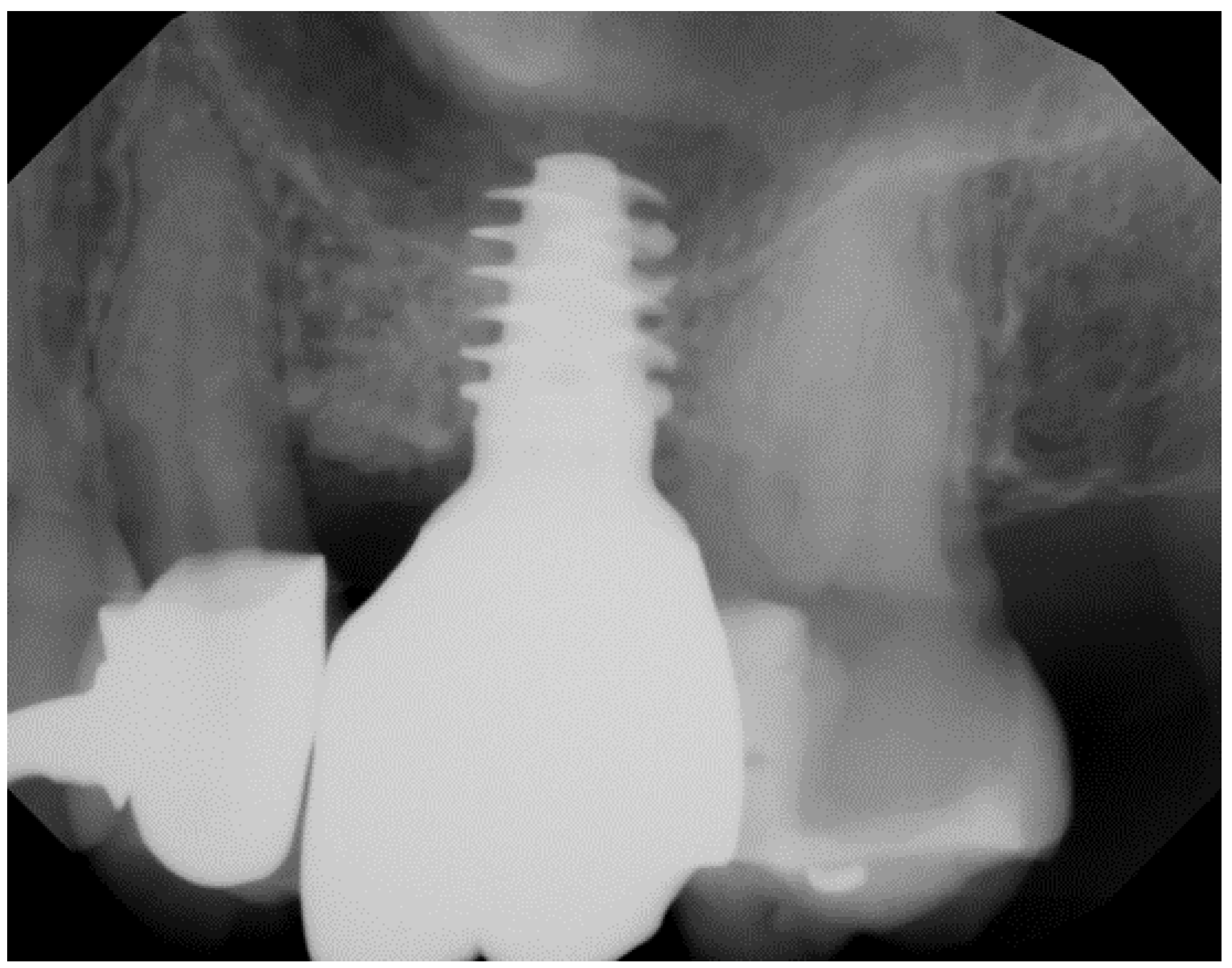
Geometry and diameter also play a significant role in the success of zirconia implants. Excessive implant thread depth is not recommended, as surgical placement might generate high bending forces on the implant body when engaging the bony perimeter of the osteotomy, especially in patients with dense bone (Figure 1) [48]. Additionally, zirconia implants with a narrow diameter (<3.75 mm) have been reported to have a higher incidence of fracture compared to implants with a regular diameter [49,50,51].
The purpose of this mapping review is to present the breadth of evidence to answer the question, “What do studies reveal about the efficacy of zirconia implants?” This includes primary preclinical and clinical research, as well as reviews.
5. Search Strategy
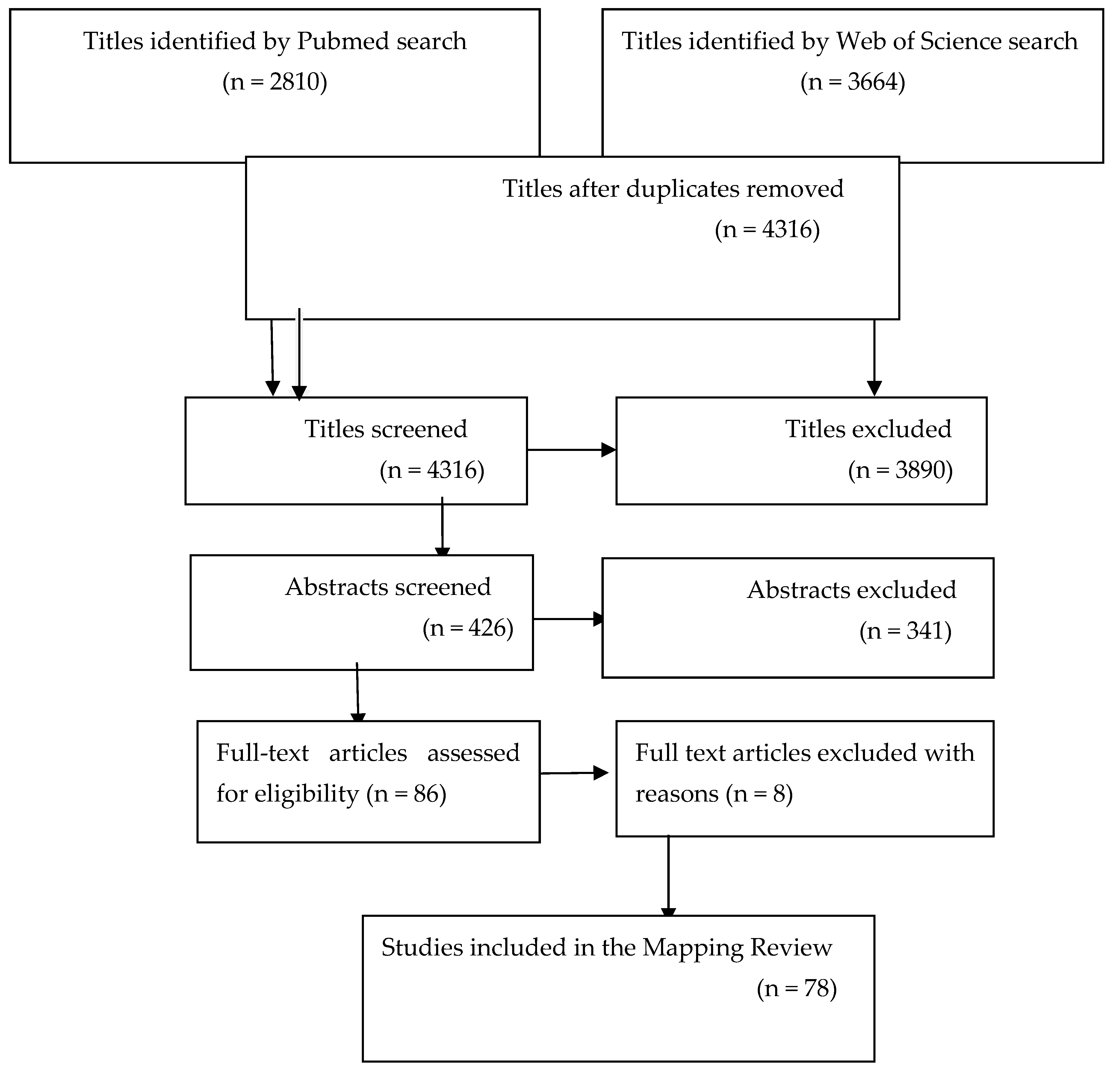
An electronic search of Pubmed and Web of Science databases was performed to select articles from 1 January 2000 to 1 October 2023, using zirconia implants as a specific search term. From 6474 titles, 426 abstracts and 85 full-text articles were screened. Additional hand searches were included from the following journals: International Journal of Oral & Maxillofacial Implants, Journal of Clinical Periodontology, Journal of Periodontology, Clinical Oral Implants Research, Clinical Implant Dentistry and Related Research, and Journal of Prosthodontics. A total of 78 articles were included (one additional article from 1992 was added for historical purposes), from the time period bracketed, that met the eligibility criteria of outcome measures with zirconia implants and stipulated the design of the implant system (Figure 2).
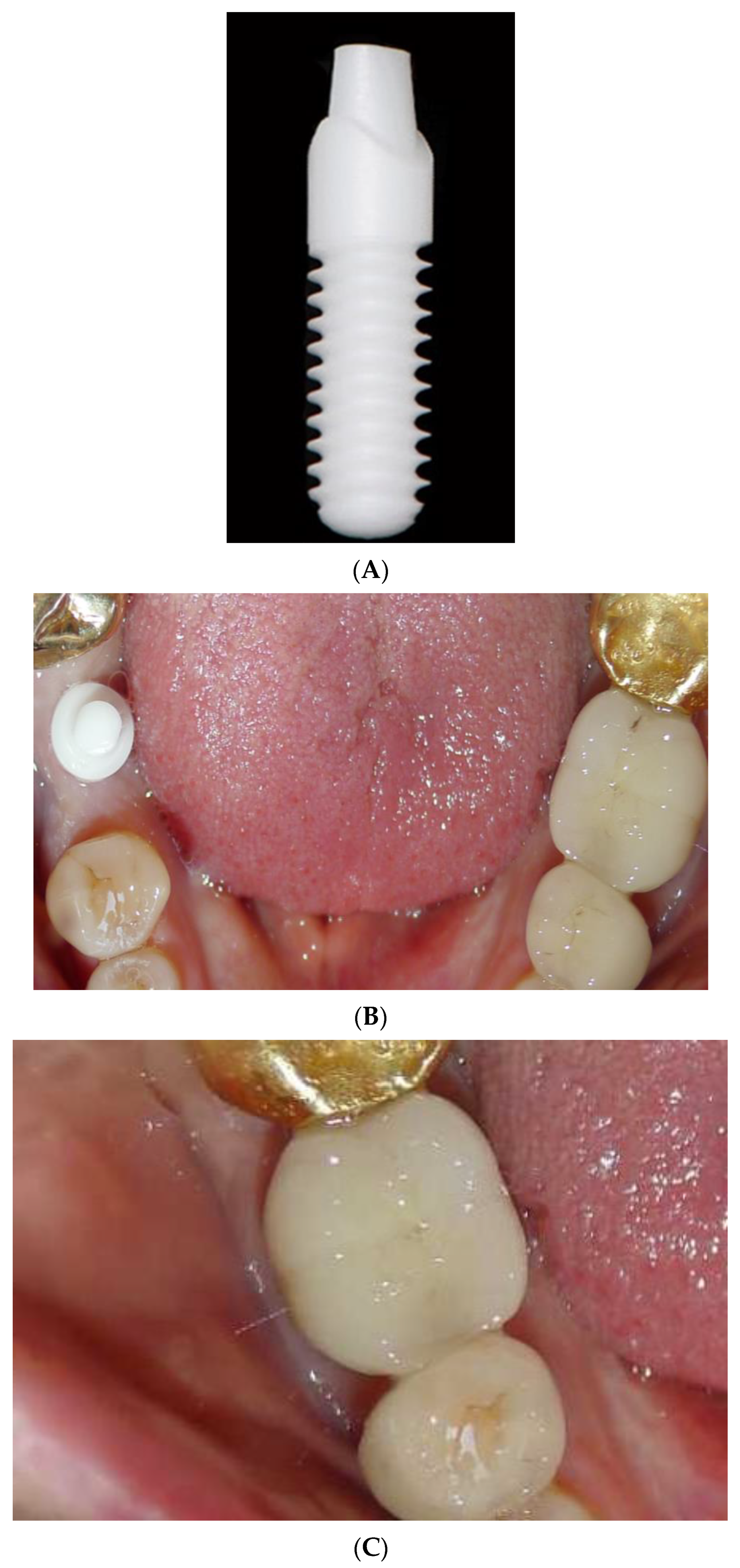
6. One-Piece Zirconia Implants
A majority of zirconia implants have been produced and clinically researched as one-piece implants (Figure 3A–C) [52]. The advantage of this design is that by eliminating the abutment–implant microgap and micromovements, marginal bone loss (MBL) may be diminished [53]. Nevertheless, the design has several drawbacks. Surgical placement is more challenging to meet both the bony compartment parameters and prosthetic design requirements. Grafting and augmentation options are also more limited. No angled abutments are available, and secondary corrections of shape by grinding must be avoided because of the impact on reducing the fracture strength (from 2084 N to 804 N when prepared) [54]. Furthermore, one-piece implants cannot be protected from mastication after placement. Patients with parafunction, or those who are not compliant with admonitions of dietary limitations, would not be candidates. One-piece implants also tend to be inserted more deeply in the esthetic zone to hide the crown margin, which compromises cement extrusion removal, potentiating peri-implant disease [55].
Balmer et al. reported 5-year results of a prospective cohort investigation with a survival rate of 98.4%, and the mean marginal bone loss was 0.7 mm ± 0.6 mm [56]. The authors observed that, after the initial mean MBL before loading the implants, no additional significant marginal bone level change was evident. Hassouna et al. compared the restorations of one-piece fusion-sputtered zirconia implants with one-piece titanium implants in a clinical and radiological study with a 5-year follow-up [57]. There was no significant difference between the marginal bone loss of the immediately loaded titanium or zirconia implants placed in the maxillary first premolar site. However, when evaluating specific survival and success data for one-piece zirconia implants after an 8-year follow-up, there were differences between immediately placed and delayed placement zirconia implants [58]. While the survival rate was 100% for both groups, the immediately placed implants had a success rate of 94.7%, while the delayed implants had a success rate of 87.5%. The PES also revealed a more favorable esthetic score for the immediate zirconia implants compared with the delayed methodology. The inclusion criteria for immediate placement of zirconia implants in this study comprised adequate bone volume and strict compliance, while the exclusion criteria included no inflammation, no hypermastication or clenching, and no untreated periodontal disease during a 3-month healing period. It must be noted that the inclusion/exclusion criteria were not as stringent for the delayed placement approach. This may help to explain why other investigations with different criteria for patient candidacy found no significant differences in survival, immediate complications, and delayed placement of titanium or zirconia implants [59,60].
Overall, a recent systematic review and meta-analysis performed on 11 studies has established, in the short term, that one-piece ceramic implants achieve osseointegration similar to titanium implants, with a stable marginal bone response [61]. For the long term, the MBL change was, on average, 1.24 mm ± 0.16 mm. The risk of implant fractures is low for current commercially available implants. The immediate loading or temporization of the implants does not appear to interfere with the course of osseointegration. However, it is important to note that significant heterogeneity, limited sample sizes, and a lack of long-term randomized clinical studies compromise definitive conclusions regarding single-piece zirconia implant comparisons with titanium implants [62].
7. Two-Piece Zirconia Implants
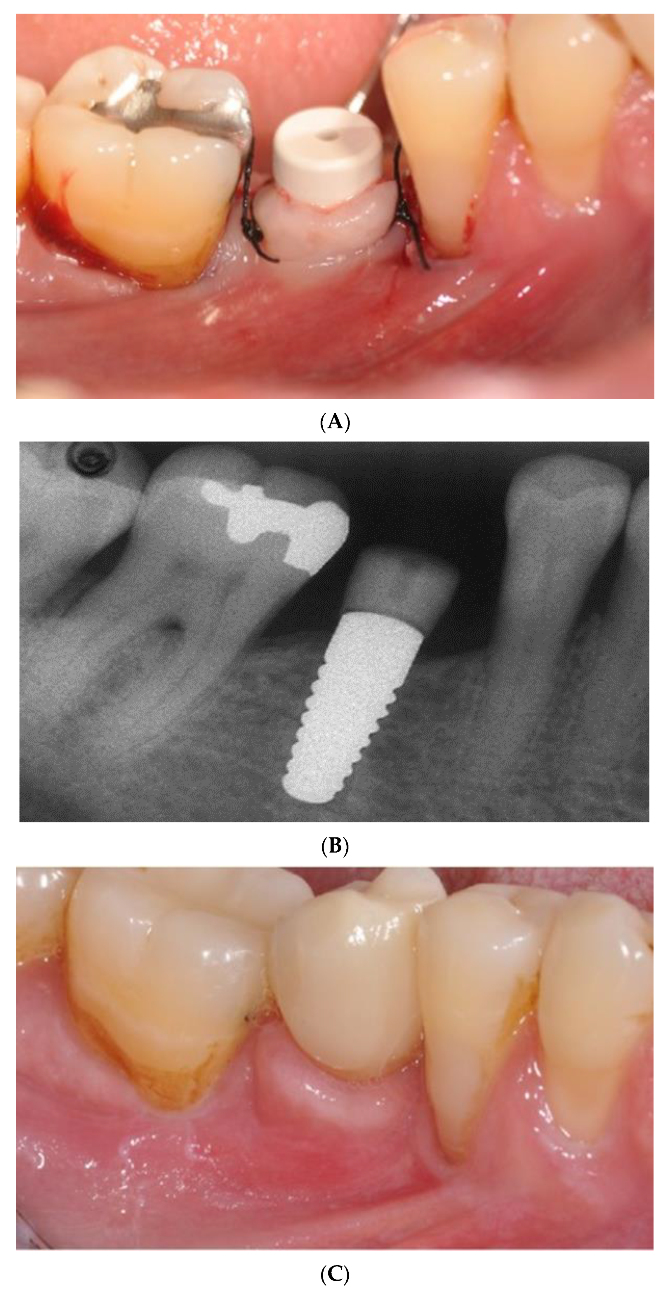
Two-piece implants (Figure 4A–D) are designed for submerged healing, which may minimize the initial bone resorption. A prospective non-randomized study evaluating 91 two-piece zirconia implants monitored for 5–12 years revealed a low prevalence of biologic and prosthetic complications [63]. A randomized clinical trial of two-piece titanium and zirconia implants followed for 80 months. Although there was a small sample size (28), the trial divulged no significant differences in peri-implant disease, bleeding on probing, MBL, PES, implant or abutment fractures, or the debonding of crowns [64]. A cohort study on 46 patients restored with two-piece zirconia implants in the posterior maxilla, and with a mean follow-up time of 9 years, reported a high survival rate [65]. However, this study did report frequent mechanical and technical complications. Furthermore, a contemporaneous prospective cohort study on two-piece zirconia implants, with at least a 6-year follow-up, revealed a troubling 83% cumulative implant survival rate and a 63% success rate [66]. Implant failure was noted with 5 patients due to “aseptic loosening” or a sudden mechanical breakdown of the interface between the bone and the implant. The fracture of a standard diameter implant and a rapid progression of peri-implantitis was responsible for 2 more implant failures. Abutment fracture, using an adhesive material for the design, was the second-most common technical complication. Using a two-piece implant design of a lobular connection mated with a PEEK abutment screwed to the implant, a retrospective cross-sectional 5-year study divulged a 73% implant survival rate and an 82% success rate (Figure 5A–D). Machined zirconia components have not been shown to reach the same precision as titanium counterparts [67]. All these studies are limited by their heterogenous methodology, lack of randomization, relatively small sample size, and lack of blinding of the participants or examiner.
8. Custom-Made Root-Analogue Zirconia Implants
The concept of replacing a tooth upon removal with an analogue that mimics the contours of its predecessor has many potential benefits. These include uncomplicated immediate implant placements, a minimally invasive surgical approach, a decreased number of surgeries, and an absence of a microgap for bacterial adherence [68,69,70,71]. While root analogue implants using titanium were introduced in 1992 by Lundgren et al. [72], zirconia has offered esthetic and biological enhancements without giving up strength. This includes no metal aura in thin mucosal biotypes, increased corrosion resistance, high biocompatibility, and hypoallergenicity [73].
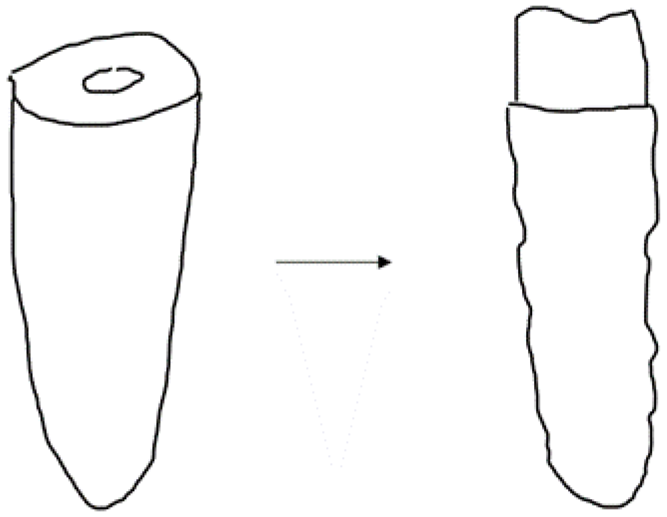
The manufacturing of a custom-made root analogue zirconia implant can be accomplished by using computer-aided design or computer-aided manufacturing, as well as additive or subtractive manufacturing combined with cone-beam computed technology [74]. This technique is limited to cases of periodontally sound teeth with a sufficiently deep socket, an atraumatic extraction, and the absence of periapical disease. The root analogue is manufactured after a crown preparation is completed, the application of micro-retentions are limited to the interdental space, and the diameter of the analogue is reduced (Figure 6) [75].
A scoping review on custom-made zirconia root analogue implants has concluded that this technique is promising and may prevent a loss of alveolar bone volume with the maintenance of the peri-implant soft tissues while attaining ideal optical and mechanical properties [70]. Nonetheless, there is a need for investigations that can assess how zirconia-based surfaces can be enhanced to promote the absorption of proteins preparatory to the migration of osteogenic cells. Since the usual acid-etching procedures cannot be used with zirconia-based materials without aging, novel microtechnologies and nanotechnologies are required to ensure the percentage of osseointegration is acceptable along the time of bone healing, especially when subtractive manufacturing is used [54]. Clinically controlled studies should follow in the long term.
9. Discussion
While a 2023 systematic review and meta-analysis has concluded that commercially available zirconia implants demonstrated reliable clinical outcomes based on survival rates, pocket depth, and MBL values, most of their studies investigated one-piece zirconia implants [76]. In fact, only one study reported technical complications with the two-piece zirconia implants (six fractures of 48 fiberglass abutments over 53.7 months) [65]. The systematic review authors also noted that at the onset of 2004, the first zirconia implants were established on the market, and many were replaced with a total of 16 current manufacturers [76]. The top manufacturers today are Straumann, Nobel Biocare, Dentsply, ZimVie, Osteem, Stryker, Smith and Nephew, Integrum, and Southern Implants. A one-year survival rate of the present commercially available zirconia implants was significantly higher than those no longer available (98.3% versus 91.2%) [41] and has fueled recent interest in zirconia implants despite an approximate 30% cost increase for zirconia implants compared to titanium implants. The ubiquity of peri-implant disease with titanium implants may have changed the calculus in terms of comparative cost, as zirconia implants demonstrate their superiority. One randomized control trial evaluated the currently marketed zirconia implants and compared them to 14 titanium implants. After a follow-up of 12 months, the survival rate was 100% for both types and the mean marginal bone loss was 2.08 mm and 1.96 mm for the zirconia and titanium implants, respectively [77]. Restorative improvements are also notable with the advent of new developments such as optimizing angle surfaces of the abutment, a groove for cement “lock”, 4.5 abutment height, and surface treatment. Still, the unanimity of conclusions from rigorous studies is that more well-designed clinical studies and randomized clinical studies comparing zirconia and titanium implants, over a longer follow-up period, are needed.
Root analogue zirconia implants offer a minimally invasive and simplified technique for replacing a tooth, but the design is limited to patients without a pre-existing periodontal defect and an atraumatic extraction. Moreover, this approach requires a cone beam computed tomography, as well as scanning and manufacturing armamentaria. Only a short-term follow-up on a small number of patients has demonstrated a 100% survival rate and minimal marginal bone loss [78]. However, there has not been a consensus that this design can achieve a high survival rate even in the short term. To this point, a recent prospective clinical study with 1-year follow-up, although with a small number of subjects, showed a mean of 48.2% implant survival [79]. The authors noted that primary stability was a significant risk factor, and that minimizing patient variables in a more rigorous long-term study would be beneficial. Here, the use of additive manufacturing may address the aging that may result in surface modification using subtractive manufacturing of the zirconia root analogue.
The three iterations of zirconia implants all offer advantages and disadvantages in their design. Overall, zirconia has outstanding biocompatibility, mechanical flexural and compressive strengths, and excellent wear resistance, and it is highly esthetic. One-piece zirconia implants eliminate a microgap that can be a source for inflammatory infiltrates. Medium- and long-term studies have corroborated the high success rate of the one-piece zirconia implants. However, they do not offer flexibility in design and require an immediate loading protocol for all patients, as well as cementation. Two-piece zirconia implants have been designed to offer surgical and restorative latitude in placement. However, the fit of machined components has been problematic and is the source of lower success rates compared to the one-piece implant. Root analogues have the advantage of a simplified surgical operation, but primary stability is still considered a risk factor.
The promise of zirconia as a biocompatible, esthetic, and fracture-resistant replacement for titanium implants with similar or greater success has not yet been realized in the literature. Future work on optimizing the surface of the zirconia implant, two-piece design improvements, and more longitudinal comparative studies with titanium implants are needed. These studies should report definitive criteria equating success and patient-related outcomes, as well as a careful evaluation of the effects of guided bone regeneration and regenerative procedures. The use of three-dimensional imaging to evaluate the bony adaptation around zirconia implants would also divulge substantive data for future improvements.
The limitations of this review include the use of heterogeneous evidence. The lack of data on the number of zirconia implants, populations, operators, randomization, types of complications, zirconia material/design, and long-term comparative follow-up with titanium implants have compromised more reliable deductions about the present state of the art of zirconia implants in its various iterations. Future directions should include more robust N-sizes for randomized clinical trials and split-mouth design methodology (limiting interpatient variables) to assess the long-term success of the 3 different iterations of zirconia implants compared to titanium implants. Additionally, zirconia implants fabricated via additive manufacturing, rather than a subtractive technique, offer a solution to surface grinding, but they have not been tested in rigorous investigations.
Another limitation of this review is the fact that it included studies on zirconia implants that are no longer marketed. This fact underscores the need for updated comparative studies of more recent iterations of zirconia implants with titanium implants enhanced with new surfaces and novel macro-thread designs.
10. Conclusions
Zirconia implants offer a potential solution to the esthetic and biological limitations of titanium implants. One-piece zirconia implants have been reported to have similar implant survival and technical complications as titanium implants but reduced biological complications while achieving superior esthetics. However, they do not offer flexibility in restorative planning and are relegated to cement fixation. Two-piece zirconia implants have not been investigated sufficiently and still appear to be more prone to technical failures until material advancements in the abutment are available. Root analogue zirconia implants offer a minimally surgically invasive approach due to an atraumatic extraction. However, the primary stability of the root analogue is still a risk factor, and long-term rigorous studies are lacking.
Funding
This research received no external funding.
Institutional Review Board Statement
Not applicable.
Informed Consent Statement
Not applicable.
Data Availability Statement
Not applicable.
Conflicts of Interest
The author declares no conflict of interest.
References
- Howe, M.S.; Keys, W.; Richards, D. Long-term (10-year) dental implant survival: A systematic review and sensitivity meta-analysis. J. Dent. 2019, 84, 9–21. [Google Scholar] [CrossRef] [PubMed]
- Sadowsky, S.J. Peri-implantitis after 40 years: Evidence, mechanisms, and implications: A mapping review. J. Prosthet. Dent. 2023; online ahead of print. [Google Scholar]
- Suárez-López Del Amo, F.; Garaicoa-Pazmiño, C.; Fretwurst, T.; Castilho, R.M.; Squarize, C.H. Dental implants-associated release of titanium particles: A systematic review. Clin. Oral Implants Res. 2018, 29, 1085–1100. [Google Scholar] [CrossRef] [PubMed]
- Fretwurst, T.; Nelson, K.; Tarnow, D.P.; Wang, H.L.; Giannobile, W.V. Is Metal Particle Release Associated with Peri-implant Bone Destruction? An Emerging Concept. J. Dent. Res. 2018, 97, 259–265. [Google Scholar] [CrossRef] [PubMed]
- Fovet, Y.; Gal, J.Y.; Toumelin-Chemla, F. Influence of pH and fluoride concentration on titanium passivating layer: Stability of titanium dioxide. Talanta 2001, 53, 1053–1063. [Google Scholar] [CrossRef] [PubMed]
- Sridhar, S.; Wang, F.; Wilson, T.G.; Palmer, K.; Valderrama, P.; Rodrigues, D.C. The role of bacterial biofilm and mechanical forces in modulating dental implant failures. J. Mech. Behav. Biomed. Mater. 2019, 92, 118–127. [Google Scholar] [CrossRef]
- Chiou, L.L.; Panariello, B.H.D.; Hamada, Y.; Gregory, R.L.; Blanchard, S.; Duarte, S. Comparison of In Vitro Biofilm Formation on Titanium and Zirconia Implants. Biomed. Res. Int. 2023, 2023, 8728499. [Google Scholar] [CrossRef]
- Nishihara, H.; Haro Adanez, M.; Att, W. Current status of zirconia implants in dentistry: Preclinical tests. J. Prosthodont. Res. 2019, 63, 1–14. [Google Scholar] [CrossRef]
- Al-Radha, A.S.; Dymock, D.; Younes, C.; O’Sullivan, D. Surface properties of titanium and zirconia dental implant materials and their effect on bacterial adhesion. J. Dent. 2012, 40, 146–153. [Google Scholar] [CrossRef]
- van Brakel, R.; Noordmans, H.J.; Frenken, J.; de Roode, R.; de Wit, G.C.; Cune, M.S. The effect of zirconia and titanium implant abutments on light reflection of the supporting soft tissues. Clin. Oral Implants Res. 2011, 22, 1172–1178. [Google Scholar] [CrossRef]
- Jung, R.E.; Becker, K.; Bienz, S.P.; Dahlin, C.; Donos, N.; Hammacher, C.; Iglhaut, G.; Linares, A.; Ortiz-Vigon, A.; Sanchez, N.; et al. Effect of peri-implant mucosal thickness on esthetic outcomes and the efficacy of soft tissue augmentation procedures. Consensus report of group 2 of SEPA/DGI, OF workshop. Clin. Oral Implants Res. 2022, 33 (Suppl. S23), 100–108. [Google Scholar] [CrossRef]
- Di Gianfilippo, R.; Valente, N.A.; Toti, P.; Wang, H.L.; Barone, A. Influence of implant mucosal thickness on early bone loss: A systematic review and metaanalysis. J. Periodontal Implant Sci. 2020, 50, 209–225. [Google Scholar] [CrossRef] [PubMed]
- Krawiec, M.; Olchowy, C.; Kubasiewicz-Ross, P.; Hadzik, J.; Dominiak, M. Role of implant loading time in prevention of marginal bone loss after implant-supported restorations: A targeted review. Dent. Med. Probl. 2022, 59, 475–481. [Google Scholar] [CrossRef] [PubMed]
- Daftary, F.; Mahallati, R.; Bahat, O.; Sullivan, R.M. Lifelong craniofacial growth and implications for osseointegrated implants. Int. J. Oral Maxillofac. Implants 2013, 28, 163–169. [Google Scholar] [CrossRef] [PubMed]
- Shapira, L.; Levin, B.P.; Stabholz, A. Long-term esthetic complications associated with anterior implant-supported restorations. Compend. Contin. Educ. Dent. 2021, 42, 358–363. [Google Scholar]
- Roehling, S.; Woelfler, H.; Hicklin, S.; Kniha, H.; Gahlert, M. A retrospective clinical study with regard to survival and success rates of zirconia implants up to and after 7 years of loading. Clin. Implant Dent. Relat. Res. 2016, 18, 545–558. [Google Scholar] [CrossRef]
- Theobald, A.H.; Wong, B.K.; Quick, A.N.; Thomson, W.M. The impact of the popular media on cosmetic dentistry. N. Z. Dent. J. 2006, 102, 58–63. [Google Scholar]
- Chevalier, J. What future for zirconia as a biomaterial? Biomaterials 2006, 27, 535–543. [Google Scholar] [CrossRef]
- Kniha, K.; Kniha, H.; Grunert, I.; Edelhoff, D.; Hölzle, F.; Modabber, A. Esthetic Evaluation of Maxillary Single-Tooth Zirconia Implants in the Esthetic Zone. Int. J. Periodontics Restor. Dent. 2019, 39, e195–e201. [Google Scholar] [CrossRef]
- Thoma, D.S.; Ioannidis, A.; Cathomen, E.; Hämmerle, C.H.; Hüsler, J.; Jung, R.E. Discoloration of the Peri-implant Mucosa Caused by Zirconia and Titanium Implants. Int. J. Periodontics Restor. Dent. 2016, 36, 39–45. [Google Scholar] [CrossRef]
- Stejskal, V.; Hudecek, R.; Stejskal, J.; Sterzl, I. Diagnosis and treatment of metal-induced side-effects. Neuro. Endocrinol. Lett. 2006, 27 (Suppl. S1), 7–16. [Google Scholar]
- Kim, K.T.; Eo, M.Y.; Nguyen, T.T.H.; Kim, S.M. General review of titanium toxicity. Int. J. Implant Dent. 2019, 5, 10. [Google Scholar] [CrossRef] [PubMed]
- Siddiqi, A.; Payne, A.G.T.; De Silva, R.K.; Duncan, W.J. Titanium allergy: Could it affect dental implant integration? Clin. Oral Implants Res. 2011, 22, 673–680. [Google Scholar] [CrossRef] [PubMed]
- Sicilia, A.; Cuesta, S.; Coma, G.; Arregui, I.; Guisasola, C.; Ruiz, E.; Maestro, A. Titanium allergy in dental implant patients: A clinical study on 1500 consecutive patients. Clin. Oral Implants Res. 2008, 19, 823–835. [Google Scholar] [CrossRef] [PubMed]
- Dawson-Amoah, K.G.; Waddell, B.S.; Prakash, R.; Alexiades, M.M. Adverse Reaction to Zirconia in a Modern Total Hip Arthroplasty with Ceramic Head. Arthroplast. Today 2020, 6, 612–616.e611. [Google Scholar] [CrossRef]
- Gahlert, M.; Kniha, H.; Laval, S.; Gellrich, N.C.; Bormann, K.H. Prospective Clinical Multicenter Study Evaluating the 5-Year Performance of Zirconia Implants in Single-Tooth Gaps. Int. J. Oral Maxillofac. Implants 2022, 37, 804–811. [Google Scholar] [CrossRef]
- Kohal, R.J.; Wolkewitz, M.; Hinze, M.; Han, J.S.; Bächle, M.; Butz, F. Biomechanical and histological behavior of zirconia implants: An experiment in the rat. Clin. Oral Implants Res. 2009, 20, 333–339. [Google Scholar] [CrossRef]
- Vagkopoulou, T.; Koutayas, S.O.; Koidis, P.; Strub, J.R. Zirconia in dentistry: Part 1. Discovering the nature of an upcoming bioceramic. Eur. J. Esthet. Dent. 2009, 4, 130–151. [Google Scholar]
- Sanon, C.; Chevalier, J.; Douillard, T.; Cattani-Lorente, M.; Scherrer, S.S.; Gremillard, L. A new testing protocol for zirconia dental implants. Dent. Mater. 2015, 31, 15–25. [Google Scholar] [CrossRef]
- Lughi, V.; Sergo, V. Low temperature degradation -aging- of zirconia: A critical review of the relevant aspects in dentistry. Dent. Mater. 2010, 26, 807–820. [Google Scholar] [CrossRef]
- Kohal, R.J.; Weng, D.; Bächle, M.; Strub, J.R. Loaded custom-made zirconia and titanium implants show similar osseointegration: An animal experiment. J. Periodontol. 2004, 75, 1262–1268. [Google Scholar] [CrossRef]
- Yamano, S.; Kwok-Yui Ma, A.; Shanti, R.M.; Kim, S.-W.; Wada, K.; Sukotjo, C. The influence of different implant materials on human gingival fibroblast morphology, proliferation, and gene expression. Int. J. Oral Maxillofac. Implants 2011, 26, 1247–1255. [Google Scholar] [PubMed]
- Noro, A.; Kaneko, M.; Murata, I.; Yoshinari, M. Influence of surface topography and surface physicochemistry on wettability of zirconia (tetragonal zirconia polycrystal). J. Biomed. Mater. Res. B Appl. Biomater. 2013, 101, 355–363. [Google Scholar] [CrossRef] [PubMed]
- Nickenig, H.-J.; Andreas Schlegel, K.; Wichmann, M.; Eitner, S. Expression of interleukin 6 and tumor necrosis factor alpha in soft tissue over ceramic and metal implant materials before uncovering: A clinical pilot study. Int. J. Oral Maxillofac. Implants 2012, 27, 671–676. [Google Scholar] [PubMed]
- Cionca, N.; Müller, N.; Mombelli, A. Two-piece zirconia implants supporting all-ceramic crowns: A prospective clinical study. Clin. Oral. Implants Res. 2015, 26, 413–418. [Google Scholar] [CrossRef] [PubMed]
- Sivaraman, K.; Chopra, A.; Narayan, A.I.; Balakrishnan, D. Is zirconia a viable alternative to titanium for oral implant? A critical review. J. Prosthodont. Res. 2018, 62, 121–133. [Google Scholar] [CrossRef] [PubMed]
- Chevalier, J.; Loh, J.; Gremillard, L.; Meille, S.; Adolfson, E. Low-temperature degradation in zirconia with a porous surface. Acta Biomater. 2011, 7, 2986–2993. [Google Scholar] [CrossRef]
- Deville, S.; Chevalier, J.; Gremillard, L. Influence of surface finish and residual stresses on the ageing sensitivity of biomedical grade zirconia. Biomaterials 2006, 27, 2186–2192. [Google Scholar] [CrossRef]
- Kohal, R.J.; Wolkewitz, M.; Mueller, C. Alumina-reinforced zirconia implants: Survival rate and fracture strength in a masticatory simulation trial. Clin. Oral Implants Res. 2010, 21, 1345–1352. [Google Scholar] [CrossRef]
- Spies, B.C.; Sperlich, M.; Fleiner, J.; Stampf, S.; Kohal, R.J. Alumina reinforced zirconia implants: 1-year results from a prospective cohort investigation. Clin. Oral Implants Res. 2016, 27, 481–490. [Google Scholar] [CrossRef]
- Roehling, S.; Schlegel, K.A.; Woelfler, H.; Gahlert, M. Performance and outcome of zirconia dental implants in clinical studies: A meta-analysis. Clin. Oral Implants Res. 2018, 29 (Suppl. S16), 135–153. [Google Scholar] [CrossRef]
- Rohr, N.; Hoda, B.; Fischer, J. Surface structure of zirconia implants: An integrative review comparing clinical results with preclinical and in vitro data. Materials 2022, 15, 3664. [Google Scholar] [CrossRef] [PubMed]
- Chopra, D.; Jayasree, A.; Guo, T.; Gulati, K.; Ivanovski, S. Advancing dental implants: Bioactive and therapeutic modifications of zirconia. Bioact. Mater. 2022, 13, 161–178. [Google Scholar] [CrossRef] [PubMed]
- Kubasiewicz, P.; Hadzik, J.; Dominiak, M. Osseointegration of zirconia implants with 3 varying surface textures and a titanium implant: A histological and micro-CT study. Adv. Clin. Exp. Med. 2018, 27, 1173–1179. [Google Scholar] [CrossRef] [PubMed]
- Kohal, R.J.; von Schierholz, C.; Nold, J.; Spies, B.C.; Adolfsson, E.; Vach, K.; Burkhardt, F. Influence of loading and aging on the fracture strength of an injection-molded two-piece zirconia implant restored with a zirconia abutment. Clin. Oral Implants Res. 2023, 34, 105–115. [Google Scholar] [CrossRef]
- Spies, B.C.; Maass, M.E.; Adolfsson, E.; Sergo, V.; Kiemle, T.; Berthold, C.; Gurian, E.; Fornasaro, S.; Vach, K.; Kohal, R.J. Long-term stability of an injection-molded zirconia bone-level implant: A testing protocol considering aging kinetics and dynamic fatigue. Dent. Mater. 2017, 33, 954–965. [Google Scholar] [CrossRef] [PubMed]
- Zhang, F.; Spies, B.C.; Willems, E.; Inokoshi, M.; Wesemann, C.; Cokic, S.M.; Hache, B.; Kohal, R.J.; Altmann, B.; Vleugels, J.; et al. 3D printed zirconia dental implants with integrated directional surface pores combine mechanical strength with favorable osteoblast response. Acta Biomater. 2022, 150, 427–441. [Google Scholar] [CrossRef] [PubMed]
- Fook, P.; Berger, D.; Riemer, O.; Karpuschewski, B. Structuring of bioceramics by micro-grindingfor dental implant applications. Micromachines 2019, 10, 312. [Google Scholar] [CrossRef]
- Gahlert, M.; Burtscher, D.; Grunert, I.; Kniha, H.; Steinhauser, E. Failure analysis of fractured dental zirconia implants. Clin. Oral Implants Res. 2012, 23, 287–293. [Google Scholar] [CrossRef]
- Schiegnitz, E.; Al-Nawas, B. Narrow-diameter implants: A systematic review and meta-analysis. Clin. Oral Implants Res. 2018, 29 (Suppl. S16), 21–40. [Google Scholar] [CrossRef]
- Atalay, P.; Öztaş, D.D. Fatigue resistance and fracture strength of narrow-diameter one-piece zirconia implants with angled abutments. J. Esthet. Restor. Dent. 2022, 34, 1060–1067. [Google Scholar] [CrossRef]
- Sadowsky, S.J. Has zirconia made a material difference in implant prosthodontics? A review. Dent. Mater. 2020, 36, 1–8. [Google Scholar] [CrossRef] [PubMed]
- Borgonovo, A.E.; Censi, R.; Vavassori, V.; Arnaboldi, O.; Maiorana, C.; Re, D. Zirconia implants in esthetic áreas: 4-year follow-up evaluation study. Int. J. Dent. 2015, 2015, 4015029. [Google Scholar] [CrossRef] [PubMed]
- Cionca, N.; Hashim, D.; Mombelli, A. Zirconia dental implants: Where are we now, and where are we heading? Periodontol. 2000 2017, 73, 241–258. [Google Scholar] [CrossRef] [PubMed]
- Staubli, N.; Walter, C.; Schmidt, J.C.; Weiger, R.; Zitzmann, N.U. Excess cement and the risk of peri-implant disease—A systematic review. Clin. Oral Implants Res. 2017, 28, 1278–1290. [Google Scholar] [CrossRef] [PubMed]
- Balmer, M.; Spies, B.C.; Kohal, R.J.; Hämmerle, C.H.; Vach, K.; Jung, R.E. Zirconia implants restored with single crowns or fixed dental prostheses: 5-year results of a prospective cohort investigation. Clin. Oral Implants Res. 2020, 31, 452–462. [Google Scholar] [CrossRef]
- Hassouna, M.; Al-Zordk, W.; Aboshilib, M.; Ghazy, M. Clinical and radiographic prospective study of customized one-piece titanium and one-piece fusion-sputtered zirconia implants: Five-year mean follow-up. BMC Oral Health 2022, 22, 531. [Google Scholar] [CrossRef]
- Kiechle, S.; Liebermann, A.; Mast, G.; Heitzer, M.; Möhlhenrich, S.C.; Kniha, H.; Kniha, K. Evaluation of one-piece zirconia dental implants: An 8-year follow-up study. Clin. Oral Investig. 2023, 27, 3415–3421. [Google Scholar] [CrossRef]
- Patel, R.; Ucer, C.; Wright, S.; Khan, R.S. Differences in dental implant survival between immediate vs. delayed placement: A systematic review and meta-analysis. Dent. J. 2023, 11, 218. [Google Scholar] [CrossRef]
- Grassi, F.R.; Capogreco, M.; Consonni, D.; Bilardi, G.; Buti, J.; Kalemaj, Z. Immediate occlusal loading of one-piece zirconia implants: Five-year radiographic and clinical evaluation. Int. J. Oral Maxillofac. Implants 2015, 30, 671–680. [Google Scholar] [CrossRef]
- Neugebauer, J.; Schoenbaum, T.R.; Pi-Anfruns, J.; Yang, M.; Lander, B.; Blatz, M.B.; Fiorellini, J.P. Ceramic dental implants: A systematic review and meta-analysis. Int. J. Oral Maxillofac. Implants 2023, 38, 30–36. [Google Scholar] [CrossRef]
- Hashim, D.; Cionca, N.; Courvoisier, D.S.; Mombelli, A. A systematic review of the clinical survival of zirconia implants. Clin. Oral Investig. 2016, 20, 1403–1417. [Google Scholar] [CrossRef] [PubMed]
- Karapataki, S.; Vegh, D.; Payer, M.; Fahrenholz, H.; Antonoglou, G.N. Clinical performance of two-piece zirconia dental implants after 5 and 12 years. Int. J. Oral Maxillofac. Implants 2023, 38, 1105–1114. [Google Scholar] [CrossRef] [PubMed]
- Koller, M.; Steyer, E.; Theisen, K.; Stagnell, S.; Jakse, N.; Payer, M. Two-piece zirconia versus titanium implants after 80 months: Clinical outcomes from a prospective randomized pilot trial. Clin. Oral Implants Res. 2020, 31, 388–396. [Google Scholar] [CrossRef] [PubMed]
- Brunello, G.; Rauch, N.; Becker, K.; Hakimi, A.R.; Schwarz, F.; Becker, J. Two-piece zirconia implants in the posterior mandible and maxilla: A cohort study with a follow-up period of 9 years. Clin. Oral Implants Res. 2022, 33, 1233–1244. [Google Scholar] [CrossRef] [PubMed]
- Cionca, N.; Hashim, D.; Mombelli, A. Two-piece zirconia implants supporting all-ceramic crowns: Six-year results of a prospective cohort study. Clin. Oral Implants Res. 2021, 32, 695–701. [Google Scholar] [CrossRef] [PubMed]
- Preis, V.; Kammermeier, A.; Handel, G.; Rosentritt, M. In vitro performance of two-piece zirconia implant systems for anterior application. Dent. Mater. 2016, 32, 765–774. [Google Scholar] [CrossRef] [PubMed]
- Pirker, W.; Kocher, A. Immediate, non-submerged, root-analogue zirconia implant in single tooth replacement. Int. J. Oral Maxillofac. Surg. 2008, 37, 293–295. [Google Scholar] [CrossRef]
- Pirker, W.; Kocher, A. Immediate, non-submerged, root-analogue zirconia implants placed into single-rooted extraction sockets: 2-year follow-up of a clinical study. Int. J. Oral Maxillofac. Surg. 2009, 38, 1127–1132. [Google Scholar] [CrossRef]
- Pessanha-Andrade, M.; Sordi, M.B.; Henriques, B.; Silva, F.S.; Teughels, W.; Souza, J.C.M. Custom-made root-analogue zirconia implants: A scoping review on mechanical and biological benefits. J. Biomed. Mater. Res. B Appl. Biomater. 2018, 106, 2888–2900. [Google Scholar] [CrossRef]
- Pirker, W.; Wiedemann, D.; Lidauer, A.; Kocher, A.A. Immediate, single stage, truly anatomic zirconia implant in lower molar replacement: A case report with 2.5 years follow-up. Int. J. Oral Maxillofac. Surg. 2011, 40, 212–216. [Google Scholar] [CrossRef]
- Lundgren, D.; Rylander, M.; Johansson, C.; Albrektsson, T. Healing-in of root analogue titanium implants placed in extraction sockets. An experimental study in the beagle dog. Clin. Oral Implants Res. 1992, 3, 136–143. [Google Scholar] [CrossRef] [PubMed]
- Van Dooren, E.; Calamita, M.; Calgaro, M.; Coachman, C.; Ferencz, J.L.; Pinho, C.; Silva, N.R. Mechanical, biological and clinical aspects of zirconia implants. Eur. J. Esthet. Dent. 2012, 7, 396–417. [Google Scholar] [PubMed]
- Regish, K.M.; Sharma, D.; Prithviraj, D.R. An overview of immediate root analogue zirconia implants. J. Oral Implantol. 2013, 39, 225–233. [Google Scholar] [CrossRef] [PubMed]
- Smeets, R.; Stadlinger, B.; Schwarz, F.; Beck-Broichsitter, B.; Jung, O.; Precht, C.; Kloss, F.; Gröbe, A.; Heiland, M.; Ebker, T. Impact of Dental Implant Surface Modifications on Osseointegration. Biomed. Res. Int. 2016, 2016, 6285620. [Google Scholar] [CrossRef]
- Roehling, S.; Gahlert, M.; Bacevic, M.; Woelfler, H.; Laleman, I. Clinical and radiographic outcomes of zirconia dental implants-A systematic review and meta-analysis. Clin. Oral Implants Res. 2023, 34 (Suppl. S26), 112–124. [Google Scholar] [CrossRef] [PubMed]
- Ruiz Henao, P.A.; Caneiro Queija LMareque, S.; Tesende Pereira, A.; Linares Gonzalez, A.; Blanco Carrion, J. Titanium vs. ceramic single dental Implants in anterior maxilla: A 12-month randomized clinical trial. Clin. Oral Implants Res. 2021, 32, 951–961. [Google Scholar] [CrossRef]
- Mangano, F.G.; De Franco, M.; Caprioglio, A.; Macchi, A.; Piattelli, A.; Mangano, C. Immediate, non-submerged, root-analogue direct laser metal sintering (DLMS) implants: A 1-year prospective study on 15 patients. Lasers Med. Sci. 2014, 29, 1321–1328. [Google Scholar] [CrossRef]
- Akkoyun, E.F.; Demirbas, A.E.; Gumus, H.O.; Alkan, B.A.; Alkan, A. Custom-made root analog immediate dental implants: A prospective clinical study with 1-year follow-up. Int. J. Oral Maxillofac. Implants 2022, 37, 1223–1231. [Google Scholar] [CrossRef]
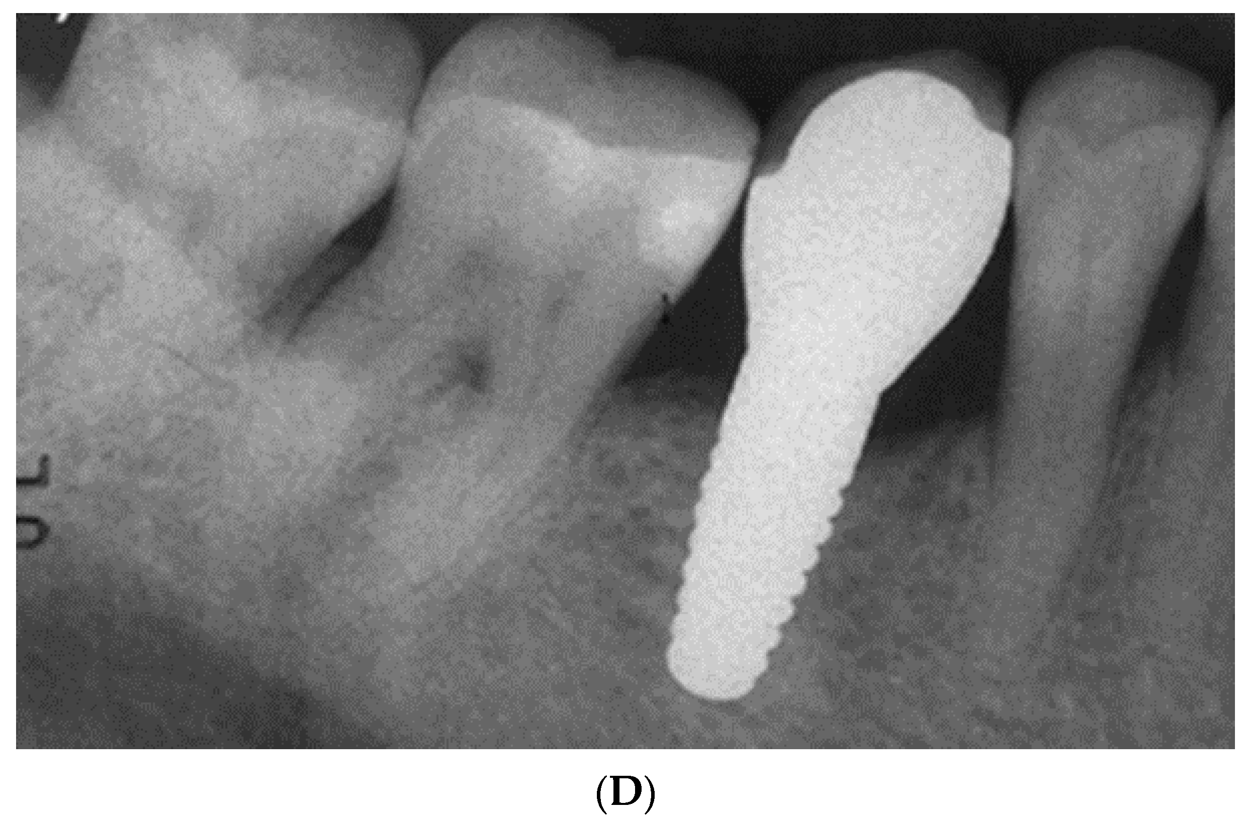
Figure 1.
Excessive thread-depth design on Straumann TLX titanium implant, which is not recommended for zirconia implants.
Figure 1.
Excessive thread-depth design on Straumann TLX titanium implant, which is not recommended for zirconia implants.

Figure 2.
Search strategy and selection process of included studies.

Figure 3.
(A) Zirconia one-piece implant. (B) One-piece zirconia implant in situ. (C) Restored one-piece zirconia implant.
Figure 3.
(A) Zirconia one-piece implant. (B) One-piece zirconia implant in situ. (C) Restored one-piece zirconia implant.

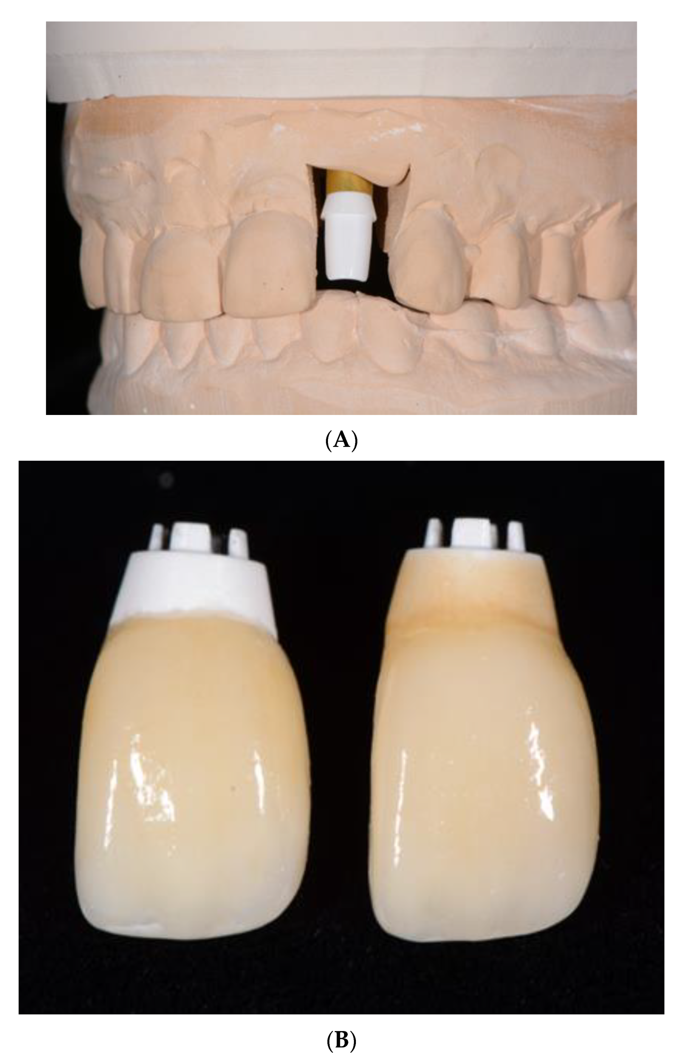
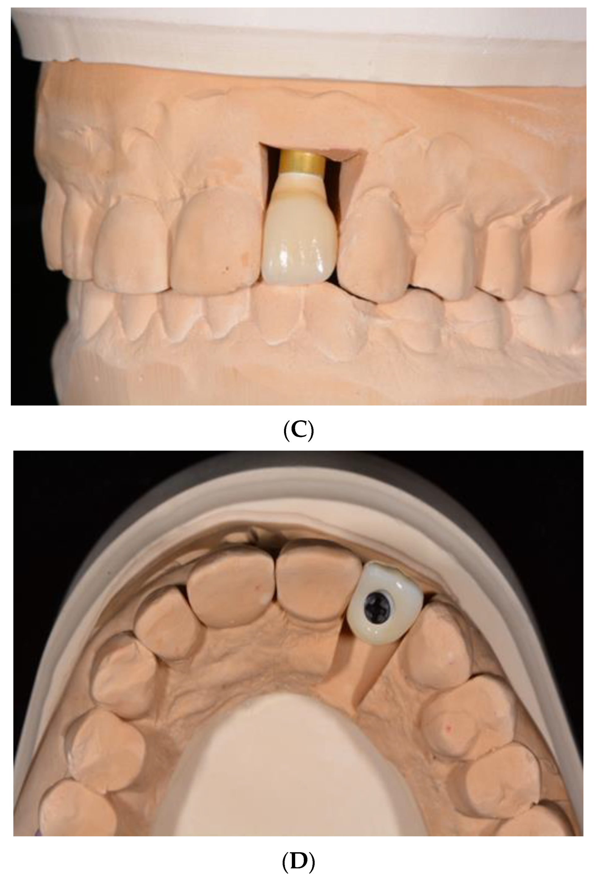
Figure 4.
(A). Two-piece zirconia implant with healing abutment in place. (B). Radiograph of two-piece zirconia implant with healing abutment in Site #29. (C). Definitive restoration on two-piece zirconia implant. (D). Radiograph of definitive crown on two-piece zirconia implant.
Figure 4.
(A). Two-piece zirconia implant with healing abutment in place. (B). Radiograph of two-piece zirconia implant with healing abutment in Site #29. (C). Definitive restoration on two-piece zirconia implant. (D). Radiograph of definitive crown on two-piece zirconia implant.


Figure 5.
(A) Two-piece zirconia implant analog with abutment in place on cast. (B) Crown seated on lobular abutment before and after staining and glazing cervical aspect of abutment. (C) 2-piece zirconia implant with crown in place and abutment shaded in cervical region. (D). Screw-retained design using 2-piece zirconia implant.


Figure 6.
Graphic representation of root analogue with macro-retentions and a prepared abutment.

Disclaimer/Publisher’s Note: The statements, opinions and data contained in all publications are solely those of the individual author(s) and contributor(s) and not of MDPI and/or the editor(s). MDPI and/or the editor(s) disclaim responsibility for any injury to people or property resulting from any ideas, methods, instructions or products referred to in the content. |
© 2023 by the author. Licensee MDPI, Basel, Switzerland. This article is an open access article distributed under the terms and conditions of the Creative Commons Attribution (CC BY) license (https://creativecommons.org/licenses/by/4.0/).
Share and Cite
MDPI and ACS Style
Sadowsky, S.J. Zirconia Implants: A Mapping Review. Oral 2024, 4, 9-22. https://doi.org/10.3390/oral4010002
AMA Style
Sadowsky SJ. Zirconia Implants: A Mapping Review. Oral. 2024; 4(1):9-22. https://doi.org/10.3390/oral4010002
Chicago/Turabian StyleSadowsky, Steven J. 2024. "Zirconia Implants: A Mapping Review" Oral 4, no. 1: 9-22. https://doi.org/10.3390/oral4010002