Article

Role of Glutathione in Neutrophil Chemotaxis in Periodontitis

by
Nurul Iman Binti Badlishah Sham
1,2 and
Melissa M. Grant
2,*
1
Faculty of Dentistry, Universiti Sains Islam Malaysia, Kuala Lumpur 55100, Malaysia
2
School of Dentistry, Institute of Clinical Sciences, University of Birmingham, 5 Mill Pool Way, Edgbaston, Birmingham B5 7EG, UK
*
Author to whom correspondence should be addressed.
Oral 2023, 3(4), 526-538; https://doi.org/10.3390/oral3040043
Submission received: 12 September 2023 / Revised: 3 November 2023 / Accepted: 7 November 2023 / Published: 23 November 2023
(This article belongs to the Special Issue Current Issues in Oral Health)

Abstract

:
Periodontitis is a common non-communicable inflammatory disease that leads to the destruction of periodontal tissues and tooth loss. Initiated by the plaque biofilm, there is a strong innate immune response with an abundance of neutrophils in the periodontium of affected individuals. Previous reports have shown that the intracellular concentration of glutathione in peripheral blood neutrophils from periodontitis patients and the chemotactic ability of these cells are compromised. Furthermore, other studies have described that in oxidative stress conditions neutrophil chemotaxis is aberrant and causes the glutathionylation of F-actin, a key player in chemotaxis. In this study, the effects of glutathione-modulating compounds were assessed in neutrophils isolated from healthy donors, showing that the perturbation of glutathione homeostasis decreases the chemotaxis of neutrophils. Following this, the intracellular glutathione status and chemotactic ability of neutrophils isolated from periodontitis patients was compared to that of age- and sex-matched controls. A decrease in glutathione and chemotactic ability were confirmed. Finally, the proteome of these neutrophils was explored, demonstrating a change in the abundance of proteins involved in glutathione homeostasis. Together these data suggest that peripheral blood neutrophils from periodontitis patients are compromised in their ability to cope with oxidative stress and move.

1. Introduction

Chronic periodontitis is an oral disease characterized by non-resolving inflammation which leads to host-mediated tissue damage and bone loss around the teeth [1]. It is initiated by the biofilm but exacerbated by the host response which leads to tissue and bone destruction [2]. Among the symptoms of periodontitis are redness and bleeding gums, swollen gums, loose teeth, pain on biting as well as bad breath. It is a common infection affecting 11% of the population in its most severe form and over 50% of people over the age of 65 years in any form [3]. The human oral tissues are always exposed to potential and harmful microorganisms and to combat the infections in this area, leucocytes are attracted to and infiltrate into the tissues from the bloodstream in response to chemoattractants [4]. A strong neutrophil tissue infiltration is a characteristic of periodontitis. Neutrophil homeostasis is required for periodontal health as these cells protect the periodontal tissues from damage and they are commonly found in the gingival crevice and epithelial area [5]. By killing various pathogenic bacteria either through non-oxidative or oxidative means in an intracellular or extracellular environment, neutrophils can protect the host tissue. A phenomenon that is associated with periodontitis is oxidative stress [6,7,8]. Matthews et al. suggested a dual role for neutrophils in the production of oxidative tissue damage, involving a potentially reversible Fcɤ receptor-mediated hyperactivity and an increase in oxygen radical release in unstimulated cells [7,8]. Indeed, the manifestation of periodontitis disease is not merely oral but systemic, as peripherally isolated neutrophils display aberrant ROS production, chemotaxis, cytokine production and altered transcriptional activity [7,8,9,10].
Neutrophils are the most abundant leucocyte and the first to arrive at a site of infection. The physiological function of neutrophils is to kill microbes: phagocytosis with the generation of reactive oxygen species (ROS) and hypochlorous acid are the main killing mechanisms for most pathogens [11,12]. For neutrophils, chemotaxis is the process that allows the cells to reach the infected area, thus, effecting phagocytosis and subsequent destruction of the microorganism via ROS and proteolytic enzymes within the phagolysosome [13]. Chemotaxis is an important element for neutrophil efficacy as it enables cells to reach the site of infection, a crucial step to alleviate disease states. During chemotaxis, filamentous actin (F-actin) is polymerized asymmetrically at the leading edge which provides the force for the projection of the plasma membrane in the proper direction, in response to a directional chemoattractant signal [14]. Conversely, when attractant concentration is uniform, the polarization response stimulates all portions of the plasma membrane at the same intensity, meanwhile, with the absence of gradient, the direction of polarity is random, resulting in undirected movement [15]. The regulation of actin dynamics is important for cell adhesion, migration and phagocytosis; therefore, it is crucial for neutrophils to fulfil their roles in innate immunity. It is of particular interest that patients suffering from chronic inflammatory disorders, such as periodontitis [9], Crohn’s disease [16] and chronic obstructive pulmonary disease (COPD) [17] display aberrant neutrophil chemotaxis.
Neutrophils are among the immune cells that are attracted to the site of infection by chemokines, such as interleukin-8 (CXCL8, IL-8), and bacteria-derived peptides, such as N-formyl-methyionylleucyl phenylalanine (fMLP) [9]. fMLP causes the strongest migration and fastest directional movement, whereas chemokine CXCL8 is a weak chemoattractant in comparison and causes a lesser amount of migration. fMLP can be detected by G protein-coupled receptors (GPCRs), and chemokines by receptors such as CXCR1: activating signaling cascades following receptors binding to initiate movement. In chemotaxis, the cells interact with the chemoattractant to localize the response and go through the processes of cell polarization, directional sensing, cell adhesion and motility [18,19].
A formation of new pseudopods at the leading edge and a retraction at the posterior edge of the cells occurs when a chemoattractant is detected [20]. Intriguingly, ROS produced by NADPH (Nicotinamide adenine dinucleotide phosphate) oxidases are generated by chemoattractant stimulation and play a role in the signal transduction of cell movement [21]. ROS derived from NADPH oxidase maintain neutrophil pseudopod formation and chemotactic behaviour through actin glutathionylation and polymerisation [22]. It has been shown that when NADPH oxidase is inhibited, chemotaxis inefficiency can occur among neutrophils from healthy donors which are exposed to a chemoattractant [22]. Similarly, Niethammer et al. [23] showed that hydrogen peroxide (H2O2) can permeate the plasma membrane and allows directional cell motility towards the chemotactic gradient. H2O2 is a secondary messenger that mediates its effects through the oxidation of cysteine thiols. Fluorescence imaging microscopy has been used to show that motile cells generate H2O2 at membranes and cell protrusions [24]. H2O2 inhibits cofilin (part of the cytoskeletal machinery) activity through the oxidation of cysteines and helps with cell motility, which indicates that H2O2 contributes to polarized cell motility through cofilin inhibition [24].
Glutathione (GSH, γ-L-glutamyl-L-cysteinylglycine) is a ubiquitous tripeptide in mammalian systems (9) and the most abundant low-molecular-weight peptide in eukaryotic cells which plays an important role as an antioxidant by acting as a nucleophilic scavenger to protect the cells and detoxifying electrophilic species [25]. GSH has a vital role in cellular functions because it is required for homeostatic and cellular processes, the proliferation of cells, the activation of leukocytes, cytokine production and chemotaxis [22,26]. GSH is responsible for maintaining redox balance, and efforts have been made to decrease or increase glutathione levels in organisms. It is synthesized throughout the body and found in high concentrations in all cells. In glutathione synthesis, first glutamine and cysteine form γ-glutamyl cysteine via the action of glutamate cysteine ligase or γ-glutamyl cysteine synthetase, and then this γ-glutamyl cysteine is converted into reduced glutathione by the addition of glycine from glutathione synthetase. Reduced glutathione can be oxidised and recycled back to its reduced form via the actions of glutathione peroxidase and reductase, respectively. Additionally, glutathione can be used in conjugation reactions by glutathione-S-transferase. Each of these enzymes can be inhibited to alter the redox balance inside cells. Figure 1 shows compounds that can be used to intervene in the production of glutathione: cellular-reduced glutathione can be depleted through the inhibition of glutathione reductase by carmutsine or bis-chloroethylnitrosourea (BCNU) preventing recycling from the oxidised form (GSSG); through the formation to a thioester conjugate with 1-chloro2,4-dinitrobenzene (CDNB) catalysed by endogenous glutathione-S-transferase (13); or through the selective inhibition of γ-glutamyl-cysteine synthetase by buthioninesulfoximine (BSO) [27] (Figure 1). It may be possible to increase glutathione levels by providing excess precursors, such as N-acetyl cysteine (NAC). NAC may also act as a sacrificial antioxidant. Furthermore, a previous study has shown that sulforaphane (SFN) can restore intracellular glutathione and reduce the hyperactivity of circulating neutrophils in periodontitis patients [28] via the stimulation of γ-glutamyl-cysteine synthetase production.
During oxidative stress, glutathione can protect critical proteins by reversibly binding to protein cysteine residues to generate glutathionylated proteins. This can then be reversed by the action of glutaredoxin and sulfiredoxin catalysed reactions. Actin is one of the most abundant proteins in the cytoskeleton of neutrophils and is a key player in chemotaxis as it is needed for cell migration. The regulation of actin dynamics is crucial for the cellular processes of cell adhesion, migration and phagocytosis. Sakai et al. [22] highlighted that NADPH oxidase-dependent ROS production induced reversible actin glutathionylation in stimulated neutrophils which negatively regulated actin polymerization in the regulation of actin dynamics.
In the chronic inflammatory condition periodontitis, there are decreased levels of extracellular glutathione in the periodontium and in gingival cervicular fluid [30,31], and there is also a low intracellular ratio of total glutathione to oxidized glutathione (GSH:GSSG ratio) in the peripheral neutrophils of periodontitis patients [28]. In addition, in comparison to healthy control donors, neutrophils isolated from blood donated by periodontitis patients have aberrant chemotaxis [9]. Others have also demonstrated that chemotaxis is directly affected by the intracellular quantity of glutathione and that during oxidative stress actin monomers can become glutathionylated, which prevents actin polymerization, and hence chemotaxis [22]. In this study, the aim was to further explore the level of protein glutathionylation and the control of the glutathione cycle in peripheral blood neutrophils isolated from periodontitis patients, in comparison to orally and systemically healthy controls.

2. Materials and Methods

2.1. Study Participants

In this study, two sets of donors gave blood for the isolation of neutrophils. For both, ethical permission was granted by the University of Birmingham and Birmingham Dental Hospital: 14/SW/1148 Project (MG_2015_02). All volunteers gave informed consent prior to sample donation.
  • Healthy volunteers were recruited from staff and students (22 ± 5 years old) of the School of Dentistry, University of Birmingham and Birmingham Dental Hospital.
  • Pilot study: Periodontitis patients (n = 4) were recruited from periodontitis new patient clinics at the University of Birmingham’s School of Dentistry and Birmingham Dental Hospital. Age- (± 5 years) and sex-matched healthy controls (n = 4) were recruited from the staff of the University of Birmingham’s School of Dentistry. Demographic data for these donors is shown in Table 1. Donors gave medical history details including age, sex, ethnicity and smoking status at recruitment. Periodontitis patients underwent detailed pocket charting. Measurements that were recorded in this clinical study included the percentages of sites of bleeding on probing (% bleeding) and the presence of plaque percentages (plaque index PI). Periodontal disease was classified as severe periodontitis based on Eke et al. (2012) [32] case definitions (severe periodontitis: >2 interproximal sites with PPD > 5 mm, loss of attachment and bleeding on probing). Controls were not examined for periodontal health but were asked for their latest classification by their general dental practitioner. Thus, they may have had some level of gingivitis. The exclusion criteria were patients lacking the capacity to consent, smokers and those with other chronic inflammatory conditions such as diabetes, arthritis and chronic kidney disease.

2.2. Neutrophil Cell Isolation

Venous blood from healthy volunteers was collected from the ante-cubital fossa into VacutainerTM lithium heparin (17 IU/mL) tubes, and neutrophils were isolated using Percoll density gradients (GE Healthcare) as previously described [7]. Briefly, two discontinuous gradients, 1.079 and 1.098, were used for neutrophil isolation with subsequent erythrocyte lysis (0.83% NH4Cl containing 1% KHCO3, 0.04% EDTA and 0.25% BSA). Isolated cells were re-suspended in PBS supplemented with glucose (1 mM) and cations (1 mM MgCl2, 1.5 mM CaCl2) at 1 × 10⁶ cells/mL.

2.3. Neutrophil Cell Viability

Neutrophil cell viability was measured via trypan blue exclusion using Luna second generation automated cell counter (Logos Biosystems, Annandale, VI, USA). Equal amounts of neutrophil suspension were mixed with trypan blue stain (10 µL) before counting, and viability was expressed as the percentage of viable cells from total cells.

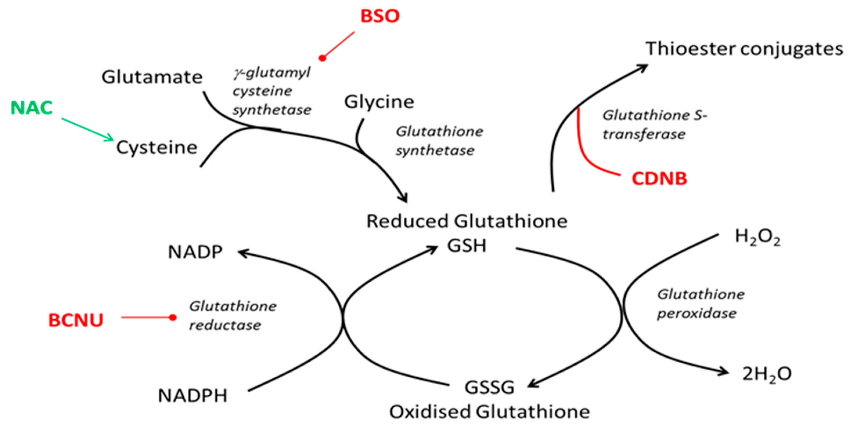
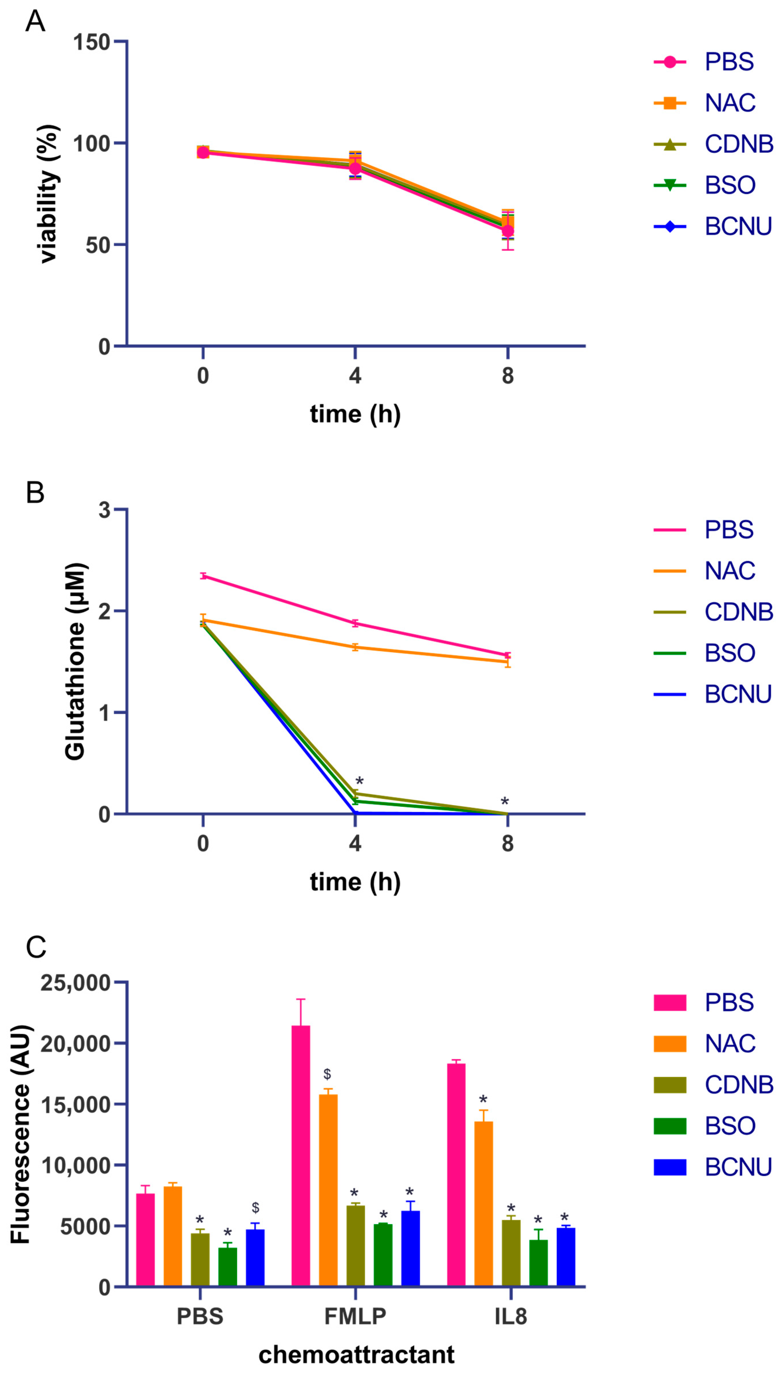
2.4. Glutathione Assays

Reduced, oxidised and total glutathione were measured using the luminescent-based GSH-Glo and GSH:GSSG Glo from Promega (Southampton, UK). Following incubation with appropriate compounds cells (1 × 10⁵ cells per mL) for 4 or 8 h, the samples were distributed in 50 μL per well in 96-well white-walled plates and luminescence was measured using a Tecan Infinite 200 PRO plate multifunctional reader, in comparison to a standard curve.

2.5. Neutrophil Cell Chemotaxis

Chemotaxis was assessed using QCM chemotaxis 3 µm 96-well cell migration chambers (Merck Millipore, Darmstadt, Germany). This method was based on the manufacturer’s protocol. Briefly, neutrophils (1 × 10⁵ cells per mL) were incubated with the 4 GSH modulating compounds for 4 h at room temperature. Following this, the cells with GSH compounds were distributed into the upper chamber of the QCM assay plate. This was placed over the lower chamber containing the chemoattractants: fMLP (Sigma, Dorset, UK) and IL8 (Sigma, UK) with a concentration of 1 ng/mL each. The assembled QCM assay plate was then incubated for another 4 h at 37 °C in a CO2 incubator. After this incubation, the upper chamber was incubated with a prewarmed cell detachment solution for 30 min at 37 °C to allow for the dissociation of any cells on the lower face of the membrane. Cells in the lower chamber and those in the cell detachment solution were combined in a 96-well plate and CyQuant dye was added. The plate was incubated for 15 min at room temperature before reading fluorescence after excitation at 480 nm and emission at 520 nm using a Tecan Infinite 200 PRO plate multifunctional reader.

2.6. Quantitative Proteomics

For the detection of all proteins, including potentially glutathionylated proteins, neutrophils were lysed with 10% SDS with 100 mM TEAB containing a protease inhibitor (Proteases Mini, Roche, Welwyn Garden City, UK) and then stored at −20 °C until all samples had been prepared. The protein content of each preparation was measured via BCA assay. Extracted proteins (100 µg) isolated from neutrophil samples were reduced, alkylated and digested overnight as previously described [33]. The TMT Label Reagents (Thermo Fisher Scientific, Loughborough, UK) were equilibrated to room temperature before use. Anhydrous acetonitrile (41 µL) was added to each tube containing individual TMT labels (0.8 mg). Then, one of the TMT label reagents (41 µL) was added to one peptide preparation. The reactions were incubated for 1 h at room temperature. To quench the reaction, hydroxylamine (8 µL, 5% in 100 mM TEAB) was added to the reactions and they were then incubated for 15 min at room temperature. All labelled samples were combined and dried via vacuum centrifugation. The sample was desalted and subsequently analysed using mass spectrometry (Q Exactive HF Orbitrap mass spectrometer, Thermo Fisher Scientific, Loughborough, UK) for peptide identification at the University of Birmingham’s Advanced Mass spectrometry Facility.

2.7. Statistical Analysis

The statistical analysis was performed using Prism 5.0 software (GraphPad, San Diego, CA, USA) tests used as described.

3. Results

3.1. Gluthathione Modulation in Healthy Neutrophils

In initial experiments, the aim was to understand the role of GSH in neutrophil chemotaxis. To undertake this, neutrophils were isolated from healthy donors and incubated for up to 8 h with the glutathione-modulating compounds BSO (10 µm), NAC (5 mM), BCNU (5 µm) and CDNB (10 µm). Figure 2A shows that the compounds do not affect the viability of neutrophils across the course of the experiment, when compared to the vehicle control, which is PBS. However, there was a decline in neutrophil viability generally over the 8 h time course.
When assessing the changes in reduced-glutathione concentrations with the compounds used, BCNU, CDNB and BSO all decreased reduced-glutathione concentrations to very low or undetectable levels by the 4 h mark (Figure 2B). NAC did not increase glutathione levels but seemed to prevent as much loss of glutathione as was seen in the vehicle control (PBS) cells.
The measurement of oxidized glutathione levels demonstrated that the neutrophils contained 0.15 ± 0.02 uM per well in unstimulated cells and that upon incubation with BSO (10 µm), BCNU (5 µm) or CDNB (10 µm) for 4 h, GSSG levels were then undetectable. After 4 h, GSSG in PBS or NAC (5 mM) treated cells did not change significantly (0.17 ± 0.007 μM per well PBS; 0.25 ± 0.01 μM per well NAC). The GSH:GSSG ratios in PBS and NAC treated cells after 4 h were 21.6 and 14.5, respectively. Due to the undetectable GSSG in the BSO, BCNU and CDNB treated cells, the ratio could not be detected.
Lastly, the effect of the glutathione-modulating compounds on chemotaxis was measured. Figure 2C shows that that BCNU, an inhibitor of glutathione reductase, CDNB, a depleter of glutathione through thioester conjugation, and BSO, an inhibitor of rate limiting γ-glutathione cysteine synthetase, each abolish the chemotactic behaviour of neutrophils responding to either FMLP or IL-8.

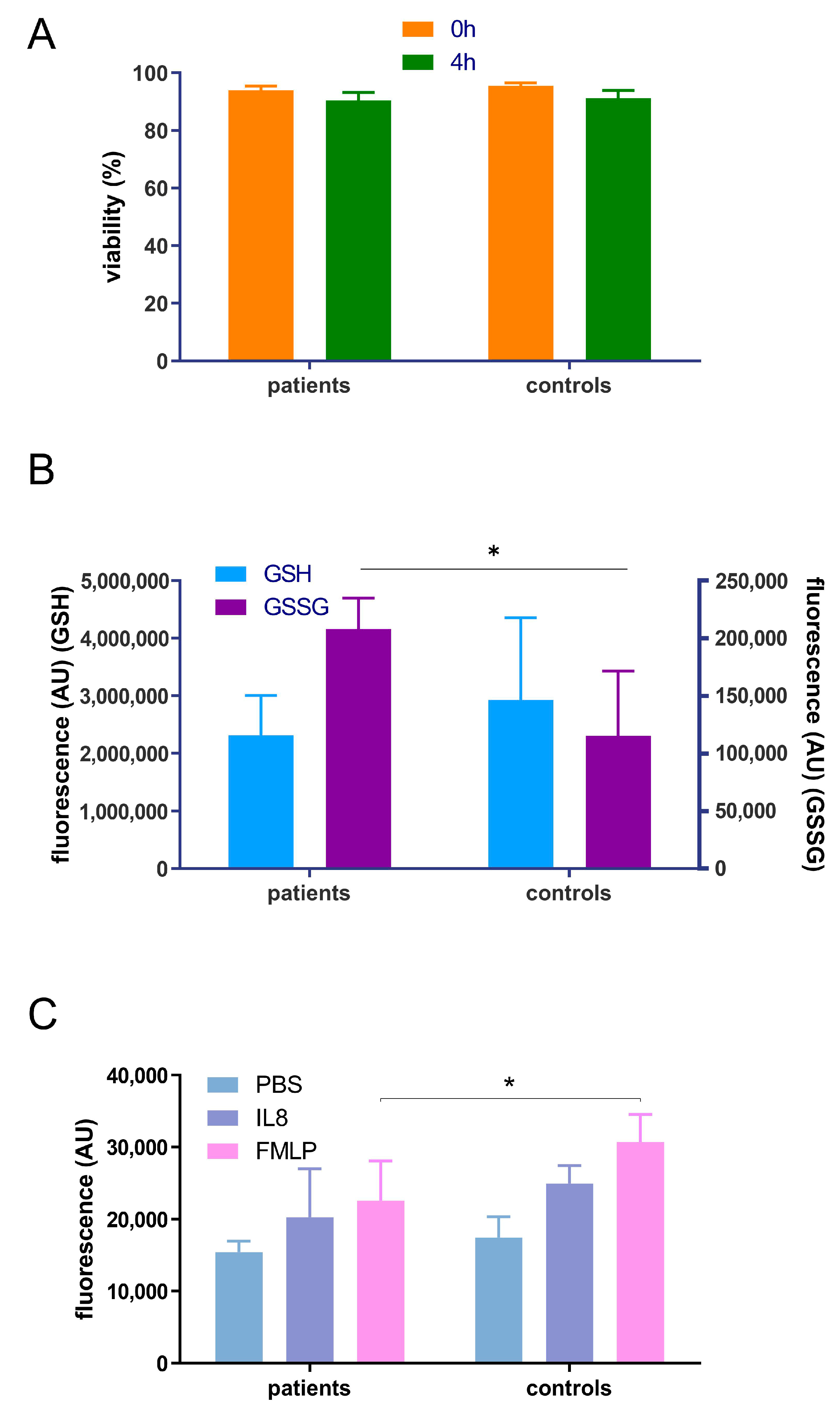
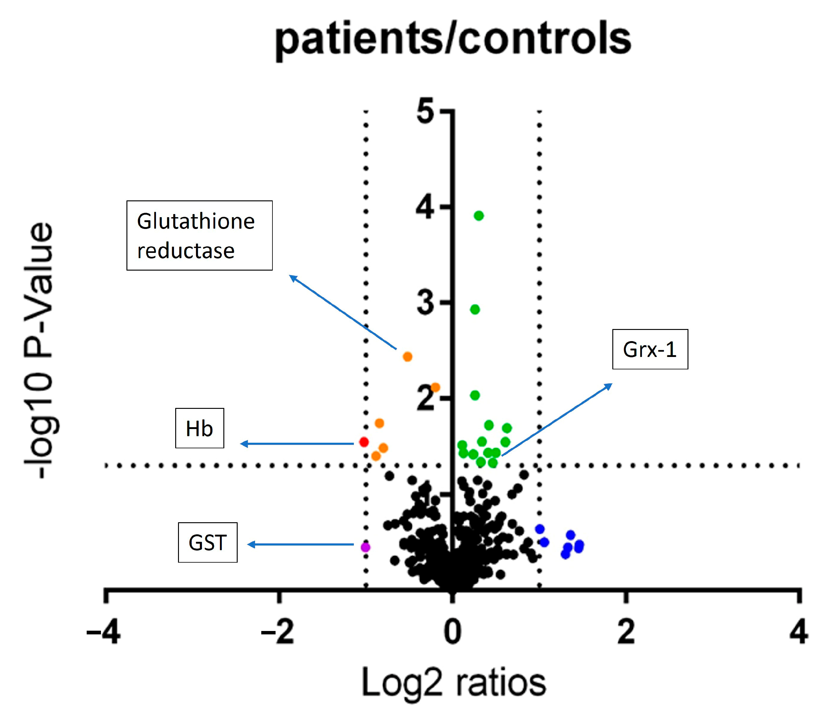
3.2. Pilot Study to Determione the Effect of Periodontitis on Neutrophil Chemotaxis, Glutathione Status and Proteome

Following the demonstration that glutathione levels within neutrophils isolated for healthy donors could be altered, and that this affects neutrophil chemotaxis, the next aim was to explore if there was evidence for altered chemotaxis, glutathione machinery and glutathionylation in patients with chronic periodontitis, in comparison to healthy controls. Following the methods of Woodi et al. [34], it was assessed whether protein glutathionylation could be detected using the instrumentation available. Haemoglobin (Hb) was used as a model protein and incubated with GSSG as previously described. Glutathionylated Hb could be detected via LS-MS. The beta chain showed a shift in mass of 305 Da, indicative of the addition of one glutathione molecule. An analysis of trypitcally digested glutathioylated Hb suggested that the glutathione adduct was found at cysteine, as previously reported [34] (see Supplementary Figures S1 and S2).
In a pilot study to explore the effects of periodontitis, four periodontitis patients and age- and sex-matched controls were recruited into the study. Each donor made one blood donation and neutrophils were isolated through density centrifugation. The isolated neutrophils were evaluated for chemotaxis and for intracellular glutathione assay. For chemotaxis, the patient neutrophils displayed less chemotaxis towards fMLP than the healthy controls. The intracellular total glutathione was similar between patients and controls with no significant differences; however, there was significantly more oxidised glutathione in patients than in controls (Figure 3).
With the knowledge that protein glutathioylation could be detected in our laboratories, proteins were isolated from peripheral blood neutrophils from both control and patient donors. The proteins were analyzed through quantitative proteomics using the tandem mass tag (TMT) method, thus identifying 495 proteins. A volcano plot was used to explore the data (Figure 4). The latter revealed a number of proteins of interest that were consistently altered between the donors.
Three proteins that are part of glutathione homeostatis were identified: glutaredoxin-1 (Grx-1) was increased significantly in a small quantity; Gluthatione-S-Transferase (GST) was decreased over 2-fold, but non-significantly, suggesting that less GSH was leaving the cell; and glutathione reductase was significantly decreased, but less than 2-fold, perhaps indicating that the GSH cycle was perturbed and more GSSG may be present in the neutrophil cell. Haemoglobin was identified as being significantly and over 2-fold decreased. In the neutrophil isolation procedure, erythrocytes were lysed. However, there may still be a small contamination of red blood cells in the neutrophils, which may be caused by handling techniques during the isolation procedure.
Other proteins of interest were highlighted (see Supplementary Table S1 for full list of proteins), either because they showed the most significant change in the analysis of DNA-apurinic or apyrimidinic site lyase; Cdc42; glycogen phosphorylase; Alcohol dehydrogenase class-3; or the highest-fold change in eosinophil peroxidase. After the identification of these proteins, post translational modification by glutathionylation was evaluated. However no glutathionylated peptides were detected via the methods used.

4. Discussion

Chemotaxis is the movement of cells towards chemoattractant in inflammatory responses. To enter an infected tissue, neutrophils require a chemoattractant produced by host cells and that microorganisms migrate to the infection site. In this study, the chemotactic behaviour of neutrophil cells was determined when exposed to glutathione-modulating compounds. Glutathione plays a key role in redox regulation [35]. One of the regulatory actions of GSH is protein glutathionylation which plays an important role in the redox regulation of protein activity and cell signaling. It is a post-translational modification in which GSH forms a disulphide bond with a cysteine of a protein. Post-translational modification is a regulatory mechanism that controls the location and activity of a wide range of proteins.
The glutathione-modulating compounds were used to manipulate the glutathione levels in primary neutrophils and to determine what effect this had on neutrophil chemotaxis. Whilst viability was unchanged in the time course used, decreases in reduced glutathione were caused by the compounds used: BCNU, CDNB and BSO all decreased glutathione concentrations to very low or undetectable levels after 4 h. NAC, as a precursor to glutathione synthesis, did not increase glutathione, but perhaps this is due to the limited time course used. It appeared to prevent as much loss of reduced glutathione across the course of the experiments as that of the vehicle control (PBS). This may have been due to the additional scavenging activities of NAC with cells that are continually producing small quantities of ROS. Due to the undetectable levels of reduced or oxidised glutathione, it was not possible to calculate the GSH:GSSG ratio in the neutrophils treated with BCNU, CDNB or BSO. PBS and NAC treated cells after 4 h had similar ratios to those found previously in primary neutrophils [28].
Neutrophil chemotaxis contributes to many inflammatory and autoimmune diseases, including rheumatoid arthritis, periodontitis, respiratory distress, and systemic inflammatory response syndromes. Previously, our group has demonstrated impaired chemotactic behaviour: much slower movement and aberrant chemotactic index in periodontitis patients [9]. Here, we show that fewer neutrophils migrated in patient isolations than controls, suggesting that they were slower. This is in agreement with video microscopy analysis of neutrophil isolated from periodontitis patients using the Insall chamber in our laboratory [9] and also similar, though less high throughput, to the Boyden chamber analysis of neutrophil movement performed by Kumar and Prakash [36]. Previously, Roberts et al. [9] had found that there was a persistent reduction in speed (movement in any direction) though an improvement in velocity (movement towards the chemoattractant) for IL8 in periodontal disease patients compared to controls even after therapy. The Boyden chamber cannot give this level of information about movement but gives a more rapid answer that could be used to gain insight into greater numbers of patients.
In the glutathione redox cycle, the oxidation of reduced glutathione by H2O2 is catalysed by glutathione peroxidase. It is then reconverted by glutathione reductase to form reduced glutathione. Therefore, glutathione reductase is important in the production of glutathione itself, as the glutathione regeneration is catalysed by glutathione reductase. In neutrophils, glutathione reductase plays an important role in the regulation of the respiratory burst and cell signaling. Glutathione reductase was significantly but slightly decreased. Decreases in this enzyme may contribute to the neutrophil status observed in periodontitis.
Sakai et al. [22] found out that impairment of neutrophil chemotaxis is caused by the disruption of glutaredoxin 1 (Grx1), which is a positive regulator of actin polymerization that causes actin deglutathionylation, an increment of actin glutathionylation and reduced actin polymerization [22]. They showed that reduced ROS leads to a deficiency of chemotaxis, and therefore, they suggested that ROS-induced actin glutathionylation is an important key regulator in neutrophil chemotaxis. Neutrophils that have Grx1 deficiency showed impaired recruitment to sites of inflammation and reduced bactericidal capability. Therefore, ROS-induced actin glutathionylation is a key regulatory mechanism that controls neutrophil function in innate immunity.
DNA-apurinic or apyrimidinic site lyase was altered in expression: a mild decrease at fold change 0.87, p = 0.007. DNA-apurinic or apyrimidinic site lyase plays a role in repairing oxidative damage to DNA bases, as it is a DNA repair enzyme. From the action of DNA glycosylases, this enzyme catalyses the excision of ribose residues at apurinic and apyrimidinic DNA sites. In neutrophils, it plays a role in the repair of oxidative DNA damage. This is important, as previous studies have shown that periodontitis patients have excess production of ROS [37] and this enzyme could help to repair the neutrophil’s DNA damage. DNA-apurinic or apyrimidinic site lyase deficient cells have been shown to have increased ROS, which is a hallmark of the periodontal neutrophil phenotype, and thus, this may be a novel mechanism in these cells requiring further study.
The cell division control protein 42 homolog, or Cdc42, regulates signaling pathways that control diverse cellular functions including cell morphology, migration, endocytosis, and cell cycle progression. Cdc42 is also involved in regulating actin cytoskeleton and processes that are dependent on actin cytoskeleton, such as phagocytosis, cell migration and chemotaxis. In actin cytoskeleton, Cdc42 regulates membrane receptors in the signal transduction pathway to form filopodia. Filopodia are membrane protrusions that extend from the plasma membrane in migratory cells that contain actin filaments and can function as sensory probes into the cellular surroundings. In neutrophils, Cdc42 is also involved in chemotaxis: an increase in Cdc42 activity can stimulate migration speed, such as that seen in neutrophils isolated from knockout mice [38].
Glycogen is accumulated in the liver and glycogen phosphorylase plays an important role in carbohydrate metabolism. In this study, glycogen phosphorylase was found to be mildly but significantly increased (1.2-fold change, p = 0.009). This protein catalyses the reaction initiating glycogen degradation, to generate glucose-1-phosphate, the main source of glucose in the neutrophil. This suggests an energy requirement in neutrophils in circulation in periodontitis patients.
Alcohol dehydrogenase class-3 (ADH3) catalyses the oxidation of long-chain primary alcohols through the oxidation of glutathione. ADH3 acts as an S-hydroxymethyl glutathione dehydrogenase, and as a glutathione-dependent formaldehyde dehydrogenase it can detoxify formaldehyde. Every cell in the human body produces formaldehyde and it is metabolized quickly to prevent accumulation. The reaction occurs at the expense of glutathione, and therefore could be a source of glutathione loss within neutrophils. The change in ADH3 was a fold change of 1.23, p = 0.0001.
Eosinophil peroxidase was increased by 2.8-fold change (p = 0.23). It is found in the primary granules in innate immune cells in humans, including neutrophils. This enzyme catalyses the peroxidase reaction and shares some similarities with myeloperoxidase, wherein its products can disrupt bacterial cell walls. Eosinophil peroxidase is also found in eosinophils, which are the most common, but of low abundance, contaminant in neutrophil isolations. A previous study [39] investigated the transcriptomic contribution of eosinophils to density centrifugation isolated neutrophils and found that the portion of eosinophil transcripts is very low. Although eosinophils were not quantified in these studies, previous experience within our lab using histological identification suggests eosinophil contamination to be very low [40].
Taken together, there is a picture of altered glutathione regulation through the changes in proteins recycling glutathione within the neutrophils of periodontitis patients. Unfortunately, it was not possible to identify any glutathionylated protein targets, one of the desired aims of this study. This is most likely due to the low abundance that was then diluted further by the addition of the TMT tags for quantitation. Future studies could make use of iodo-TMT tags that could isolate glutathionylated proteins only [41]. There are a number of controlled steps required to ensure that glutathionylated proteins are isolated instead of other cysteine modifications or oxidations.
This study has the limitation that only four periodontitis patients and four controls were used in this pilot phase to explore the changes in neutrophil physiology proteome. In a larger study, a power calculation using the data here would suggest that sixteen individuals would be needed in each group. To achieve the suggested proteomic analysis there would need to be a change to a label-free approach to realize these numbers.

5. Conclusions

In conclusion, in this study glutathione-depleting compounds (BCNU, CDNB or BSO) decreased the chemotactic ability of primary neutrophils towards FMLP and IL-8. The redox balance in periodontitis patients was perturbed and appeared to affect the glutathione antioxidant and chemotactic ability of neutrophils. In addition, the glutathione enzyme systems also appeared to be perturbed within these neutrophils, perhaps giving some hints to the mechanisms of the glutathione homeostasis perturbation.

Supplementary Materials

The following supporting information can be downloaded at: https://www.mdpi.com/article/10.3390/oral3040043/s1, Figure S1: Mass spectrum of haemoglobin by direct infusion method showing alpha and glutathionylated beta peaks, Figure S2: Sequence of beta haemoglobin subunit with cysteine modified with glutathionylation highlighted in green, Table S1: Accession number, description of protein and log fold change and p value for comparing the proteins isolated from neutrophils and identified by TMT based quantitative mass spectrometry from periodontitis patients and healthy controls.

Author Contributions

Conceptualization, methodology, formal analysis, resources, data curation, writing—original draft preparation.; writing—review and editing; visualization all N.I.B.B.S., M.M.G.; supervision, M.M.G.; project administration, M.M.G.; funding acquisition, N.I.B.B.S., M.M.G. All authors have read and agreed to the published version of the manuscript.

Funding

This research was funded by the Ministry of Higher Education, Malaysia and Universiti Sains Islam Malaysia.

Institutional Review Board Statement

The study was conducted in accordance with the Declaration of Helsinki, and approved by the South West—Central Bristol Research Ethics Committee 14/SW/1148, and the project was approved by the University of Birmingham and Birmingham Dental Hospital: MG_2015_02.

Informed Consent Statement

Informed consent was obtained from all subjects involved in the study.

Data Availability Statement

The data presented in this study are available in supplementary material.

Conflicts of Interest

The authors declare no conflict of interest.

References

  1. Hasturk, H.; Kantarci, A. Activation and resolution of periodontal inflammation and its systemic impact. Periodontol. 2000 2015, 69, 255–273. [Google Scholar] [CrossRef] [PubMed]
  2. Graves, D.T.; Cochran, D. The contribution of interleukin-1 and tumor necrosis factor to periodontal tissue destruction. J. Periodontol. 2003, 74, 391–401. [Google Scholar] [CrossRef] [PubMed]
  3. Nazir, M.A. Prevalence of periodontal disease, its association with systemic diseases and prevention. Int. J. Health Sci. 2017, 11, 72–80. [Google Scholar]
  4. Gamonal, J.; Acevedo, A.; Bascones, A.; Jorge, O.; Silva, A. Characterization of cellular infiltrate, detection of chemokine receptor CCR5 and interleukin-8 and RANTES chemokines in adult periodontitis. J. Periodontal Res. 2001, 36, 194–203. [Google Scholar] [CrossRef]
  5. Hajishengallis, E.; Hajishengallis, G. Neutrophil homeostasis and periodontal health in children and adults. J. Dent. Res. 2014, 93, 231–237. [Google Scholar] [CrossRef]
  6. Dias, I.H.; Matthews, J.B.; Chapple, I.L.; Wright, H.J.; Dunston, C.R.; Griffiths, H.R. Activation of the neutrophil respiratory burst by plasma from periodontitis patients is mediated by pro-inflammatory cytokines. J. Clin. Periodontol. 2011, 38, 1–7. [Google Scholar] [CrossRef]
  7. Matthews, J.B.; Wright, H.J.; Roberts, A.; Cooper, P.R.; Chapple, I.L. Hyperactivity and reactivity of peripheral blood neutrophils in chronic periodontitis. Clin. Exp. Immunol. 2007, 147, 255–264. [Google Scholar] [CrossRef]
  8. Matthews, J.B.; Wright, H.J.; Roberts, A.; Ling-Mountford, N.; Cooper, P.R.; Chapple, I.L. Neutrophil hyper-responsiveness in periodontitis. J. Dent. Res. 2007, 86, 718–722. [Google Scholar] [CrossRef]
  9. Roberts, H.M.; Ling, M.R.; Insall, R.; Kalna, G.; Spengler, J.; Grant, M.M.; Chapple, I.L. Impaired neutrophil directional chemotactic accuracy in chronic periodontitis patients. J. Clin. Periodontol. 2015, 42, 1–11. [Google Scholar] [CrossRef]
  10. Ling, M.R.; Chapple, I.L.; Matthews, J.B. Neutrophil superoxide release and plasma C-reactive protein levels pre- and post-periodontal therapy. J. Clin. Periodontol. 2016, 43, 652–658. [Google Scholar] [CrossRef]
  11. Weiss, S.J. Tissue destruction by neutrophils. N. Engl. J. Med. 1989, 320, 365–376. [Google Scholar] [CrossRef]
  12. Chapman, A.L.; Hampton, M.B.; Senthilmohan, R.; Winterbourn, C.C.; Kettle, A.J. Chlorination of bacterial and neutrophil proteins during phagocytosis and killing of Staphylococcus aureus. J. Biol. Chem. 2002, 277, 9757–9762. [Google Scholar] [CrossRef] [PubMed]
  13. Cooper, P.R.; Palmer, L.J.; Chapple, I.L. Neutrophil extracellular traps as a new paradigm in innate immunity: Friend or foe? Periodontol. 2000 2013, 63, 165–197. [Google Scholar] [CrossRef] [PubMed]
  14. Li, R.; Bowerman, B. Symmetry breaking in biology. Cold Spring Harb. Perspect. Biol. 2010, 2, a003475. [Google Scholar] [CrossRef] [PubMed]
  15. Wang, F. The signaling mechanisms underlying cell polarity and chemotaxis. Cold Spring Harb. Perspect. Biol. 2009, 1, a002980. [Google Scholar] [CrossRef]
  16. Harbord, M.W.; Marks, D.J.; Forbes, A.; Bloom, S.L.; Day, R.M.; Segal, A.W. Impaired neutrophil chemotaxis in Crohn’s disease relates to reduced production of chemokines and can be augmented by granulocyte-colony stimulating factor. Aliment. Pharmacol. Ther. 2006, 24, 651–660. [Google Scholar] [CrossRef]
  17. Yoshikawa, T.; Dent, G.; Ward, J.; Angco, G.; Nong, G.; Nomura, N.; Hirata, K.; Djukanovic, R. Impaired neutrophil chemotaxis in chronic obstructive pulmonary disease. Am. J. Respir. Crit. Care Med. 2007, 175, 473–479. [Google Scholar] [CrossRef]
  18. Janetopoulos, C.; Firtel, R.A. Directional sensing during chemotaxis. FEBS Lett. 2008, 582, 2075–2085. [Google Scholar] [CrossRef]
  19. Kolaczkowska, E.; Kubes, P. Neutrophil recruitment and function in health and inflammation. Nat. Rev. Immunol. 2013, 13, 159–175. [Google Scholar] [CrossRef]
  20. Andrew, N.; Insall, R.H. Chemotaxis in shallow gradients is mediated independently of PtdIns 3-kinase by biased choices between random protrusions. Nat. Cell Biol. 2007, 9, 193–200. [Google Scholar] [CrossRef]
  21. Dickinson, B.C.; Chang, C.J. Chemistry and biology of reactive oxygen species in signaling or stress responses. Nat. Chem. Biol. 2011, 7, 504–511. [Google Scholar] [CrossRef] [PubMed]
  22. Sakai, J.; Li, J.; Subramanian, K.K.; Mondal, S.; Bajrami, B.; Hattori, H.; Jia, Y.; Dickinson, B.C.; Zhong, J.; Ye, K.; et al. Reactive oxygen species-induced actin glutathionylation controls actin dynamics in neutrophils. Immunity 2012, 37, 1037–1049. [Google Scholar] [CrossRef] [PubMed]
  23. Niethammer, P.; Grabher, C.; Look, A.T.; Mitchison, T.J. A tissue-scale gradient of hydrogen peroxide mediates rapid wound detection in zebrafish. Nature 2009, 459, 996–999. [Google Scholar] [CrossRef] [PubMed]
  24. Cameron, J.M.; Gabrielsen, M.; Chim, Y.H.; Munro, J.; McGhee, E.J.; Sumpton, D.; Eaton, P.; Anderson, K.I.; Yin, H.; Olson, M.F. Polarized cell motility induces hydrogen peroxide to inhibit cofilin via cysteine oxidation. Curr. Biol. 2015, 25, 1520–1525. [Google Scholar] [CrossRef]
  25. Johansson Mali’n, T.; Lindberg, S.; Astot, C. Novel glutathione conjugates of phenyl isocyanate identified by ultra-performance liquid chromatography/electrospray ionization mass spectrometry and nuclear magnetic resonance. J. Mass Spectrom. 2014, 49, 68–79. [Google Scholar] [CrossRef]
  26. Sies, H. Glutathione and its role in cellular functions. Free Radic. Biol. Med. 1999, 27, 916–921. [Google Scholar] [CrossRef]
  27. Meister, A. Selective modification of glutathione metabolism. Science 1983, 220, 472–477. [Google Scholar] [CrossRef]
  28. Dias, I.H.; Chapple, I.L.; Milward, M.; Grant, M.M.; Hill, E.; Brown, J.; Griffiths, H.R. Sulforaphane restores cellular glutathione levels and reduces chronic periodontitis neutrophil hyperactivity in vitro. PLoS ONE 2013, 8, e66407. [Google Scholar] [CrossRef]
  29. Harlan, J.M.; Levine, J.D.; Callahan, K.S.; Schwartz, B.R.; Harker, L.A. Glutathione redox cycle protects cultured endothelial cells against lysis by extracellularly generated hydrogen peroxide. J. Clin. Investig. 1984, 73, 706–713. [Google Scholar] [CrossRef]
  30. Grant, M.M.; Brock, G.R.; Matthews, J.B.; Chapple, I.L. Crevicular fluid glutathione levels in periodontitis and the effect of non-surgical therapy. J. Clin. Periodontol. 2010, 37, 17–23. [Google Scholar] [CrossRef]
  31. Brock, G.R.; Butterworth, C.J.; Matthews, J.B.; Chapple, I.L. Local and systemic total antioxidant capacity in periodontitis and health. J. Clin. Periodontol. 2004, 31, 515–521. [Google Scholar] [CrossRef] [PubMed]
  32. Eke, P.I.; Page, R.C.; Wei, L.; Thornton-Evans, G.; Genco, R.J. Update of the Case Definitions for Population-Based Surveillance of Periodontitis. J. Periodontol. 2012, 83, 1449–1454. [Google Scholar] [CrossRef] [PubMed]
  33. Pasha, S.; Inui, T.; Chapple, I.; Harris, S.; Holcombe, L.; Grant, M.M. The Saliva Proteome of Dogs: Variations within and between Breeds and between Species. Proteomics 2018, 18, 1700293. [Google Scholar] [CrossRef] [PubMed]
  34. Woodi, M.; Mondal, A.K.; Padmanabhan, B.; Rajagopalan, K.P. Analysis of protein posttranslational modifications by mass spectrometry: With special reference to haemoglobin. Indian J. Clin. Biochem. 2009, 24, 23–29. [Google Scholar] [CrossRef] [PubMed]
  35. Trachootham, D.; Lu, W.; Ogasawara, M.A.; Nilsa, R.D.; Huang, P. Redox regulation of cell survival. Antioxid. Redox Signal. 2008, 10, 1343–1374. [Google Scholar] [CrossRef]
  36. Kumar, R.S.; Prakash, S. Impaired neutrophil and monocyte chemotaxis in chronic and aggressive periodontitis and effects of periodontal therapy. Indian J. Dent. Res. 2012, 23, 69–74. [Google Scholar] [CrossRef]
  37. Dahiya, P.; Kamal, R.; Gupta, R.; Bhardwaj, R.; Chaudhary, K.; Kaur, S. Reactive oxygen species in periodontitis. J. Indian Soc. Periodontol. 2013, 17, 411–416. [Google Scholar] [CrossRef]
  38. Szczur, K.; Xu, H.; Atkinson, S.; Zheng, Y.; Filippi, M.D. Rho GTPase CDC42 regulates directionality and random movement via distinct MAPK pathways in neutrophils. Blood 2006, 108, 4205–4213. [Google Scholar] [CrossRef]
  39. Thomas, H.B.; Moots, R.J.; Edwards, S.W.; Wright, H.L. Whose Gene Is It Anyway? The Effect of Preparation Purity on Neutrophil Transcriptome Studies. PLoS ONE 2015, 10, e0138982. [Google Scholar] [CrossRef]
  40. Roberts, H.; White, P.; Dias, I.; McKaig, S.; Veeramachaneni, R.; Thakker, N.; Grant, M.; Chapple, I. Characterization of neutrophil function in Papillon-Lefevre syndrome. J. Leukoc. Biol. 2016, 100, 433–444. [Google Scholar] [CrossRef]
  41. Pan, K.T.; Chen, Y.Y.; Pu, T.H.; Chao, Y.S.; Yang, C.Y.; Bomgarden, R.D.; Rogers, J.C.; Meng, T.C.; Khoo, K.H. Mass spectrometry-based quantitative proteomics for dissecting multiplexed redox cysteine modifications in nitric oxide-protected cardiomyocyte under hypoxia. Antioxid. Redox Signal. 2014, 20, 1365–1381. [Google Scholar] [CrossRef] [PubMed]
Figure 1. Pharmacologic modification of the glutathione redox cycle in vitro. Adapted from Harlan et al., 1984. [29] Abbreviations BCNU: carmutsine or bis-chloroethylnitrosourea; CDNB: 1-chloro2,4-dinitrobenzene; BSO: buthioninesulfoximine; NAC: N-acetyl cysteine.
Oral 03 00043 g001
Figure 2. Isolated neutrophils were incubated with BSO (10 µm), NAC (5 mM), BCNU (5 µm) or CDNB (10 µm) for 4 or 8 h. (A). Neutrophil cell viability was determined via trypan blue exclusion. (B). Neutrophil intracellular-reduced glutathione determined using GSH-Glo assay. (C). Neutrophil cell chemotaxis with glutathione-modulating compounds. Isolated neutrophils were incubated with BSO (10 µm), NAC (5 mM), BCNU (5 µm) or CDNB (10 µm) for 4 h prior to distribution into 96-well QCM migration assay plates. Cells 1 × 10⁵ per mL were then allowed to respond to chemoattractants FMLP or IL8/CXCL-8 for a further 4 h before measurement of the number of cells that had migrated. Data are represented as mean ±standard deviation (n = 4 donors). Significance was measured by One-way ANOVA with Tukey’s post-test * p < 0.0001, $ p < 0.001 (comparisons are to PBS treatment for each chemoattractant or timepoint).
Oral 03 00043 g002
Figure 3. Neutrophils were isolated from patients with periodontitis and from age- and sex-matched healthy controls. (A). Neutrophil cell viability was determined via trypan blue exclusion. (B). Neutrophil intracellular-reduced and oxidized glutathione were determined using GSH-Glo or GSSG-Glo assays. (C). Neutrophil cell chemotaxis. Cells 1 × 10⁵ per mL responded to chemoattractants FMLP or IL8/CXCL-8 for 4 h before measurement of the number of cells that had migrated. Data are represented as mean ± standard deviation (n = 4 donors). Significance was measured by t-test. * p < 0.05.
Oral 03 00043 g003
Figure 4. Volcano plot of all periodontal patients versus controls. Mean changes between age- and sex-matched pairs were calculated and assessed. The colours represent changes that are either more than 2-fold changed (blue and violet), or have significance less than 0.05 (orange or green) or that are both greater than 2-fold and less than 0.05 (red).
Oral 03 00043 g004
Table 1. Profile of periodontal patients and controls in pilot study. “-” signifies that no information was recorded.
Control GroupPeriodontitis Group
Number44
Mean age (range)40.5 ± 4.5 years old40.75 ± 2.5 years old
Sex (% female)50%50%
Number of teeth-30 ± 2
Percentage bleeding-18 + ±7%
Percentage plaque surfaces-27 ± 21%
PPD Mean ± SD (mm)-3 ± 1 mm
Max PPD (mm)-10 ± 3 mm
Teeth > 4 mm-16 ± 6
CAL Mean ± SD-3 ± 1 mm
% sites > 4 mm-23 ± 11%
Disclaimer/Publisher’s Note: The statements, opinions and data contained in all publications are solely those of the individual author(s) and contributor(s) and not of MDPI and/or the editor(s). MDPI and/or the editor(s) disclaim responsibility for any injury to people or property resulting from any ideas, methods, instructions or products referred to in the content.

