Article

Effects of Ceramic Translucency and Thickness on Polymerization of a Photosensitive Resin Cement

1
School of Dentistry, University of Adelaide, Adelaide 5000, SA, Australia
2
Melbourne Dental School, University of Melbourne, Melbourne 3053, VIC, Australia
3
Sir John Walsh Research Institute, Faculty of Dentistry, University of Otago, Dunedin 9016, New Zealand
*
Author to whom correspondence should be addressed.
Oral 2023, 3(4), 490-498; https://doi.org/10.3390/oral3040040
Submission received: 22 May 2023 / Revised: 21 October 2023 / Accepted: 25 October 2023 / Published: 30 October 2023
(This article belongs to the Special Issue Advances in Dental Composite Materials)

Abstract

:
We investigated the effects of lithium disilicate ceramic thickness and translucency on the degree of polymerization of light-cured resin cement using the measure of hardness. Lithium disilicate specimens of three translucencies (low, medium and high) were prepared to four thicknesses (0.5, 1.0, 2.0 and 3.0 mm). A light-cured resin cement was cured through each of the ceramic specimens using a handheld curing light for 50 s. A 3D printed jig was used to achieve a uniform thickness of the resin cement. Directly cured resin cement was used as the control. Hardness was measured using nano-indentation to determine the degree of polymerization of the resin cement. Two-way ANOVA and Tukey post hoc tests were used to evaluate interaction between translucency and thickness. Hardness values from control specimens were assessed using the two-tailed t-test with the Bonferroni approach. The translucency of the specimens significantly influenced the hardness (p < 0.001), where a negative linear relationship between cement hardness and ceramic thickness was present for low translucency and high translucency. However, at a 0.5 mm thickness, all specimens showed similar hardness regardless of the translucency. The translucency of ceramics affected the hardness, and hence polymerization, of light-cure resin cement. However, the effect of increased thickness was a more significant factor.

1. Introduction

Contemporary dental ceramics and bonding technologies facilitate restoring dentition with more predictable, esthetic and conservative outcomes [1]. Indirect ceramic restorations offer superior physical properties, wear resistance, reduced polymerization shrinkage and more precise control of contours and contacts [2]. In addition, ceramics allow diffuse transmission and reflectance of light, which reproduce depth of translucency. Eventually, the shade of ceramic restoration is likely to mimic the shade of natural teeth that results in a superior esthetic outcome [3]. Glass-based ceramic veneers and crowns have been well researched in regards to their clinical longevity, patient satisfaction and excellent success rate [4]. A lithium disilicate ceramic is widely used in contemporary dentistry because of its esthetic outcome and favorable survival rates that were reported to be more than 95% in an observation period of 5 years [5].
However, there are several factors that affect overall success [6], and the cementation procedure is one such factor, directly affecting fatigue resistance and retention [7,8,9]. The adhesion of ceramic restorations to the tooth structure is a critical step that affects longevity and success of treatment. A strong, durable bond achieved with well-polymerized resin cement provides high retention, improved marginal adaptation and increased ceramic fracture resistance [7]. Insufficient polymerization has been associated with microleakage, recurrent caries, post-operative sensitivity, discoloration and decreased mechanical properties [10].
Resin cements have been classified into different categories depending on their adhesive procedures and chemical components [7]. One type of classification is according to the mode of polymerization, chemical cure, light cure and dual cure. Chemical cure resin cements set via chemical reactions initiated with a catalyst, most commonly the tertiary amine, benzoyl peroxide [11]. Light-cured resin cement contains a photo-initiator, most commonly camphorquinone, which absorbs blue light to generate free radicals that initiate polymerization. Although light cure resin cements offer clinical advantages of extended working time and setting on demand, they have greater potential for incomplete polymerization because ceramic material characteristics may inhibit sufficient light energy being transmitted through the ceramic to the cement. The extent of light attenuation depends on the ceramic composition, thickness and translucency [11,12]. One study found that dental ceramics absorb 40 to 50% of the curing light intensity, and increased ceramic thickness requires increased exposure times beyond 30 to 40 s for adequate resin curing [13]. Another study showed that irradiances of two curing units decreased by >80% through 1.5 mm discs of various glass ceramics [14]. Dual-cure resin cements containing both chemical and photo-initiators were developed in an attempt to overcomes these effects. The initial set is achieved with light curing to quickly seal the margins, and the chemical curing component ensures that the cement will cure underneath the restoration [8]. Dual-cure resin cements may be useful when the ceramic is too thick or too opaque to allow sufficient light transmission. However, studies have found that chemical-only-cured resin cements demonstrated inferior physical properties compared to partially or fully photo-polymerized cements [15]. Furthermore, the reactive amine groups contained in dual- and chemical-cure resin cements have been associated with poor color stability [16]. Hence, use of solely light-cure resin cements is clearly more advantageous; however, the variability of cure due to light penetration through the prosthesis remains a potential issue.
The aim of this study was to evaluate the effect of lithium disilicate translucency and thickness on the polymerization of a light-cured resin cement using hardness as an indicator of the degree of polymerization [17]. The null hypothesis is that the ceramic thickness and translucency do not affect the hardness of the underlying resin cement. The significance of the study is to provide a guide for clinicians as to the thickness and translucency of lithium disilicate veneers at which it is still effective to use light-initiated-only resin cement.

2. Materials and Methods

Three lithium disilicate ceramic blocks (IPS e.max CAD, Ivoclar Vivodent, Liechtenstein) of three different translucencies, low (LT), medium (MT) and high (HT), were used in this study. All the ceramic specimens had shade A2. The lithium disilicate had content of SiO2 at 57.0–80.0 and Li2O at 11.0–19.0. A single light-cured resin cement (RelyX Veneer Cement; 3M ESPE, Seefeld, Germany) was used in the study. The cement contents are summarized in Table 1 below.

2.1. Specimen Preparation
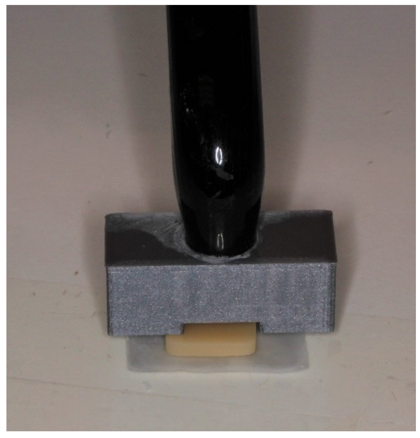
Using a diamond saw and under water cooling (Accutom 50; Struers, Copenhagen, Denmark), lithium disilicate blocks of the different translucencies were sliced by a single operator. The specimens were cut into flat and square smaller blocks (5 mm × 5 mm). Four different thicknesses were included, 0.5, 1.0, 2.0 and 3.0 mm, to mimic the range of thicknesses ceramic crowns are usually fabricated at. The tolerance of the diamond saw was 0.05 mm. The slices were polished sequentially on one surface down to 2200 grit sandpaper to ensure a smooth surface. The specimens were then sintered using a sintering oven (CEREC speed fire; Dentsply Sirona, York, PA, USA) using pre-set parameters. A digital caliper was used to confirm the thickness of each slice to be within the desired range. The specimens were placed in dry storage at room temperature for 24 h until further testing was carried out.

2.2. Curing Jigs

In order to control cement thickness and the position of the curing light tip, two 3D-printed curing jigs were used in the study. To ensure consistent spacing for the resin cement, a cement spacer jig was designed and then 3D printed. The jig was placed below the specimen to achieve clinically appropriate cement thickness of 100 μm and allow extrusion of excess cement under pressure. To ensure consistency in position and distance of the light-cure tip, a curing-tip jig was designed and 3D printed to befit over the 8-mm-wide tip of the curing light to keep the light at a constant distance to the ceramic.

2.3. Light-Curing Protocol

The cement spacer jig was placed on a flat, clean glass surface and the light-cured resin cement (RelyX Veneer Cement) was directly applied from the syringe in the center. The ceramic specimen was then placed over the uncured cement and even finger pressure applied for 3 s. The same operator placed the curing-tip jig over the ceramic block on which the LED curing light (DemiPlus; Kerr, Orange, CA, USA), which had a flat tip with an output intensity of 1200 mW/cm2, was applied as per manufacturer’s instructions (20 s spot cure and then 30 s final cure). Figure 1 and Figure 2 show the layout of the light curing step. The cement did not bond to the glass surface. The output of the curing light was verified using checkMARC (KaVo Kerr; Kerr, Orange, CA, USA) prior to the study. Resin cement on a flat surface was cured without a ceramic layer to serve as the control. Following light curing, the ceramic–cement specimens were placed into dry, light-proof containers to prevent further polymerization.

2.4. Measurement of Resin Polymerization

The specimen was removed from the light-proof container and set up for testing, and the resin cement bonded to the ceramic specimen was then subjected to hardness testing. The nano-indentation (UMIS2000; Semilabs, Tampa, FL, USA) hardness test of the cement layer was applied to a maximum of 50 mN and held for 1 s using a standard Berkovich indenter and analyzed with IBIS2 software (Semilabs). Power calculation was carried out for 12 testing groups, with 20 indents at 50 μm spacing made on the cement surface opposite to the ceramic to represent the tooth–cement interface (n = 20).

2.5. Statistical Analysis
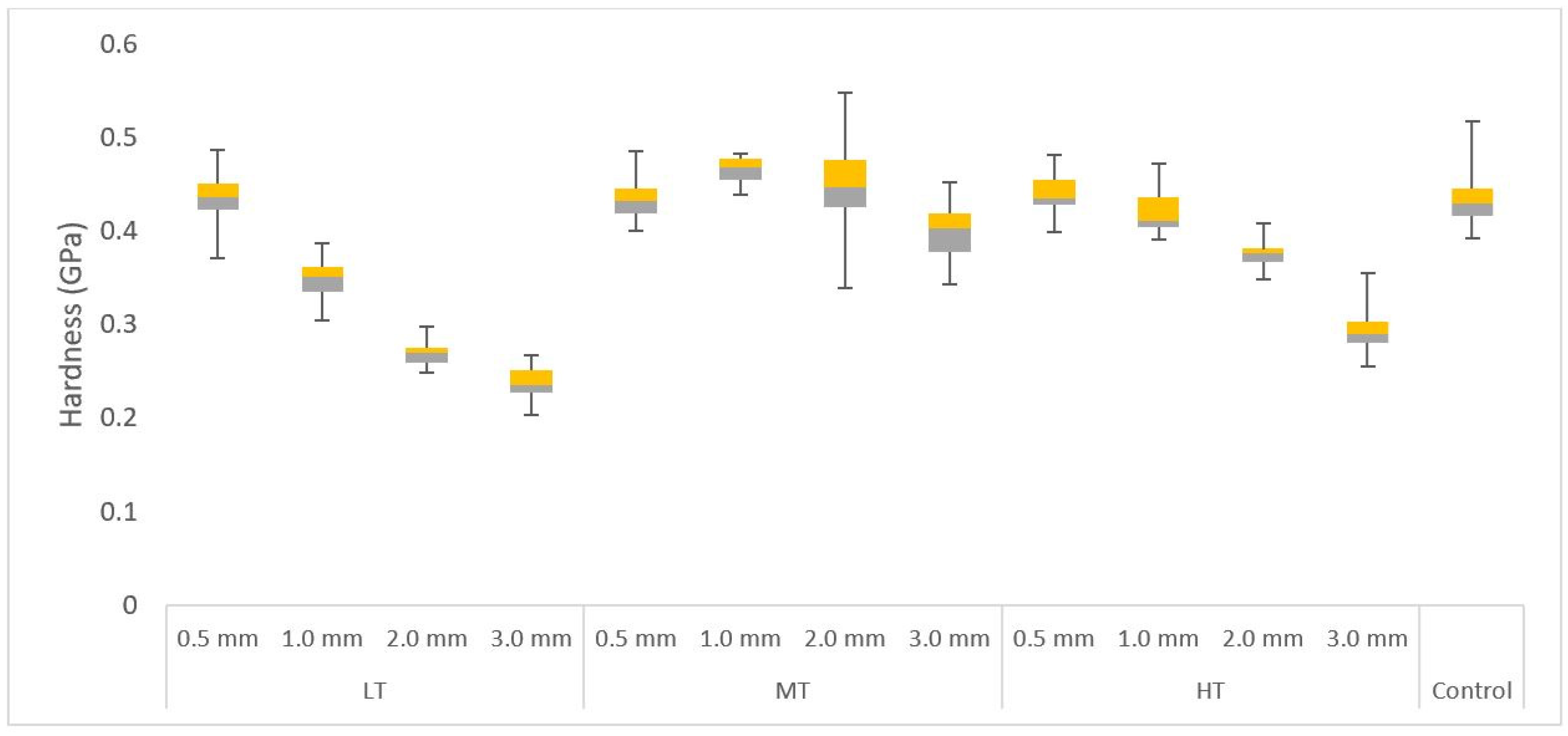
The normality of the hardness data was confirmed with the Kolmogorov–Smirnov test. The interaction between translucency and thickness was evaluated with the two-way ANOVA test and Tukey post hoc test. In addition, the mean hardness values were calculated for each ceramic–cement specimen and compared with the hardness values from control specimens using the two-tailed t-test with the Bonferroni approach. All statistical analyses were completed with the SPSS software package (IBM SPSS Statistics, version 22, SPSS Inc., Chicago, IL, USA) at the 0.05 level of significance.

3. Results

Table 2 represents the mean nano-indentation hardness (GPa) values for all the tested specimens. The LT had the lowest hardness values at all thicknesses compared to the MT and HT. The translucency of the specimens significantly influenced the hardness (p < 0.001), where the highest hardness was generally observed for MT followed by HT and LT, respectively. Likewise, the thickness had a significant effect on the hardness (p < 0.001), with thicker specimens exhibiting less hardness values. A significant interaction occurred between the translucency and thickness (p < 0.001). A negative linear relationship between cement hardness and ceramic thickness was present for LT and HT (Figure 3), where a statistically significant difference was observed between the different thicknesses (p < 0.05). In general, the thickness of the ceramic appears to influence the hardness more than the translucency. The highest hardness for MT was for 1.0 mm followed by 2.0 mm, 0.5 mm and 3.0 mm, respectively. Significant differences among the MT specimens were observed between 0.5 mm and 1.0 mm (p = 0.03), 0.5 mm and 3.0 mm (p = 0.004), 1.0 mm and 3.0 mm (p < 0.001) and 2.0 and 3.0 mm (p < 0.001).
The comparisons of each specimen group against the control group are shown in Table 3. Regardless of the translucency, all of them showed similar hardness to the control group at a 0.5 mm thickness. At a 1 mm thickness, MT and HT provided a similar outcome to the control group. For the LT, there was no difference between the hardness of resin cement light cured directly or through a 0.5 mm thickness (p = 0.74), but the decrease in hardness for 1.0, 2.0 and 3.0 mm thicknesses was statistically significant (p < 0.01). The MT group showed similar hardness to the control at 0.5 mm (p = 0.92) and 2.0 mm (p = 0.40). At a 1.0 mm thickness, the hardness for the MT group was significantly greater than the control (p < 0.001), and at 3.0 mm, it was significantly less than the control (p < 0.001). For the HT, 0.5 and 1.0 mm thicknesses did not significantly affect the hardness of the cement (p > 0.05). However, the reduction in hardness was significant for 2.0 and 3.0 mm (p < 0.001).

4. Discussion

The results of this study revealed that a lithium disilicate ceramic with a thickness of 0.5 mm allowed for similar hardness of cement regardless of translucency. As the thickness increases beyond 0.5 mm for low translucency and 1.0 mm for high translucency, statistically significant lower cement hardness than the control was observed. There was a negative linear relationship between increasing thickness of low- and high-translucency ceramics and the hardness of the cured resin cement. The observed negative relationship is in agreement with previous studies [17,18,19,20,21]. However, such relationship was inconsistent for the medium-translucency material. Hence, we reject the null hypothesis that ceramic thickness and translucency do not affect the hardness of the underlying resin cement.
The extent of light penetration through a material, such as lithium disilicate, is largely related to the extent of scattering and absorption of light. This is a phenomenon in which the light changes direction or is absorbed after interacting with a medium, which decreases the intensity of light that is transmitted through a medium [12]. The amount of light reflected, absorbed and transmitted using a restorative material depends on the number and size of the particles and pores within the material. Previous studies have shown that the smaller the particle size, the greater the light scattering, and the ceramics with bigger particles allowed for deeper resin polymerization [22].
Lithium disilicate ceramics have a structure of randomly oriented needle-like crystals [23]. The HT lithium disilicate is distinguished with the presence of a small number of large lithium metasilicate crystals in the pre-crystallized state compared to the LT, which contains a large number of smaller crystals [24]. After full crystallization heat treatment, the HT is composed of lithium disilicate crystals in a glassy matrix. On the contrary, the fully crystallized LT material exhibits a higher density of small interlocked lithium disilicate crystals. Such a structure, together with spherical pores of highly soluble lithium phosphate crystals, may restrict the extent of light penetration [25] while the presence of residual pores increases scattering [26]. Excluding experimental error, the hardness of cement on MT material may be the result of dimensional, structural or chemical differences in the crystals and crystal boundaries that yielded less absorption and scattering of light. Further research, such as a crystallographic analysis for the lithium disilicate structure of different translucencies, using scanning electron microscopy may assist in explaining these results.
In accordance with the present study, previous studies have examined how the translucency of dental ceramics is also influenced by the thickness of the material. One study measured the translucency of glass ceramics ranging from 0.6 to 2.0 mm and confirmed the exponential increase in the translucency as the thickness decreased [21]. Furthermore, it has been reported that ceramic thickness has a more profound effect on resin polymerization compared to translucency and shade [26,27,28]. One study measured the light transmission through a leucite–glass ceramic of 1, 2, 3 and 4 mm thicknesses. The Vickers hardness testing on the resin cement specimens revealed that a ceramic thickness of 3 mm and above significantly reduced the micro-hardness values for light- and dual-cure cements, with a significant correlation being found between the amount of light transmitted and hardness [18]. Some studies found that thickness and translucency were not a major factor in the extent of polymerization of the resin cement. However, these studies examined specimens that were relatively thin (1.5 mm or less) and hence the effects of the differing translucencies may not have been as significant [29,30]. Others had other issues with not ensuring that the curing light was calibrated as the output of light can deteriorate over time [31,32]. Some of the findings of those studies are undermined as light intensity has a direct impact on the extent of polymerization regardless of thickness or translucency.
Direct light curing of resin cement is expected to achieve the greatest degree of polymerization compared to when light is cured through a ceramic. However, the present study found higher values of hardness for resin cement through the MT material that is less than 3.0 mm in thickness, compared to the control. A similar observation was made in another study, which found that for some cements direct light curing did not result in a higher degree of conversion compared to when light cured through ceramic discs [12]. In addition to material characteristics, the delivered amount of light energy is dependent on the distance of the light tip from the resin cement, movement of light tip during curing, condition of the light-curing unit as well as thickness of the resin being cured. The distance of the light tip from the resin cement and the thickness of the cement film were kept constant using a 3D-printed jig. The output of the curing light used was verified prior to the experiment but light density may have been an additional variable to consider. The use of a handheld curing light may have introduced experimental error due to minor movement and changes in angulation.
There are a number of potential limitations to this study. One is the method used to assess the extent of resin cement polymerization. Spectrometry type testing can directly determine the degree of cement conversion and is generally more sensitive when used correctly. Still, using hardness as a measure of resin polymerization or conversion is still a reliable method. Several studies have found that increased hardness correlates well to the degree of conversion and hence the performance of the resin [33,34,35,36]. However, indirectly determining the degree of polymerization via the hardness test, compared to direct measurement of the degree of polymerization, may affect the limit comparison among resins that prevents generalization of the results to all light-cured resin cements. In the dental literature, hardness values represent a good correlation with the degree of conversion for resins and have been shown to be a reliable method when specimen thickness was as low as 0.5 mm [17]. The use of the nano-indentation technique to measure the hardness does offer the advantage of consistency of measurement methods while other techniques, such as optical and scraping techniques, tend to overestimate the hardness of the cured resin cement [37]. Previous studies have shown that translucency of the ceramic has a greater effect than shade [38] but darker shades have been associated with decreased light transmission [39]. The 100 μm cement thickness was chosen as it is the approximate average of the expected internal gap as reported in the literature. However, cemented ceramic restorations may have variable cement thicknesses and different cement thicknesses may need to be explored further for different manufacturing methods of the ceramic [40]. Further studies are needed to test the effect of geometry on the light transmission and extent of polymerization of light-cure resin cement, and also clinical medium-to-long-term studies on the effect of such a bonding protocol on success and patient satisfaction.
The results of this study provide a guide to clinicians on thicknesses and translucency of lithium disilicate ceramics at which fully light-cured resin cement is indicated to retain a ceramic restoration. The use of nano-indentation and commonly used ceramic and resin cements makes the findings more relatable to the clinical setting. Further investigations are needed to clarify the relationship between the polymerization of light-cured resin cement and MT lithium disilicate material.

5. Conclusions

Considering the results obtained and the limitations of this study, the following conclusions may be drawn:
  • The thickness of low- and high-translucency lithium disilicate ceramics negatively affects the polymerization of light-cured resin cement.
  • The thickness of ceramic materials has a greater effect on the polymerization of light-cured resin cement than translucency.

Author Contributions

Conceptualization, A.B., Y.M.-J.L., M.-S.L. and K.C.L.; Methodology, A.B., Y.M.-J.L., M.-S.L. and K.C.L.; Software, K.C.L.; Formal Analysis, A.B., Y.M.-J.L., M.-S.L. and K.C.L.; Investigation, Y.M.-J.L., M.-S.L. and K.C.L.; Data Curation, A.B., Y.M.-J.L., M.-S.L. and K.C.L.; Writing—Original Draft Preparation, A.B., Y.M.-J.L., M.-S.L. and K.C.L.; Writing—Review and Editing, A.B., J.A., Y.M.-J.L., M.-S.L. and K.C.L.; Supervision, A.B.; Funding Acquisition, A.B. and J.A. All authors have read and agreed to the published version of the manuscript.

Funding

This research received no external funding.

Institutional Review Board Statement

Not applicable.

Informed Consent Statement

Not applicable.

Data Availability Statement

Not applicable.

Acknowledgments

The authors thank 3M ESPE and Ivoclar Vivadent for the donation of the materials used in the project.

Conflicts of Interest

The authors declare no conflict of interest.

References

  1. Jurado, C.A.; Villalobos-Tinoco, J.; Tsujimoto, A.; Castro, P.; Torrealba, Y. The art of minimal tooth reduction for veneer restorations. Eur. J. Gen. Dent. 2020, 9, 45–52. [Google Scholar] [CrossRef]
  2. Ritter, A.V.; Baratieri, L.N. Ceramic restorations for posterior teeth: Guidelines for the clinician. J. Esthet. Restor. Dent. 1999, 11, 72–86. [Google Scholar] [CrossRef] [PubMed]
  3. Brodbelt, R.H.W.; O’Brien, W.J.; Fan, P.L. Translucency of dental porcelains. J. Dent. Res. 1980, 59, 70–75. [Google Scholar] [CrossRef] [PubMed]
  4. Assaf, A.; Azer, S.S.; Sfeir, A.; Al-Haj Husain, N.; Özcan, M. Risk Factors with Porcelain Laminate Veneers Experienced during Cementation: A Review. Materials 2023, 16, 4932. [Google Scholar] [CrossRef] [PubMed]
  5. Bonna, A.B.; Kelly, J.R. The clinical success of all-ceramic restorations. J. Prosthodont. 2008, 139, 8–13. [Google Scholar] [CrossRef] [PubMed]
  6. Aziz, A.; El-Mowafy, O. Six-year clinical performance of lithium disilicate glass-ceramic CAD-CAM versus metal-ceramic crowns. J. Adv. Prosthodont. 2023, 15, 44. [Google Scholar] [CrossRef] [PubMed]
  7. Gierthmuehlen, P.C.; Spitznagel, F.A.; Koschate, M.; Bonfante, E.A.; Prott, L.S. Influence of ceramic thickness and dental substrate on the survival rate and failure load of non-retentive occlusal veneers after fatigue. J. Esthet. Restor. Dent. 2023, 1–8. [Google Scholar] [CrossRef]
  8. Dapieve, K.S.; Pereira, G.K.; Venturini, A.B.; Daudt, N.; Valcanaia, A.; Bottino, M.C.; Valandro, L.F. Do resin cement viscosity and ceramic surface etching influence the fatigue performance of bonded lithium disilicate glass-ceramic crowns? Dent. Mater. 2022, 38, e59–e67. [Google Scholar] [CrossRef]
  9. Blatz, M.B.; Sadan, A.; Kern, M. Resin-ceramic bonding: A review of the literature. J. Prosthet. Dent. 2003, 89, 268–274. [Google Scholar] [CrossRef]
  10. Braga, R.R.; Cesar, P.F.; Gonzaga, C.C. Mechanical properties of resin cements with different activation modes. J. Oral Rehabil. 2002, 29, 257–262. [Google Scholar] [CrossRef]
  11. Sunico-Segarra, M.; Segarra, A. A Practical Clinical Guide to Resin Cements; Springer: Berlin, Germany, 2014. [Google Scholar]
  12. Anusavice, K.J.; Phillips, R.W.; Shen, C.; Rawls, H.R. Phillip’s Science of Dental Materials, 12th ed.; Elsevier/Saunders: St. Louis, MO, USA, 2013. [Google Scholar]
  13. Strang, R.; Macdonald, I.; O’Hagan, S.; Murray, J.; Stephen, K.W. Variations in performance of curing light units by determination of composite resin setting time. Br. Dent. J. 1987, 162, 63–65. [Google Scholar] [CrossRef]
  14. Flury, S.; Lussi, A.; Hickel, R.; Ilie, N. Light curing through glass ceramics with a second- and a third-generation LED curing unit: Effect of curing mode on the degree of conversion of dual-curing resin cements. Clin. Oral Investig. 2013, 17, 2127–2137. [Google Scholar] [CrossRef] [PubMed]
  15. El-Badrawy, W.A.; El-Mowafy, O.M. Chemical versus dual curing of resin inlay cements. J. Prosthet. Dent. 1995, 73, 515–524. [Google Scholar] [CrossRef]
  16. Pissaia, J.F.; Guanaes, B.K.; Kintopp, C.C.; Correr, G.M.; da Cunha, L.F.; Gonzaga, C.C. Color stability of ceramic veneers as a function of resin cement curing mode and shade: 3-year follow-up. PLoS ONE 2019, 14, e0219183. [Google Scholar] [CrossRef]
  17. Ferracane, J.L. Correlation between hardness and degree of conversion during the setting reaction of unfilled dental restorative resins. Dent. Mater 1985, 1, 11–14. [Google Scholar] [CrossRef] [PubMed]
  18. Kilinc, E.; Antonson, S.A.; Hardigan, P.C.; Kesercioglu, A. The effect of ceramic restoration shade and thickness on the polymerization of light- and dual-cure resin cements. Oper. Dent. 2011, 36, 661–669. [Google Scholar] [CrossRef]
  19. Ilie, N.; Hickel, R. Correlation between ceramics translucency and polymerization efficiency through ceramics. Dent. Mater. 2008, 24, 908–914. [Google Scholar] [CrossRef] [PubMed]
  20. Aldryhim, H.; El-Mowafy, O.; McDermott, P.; Prakki, A. Hardness of resin cements polymerized through glass-ceramic veneers. Dent. J. 2021, 9, 92. [Google Scholar] [CrossRef]
  21. Wang, F.; Takahashi, H.; Iwasaki, N. Translucency of dental ceramics with different thicknesses. J. Prosthet. Dent. 2013, 110, 14–20. [Google Scholar] [CrossRef]
  22. Pilo, R.; Cardash, H.S. Post-irradiation polymerization of different anterior and posterior visible light-activated resin composites. Dent. Mater. 1992, 8, 299–304. [Google Scholar] [CrossRef]
  23. Shenoy, A.; Shenoy, N. Dental ceramics: An update. J. Conserv. Dent. 2010, 13, 195–203. [Google Scholar] [CrossRef]
  24. Al Ben Ali, B.; Kang, K.; Finkelman, M.D.; Zandparsa, R.; Hirayama, H. The effect of variations in translucency and background on colour differences in CAD/CAM lithium disilicate glass ceramics. J. Prosthodont. 2014, 23, 213–220. [Google Scholar] [CrossRef]
  25. Denry, I.; Holloway, J.A. Ceramics for dental applications: A review. Materials 2010, 3, 351–368. [Google Scholar] [CrossRef]
  26. Tsukuma, K.; Yamashita, I.; Kusunose, T. Transparent 8 mol% Y2O3-ZrO2 (8Y) ceramics. J. Am. Ceram. Soc. 2008, 91, 813–818. [Google Scholar] [CrossRef]
  27. O’Keefe, K.L.; Pease, P.L.; Herrin, H.K. Variables affecting the spectral transmittance of light through porcelain veneer samples. J. Prosthet. Dent. 1991, 66, 434–438. [Google Scholar] [CrossRef]
  28. Peixoto, R.T.; Paulinelli, V.M.; Sander, H.H.; Lanza, D.M.; Cury, L.A.; Poletto, L.T. Light transmission through porcelain. Dent. Mater. 2007, 23, 1363–1368. [Google Scholar] [CrossRef]
  29. Yuh, C.-S.; Kim, J.-H.; Kim, S.-J.; Lee, Y.-K.; Shim, J.-S. Comparison of the degree of conversion of light-cured resin cement in regard to porcelain laminate thickness, light source and curing time using FT-IR. J. Korean Acad. Prosthodont. 2009, 47, 416–423. [Google Scholar] [CrossRef]
  30. Furuse, A.Y.; Glir, D.H.; Rizzante, F.A.; Prochnow, R.; Borges, A.F.; Gonzaga, C.C. Degree of conversion of a flowable composite light-activated through ceramics of different shades and thicknesses. Br. J. Oral Sci. 2015, 14, 230–233. [Google Scholar] [CrossRef]
  31. Donmez, M.B.; Yucel, M.T. Effect of monolithic CAD-CAM ceramic thickness on resin cement polymerization: An in-vitro study. Am. J. Dent. 2019, 32, 240–244. [Google Scholar] [PubMed]
  32. Liporoni, P.C.; Ponce, A.C.; de Freitas, M.R.; Zanatta, R.F.; Pereira, M.C.; Catelan, A. Influence of thickness and translucency of lithium disilicate ceramic on degree of conversion of resinous materials. J. Clin. Exp. Dent. 2020, 12, e745–e748. [Google Scholar] [CrossRef] [PubMed]
  33. Da Silva, G.R.; Simamoto-Junior, P.C.; Da Mota, A.S.; Soares, C.J. Mechanical properties of light-curing composites polymerized with different laboratory photo-curing units. Dent. Mater. J. 2007, 26, 217–223. [Google Scholar] [CrossRef]
  34. Da Rocha Svizero, N.; Silva, M.S.; Alonso, R.C.; Rodrigues, F.P.; Di Hipólito, V.; Carvalho, R.M.; D’Alpino, P.H. Effects of curing protocols on fluid kinetics and hardness of resin cements. Dent. Mater. J. 2013, 32, 32–41. [Google Scholar] [CrossRef] [PubMed]
  35. Ortiz-Magdaleno, M.; Bogarin-Topete, E.R.; Cerda-Cristerna, B.I.; Gutiérrez-Sánchez, M. Effect of degree of conversion on the surface properties of polymerized resin cements used for luting glass fiber posts. J. Prosth Dent. 2023, 130, 256.e1–256.e12. [Google Scholar] [CrossRef]
  36. De Jesus, R.H.; Quirino, A.S.; Salgado, V.; Cavalcante, L.M.; Palin, W.M.; Schneider, L.F. Does ceramic translucency affect the degree of conversion of luting agents? Appl. Adhes. Sci. 2020, 8, 4. [Google Scholar] [CrossRef]
  37. Dewald, J.P.; Ferracane, J.L. A comparison of four modes of evaluating depth of cure of light-activated composites. J. Dent. Res. 1987, 66, 727–730. [Google Scholar] [CrossRef]
  38. Vohra, F.; Al-Rifaiy, M.; Al Qahtani, M. Factors affecting resin polymerization of bonded all ceramic restorations. Review of literature. J. Dow Univ. Health Sci. 2013, 7, 80–86. [Google Scholar]
  39. Cardash, H.S.; Baharav, H.; Pilo, R.; Ben-Amar, A. The effect of porcelain color on the hardness of luting composite resin cement. J. Prosthet. Dent. 1993, 69, 620–623. [Google Scholar] [CrossRef] [PubMed]
  40. Boitelle, P.; Mawussi, B.; Tapie, L.; Fromentin, O. A systematic review of CAD/CAM fit restoration evaluations. J. Oral Rehabil. 2014, 41, 853–874. [Google Scholar] [CrossRef]
Figure 1. A representative diagram of cross-section of study configuration: the light jig to maintain even distance for each specimen, the ceramic and cement as well as the cement jig to ensure even layer of cement for each of the specimens.
Oral 03 00040 g001
Figure 2. A photo of the jig and light used to conduct the light-curing step of the experiment.
Oral 03 00040 g002
Figure 3. Box and whisker plot illustrating the hardness values of ceramic specimens with different thickness and translucencies. A negative linear relationship is noticeable for LT and HT groups.
Oral 03 00040 g003
Table 1. Relyx Veneer cement content.
Content
Filler ContentZirconia and fumed silica
MonomerTEGDMA/BisGMA
(BisGMA), dimethacrylate triethylene; (TEGDMA) glycol dimethacrylate.
Table 2. Mean (SD) hardness (Gpa) values of the resin cement light cured through different ceramic translucencies and thicknesses.
Thickness
0.5 mm1.0 mm2.0 mm3.0 mm
TranslucencyLT0.44 (0.03)0.35 (0.02)0.27 (0.01)0.24 (0.02)
MT0.44 (0.02)0.47 (0.01)0.45 (0.06)0.40 (0.03)
HT0.44 (0.02)0.42 (0.02)0.38 (0.01)0.29 (0.02)
Control0.44 (0.03)
Table 3. Statistical differences between the specimens with different translucencies and thicknesses and the control group.
TranslucencyThicknessp-Value
LT0.5 mm0.74
1.0 mm<0.001
2.0 mm<0.001
3.0 mm<0.001
MT0.5 mm0.92
1.0 mm<0.001
2.0 mm0.40
3.0 mm<0.001
HT0.5 mm0.44
1.0 mm0.07
2.0 mm<0.001
3.0 mm<0.001
Disclaimer/Publisher’s Note: The statements, opinions and data contained in all publications are solely those of the individual author(s) and contributor(s) and not of MDPI and/or the editor(s). MDPI and/or the editor(s) disclaim responsibility for any injury to people or property resulting from any ideas, methods, instructions or products referred to in the content.

