Article

Effectiveness of Passive Ultrasonic Irrigation Protocols in Simulated Complex Root Canal Cavities

by
Flávia A. Plazza
1,
Renan Dal-Fabbro
2,
Leopoldo Cosme-Silva
3,
Paulo C. T. Duarte
1,
Caroline Loureiro
1,
Vitória Z. Custódio
1,
Luciano T. A. Cintra
1,
Marco A. H. Duarte
4 and
João Eduardo Gomes-Filho
1,*
1
Department of Preventive and Restorative Dentistry, School of Dentistry, São Paulo State University (UNESP), Araçatuba 16015-050, SP, Brazil
2
Department of Cariology, Restorative Sciences, and Endodontics, School of Dentistry, University of Michigan, Ann Arbor, MI 48103, USA
3
Department of Restorative Dentistry and Endodontics, School of Dentistry, Federal University of Alagoas (UFAL), Maceió 57072-900, AL, Brazil
4
Department of Dentistry, Endodontics and Dental Materials, Bauru School of Dentistry, University of São Paulo (FOB/USP), Bauru 17012-901, SP, Brazil
*
Author to whom correspondence should be addressed.
Oral 2023, 3(1), 1-10; https://doi.org/10.3390/oral3010001
Submission received: 14 November 2022 / Revised: 13 December 2022 / Accepted: 15 December 2022 / Published: 20 December 2022

Abstract

:
This study aimed to evaluate the effectiveness of different Passive Ultrasonic Irrigation (PUI) protocols on debris removal and exposure of dentinal tubules in simulated complex root canal cavities. Twenty single-rooted human mandibular premolars with simulated root canal cavities were filled with the debris and randomly divided into ten groups based on the final irrigation protocol: 1—positive control; 2—negative control; 3—conventional irrigation (CI) with 2.5% sodium hypochlorite (NaOCl); 4—CI with 17% ethylenediaminetetraacetic acid (EDTA) followed by NaOCl; 5—three cycles of PUI for 20 s (NaOCl-NaOCl-NaOCl); 6—three cycles of PUI for 20 s (NaOCl-EDTA-NaOCl); 7—one 60 s PUI cycle (NaOCl); 8—one PUI 180 s cycle (NaOCl); 9—two cycles of PUI for 60 s (EDTA-NaOCl); and 10—two cycles of PUI for 60 s (NaOCl-EDTA). The groups were analyzed by SEM. The Kruskal-Wallis test was used at a 5% level. PUI showed a higher reduction of debris, similar to the positive control group (p > 0.05) and higher than the CI and negative control groups (p < 0.05). Regarding the exposure tubules, the CI groups were similar to the negative control group in all cavities (p > 0.05). The PUI groups were similar to the positive control group (p > 0.05). However, only groups 6, 7, and 10 were statistically different from the CI and negative control (p < 0.05). The protocols using PUI, comprising groups with three cycles of 20 s (NaOCl-EDTA-NaOCl), two cycles of 60 s (EDTA-NaOCl), or one cycle of 60 s (NaOCl), were more effective at removing debris and increasing the exposure of dentinal tubules.

1. Introduction

Removing root canal debris is a challenging step during root canal treatment; irrigation is a fundamental technique to achieve this objective once it favors cleaning areas where the mechanical instrumentation cannot reach [1]. Conventional irrigation (CI) is the most widely used method; however, it is inefficient for cleaning the apical portion of the root canal and isthmus since, in the best scenario, it carries the solution just 1 mm beyond the needle tip [2]. This inappropriate disinfection approach leaves microbes alive that thrive after treatment, leading to persistent apical lesions and root canal treatment failure [3].
Recently, Passive Ultrasonic Irrigation (PUI) has been used to improve root canal system cleaning [4]. This technique uses an ultrasonic device to promote the movement of the irrigation solution within the root canal through ultrasonic waves produced by acoustic energy, facilitating the contact of the irrigation solution with irregularities and the apical portion of the root canal [4]. The acoustic flow promoted by PUI leads to the rupture of bacterial aggregations and the removal of the smear layer or biofilm, associated with minimal periapical extrusion. However, conflicting results in the literature are reported compared to CI [5,6,7,8]. One possible explanation for these contradictory findings is that the PUI protocols are largely flexible, ranging from the type of irrigant used and the concentration of the solution to the application time of the ultrasonic device.
PUI has been employed in several protocols. Some studies use intermittent activation of three cycles of 20 s each [9,10]. On the other hand, it can be used by continuous activation of only one cycle of 60 s [11,12]. Continuous activation of the 3-min ultrasound has also been proposed [5,13]. In addition, there still needs to be a standard regarding the use of EDTA in the ultrasonic activation protocol. Some studies did not include it in the PUI protocol [8,14], while others did [15,16]. The efficacy of the use of EDTA in the PUI protocol has been controversial in the literature. For some authors, the debris removal efficiency was increased with the help of EDTA in the PUI protocols [7,17]. In contrast, other authors did not obtain the same results, showing no difference in the results with or without EDTA [17,18]. Considering this information, it is clear that a paramount protocol still needs to be standardized.
This study aimed to evaluate the effectiveness of different CI and PUI protocols on cleaning ability by evaluating debris removal and the exposure of dentinal tubules in simulated complex root canal cavities using scanning electron microscopy (SEM). Two null hypotheses were tested: (1) PUI would not be more effective than CI in removing debris; and (2) Different PUI protocols would not promote a statistically significant difference between the groups.

2. Materials and Methods

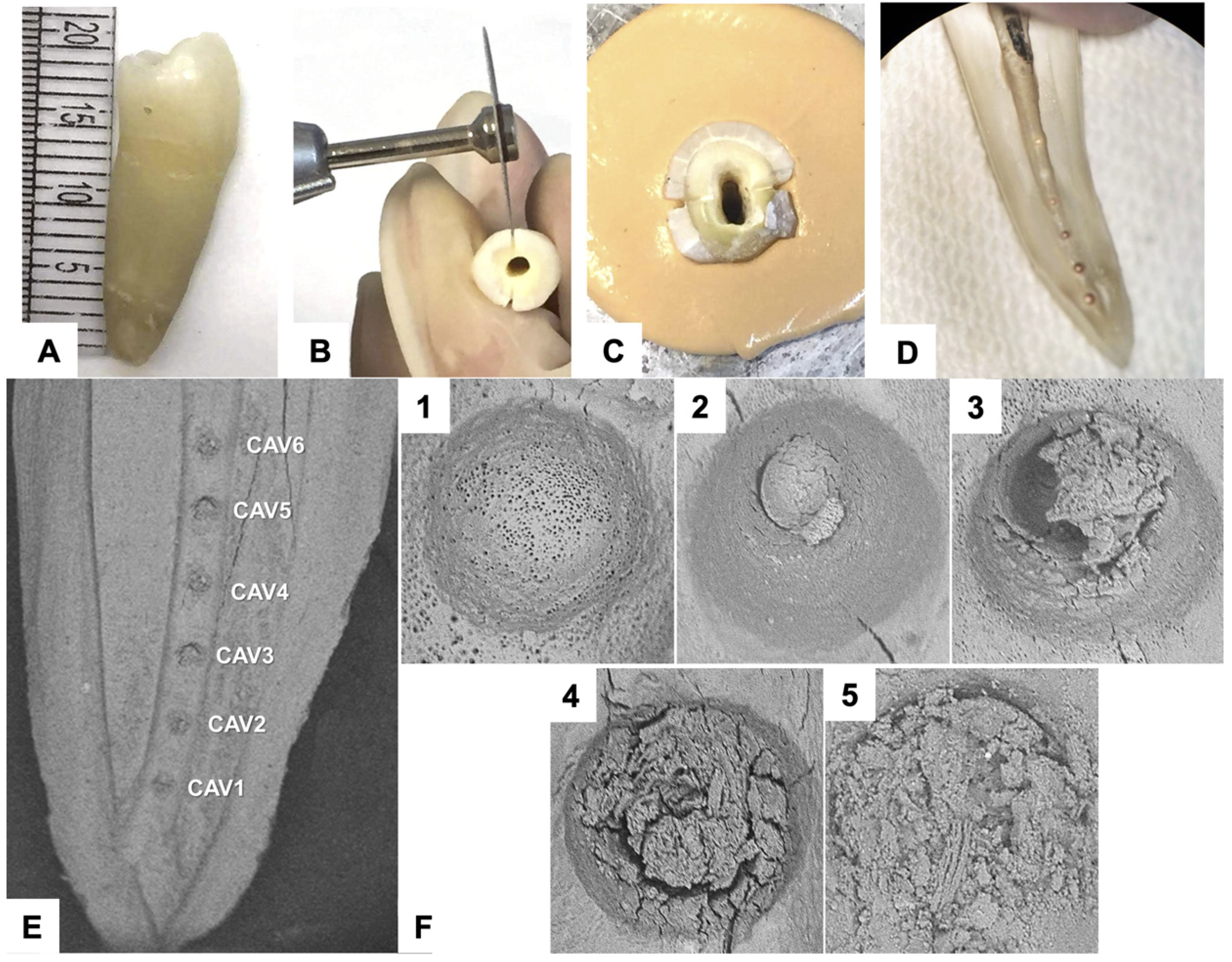
2.1. Specimens Selection and Preparation

This study was approved by the Research Ethics Committee (No 10320619.3.0000.5420). Twenty single-rooted human mandibular premolars were selected. Mesiodistal and buccolingual digital periapical radiographs determined the presence of a single root canal. The angle of curvature (≥15° ≤ 20°) was determined based on the methodology described by Schneider [19].
The teeth preparation protocol was adapted from a previous study [20]. Briefly, all teeth were worn horizontally with a diamond disk to a standard length of 19 mm. The root canals were instrumented with the ProDesign Logic (Easy Dental Equipment, Belo Horizonte, Brazil) rotary system, with a working length of 18 mm. Initially, a #10 K-file was inserted into the working length, followed by a #40/01 Glide Path file with the same length using an electric moto, and finalized with a #40/05 final modeling file. For each instrument change, 5 mL of 2.5% NaOCl was used for irrigation.
After instrumentation, two longitudinal grooves were made with a 0.08 diamond disk under eight times magnification using a dental operating microscope (DF Vasconcelos, São Paulo, Brazil) to a depth close to the root canal. The root canals were embedded in a heavy-bodied silicone (Optosil Comfort Putty, Heraeus Kulzer GmbH, Hanau, Germany), which prevented the extrusion of the irrigant and simulated a closed irrigation and aspiration system up to the cement-enamel junction level. After being embedded, a #24 spatula (SSWhite Duflex, Rio de Janeiro, Brazil) was used in the previously made groove, and a vertical force was applied to divide the sample into two halves. The vestibular part was removed, and six hemispherical cavities of predefined levels (C1, C2, C3, C4, C5, and C6) of approximately 0.05 mm in depth were made using a flame-shaped amalgam polishing drill at low speed (Wilcos Dental Products, Petrópolis—Rio de Janeiro, Brazil). The spacing between the cavities was made at an interval of 1 mm, starting 1 mm from the apex of the tooth (Figure 1A).
The specimens were washed for 1 min in running water to remove the debris, immersed in an ultrasonic bath containing 2.5% NaOCl solution for 3 min, and then immersed in 17% EDTA for 3 min. Samples were washed with distilled water for 1 min, dried in an oven at 80 °C for 3 min, and mounted on the silicone system after the cavities had been filled with the debris. The debris was prepared according to the modified protocol (already published), with a mixture of 0.025 g of dentin debris to 0.1 mL of 2.5% NaOCl for 5 min [17]. In this study, the same prepared tooth with cavities was used in five different groups to reduce the interference of anatomical variation in the results.

2.2. Control and Experimental Group Design

The sample size was based on a previous study [20]. The present study was composed of ten groups elaborated from the final irrigation protocols most evidenced in the literature, as follows:
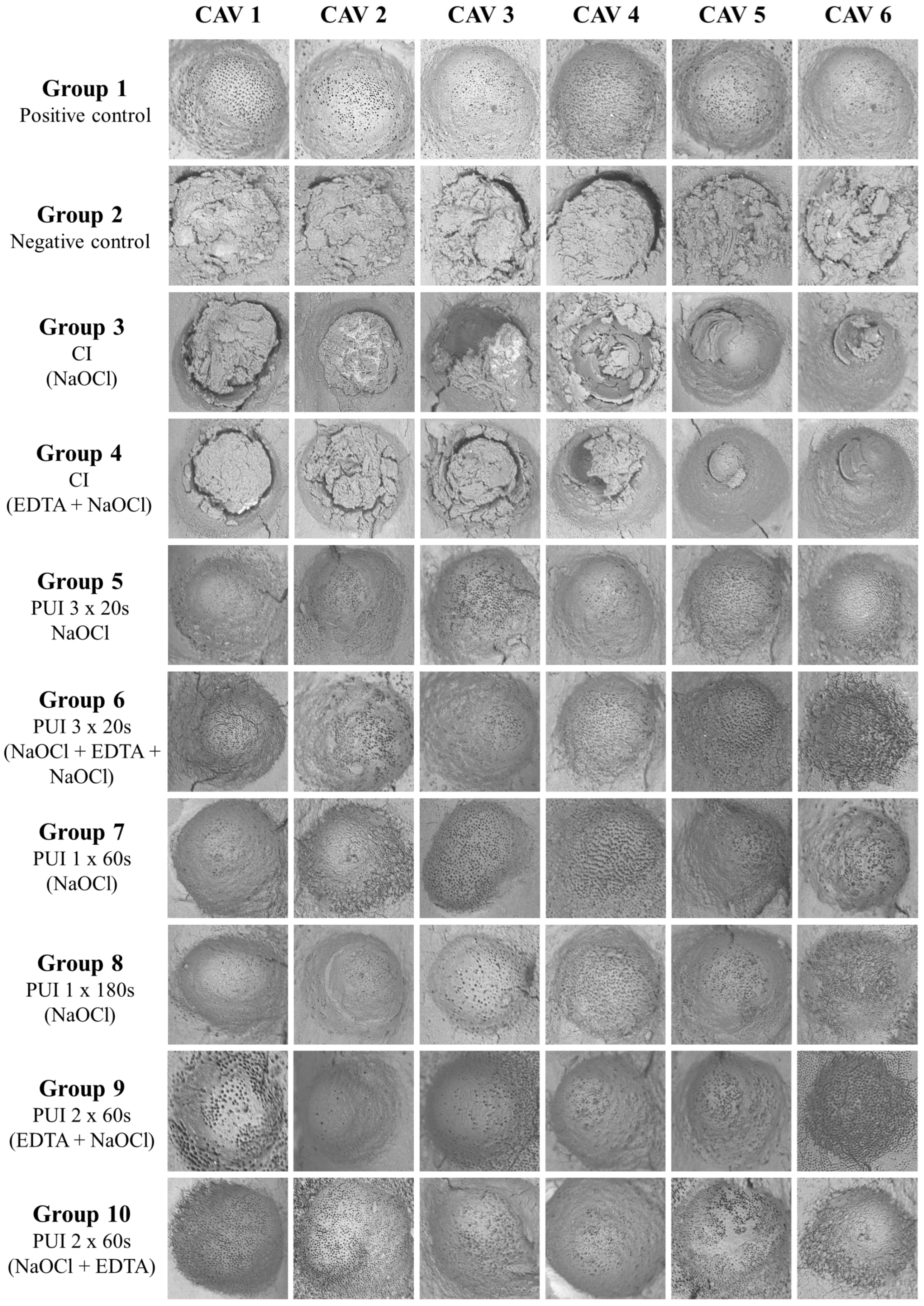
  • Group 1—Positive control: Cavities were prepared as described above and kept debris-free. No irrigation protocol was carried out.
  • Group 2—Negative control: The artificial cavities were filled with debris, and no irrigation protocol was performed.
  • Group 3—CI with 15 mL of 2.5% NaOCl.
  • Group 4—CI with 5 mL of NaOCl, 5 mL of 17% EDTA, and new 5 mL 2.5% NaOCl.
  • Group 5—three cycles of PUI for 20 s using 2.5% NaOCl.
  • Group 6—three cycles of PUI for 20 s using 2.5% NaOCl, 17% EDTA, and 2.5% NaOCl.
  • Group 7—one PUI cycle for 60 s using 2.5% NaOCl.
  • Group 8—one PUI cycle for 180 s using 2.5% NaOCl.
  • Group 9—two PUI cycles for 60 s using 17% EDTA, and 2.5% NaOCl.
  • Group 10—two PUI cycles for 60 s using 2.5% NaOCl, and 17% EDTA.
The total volume of irrigating solutions used in the experimental groups was standardized to 15 mL. It is important to highlight that in the groups where EDTA was used, regardless of the time of ultrasonic activation, the chelating solution remained for 60 s within the root canal. PUI was performed with a modification of the previously described technique [9]. An Irrisonic Power tip (Helse Industria e Comercio, Santa Rosa de Viterbo, Brazil) was mounted on a Gnatus ultrasonic handpiece (Medical-Dental Equipment, Brazil), adjusted at power 1, and placed 1 mm before the working length. The technique was modified once the ultrasound tip was oscillating in the vestibular-lingual direction to improve the cleaning efficiency [9].
Analyses were performed on the images obtained in an SEM of low vacuum (PSEM, Express™, Aspex Corporation, Delmont, PA, USA) of indentations with a 500-fold increase and 20 kV. The images were classified by score according to the amount of debris present in each cavity by adapting the methodology established in [20]: 1—without debris but with exposure of the dentinal tubules; 2—without debris and without exposure of the dentinal tubules; 3—debris covering an area smaller than 50% of the dentinal tubules; 4—debris covering an area greater than 50% of the dentinal tubules; and 5—dentinal tubules entirely covered by debris. Figure 1B shows representative images of the scores. All images from the control and experimental groups were analyzed by two independent examiners, previously calibrated and blinded for the study, assigning scores to the images according to the evaluation criteria described previously.

2.3. Statistical Analysis

The data collected were statistically analyzed using Sigma Plot 12.0 software for Windows (Systat Software Inc, San Jose, CA, USA). For the statistical analysis, the Kappa test was used in the inter-examiner concordance analysis. The Kruskal-Wallis test was used to compare data on cleaning effectiveness. The significance level was set at 5% (p < 0.05).

3. Results

3.1. Protocols × Samples

Statistical analysis was performed evaluating the general cleaning and dentinal tubules exposure abilities of the protocols, including all cavities (Table 1). The CI groups (Groups 3 and 4) showed better debris removal than the negative control group (Group 2). All groups using PUI (Groups 5, 6, 7, 8, 9, and 10) were more effective in removing the debris compared to the negative control group (Group 2) and CI (Groups 3 and 4) and similar to the positive control group (Group 1).
The CI groups did not promote exposure of the dentinal tubules. In contrast, the PUI groups (Groups 5, 6, 7, 8, 9, and 10) promoted exposure of dentinal tubules similar to the positive control (p > 0.05) (Table 1). Moreover, Group 6 (three cycles 20 s NaOCl-EDTA-NaOCl) was more effective in exposing dentinal tubules than Group 5 (three cycles 20 s NaOCl), showing better protocol efficacy when EDTA was associated. In contrast, Group 6 was similar to Group 7 (once cycle 60 s NaOCl) and 10 (two cycles 60 s NaOCl-EDTA), showing the importance of PUI activation time for cleaning. Along with Group 5, Groups 8 (once cycle 180 s NaOCl) and 9 (two cycles 60 s EDTA-NaOCl) were similar to the other PUI groups.

3.2. Protocols × Cavities

The six cavities in each sample were evaluated according to the score of cleaning the debris obtained with the different protocols (Table 2). By comparing the cavities, it was observed that all the PUI protocols provided better cleaning than the negative control group and the CI groups (p < 0.05). Cavities 1 and 2 underwent better cleaning with the PUI protocols. The group of three cycles of 20 s using only NaOCl (Group 5) showed no significant difference compared to the CI groups (Groups 3 and 4), showing the importance of using EDTA in short-time protocols. In addition, CI groups were similar to the negative control group (Group 2). In cavity 3, all PUI groups were superior to the CI groups, which were equivalent to the negative control group (Group 2). Cavities 4, 5, and 6 showed effective cleaning with all groups, including the CI groups. The CI group with NaOCl (Group 3) did not show a statistical difference from the negative control group (Group 2) in cavity 4 but did not show differences between the other groups. The PUI groups (Groups 5, 6, 7, 8, 9, and 10) obtained a similar result to the positive control group (Group 1) in all cavities.
Regarding the exposure of the dentinal tubules in all cavities, the groups where PUI was used were equivalent to the positive control (p > 0.05) and more effective than the CI and negative control groups (p < 0.05). However, Group 5 was similar to CI groups and negative control (p > 0.05). In cavities 1, 2, 4, and 5, Group 8 was equivalent to CI groups and negative control (p > 0.05) (Table 3). Figure 2 shows the cleaning characteristics of each group.

4. Discussion

The present study investigated the effectiveness of different PUI protocols on debris removal and the exposure of dentinal tubules in simulated complex root canal cavities using SEM. All groups employing PUI protocols differed significantly in terms of their abilities to remove debris compared to CI, rejecting the first null hypothesis. Using different PUI protocols did not show statistically significant differences between groups, thus leading to the acceptance of the second null hypothesis.
In this study, the same tooth was used in five different groups to reduce the interference of anatomical variation in the results. In addition, it has been shown that the same dental element can be used more than five times without damaging its structure [9,21]. Tooth instrumentation was performed using a #40 file to facilitate the irrigant penetration into the root canal apical third [22]. The cavities were added in an attempt to standardize and focus the amount of debris being cleaned and analyzed in SEM [20].
Sodium hypochlorite at 2.5% concentration was chosen as an irrigating solution because of its physicochemical and biological properties [4]. EDTA has been advocated for its chelating capacity, as it acts in the inorganic portion of the smear layer [22]. The importance of debris removal and exposure of dentinal tubules has been described by several authors in the past, assisting in the disinfection of the root canal system, in addition to facilitating the penetration of intracanal medication and endodontic cement [23,24].
In the general analysis of the samples concerning the protocols, it could be observed that the CI and PUI groups were superior to the negative control group in removing debris, with only the PUI groups being similar to the positive control. Moreover, the PUI groups effectively opened the dentinal tubules, showing the importance of physical activity in the root canal cleaning [25,26]. Many studies have been performed using PUI with different protocols, and the results have been divergent. Some authors did not observe any difference between the methods [7,8]. The non-standardization of the applied method, e.g., in terms of sample preparation, ultrasound power, presence or absence of cavities, and area of choice for analysis, among the different studies can explain this.
In the analysis of debris removal from each cavity individually, it could be observed that all PUI groups were better than CI and negative control groups in cavities 1, 2, and 3. This finding shows the importance of the physical action of irrigation for improving the apical third cleaning [27,28]. The greater effectiveness of PUI in cleaning the root canal apical third was observed in some studies [5,6]. However, the results were similar to CI groups in other studies [7,8].
An interesting finding was the superiority of Group 6 (three cycles 20 s NaOCl-EDTA-NaOCl) compared to Group 5 (three cycles 20 s NaOCl), indicating the importance of using EDTA as a chemical agent to aid the physical activity of shorter-time agitation protocols. Additionally, Group 7 (one cycle 60 s NaOCl) was similar to Group 6 (three cycles 20 s NaOCl-EDTA-NaOCl) and Group 10 (two cycles 60 s NaOCl-EDTA), which may be explained by the more extended physical action of the irrigant. This finding suggests that longer-time agitation protocols using only NaOCl have equivalent efficiency to protocols associated with the use of EDTA.
Regarding the exposure of dentinal tubules, it was observed that the CI groups were similar to the negative control group in all cavities, showing the importance of seeking physical means of enhancing the action of the irrigant. However, all PUI groups were similar to the positive control group, showing its effectiveness in opening the dentinal tubules. Group 5 (three cycles 20 s NaOCl) was inferior to the other PUI groups, again showing the importance of chelating to improve dentinal exposure. Group 8 (one cycle 180 s NaOCl) was also lower, which may be explained by the formation of new debris due to irrigating liquid vaporization in a longer time protocol (superior to 60 s) without renovation [29,30,31].
The flow generated by ultrasound was more effective in removing the smear layer than CI associated with EDTA, showing the importance of the physical action of root canal irrigation. Some studies showed the importance of using ultrasound to improve dentinal tubular exposure [17,32]. However, others did not show differences between ultrasound and CI regarding dentinal exposure [7,15,18]. Although the present investigation elucidates the PUI protocols, it presents limitations regarding the lack of an evaluation of the antibacterial reduction evoked by the different tested protocols.

5. Conclusions

Based on this study, it can be concluded that the protocols using PUI in three cycles of 20 s (NaOCl-EDTA-NaOCl), two cycles of 60 s (EDTA-NaOCl), or one cycle of 60 s (NaOCl) were more effective at removing debris and increasing the exposure of the dentinal tubules.

Author Contributions

Conceptualization, F.A.P. and J.E.G.-F.; methodology, F.A.P., C.L. and V.Z.C.; software, R.D.-F. and L.C.-S.; validation, R.D.-F., L.C.-S. and P.C.T.D.; formal analysis, L.T.A.C.; investigation, F.A.P., C.L. and V.Z.C.; resources, M.A.H.D. and J.E.G.-F.; data curation, R.D.-F. and L.C.-S.; writing—original draft preparation, F.A.P., P.C.T.D., C.L. and V.Z.C.; writing—review and editing, R.D.-F.; visualization, R.D.-F. and J.E.G.-F.; supervision, L.T.A.C. and J.E.G.-F.; project administration, M.A.H.D. and J.E.G.-F.; funding acquisition, J.E.G.-F. All authors have read and agreed to the published version of the manuscript.

Funding

This research was funded by FAPESP (São Paulo State Research Support Foundation) through the process 2017/14112-6, and by the companies Helse Ultrasonic (Santa Rosa de Viterbo, SP, Brazil) and Easy Dental Equipment (Belo Horizonte, MG, Brazil).

Institutional Review Board Statement

The study was conducted in accordance with the Declaration of Helsinki, and approved by the Institutional Ethics Committee of São Paulo State University (protocol code 10320619.3.0000.5420 and date of approval 09/13/2019).

Informed Consent Statement

Informed consent was obtained from all subjects involved in the study.

Data Availability Statement

Not applicable.

Acknowledgments

The authors would like to thank the Bauru School of Dentistry (USP) for their cooperation in this study and Helse Ultrasonic and Easy Dental Equipment for materials donations.

Conflicts of Interest

The authors declare no conflict of interest.

References

  1. de Gregorio, C.; Estevez, R.; Cisneros, R.; Paranjpe, A.; Cohenca, N. Efficacy of different irrigation and activation systems on the penetration of sodium hypochlorite into simulated lateral canals and up to working length: An in vitro study. J. Endod. 2010, 36, 1216–1221. [Google Scholar] [CrossRef] [PubMed]
  2. Versiani, M.A.; De-Deus, G.; Vera, J.; Souza, E.; Steier, L.; Pécora, J.D.; Sousa-Neto, M.D. 3D mapping of the irrigated areas of the root canal space using micro-computed tomography. Clin. Oral Investig. 2015, 19, 859–866. [Google Scholar] [CrossRef] [PubMed]
  3. Nair, P.N. On the causes of persistent apical periodontitis: A review. Int. Endod. J. 2006, 39, 249–281. [Google Scholar] [CrossRef] [PubMed]
  4. Orlowski, N.B.; Schimdt, T.F.; Teixeira, C.D.S.; Garcia, L.; Savaris, J.M.; Tay, F.R.; Bortoluzzi, E.A. Smear Layer Removal Using Passive Ultrasonic Irrigation and Different Concentrations of Sodium Hypochlorite. J. Endod. 2020, 46, 1738–1744. [Google Scholar] [CrossRef]
  5. Agrawal, V.S.; Kapoor, S. An in vitro scanning electron microscopic study comparing the efficacy of passive ultrasonic and syringe irrigation methods using sodium hypochlorite in removal of debris from the root canal system. J. Ir. Dent. Assoc. 2012, 58, 156–161. [Google Scholar]
  6. Urban, K.; Donnermeyer, D.; Schäfer, E.; Bürklein, S. Canal cleanliness using different irrigation activation systems: A SEM evaluation. Clin. Oral Investig. 2017, 21, 2681–2687. [Google Scholar] [CrossRef]
  7. Castagna, F.; Rizzon, P.; da Rosa, R.A.; Santini, M.F.; Barreto, M.S.; Duarte, M.A.; Só, M.V. Effect of passive ultrassonic instrumentation as a final irrigation protocol on debris and smear layer removal--a SEM analysis. Microsc. Res. Tech. 2013, 76, 496–502. [Google Scholar] [CrossRef]
  8. Sahar-Helft, S.; Sarp, A.S.; Stabholtz, A.; Gutkin, V.; Redenski, I.; Steinberg, D. Comparison of positive-pressure, passive ultrasonic, and laser-activated irrigations on smear-layer removal from the root canal surface. Photomed. Laser Surg. 2015, 33, 129–135. [Google Scholar] [CrossRef]
  9. Jiang, L.M.; Verhaagen, B.; Versluis, M.; van der Sluis, L.W. Influence of the oscillation direction of an ultrasonic file on the cleaning efficacy of passive ultrasonic irrigation. J. Endod. 2010, 36, 1372–1376. [Google Scholar] [CrossRef]
  10. Leoni, G.B.; Versiani, M.A.; Silva-Sousa, Y.T.; Bruniera, J.F.; Pécora, J.D.; Sousa-Neto, M.D. Ex vivo evaluation of four final irrigation protocols on the removal of hard-tissue debris from the mesial root canal system of mandibular first molars. Int. Endod. J. 2017, 50, 398–406. [Google Scholar] [CrossRef]
  11. Lee, S.J.; Wu, M.K.; Wesselink, P.R. The effectiveness of syringe irrigation and ultrasonics to remove debris from simulated irregularities within prepared root canal walls. Int. Endod. J. 2004, 37, 672–678. [Google Scholar] [CrossRef] [PubMed]
  12. Mancini, M.; Cerroni, L.; Iorio, L.; Armellin, E.; Conte, G.; Cianconi, L. Smear layer removal and canal cleanliness using different irrigation systems (EndoActivator, EndoVac, and passive ultrasonic irrigation): Field emission scanning electron microscopic evaluation in an in vitro study. J. Endod. 2013, 39, 1456–1460. [Google Scholar] [CrossRef] [PubMed] [Green Version]
  13. Ferreira, R.B.; Marchesan, M.A.; Silva-Sousa, Y.T.; Sousa-Neto, M. Effectiveness of root canal debris removal using passive ultrasound irrigation with chlorhexidine digluconate or sodium hypochlorite individually or in combination as irrigants. J. Contemp. Dent. Pract. 2008, 9, 68–75. [Google Scholar] [CrossRef] [PubMed] [Green Version]
  14. Khalap, N.D.; Kokate, S.; Hegde, V. Ultrasonic versus sonic activation of the final irrigant in root canals instrumented with rotary/reciprocating files: An in-vitro scanning electron microscopy analysis. J. Conserv. Dent. 2016, 19, 368–372. [Google Scholar] [CrossRef] [PubMed]
  15. de Castro, F.P.; Pinheiro, S.L.; Duarte, M.A.; Duque, J.A.; Fernandes, S.L.; Anchieta, R.B.; da Silveira Bueno, C.E. Effect of time and ultrasonic activation on ethylenediaminetetraacetic acid on smear layer removal of the root canal. Microsc. Res. Tech. 2016, 79, 1062–1068. [Google Scholar] [CrossRef]
  16. Koçak, S.; Bağcı, N.; Çiçek, E.; Türker, S.A.; Can Sağlam, B.; Koçak, M.M. Influence of passive ultrasonic irrigation on the efficiency of various irrigation solutions in removing smear layer: A scanning electron microscope study. Microsc. Res. Tech. 2017, 80, 537–542. [Google Scholar] [CrossRef] [PubMed]
  17. Martins Justo, A.; Abreu da Rosa, R.; Santini, M.F.; Cardoso Ferreira, M.B.; Pereira, J.R.; Hungaro Duarte, M.A.; Reis So, M.V. Effectiveness of final irrigant protocols for debris removal from simulated canal irregularities. J. Endod. 2014, 40, 2009–2014. [Google Scholar] [CrossRef]
  18. Andrabi, S.M.; Kumar, A.; Zia, A.; Iftekhar, H.; Alam, S.; Siddiqui, S. Effect of passive ultrasonic irrigation and manual dynamic irrigation on smear layer removal from root canals in a closed apex in vitro model. J. Investig. Clin. Dent. 2014, 5, 188–193. [Google Scholar] [CrossRef]
  19. Schneider, S.W. A comparison of canal preparations in straight and curved root canals. Oral Surg. Oral Med. Oral Pathol. 1971, 32, 271–275. [Google Scholar] [CrossRef]
  20. Kato, A.S.; Cunha, R.S.; da Silveira Bueno, C.E.; Pelegrine, R.A.; Fontana, C.E.; de Martin, A.S. Investigation of the Efficacy of Passive Ultrasonic Irrigation Versus Irrigation with Reciprocating Activation: An Environmental Scanning Electron Microscopic Study. J. Endod. 2016, 42, 659–663. [Google Scholar] [CrossRef]
  21. Rodig, T.; Bozkurt, M.; Konietschke, F.; Hulsmann, M. Comparison of the Vibringe system with syringe and passive ultrasonic irrigation in removing debris from simulated root canal irregularities. J. Endod. 2010, 36, 1410–1413. [Google Scholar] [CrossRef] [PubMed]
  22. Haapasalo, M.; Shen, Y.; Qian, W.; Gao, Y. Irrigation in endodontics. Dent. Clin. N. Am. 2010, 54, 291–312. [Google Scholar] [CrossRef] [PubMed]
  23. Boutsioukis, C.; Arias-Moliz, M.T.; Chávez de Paz, L.E. A critical analysis of research methods and experimental models to study irrigants and irrigation systems. Int. Endod. J. 2022, 55 (Suppl. S2), 295–329. [Google Scholar] [CrossRef] [PubMed]
  24. Rossi-Fedele, G.; Rödig, T. Effectiveness of root canal irrigation and dressing for the treatment of apical periodontitis: A systematic review and meta-analysis of clinical trials. Int. Endod. J. 2022. Online ahead of print. [Google Scholar] [CrossRef]
  25. Souza, C.C.; Bueno, C.E.; Kato, A.S.; Limoeiro, A.G.; Fontana, C.E.; Pelegrine, R.A. Efficacy of passive ultrasonic irrigation, continuous ultrasonic irrigation versus irrigation with reciprocating activation device in penetration into main and simulated lateral canals. J. Conserv. Dent. 2019, 22, 155–159. [Google Scholar] [CrossRef]
  26. Barbosa, A.F.A.; Lima, C.O.; Sassone, L.M.; Fares, R.D.; Fidalgo, T.; Silva, E. Effect of passive ultrasonic irrigation on hard tissue debris removal: A systematic review and meta-analysis. Braz. Oral. Res. 2021, 35, e123. [Google Scholar] [CrossRef]
  27. Kluge, M.; Trüschler, J.; Elamin, F.; Anderson, A.; Hellwig, E.; Altenburger, M.; Vach, K.; Wittmer, A.; Al-Ahmad, A. Improving the microbial sampling and analysis of secondary infected root canals by passive ultrasonic irrigation. Clin. Oral Investig. 2022, 26, 4575–4586. [Google Scholar] [CrossRef]
  28. Li, Q.; Zhang, Q.; Zou, X.; Yue, L. Evaluation of four final irrigation protocols for cleaning root canal walls. Int. J. Oral. Sci. 2020, 12, 29. [Google Scholar] [CrossRef]
  29. Ali, A.; Bhosale, A.; Pawar, S.; Kakti, A.; Bichpuriya, A.; Agwan, M.A. Current Trends in Root Canal Irrigation. Cureus 2022, 14, e24833. [Google Scholar] [CrossRef]
  30. Boutsioukis, C.; Arias-Moliz, M.T. Present status and future directions-irrigants and irrigation methods. Int. Endod. J. 2022, 55 (Suppl. S3), 588–612. [Google Scholar] [CrossRef]
  31. Cheung, A.W.T.; Lee, A.H.C.; Cheung, G.S.P. Clinical efficacy of activated irrigation in endodontics: A focused review. Restor. Dent. Endod. 2021, 46, e10. [Google Scholar] [CrossRef] [PubMed]
  32. Çapar, İ.D.; Aydinbelge, H.A. Effectiveness of various irrigation activation protocols and the self-adjusting file system on smear layer and debris removal. Scanning 2014, 36, 640–647. [Google Scholar] [CrossRef] [PubMed]
Figure 1. Step-by-step preparation. (A) Tooth selection. (B) Initial section. (C) Tooth embedding. (D) Final aspect of the cavities. (E) Representative SEM image of the cavities. All hemispherical cavities shown are 1 mm apart. (F) Representative images of scores (1, 2, 3, 4, and 5).
Oral 03 00001 g001
Figure 2. Representative images of each group in 500× magnification in the Scanning Electron Microscope of low vacuum.
Oral 03 00001 g002
Table 1. Analysis of the general cleaning achieved with each protocol concerning the samples and the dentinal tubules (p < 0.05).
GroupsCleaning
(Debris Removal)
Lower 95% CI of MeanUpper 95% CI of MeanCleaning
(Tubules Exposure)
Lower 95% CI of MeanUpper 95% CI of Mean
1- Positive Control1b0.971.121bc11.13
2- Negative Control4c442a22
3- CI NaOCl + NaOCl + NaOCl2a1.902.432a22
4- CI NaOCl + EDTA + NaOCl3a2.132.672a1.952
5- PUI 3 × 20 s NaOCl1b1.021.171b1.221.47
6- PUI 3 × 20 s NaOCl + EDTA + NaOCl1b0.971.121c0.991.10
7- PUI 1 × 60 s NaOCl1b0.961.101c0.981.05
8- PUI 1 × 180 s NaOCl1b0.981.081bc1.151.38
9- PUI 2 × 60 s EDTA + NaOCl1b0.981.081bc1.011.15
10- PUI two cycles 60 s NaOCl + EDTA1b0.981.051c0.991.10
* The column on the left side (debris removal) assesses the general removal of the smear layer, while in the right column (dentinal tube exposure), a higher magnification SEM was employed to see if the dentinal tubes were, in fact, clean. The numbers are related to the median, lower, and upper 95% confidence intervals of each group. Different overlapping letters indicate a statistical difference between groups (analysis by column).
Table 2. Analysis of the general cleaning achieved for each protocol used in every individual cavity (p < 0.05).
GroupsCAV1CAV2CAV3CAV4CAV5CAV6
1- Positive Control1Ab1Aa1Ab1Aa1Aa1Aa
2- Negative Control4Ac4Ab4Aa4Ab4Ac4Ab
3- IC NaOCl + NaOCl + NaOCl3Aac2.5Ababc2ABa2ABab1.5Bab1Ba
4- IC NaOCl + EDTA + NaOCl3Aac3ABbc3ABa1Ba2Bbc1.5Ba
5- PUI 3 × 20 s NaOCl1Aab1Aac1Ab1Aa1Aa1Aa
6- PUI 3 × 20 s NaOCl + EDTA + NaOCl1Ab1Aa1Ab1Aa1Aa1Aa
7- PUI 1 × 60 s NaOCl1Ab1Aa1Ab1Aa1Aab1Aa
8- PUI 1 × 180 s NaOCl1Ab1Aa1Ab1Aa1Aa1Aa
9- PUI 2 × 60 s EDTA + NaOCl1Ab1Aa1Ab1Aa1Aa1Aa
10- PUI two cycles 60 s NaOCl + EDTA1Ab1Aa1Ab1Aa1Aa1Aa
* Analysis was performed vertically (lower case letters) and horizontally (upper case letters). Different overlapping letters indicate a statistical difference between the groups.
Table 3. Analysis of the dentin tubule exposure for each protocol used in every individual cavity (p < 0.05).
GroupsCAV1CAV2CAV3CAV4CAV5CAV6
1- Positive Control1b1b1b1b1b1b
2- Negative Control2a2a2a2a2a2a
3- IC NaOCl + NaOCl + NaOCl2a2a2a2a2a2a
4- IC NaOCl + EDTA + NaOCl2a2a2a2a2a2a
5- PUI 3 × 20 s NaOCl1.5ab1ab1ab1ab1ab1ab
6- PUI 3 × 20 s NaOCl + EDTA + NaOCl1b1b1b1b1b1b
7- PUI 1 × 60 s NaOCl1b1b1b1b1b1b
8- PUI 1 × 180 s NaOCl1ab1ab1b1ab1ab1b
9- PUI 2 × 60 s EDTA + NaOCl1b1b1b1b1b1b
10- PUI two cycles 60 s NaOCl + EDTA1b1b1b1b1b1b
* At the opening of the tubules, number 1 indicates exposed tubules while number 2 indicates tubules that were not exposed. Different overlapping letters indicate a statistical difference between groups (analysis by column).
Disclaimer/Publisher’s Note: The statements, opinions and data contained in all publications are solely those of the individual author(s) and contributor(s) and not of MDPI and/or the editor(s). MDPI and/or the editor(s) disclaim responsibility for any injury to people or property resulting from any ideas, methods, instructions or products referred to in the content.

