Case Report

Oral Squamous Cell Carcinoma Mimicking Medication-Related Osteonecrosis of the Jaws (MRONJ): A Case Series

1
Department of Surgical, Oncological and Oral Sciences (Di.Chir.On.S.), University of Palermo, 90100 Palermo, Italy
2
Department of Biomedical, Dental Sciences, Morphological and Functional Images, University of Messina, 98100 Messina, Italy
3
Department of Dental Surgery, Faculty of Dental Surgery, University of Malta, MSD 1752 Msida, Malta
4
Maxillofacial Surgery Unit, Clinica del Mediterraneo di Ragusa, 97100 Ragusa, Italy
5
Department of Health Promotion, Mother and Child Care, Internal Medicine and Medical Specialties (ProMISE), University of Palermo, 90100 Palermo, Italy
*
Author to whom correspondence should be addressed.
Oral 2021, 1(4), 326-331; https://doi.org/10.3390/oral1040032
Submission received: 26 August 2021 / Revised: 14 October 2021 / Accepted: 29 October 2021 / Published: 3 November 2021

Abstract

:
(1) Background: Medication-related osteonecrosis of the jaw (MRONJ) is a potential adverse drug reaction of antiresorptive and/or antiangiogenic treatment. MRONJ is mostly diagnosed by anamnestic data, clinical examination and radiological findings, with signs and symptoms often unspecific. On the other hand, oral squamous cell carcinoma (OSCC) is characteristic for its pleomorphic appearance (e.g., ulcer, mucous dehiscence, non-healing post-extractive socket). We report three cases where OSCC mimicked MRONJ lesions. (2) Patients: Three patients undergoing amino-bisphosphonate treatment for osteoporosis presented with areas of intraorally exposed jawbone and unspecific radiological signs compatible with MRONJ. Due to the clinical suspicious of malignant lesion, incisional biopsy for histological examination was also performed. (3) Results: Histological examination of the tissue specimen revealed the presence of OSCC. All patients underwent cancer treatment. (4) Conclusions: Several signs and symptoms of OSCC may simulate, in patients with a history of anti-resorptive, MRONJ; for these reasons, it is important to perform histologic analysis when clinicians are facing a suspicious malignant lesion.

1. Introduction

Osteonecrosis of the jaw related to medication (MRONJ) has been defined as “an adverse drug reaction described as the progressive destruction and death of bone that affects the mandible and maxilla of patients exposed to the treatment with medications known to increase the risk of disease, in the absence of a previous radiation treatment” [1,2,3].
The diagnosis of MRONJ is commonly based both on clinical and radiographic findings, because patients may present with other common clinical disorders not to be confused with MRONJ; among these are alveolar osteitis, sinusitis, periodontitis, chronic sclerosing osteomyelitis or benign fibro-osseous lesions. However, differential diagnosis may also consider bone metastases and oral squamous cell carcinoma (OSCC) [3,4,5].
In particular, OSCC can appear through bone exposure, mucous dehiscence, sudden increased dental mobility, lack of post-extractive alveolar repair and pain, mimicking MRONJ [6,7,8].
The aim of this paper is to report three cases of patients at risk of MRONJ but found to suffer from OSCC and use them to discuss some issues of the current diagnostic workflow of both diseases.

2. Case Presentation

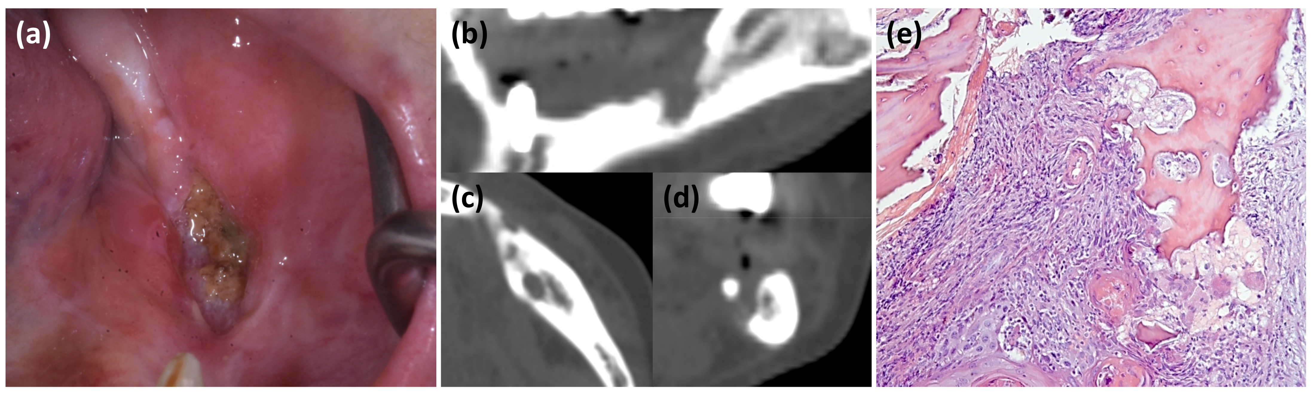
2.1. Clinical Case 1

A 62-year-old non-smoker woman was referred to the sector of Oral Medicine at the University Hospital in October 2020 for swelling in his left mandible and severe pain.
In 2006, the patients had been diagnosed with a OSCC (grade 1) on the posterior part of the left body of the mandible in a different institution; afterwards, she underwent marginal mandibular resection.
Additionally, she was affected by chronic drug-induced pancreatitis and osteoporosis, and she had been treated from 2016 to 2018 with alendronate (once-weekly oral alendronate 70 mg), and steroids.
The patient was partially edentulous, she reported mandibular rehabilitative treatment by 3 dental implants in 2019; all of them failed to integrate after 2 months from implant placement. The patient reported that she developed numerous episodes of swelling and pain in the implant site after fixtures’ loss.
Intra-oral examination revealed a large area of exposed necrotic bone of the posterior left part of the mandible (Figure 1a). Computed tomography (CT) showed a diffuse osteosclerotic pattern, non-healing post-surgical site and cortical disruption (Figure 1b–d).
All clinical and radiological features were compatible with a diagnosis of MRONJ; however, due to the clinical aspects of the lesions and the medical history (i.e., previous OSCC), incisional biopsy for histological examination was promptly performed. Histological examination showed a squamous cell carcinoma infiltrating lamellae of cortical bone (Figure 1e).
The histological findings confirmed the diagnosis of OSCC; appropriate oncological management has therefore been started.

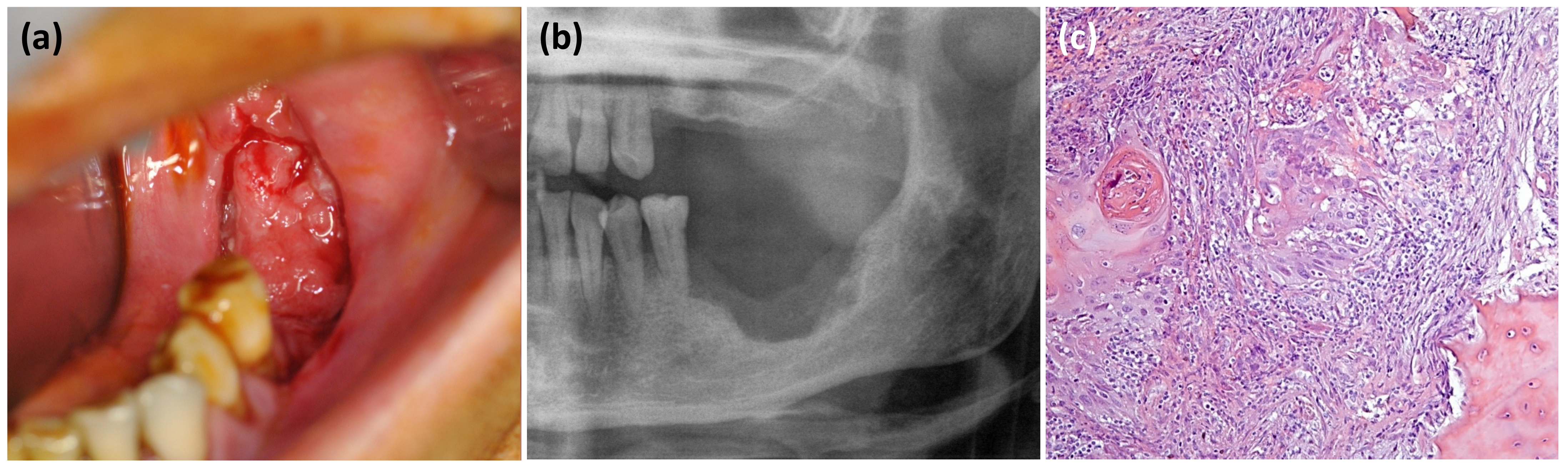
2.2. Clinical Case 2

A 90-year-old non-smoker woman with osteoporosis was treated with ibandronate (once-monthly oral ibandronate 150 mg) for 10 years.
Six months before examination, she underwent dental extraction of tooth 3.6. After this surgery, due to the presence of a non-healing post-extraction socket, she underwent bony curettage and antiseptic mouthwashes at a different institution for 3 months.
In January 2019, she was referred to the Maxillofacial Surgery Unit of Ragusa, where extra-oral examination revealed the presence of soft tissue swelling and high-volume lymph nodes in the left neck. Intra-oral examination highlighted bone exposure and high granulation tissue of the posterior left portion of the mandible; additionally, she referred the paraesthesia of the inferior alveolar nerve (Figure 2a).
Orthopantomography showed extensive osteolysis, with pathological fracture of the left mandibular body (Figure 2b).
Incisional biopsy was performed; the histological examination showed islets of squamous cell carcinoma surrounded by granulation tissue; bone tissue was not included in the sample (Figure 1c). Subsequently, the patient was referred to the Department of Oncology for management.

2.3. Clinical Case 3

A 72-year-old non-smoker woman with osteoporosis was treated with alendronate for more than 15 years (once-weekly oral alendronate 70 mg)
She reported having undergone dental extractions about 5 months before the referral (tooth 3.6 and 3.7). Due to the presence of a non-healing post-extraction socket, she underwent several bony curettage and antiseptic mouthwashes at another institution for 2 months.
Extra-oral examination showed the presence of soft tissue swelling and high-volume lymph nodes in the left neck. Intra-oral examination revealed high granulation tissue of the posterior left portion of the mandible. She did not report the paresthesia of the inferior alveolar nerve (Figure 3a).
Orthopantomography revealed an extensive osteolysis of the posterior left portion of the mandible (Figure 3b).
Incisional biopsy was carried out; histological examinations detected a squamous cell carcinoma, accompanied by granulation tissue, with focal infiltration of the cortical bone fragments included in the sample (Figure 3c). Subsequently, the patient underwent oncological treatment.

3. Discussion

Almost 20 years after the first mention of osteonecrosis of the jaws related to antiresorptive medication (MRONJ) [9], it remains a current topic within the scientific community. Through the last years, increasing information regarding the possible etiology, risk factors, and diagnostic strategy have been obtained.
MRONJ is a potentially severe and debilitating adverse reaction to antiresorptive and/or antiangiogenic agents [3,4,10,11].
The risk of developing MRONJ ranges from 0.01% in patients with osteoporosis, and up to 15% in patients taking antiresorptive and/or antiangiogenic medication for malignancy [3,4].
MRONJ typically manifests as an infected area of necrotic bone, and it may be related to severe chronic pain and facial deformity, which can adversely affect the quality of life [5].
MRONJ is usually diagnosed by clinical examination and radiological findings without performing a biopsy for histological investigations [4,5,10,12]. However, it is important to remember that clinical and radiological criteria of MRONJ are often unspecific. Indeed, patients at risk for or with established MRONJ also can present with other common clinical conditions, both benign and malignant disorders.
Indeed, OSCC that involves the mandibular alveolus may exhibit themselves through several clinical presentations that may mimic MRONJ, such us mucous dehiscence, sudden increased dental mobility, lack of post-extractive alveolar repair, pain and paresthesia [6,7,8,13,14]. Cancer metastasis should also be included in the differential diagnosis for the bone lesion, especially in cancer patients at risk of MRONJ, as well as OSCC (Table 1) [6,7,15].
Only few studies described OSCC in patients assuming MRONJ-related medication. Gander et al. described three cases of malignancy (OSCC and cancer metastasis) mimicking bisphosphonate-associated osteonecrosis of the jaw [7].
Arduino et al. reported on an OSCC that appeared adjacent to an area diagnosed as MRONJ [13]. Tocaciu et al. showed two cases in which an osteolytic lesion caused a diagnostic dilemma between MRONJ and OSCC; similarly, Terenzi et al. [8,14].
The gold standard for the diagnosis of OSCC is still biopsy and histopathological examination [17,18].
The role of biopsy in MRONJ is controversial; biopsy should be performed only in doubtful cases with confounding patterns for differential diagnosis, because any surgical procedure may lead to progression of the underlying disease [6]. Some authors proposed that bone fragment investigation may be evaluated to rule out malignancy and confirm MRONJ at the discretion of the surgeon and oncologist [10,15]. Thus, a conservative approach is currently recommended; however, it seems prudent therefore to biopsy only suspicious malignant osteolytic lesions in patients taking MRONJ-related drugs, with MRONJ being reserved as a diagnosis of exclusion [8].
It should also be considered that it may be sometimes difficult to distinguish between mucosal lesions deriving from MRONJ, OSCC or metastasis by primary disease, since the appearance of pseudoepitheliomatous hyperplasia in MRONJ may often mimic OSCC, leading to a misdiagnosis of malignancy [6,7,8,13]. For this reason, it is important to inform the pathologist about the history of MRONJ-related drugs when a biopsy is sent to them for histopathological analysis. Additionally, the type and amount of tissue submitted should be appropriate: specimens that comprise only necrotic bone are difficult to evaluate; due to the possible presence of pseudoepitheliomatous hyperplasia in MRONJ lesions, a proper biopsy specimen should also contain both abnormal and normal soft tissues [8].

4. Conclusions

In the last decades, an increasing use of MRONJ-related drugs has been documented in the patients’ community, and this trend is going to increase in the coming years.
Even if a clinician should be aware of the importance of early diagnosis of MRONJ, it should be reminded that several signs and symptoms of OSCC may simulate MRONJ. Indeed, the rapid onset of high dental mobility or the lack of post-extraction alveolar repair may suggest a diagnosis of MRONJ, especially in the presence of local and systemic confounding factors
For these reasons, it is important to perform mucosal and/or jaw bone biopsies if there is a suspicion of OSCC in any kind of patients or metastases in cancer patients in order to exclude malignant lesions.
A meticulous selection of the biopsy site will be mandatory to reach a correct diagnosis as well as the appropriate communication with the pathologist regarding the patient’s medical history.

Author Contributions

Conceptualization, R.M. and G.C.; methodology, G.C.; validation, G.C.; investigation, R.M., C.T., V.P., M.I.C. and V.R.; data curation, R.M. and C.T.; writing—original draft preparation, M.I.C.; writing—review and editing, R.M.; visualization, R.M.; project administration, G.C. All authors have read and agreed to the published version of the manuscript.

Funding

R.M. is supported by the Ministero dell’Istruzione dell’Università e della Ricerca (MIUR)—PON-AIM Line 1 (Id. AIM1892002).

Institutional Review Board Statement

The study was conducted according to the guidelines of the Declaration of Helsinki. Ethical review and approval were waived for this study, since the study is based on common clinical practices.

Informed Consent Statement

Informed consent was obtained from all subjects involved in the study.

Data Availability Statement

Data are contained within the article.

Conflicts of Interest

The authors declare no conflict of interest.

References

  1. Bedogni, A.; Campisi, G.; Fusco, V. Medication Related Osteonecrosis of the Jaw (MRONJ); Qeios: London, UK, 2018. [Google Scholar]
  2. Campisi, G.; Mauceri, R.; Bertoldo, F.; Bettini, G.; Biasotto, M.; Colella, G.; Consolo, U.; Di Fede, O.; Favia, G.; Fusco, V.; et al. Medication-related osteonecrosis of jaws (MRONJ) prevention and diagnosis: Italian consensus update 2020. Int. J. Environ. Res. Public Health 2020, 17, 5998. [Google Scholar] [CrossRef]
  3. Campisi, G.; Bedogni, A.; Fusco, V. Raccomandazioni Clinico-Terapeutiche Sull’osteonecrosi Delle Ossa Mascellari (ONJ) Farmaco-Relata E Sua Prevenzione; Srl, N.D.F., Ed.; Palermo University Press: Palermo, Italy, 2020; ISBN 978-88-5509-148-0. [Google Scholar]
  4. Ruggiero, S.L.; Dodson, T.B.; Fantasia, J.; Goodday, R.; Aghaloo, T.; Mehrotra, B.; O’Ryan, F. American Association of Oral and Maxillofacial Surgeons position paper on medication-related osteonecrosis of the jaw—2014 update. J. Oral Maxillofac. Surg. 2014, 72, 1938–1956. [Google Scholar] [CrossRef] [PubMed]
  5. Campisi, G.; Fedele, S.; Fusco, V.; Pizzo, G.; Di Fede, O.; Bedogni, A. Epidemiology, clinical manifestations, risk reduction and treatment strategies of jaw osteonecrosis in cancer patients exposed to antiresorptive agents. Futur. Oncol. 2014, 10, 257–275. [Google Scholar] [CrossRef] [PubMed] [Green Version]
  6. Bedogni, A.; Saia, G.; Ragazzo, M.; Bettini, G.; Capelli, P.; D’Alessandro, E.; Nocini, P.F.; Lo Russo, L.; Lo Muzio, L.; Blandamura, S. Bisphosphonate-associated osteonecrosis can hide jaw metastases. Bone 2007, 41, 942–945. [Google Scholar] [CrossRef] [PubMed]
  7. Gander, T.; Obwegeser, J.A.; Zemann, W.; Grätz, K.W.; Jacobsen, C. Malignancy mimicking bisphosphonate-associated osteonecrosis of the jaw: A case series and literature review. Oral Surg. Oral Med. Oral Pathol. Oral Radiol. 2014, 117, 32–36. [Google Scholar] [CrossRef] [PubMed]
  8. Tocaciu, S.; Breik, O.; Lim, B.; Angel, C.; Rutherford, N. Diagnostic dilemma between medication-related osteonecrosis and oral squamous cell carcinoma in a mandibular lytic lesion. Br. J. Oral Maxillofac. Surg. 2017, 55, e53–e57. [Google Scholar] [CrossRef] [PubMed]
  9. Marx, R.E. Pamidronate (Aredia) and zoledronate (Zometa) induced avascular necrosis of the jaws: A growing epidemic. J. Oral Maxillofac. Surg. 2003, 61, 1115–1117. [Google Scholar] [CrossRef]
  10. Yarom, N.; Shapiro, C.L.; Peterson, D.E.; Van Poznak, C.H.; Bohlke, K.; Ruggiero, S.L.; Migliorati, C.A.; Khan, A.; Morrison, A.; Anderson, H.; et al. Medication-related osteonecrosis of the jaw: MASCC/ISOO/ASCO clinical practice guideline. J. Clin. Oncol. 2019, 37, 2270–2290. [Google Scholar] [CrossRef] [PubMed]
  11. Fusco, V.; Santini, D.; Armento, G.; Tonini, G.; Campisi, G. Osteonecrosis of jaw beyond antiresorptive (bone-targeted) agents: New horizons in oncology. Expert Opin. Drug Saf. 2016, 15, 925–935. [Google Scholar] [CrossRef] [PubMed] [Green Version]
  12. Fedele, S.; Bedogni, G.; Scoletta, M.; Favia, G.; Colella, G.; Agrillo, A.; Bettini, G.; Di Fede, O.; Oteri, G.; Fusco, V.; et al. Up to a quarter of patients with osteonecrosis of the jaw associated with antiresorptive agents remain undiagnosed. Br. J. Oral Maxillofac. Surg. 2015, 53, 13–17. [Google Scholar] [CrossRef] [PubMed]
  13. Arduino, P.G.; Scully, C.; Chiusa, L.; Broccoletti, R. Oral squamous cell carcinoma arising in a patient after hematopoietic stem cell transplantation with bisphosphonate-related osteonecrosis of the jaws. Case Rep. Dent. 2015, 2015, 4–6. [Google Scholar] [CrossRef] [PubMed]
  14. Terenzi, V.; Cassoni, A.; Coiante, E.; Spadoni, D.; Della Rocca, C.; Pernazza, A.; Valentini, V. The possible contemporary presence of BRONJ and oral squamous cell carcinoma. Oral Oncol. 2018, 83, 160–161. [Google Scholar] [CrossRef] [PubMed]
  15. Carlson, E.R.; Fleisher, K.E.; Ruggiero, S.L. Metastatic cancer identified in osteonecrosis specimens of the jaws in patients receiving intravenous bisphosphonate medications. J. Oral Maxillofac. Surg. 2013, 71, 2077–2086. [Google Scholar] [CrossRef] [PubMed]
  16. Scully, C.; Bagan, J.V.; Hopper, C.; Epstein, J.B. Oral cancer: Current and future diagnostic techniques. Am. J. Dent. 2008, 21, 199–209. [Google Scholar] [PubMed]
  17. Carreras-Torras, C.; Gay-Escoda, C. Techniques for early diagnosis of oral squamous cell carcinoma: Systematic review. Med. Oral Patol. Oral Cir. Bucal 2015, 20, e305–e315. [Google Scholar] [CrossRef] [PubMed]
  18. Cristaldi, M.; Mauceri, R.; Di Fede, O.; Giuliana, G.; Campisi, G.; Panzarella, V. Salivary Biomarkers for Oral Squamous Cell Carcinoma Diagnosis and Follow-Up: Current Status and Perspectives; Frontiers Media S.A.: Lausanne, Switzerland, 2019; Volume 10. [Google Scholar]
Figure 1. Left-sided mandibular OSCC: (a) clinical view; (bd) computed tomography scan sections; (e) magnification of histological findings.
Oral 01 00032 g001
Figure 2. Left-sided mandibular OSCC: (a) clinical view; (b) detail of panoramic X-ray; (c) magnification of histological findings.
Oral 01 00032 g002
Figure 3. Left-sided mandibular OSCC: (a) clinical view; (b) detail of panoramic X-ray; (c) magnification of histological findings.
Oral 01 00032 g003
Table 1. Shared and unshared main clinical features between MRONJ and OSCC (modified from Campisi et al. and Scully et al. [3,16]).
Shared Clinical CriteriaUnshared Clinical Criteria
MRONJ
  • Bone exposure
  • Halitosis
  • Hypoesthesia/paresthesia of the lips
  • Jaw pain
  • Mandibular asymmetry
  • Non-healing post-extraction socket
  • Soft tissue swelling
  • Sudden dental mobility or loose tooth
  • Lymph node enlargement
  • Abscess formation (with/without purulent discharge)
  • Hyperemia of the mucous membranes
  • Intra- or extra-oral fistula
  • Nasal leakage of fluid
  • Spontaneous sequestrum formation
OSCC
  • Indurated lump/ulcer
  • Granular ulcer with fissuring or raised exophytic margins
  • Red lesion (erythroplasia)
  • Mixed red/white lesion; irregular white lesion
  • Dysphagia
Publisher’s Note: MDPI stays neutral with regard to jurisdictional claims in published maps and institutional affiliations.

