
A Case of Paraneoplastic Anti-TIF1-γ Antibody-Positive Dermatomyositis Presenting with Generalized Edema and Associated with Aortic Aneurysm

1. Introduction
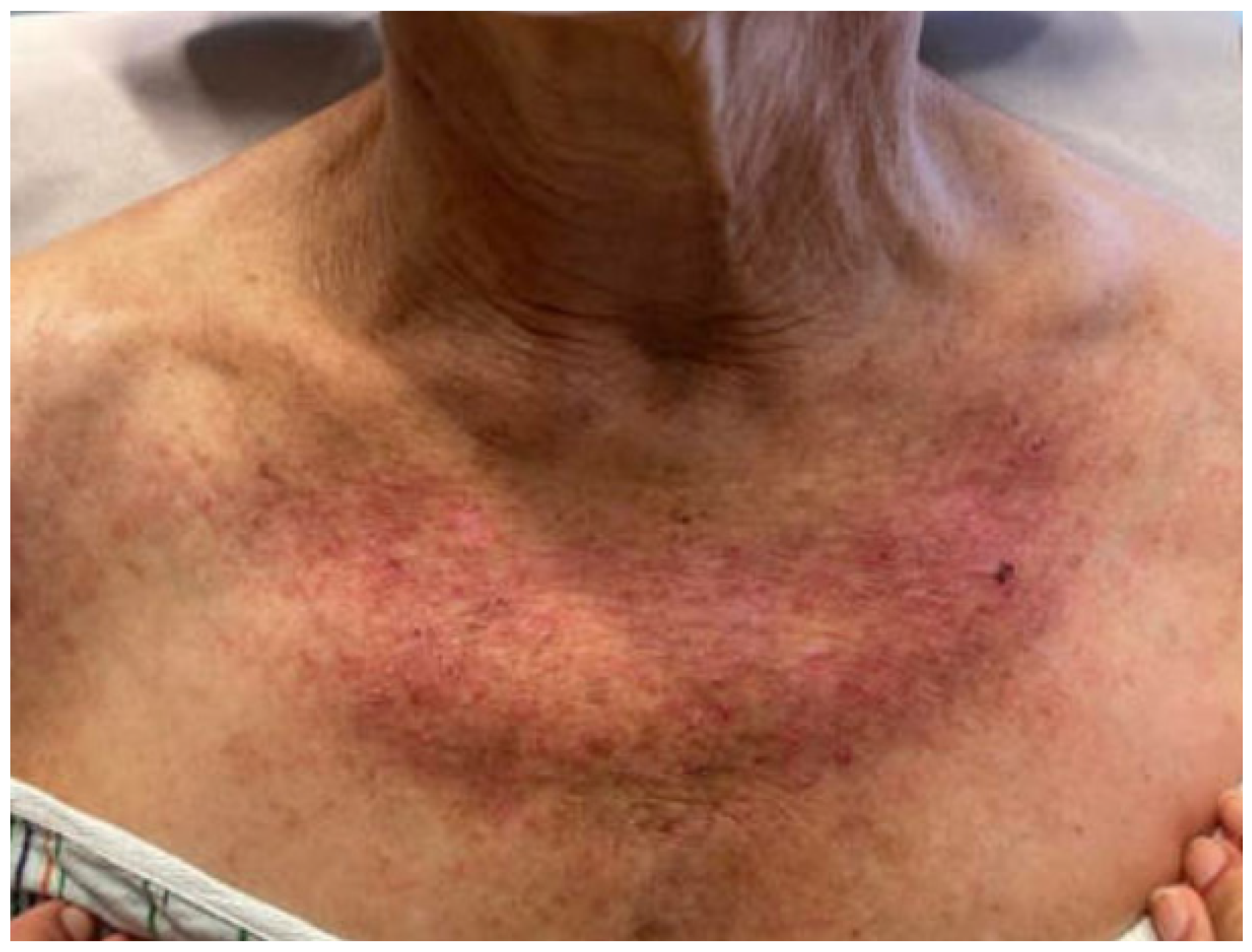
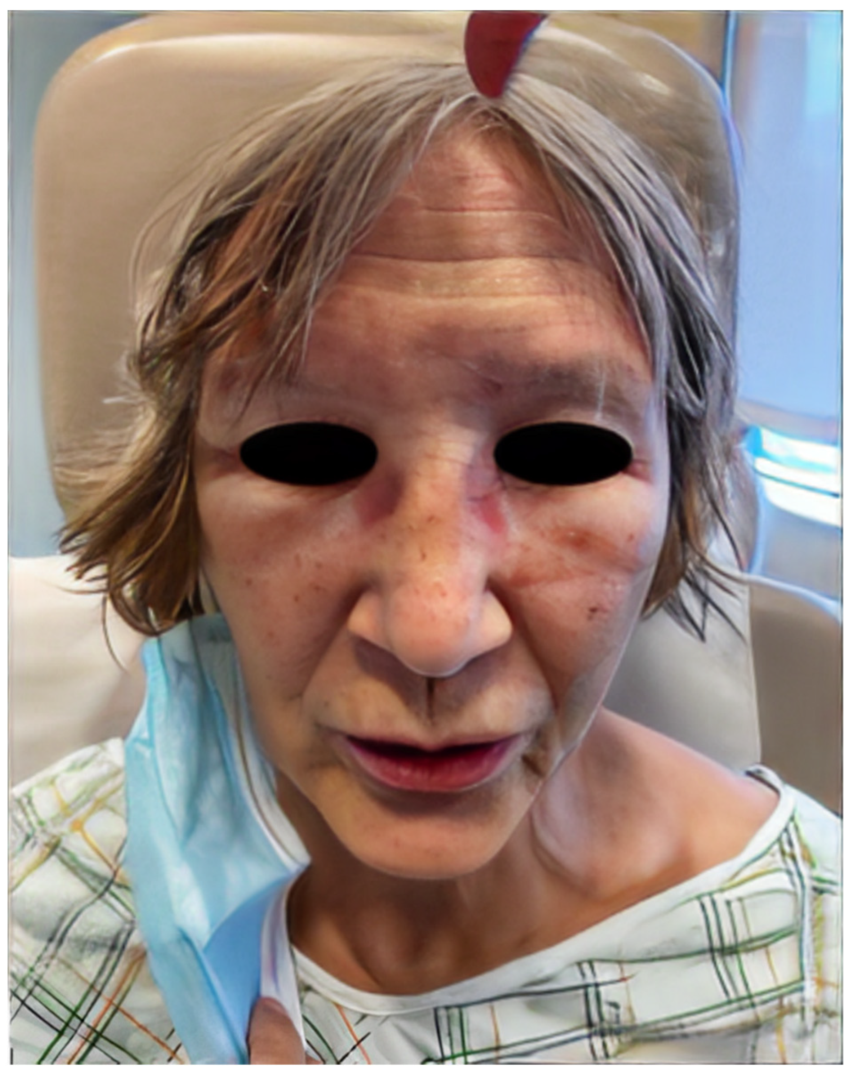
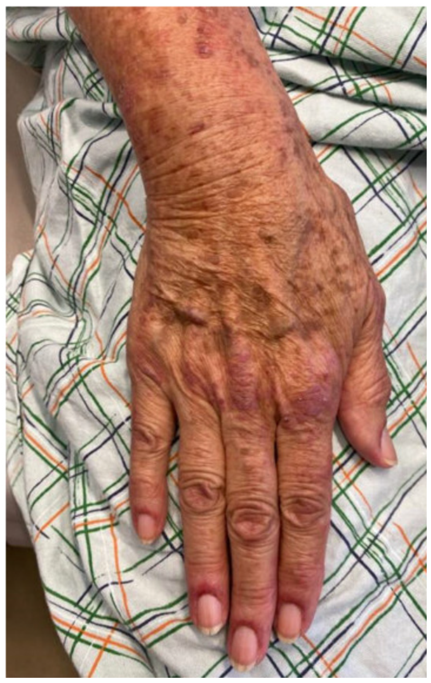
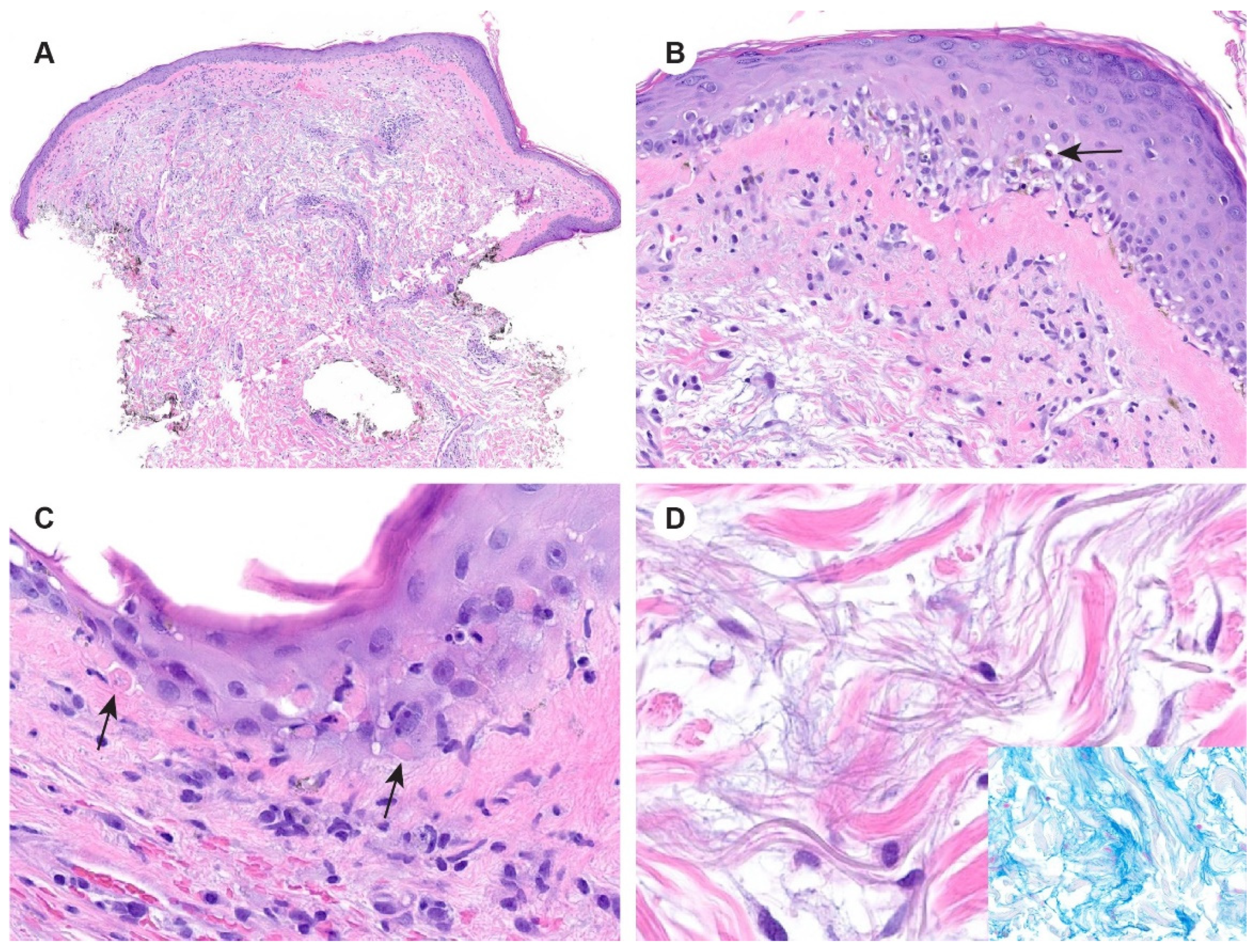
2. Case Report
3. Discussion
Author Contributions
Funding
Institutional Review Board Statement
Informed Consent Statement
Data Availability Statement
Conflicts of Interest
References
- Milisenda, J.C.; Doti, P.I.; Prieto-González, S.; Grau, J.M. Dermatomyositis presenting with severe subcutaneous edema: Five additional cases and review of the literature. Semin. Arthritis Rheum. 2014, 44, 228–233. [Google Scholar] [CrossRef]
- Ghirardello, A.; Borella, E.; Beggio, M.; Franceschini, F.; Fredi, M.; Doria, A. Myositis autoantibodies and clinical phenotypes. Autoimmun. Highlights 2014, 5, 69–75. [Google Scholar] [CrossRef]
- Fiorentino, D.F.; Chung, L.S.; Christopher-Stine, L.; Zaba, L.; Li, S.; Mammen, A.L.; Rosen, A.; Casciola-Rosen, L. Most patients with cancer-associated dermatomyositis have antibodies to nuclear matrix protein NXP-2 or transcription intermediary factor 1γ. Arthritis Rheum. 2013, 65, 2954–2962. [Google Scholar] [CrossRef] [PubMed]
- Goussot, R.; Wettlé, C.; Le Coz, C.; Cribier, B.; Lipsker, D. Dermatomyosite œdémateuse sévère [Severe edematous dermatomyositis]. Ann. Dermatol. Venereol. 2016, 143, 202–209. [Google Scholar] [CrossRef] [PubMed]
- Dunkley, L.; Jawad, A.S.M. Subcutaneous oedema as a presenting feature of polymyositis/dermatomyositis: A poor prognostic indicator? Grand Rounds 2007, 7, 21–25. [Google Scholar] [CrossRef]
- Andersen, M.F.; Longhurst, H.J.; Rasmussen, E.R.; Bygum, A. How Not to Be Misled by Disorders Mimicking Angioedema: A Review of Pseudoangioedema. Int. Arch. Allergy Immunol. 2016, 169, 163–170. [Google Scholar] [CrossRef] [PubMed]
- Papadopoulou, C.; McCann, L.J. The Vasculopathy of Juvenile Dermatomyositis. Front. Pediatr. 2018, 6, 284. [Google Scholar] [CrossRef] [PubMed]
- Ito, Y.; Kawabata, D.; Yukawa, N.; Yoshifuji, H.; Usui, T.; Tanaka, M.; Fujii, T.; Mimori, T. Severe subcutaneous generalized edema in a patient with dermatomyositis. Mod Rheumatol. 2007, 17, 171–173. [Google Scholar] [CrossRef]
- Qudsiya, Z.; Waseem, M. Dermatomyositis. In StatPearls [Internet]; StatPearls Publishing: Treasure Island, FL, USA, 2023; Available online: https://www.ncbi.nlm.nih.gov/books/NBK558917/ (accessed on 8 August 2023).
- Wainger, C.K.; Lever, W.F. Dermatomyositis; report of three cases with postmortem observations. Arch. Derm. Syphilol. 1949, 59, 196–208. [Google Scholar] [CrossRef]
- Eimer, M.J.; Brickman, W.J.; Seshadri, R.; Ramsey-Goldman, R.; McPherson, D.D.; Smulevitz, B.; Stone, N.J.; Pachman, L.M. Clinical status and cardiovascular risk profile of adults with a history of juvenile dermatomyositis. J. Pediatr. 2011, 159, 795–801. [Google Scholar] [CrossRef]
- Tisseverasinghe, A.; Bernatsky, S.; Pineau, C.A. Arterial events in persons with dermatomyositis and polymyositis. J. Rheumatol. 2009, 36, 1943–1946. [Google Scholar] [CrossRef]
- Zöller, B.; Li, X.; Sundquist, J.; Sundquist, K. Risk of subsequent coronary heart disease in patients hospitalized for immune-mediated diseases: A nationwide follow-up study from Sweden. PLoS ONE 2012, 7, e33442. [Google Scholar] [CrossRef] [PubMed]
- Roman, M.J.; Shanker, B.A.; Davis, A.; Lockshin, M.D.; Sammaritano, L.; Simantov, R.; Crow, M.K.; Schwartz, J.E.; Paget, S.A.; Devereux, R.B.; et al. Prevalence and correlates of accelerated atherosclerosis in systemic lupus erythematosus. N. Engl. J. Med. 2003, 349, 2399–2406. [Google Scholar] [CrossRef]
- Zhang, L.; Wang, H.H. The genetics and pathogenesis of thoracic aortic aneurysm disorder and dissections. Clin. Genet. 2016, 89, 639–646. [Google Scholar] [CrossRef]
- Leone, O.; Corsini, A.; Pacini, D.; Corti, B.; Lorenzini, M.; Laus, V.; Foà, A.; Reggiani, M.L.B.; Di Marco, L.; Rapezzi, C. The complex interplay among atherosclerosis, inflammation, and degeneration in ascending thoracic aortic aneurysms. J. Thorac. Cardiovasc. Surg. 2020, 160, 1434–1443.e6. [Google Scholar] [CrossRef]
- Pakpoor, J. Risk of Abdominal Aortic Aneurysm in People Admitted to Hospital with Selected Immune-mediated Diseases: Record-linkage Studies. Eur. J. Vasc. Endovasc. Surg. 2014, 47, 689. [Google Scholar] [CrossRef]
- Betteridge, Z.; McHugh, N. Myositis-specific autoantibodies: An important tool to support diagnosis of myositis. J. Intern. Med. 2016, 280, 8–23. [Google Scholar] [CrossRef] [PubMed]
- Lim, D.; Fiorentino, D.; Werth, V. Current concepts and advances in dermatomyositis: A dermatological perspective. Clin. Exp. Rheumatol. 2023, 41, 359–369. [Google Scholar] [CrossRef]
- Satoh, M.; Tanaka, S.; Ceribelli, A.; Calise, S.J.; Chan, E.K. A Comprehensive Overview on Myositis-Specific Antibodies: New and Old Biomarkers in Idiopathic Inflammatory Myopathy. Clin. Rev. Allergy Immunol. 2017, 52, 1–19. [Google Scholar] [CrossRef]
- Ge, Y.; Li, S.; Tian, X.; He, L.; Lu, X.; Wang, G. Anti-melanoma differentiation-associated gene 5 (MDA5) antibody-positive dermatomyositis responds to rituximab therapy. Clin. Rheumatol. 2021, 40, 2311–2317. [Google Scholar] [CrossRef] [PubMed]
- Duchesne, M.; Leonard-Louis, S.; Landon-Cardinal, O.; Anquetil, C.; Mariampillai, K.; Monzani, Q.; Benveniste, O.; Allenbach, Y. Edematous myositis: A clinical presentation first suggesting dermatomyositis diagnosis. Brain Pathol. 2020, 30, 867–876. [Google Scholar] [CrossRef] [PubMed]
- Sunny, N.; Kashyap, K.; Kumar, A.; Parchani, A.; Dhar, M. Dermatomyositis Masquerading As Generalized Body Swelling: A Case Report. Cureus 2023, 15, e38895. [Google Scholar] [CrossRef] [PubMed]
- De Vooght, J.; Vulsteke, J.B.; De Haes, P.; Bossuyt, X.; Lories, R.; De Langhe, E. Anti-TIF1-γ autoantibodies: Warning lights of a tumour autoantigen. Rheumatology 2020, 59, 469–477. [Google Scholar] [CrossRef]
- Oldroyd, A.; Sergeant, J.C.; New, P.; McHugh, N.J.; Betteridge, Z.; Lamb, J.A.; Ollier, W.E.; Cooper, R.G.; Chinoy, H. The temporal relationship between cancer and adult onset anti-transcriptional intermediary factor 1 antibody-positive dermatomyositis. Rheumatology 2019, 58, 650–655. [Google Scholar] [CrossRef] [PubMed]
- Venhuizen, A.C.; Martens, J.E.; van der Linden, P.J. Dermatomyositis as first presentation of ovarian cancer. Acta Obstet. Gynecol. Scand. 2006, 85, 1271–1272. [Google Scholar] [CrossRef] [PubMed]
- Fiorentino, D.; Casciola-Rosen, L. Autoantibodies to transcription intermediary factor 1 in dermatomyositis shed insight into the cancer-myositis connection. Arthritis Rheum. 2012, 64, 346–349. [Google Scholar] [CrossRef]
- Ikeda, N.; Yamaguchi, Y.; Kanaoka, M.; Ototake, Y.; Akita, A.; Watanabe, T.; Aihara, M. Clinical significance of serum levels of anti-transcriptional intermediary factor 1-γ antibody in patients with dermatomyositis. J. Dermatol. 2020, 47, 490–496. [Google Scholar] [CrossRef]
- Thompson, C.; Piguet, V.; Choy, E. The pathogenesis of dermatomyositis. Br. J. Dermatol. 2018, 179, 1256–1262. [Google Scholar] [CrossRef]
- Okiyama, N.; Ichimura, Y.; Shobo, M.; Tanaka, R.; Kubota, N.; Saito, A.; Ishitsuka, Y.; Watanabe, R.; Fujisawa, Y.; Nakamura, Y.; et al. Immune response to dermatomyositis-specific autoantigen, transcriptional intermediary factor 1γ can result in experimental myositis. Ann. Rheum. Dis. 2021, 80, 1201–1208. [Google Scholar] [CrossRef]
- O’Connell, E.W.; Reams, J.; Denio, A.E. Aortitis as a Harbinger of Occult Malignancy. Case Rep. Rheumatol. 2019, 2019, 8385630. [Google Scholar] [CrossRef]
- Fleming, S.; Hellström-Lindberg, E.; Burbury, K.; Seymour, J.F. Paraneoplastic large vessel arteritis complicating myelodysplastic syndrome. Leuk. Lymphoma 2012, 53, 1613–1616. [Google Scholar] [CrossRef] [PubMed]


Disclaimer/Publisher’s Note: The statements, opinions and data contained in all publications are solely those of the individual author(s) and contributor(s) and not of MDPI and/or the editor(s). MDPI and/or the editor(s) disclaim responsibility for any injury to people or property resulting from any ideas, methods, instructions or products referred to in the content. |
© 2023 by the authors. Licensee MDPI, Basel, Switzerland. This article is an open access article distributed under the terms and conditions of the Creative Commons Attribution (CC BY) license (https://creativecommons.org/licenses/by/4.0/).
Share and Cite
Bennett, R.; Bradley, K.; Salem, I.; Weiner, D.; Patel, D.; Cloutier, J.; Pace, N.; Barton, D. A Case of Paraneoplastic Anti-TIF1-γ Antibody-Positive Dermatomyositis Presenting with Generalized Edema and Associated with Aortic Aneurysm. Dermato 2023, 3, 232-240. https://doi.org/10.3390/dermato3040018
Bennett R, Bradley K, Salem I, Weiner D, Patel D, Cloutier J, Pace N, Barton D. A Case of Paraneoplastic Anti-TIF1-γ Antibody-Positive Dermatomyositis Presenting with Generalized Edema and Associated with Aortic Aneurysm. Dermato. 2023; 3(4):232-240. https://doi.org/10.3390/dermato3040018
Chicago/Turabian StyleBennett, Raven, Katherine Bradley, Iman Salem, David Weiner, Dhrumil Patel, Jeffrey Cloutier, Nicole Pace, and Dorothea Barton. 2023. "A Case of Paraneoplastic Anti-TIF1-γ Antibody-Positive Dermatomyositis Presenting with Generalized Edema and Associated with Aortic Aneurysm" Dermato 3, no. 4: 232-240. https://doi.org/10.3390/dermato3040018