
Hyperpigmented Scleroderma-like Lesions under Combined Pembrolizumab and Pemetrexed Treatment of Non-Small Lung Cancer


Abstract
:1. Introduction
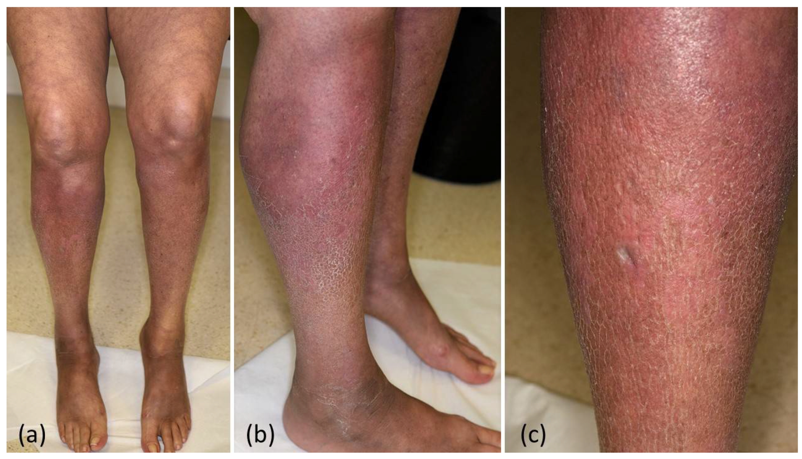
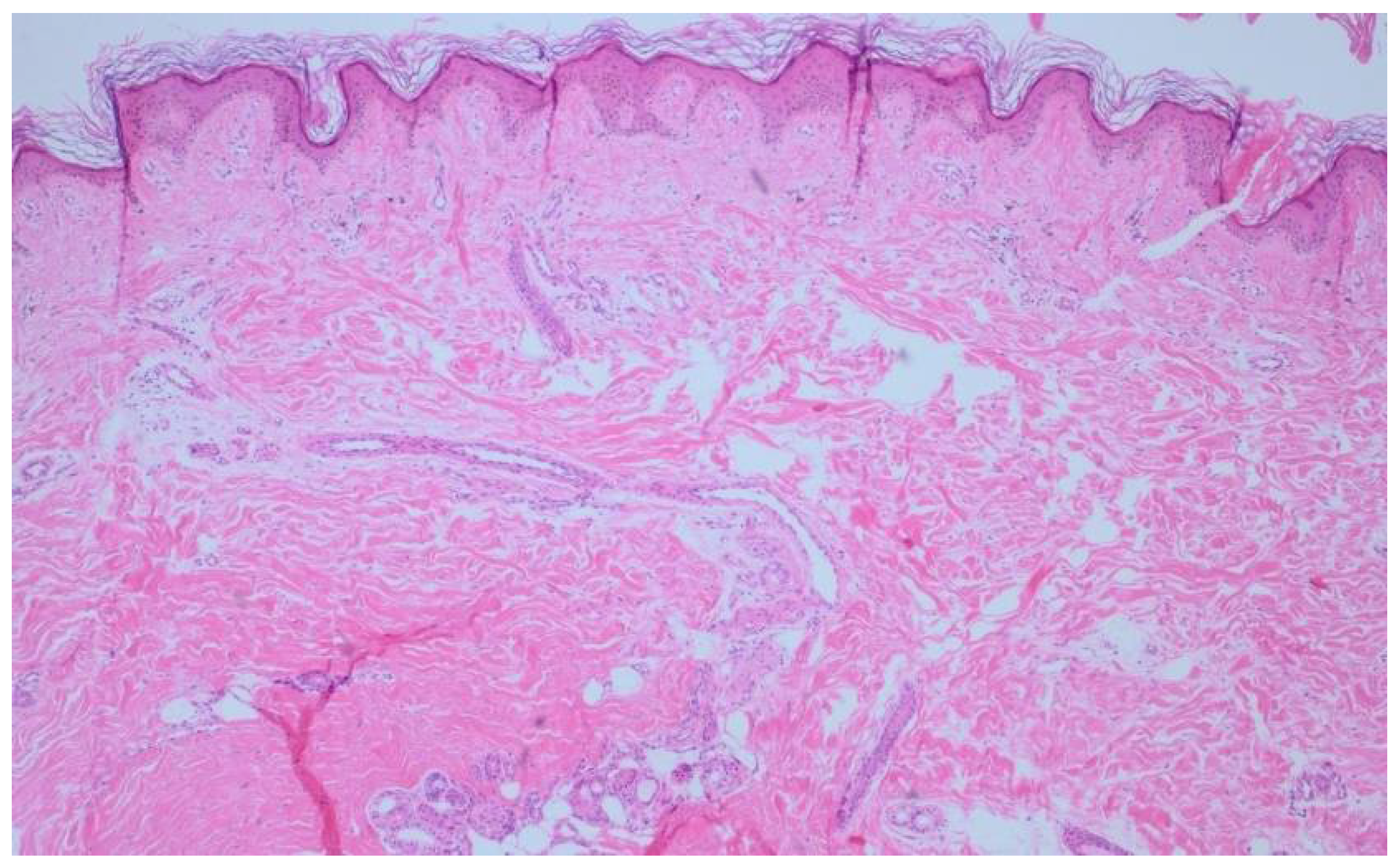
2. Case Report
3. Discussion
4. Conclusions
Author Contributions
Funding
Institutional Review Board Statement
Informed Consent Statement
Data Availability Statement
Conflicts of Interest
References
- Apalla, Z.; Nikolaou, V.; Fattore, D.; Fabbrocini, G.; Freites-Martinez, A.; Sollena, P.; Lacouture, M.; Kraehenbuehl, L.; Stratigos, A.; Peris, K.; et al. European Recommendations for Management of Immune Checkpoint Inhibitors-Derived Dermatologic Adverse Events. The EADV Task Force “Dermatology for Cancer Patients” Position Statement. J. Eur. Acad. Dermatol. Venereol. Jeadv. 2021, 36, 332–350. [Google Scholar] [CrossRef] [PubMed]
- Gambichler, T.; Scheel, C.H.; Reuther, J.; Susok, L. Management of Immune-related Adverse Events in Anti-PD-1-treated Patients with Advanced Cutaneous Squamous Cell Carcinoma. J. Eur. Acad. Dermatol. 2022, 36, 23–28. [Google Scholar] [CrossRef]
- Le, T.K.; Kaul, S.; Cappelli, L.C.; Naidoo, J.; Semenov, Y.R.; Kwatra, S.G. Cutaneous Adverse Events of Immune Checkpoint Inhibitor Therapy: Incidence and Types of Reactive Dermatoses. J. Dermatol. Treat. 2021, 1–5. [Google Scholar] [CrossRef]
- Terrier, B.; Humbert, S.; Preta, L.-H.; Delage, L.; Razanamahery, J.; Laurent-Roussel, S.; Mestiri, R.; Beaudeau, L.; Legendre, P.; Goupil, F.; et al. Risk of Scleroderma According to the Type of Immune Checkpoint Inhibitors. Autoimmun. Rev. 2020, 19, 102596. [Google Scholar] [CrossRef] [PubMed]
- Hamaguchi, Y. Drug-Induced Scleroderma-like Lesion. Allergol. Int. 2021. [Google Scholar] [CrossRef] [PubMed]
- Ishikawa, K.; Sakai, T.; Saito-Shono, T.; Miyawaki, M.; Osoegawa, A.; Sugio, K.; Ono, A.; Mori, H.; Nishida, H.; Yokoyama, S.; et al. Pemetrexed-induced Scleroderma-like Conditions in the Lower Legs of a Patient with Non-small Cell Lung Carcinoma. J. Dermatol. 2016, 43, 1071–1074. [Google Scholar] [CrossRef] [PubMed]
- Gambichler, T.; Schmitz, L. Ultraviolet A1 Phototherapy for Fibrosing Conditions. Front. Med. 2018, 5, 237. [Google Scholar] [CrossRef] [PubMed]
- Gambichler, T.; Doerler, M.; Scheel, C.H. Onset of Subacute Cutaneous Lupus Erythematosus after the Initiation of Immune Checkpoint Inhibitor Therapy of Cancer. Lupus 2001, 30, 531–533. [Google Scholar] [CrossRef] [PubMed]
- Acar, A.; Oraloglu, G.; Yaman, B.; Karaarslan, I. Nivolumab-induced Plaque Morphea in a Malign Melanoma Patient. J. Cosmet. Dermatol. 2021, 20, 2645–2647. [Google Scholar] [CrossRef] [PubMed]
- Cho, M.; Nonomura, Y.; Kaku, Y.; Nakabo, S.; Endo, Y.; Otsuka, A.; Kabashima, K. Scleroderma-like Syndrome Associated with Nivolumab Treatment in Malignant Melanoma. J. Dermatol. 2019, 46, e43–e44. [Google Scholar] [CrossRef] [PubMed]
- Tjarks, B.J.; Kerkvliet, A.M.; Jassim, A.D.; Bleeker, J.S. Scleroderma-like Skin Changes Induced by Checkpoint Inhibitor Therapy. J. Cutan. Pathol. 2018, 45, 615–618. [Google Scholar] [CrossRef] [PubMed]
- Alegre-Sánchez, A.; Fonda-Pascual, P.; Saceda-Corralo, D.; de las Heras-Alonso, E. Relapse of Morphea during Nivolumab Therapy for Lung Adenocarcinoma. Actas Dermo-Sifiliográficas Engl. Ed. 2017, 108, 69–70. [Google Scholar] [CrossRef] [PubMed]
- Shenoy, N.; Esplin, B.; Barbosa, N.; Wieland, C.; Thanarajasingam, U.; Markovic, S. Pembrolizumab Induced Severe Sclerodermoid Reaction. Ann. Oncol. 2017, 28, 432–433. [Google Scholar] [CrossRef] [PubMed]
- Barbosa, N.S.; Wetter, D.A.; Wieland, C.N.; Shenoy, N.K.; Markovic, S.N.; Thanarajasingam, U. Scleroderma Induced by Pembrolizumab: A Case Series. Mayo Clin. Proc. 2017, 92, 1158–1163. [Google Scholar] [CrossRef] [Green Version]
- Cheng, M.W.; Hisaw, L.D.; Bernet, L. Generalized Morphea in the Setting of Pembrolizumab. Int. J. Dermatol. 2019, 58, 736–738. [Google Scholar] [CrossRef] [PubMed]
- Herrscher, H.; Tomasic, G.; Gordon, A.C. Generalised Morphea Induced by Pembrolizumab. Eur. J. Cancer 2019, 116, 178–181. [Google Scholar] [CrossRef] [PubMed]
- Salamaliki, C.; Solomou, E.E.; Liossis, S.-N.C. Immune Checkpoint Inhibitor-Associated Scleroderma-Like Syndrome: A Report of a Pembrolizumab-Induced “Eosinophilic Fasciitis-Like” Case and a Review of the Literature. Rheumatol. Ther. 2020, 7, 1045–1052. [Google Scholar] [CrossRef] [PubMed]
- Langan, E.A.; Budner, K.; Zillikens, D.; Terheyden, P. Generalized Morphoea in the Setting of Combined Immune Checkpoint Inhibitor Therapy for Metastatic Melanoma. Medicine 2021, 100, e25513. [Google Scholar] [CrossRef]
- Grant, C.; Chalasani, V.; Uchin, J.M.; Dore, A. Atezolizumab-Induced Scleroderma: A Rare Complication. BMJ Case Rep. 2021, 14, e244968. [Google Scholar] [CrossRef] [PubMed]
- Das, S.; Johnson, D.B. Immune-Related Adverse Events and Anti-Tumor Efficacy of Immune Checkpoint Inhibitors. J. Immunother. Cancer 2019, 7, 306. [Google Scholar] [CrossRef] [PubMed]
- Panhaleux, M.; Espitia, O.; Terrier, B.; Manson, G.; Maria, A.; Humbert, S.; Godbert, B.; Perrin, J.; Achille, A.; Arrondeau, J.; et al. Anti-Programmed Death Ligand 1 Immunotherapies in Cancer Patients with Pre-Existing Systemic Sclerosis: A Postmarketed Phase IV Safety Assessment Study. Eur. J. Cancer 2021, 160, 134–139. [Google Scholar] [CrossRef] [PubMed]
- Shih, C.; Chen, V.J.; Gossett, L.S.; Gates, S.B.; MacKellar, W.C.; Habeck, L.L.; Shackelford, K.A.; Mendelsohn, L.G.; Soose, D.J.; Patel, V.F.; et al. LY231514, a Pyrrolo[2,3-d]Pyrimidine-Based Antifolate That Inhibits Multiple Folate-Requiring Enzymes. Cancer Res. 1997, 57, 1116–1123. [Google Scholar]
- Piérard-Franchimont, C.; Quatresooz, P.; Reginster, M.-A.; Piérard, G.E. Revisiting Cutaneous Adverse Reactions to Pemetrexed. Oncol. Lett. 2011, 2, 769–772. [Google Scholar] [CrossRef]
- Racanelli, A.C.; Rothbart, S.B.; Heyer, C.L.; Moran, R.G. Therapeutics by Cytotoxic Metabolite Accumulation: Pemetrexed Causes ZMP Accumulation, AMPK Activation, and Mammalian Target of Rapamycin Inhibition. Cancer Res. 2009, 69, 5467–5474. [Google Scholar] [CrossRef] [PubMed] [Green Version]
- Vitiello, M.; Romanelli, P.; Kerdel, F.A. Painful Generalized Erythematous Patches: A Severe and Unusual Cutaneous Reaction to Pemetrexed. J. Am. Acad. Dermatol. 2011, 65, 243–244. [Google Scholar] [CrossRef]
- Eguia, B.; Ruppert, A.-M.; Fillon, J.; Lavolé, A.; Gounant, V.; Epaud, C.; Milleron, B.; Moguelet, P.; Wislez, M.; Frances, C.; et al. Skin Toxicities Compromise Prolonged Pemetrexed Treatment. J. Thorac. Oncol. 2011, 6, 2083–2089. [Google Scholar] [CrossRef] [PubMed] [Green Version]
- Merklen-Djafri, C.; Imbert, E.; Courouge-Dorcier, D.; Schott, R.; Méraud, J.-P.; Muller, C.; Tebacher, M.; Springinsfeld, G.; Cribier, B.; Lipsker, D. Pemetrexed-Induced Skin Sclerosis. Clin. Oncol. 2012, 24, 452–453. [Google Scholar] [CrossRef] [PubMed]
- Corbaux, C.; Marie, J.; Meraud, J.-P.; Lacroix, S.; Delhoume, J.-Y.; Jouary, T.; Madoui, S. Pemetrexed-Induced Scleroderma-like Changes in the Lower Legs. Ann. De Dermatol. Et De Vénéréologie 2014, 142, 115–120. [Google Scholar] [CrossRef]
- Shuster, M.; Morley, K.; Logan, J.; Sequist, L.V.; Shaw, A.; Fidias, P.; Kroshinsky, D. Lipodermatosclerosis Secondary to Pemetrexed Use. J. Thorac. Oncol. 2015, 10, e11–e12. [Google Scholar] [CrossRef] [PubMed] [Green Version]
- Szczepanek, M.; Frątczak, A.; Lis-Święty, A. Narrow-Band Reflectance Spectrophotometry for the Assessment of Erythematous and Hyperpigmented Skin Lesions in Localized Scleroderma: A Preliminary Study. Clin. Cosmet. Investig. Dermatol. 2021, 14, 575–580. [Google Scholar] [CrossRef] [PubMed]
- Pope, J.E.; Shum, D.T.; Gottschalk, R.; Stevens, A.; McManus, R. Increased Pigmentation in Scleroderma. J. Rheumatol. 1996, 23, 1912–1916. [Google Scholar] [PubMed]
- Tiniakou, E.; Crawford, J.; Darrah, E. Insights into Origins and Specificities of Autoantibodies in Systemic Sclerosis. Curr. Opin. Rheumatol. 2021, 33, 486–494. [Google Scholar] [CrossRef] [PubMed]
- Morimoto, K.; Yamada, T.; Yokoi, T.; Kijima, T.; Goto, Y.; Nakao, A.; Hibino, M.; Takeda, T.; Yamaguchi, H.; Takumi, C.; et al. Clinical Impact of Pembrolizumab Combined with Chemotherapy in Elderly Patients with Advanced Non-Small-Cell Lung Cancer. Lung Cancer 2021, 161, 26–33. [Google Scholar] [CrossRef]
- Maria, A.T.J.; Partouche, L.; Goulabchand, R.; Rivière, S.; Rozier, P.; Bourgier, C.; Quellec, A.L.; Morel, J.; Noël, D.; Guilpain, P. Intriguing Relationships Between Cancer and Systemic Sclerosis: Role of the Immune System and Other Contributors. Front. Immunol. 2019, 9, 3112. [Google Scholar] [CrossRef] [PubMed] [Green Version]


| Target | ICI | Entity | Auto-ab | Treatment | Ref. |
|---|---|---|---|---|---|
| PD-1 | Nivolumab | Melanoma | negative | topical steroids + calcipotriol | [9] |
| Melanoma | negative | systemic steroids | [10] | ||
| RCC | negative | systemic steroids | [11] | ||
| NSCLC | not reported | spontaneous remission | [12] | ||
| Pembrolizumab | Melanoma | not reported | systemic steroids | [13] | |
| Melanoma | negative | hydroxychloroquine + systemic steroids | [14] | ||
| Choroidal melanoma | not reported | systemic steroids | [15] | ||
| Melanoma | negative | colchicine, topical steroids, systemic steroids, cyclophosphamide, infliximab | [16] | ||
| NSCLC | ANA | systemic steroids | [17] | ||
| PD-1 + CTLA-4 | Pembrolizumab + ipilimumab | Melanoma | ANA | systemic followed by topical steroids | [18] |
| PD-L1 | Atezolizumab | NSCLC | PM/SCL-75 | mycophenolate mofetil | [19] |
Publisher’s Note: MDPI stays neutral with regard to jurisdictional claims in published maps and institutional affiliations. |
© 2022 by the authors. Licensee MDPI, Basel, Switzerland. This article is an open access article distributed under the terms and conditions of the Creative Commons Attribution (CC BY) license (https://creativecommons.org/licenses/by/4.0/).
Share and Cite
Gambichler, T.; Lee, Y.-P.; Barras, M.; Scheel, C.H.; Susok, L. Hyperpigmented Scleroderma-like Lesions under Combined Pembrolizumab and Pemetrexed Treatment of Non-Small Lung Cancer. Dermato 2022, 2, 8-13. https://doi.org/10.3390/dermato2010002
Gambichler T, Lee Y-P, Barras M, Scheel CH, Susok L. Hyperpigmented Scleroderma-like Lesions under Combined Pembrolizumab and Pemetrexed Treatment of Non-Small Lung Cancer. Dermato. 2022; 2(1):8-13. https://doi.org/10.3390/dermato2010002
Chicago/Turabian StyleGambichler, Thilo, Yi-Pei Lee, Milan Barras, Christina H. Scheel, and Laura Susok. 2022. "Hyperpigmented Scleroderma-like Lesions under Combined Pembrolizumab and Pemetrexed Treatment of Non-Small Lung Cancer" Dermato 2, no. 1: 8-13. https://doi.org/10.3390/dermato2010002