Review

The Role of Macrophages in Cardiac Function and Disease

by
Nella Prevete
1,2 and
Daniela Sorriento
3,4,*
1
Department of Translational Medical Sciences, Federico II University, 80131 Naples, Italy
2
Institute of Experimental Endocrinology and Oncology (IEOS), National Research Council (CNR), 80131 Naples, Italy
3
Department of Advanced Biomedical Sciences, Federico II University, 80131 Naples, Italy
4
Hypertension Research Center (CIRIAPA), Federico II University, 80131 Naples, Italy
*
Author to whom correspondence should be addressed.
J. Mol. Pathol. 2023, 4(4), 318-332; https://doi.org/10.3390/jmp4040026
Submission received: 31 October 2023 / Revised: 28 November 2023 / Accepted: 5 December 2023 / Published: 7 December 2023

Abstract

:
A tight association between inflammation and cardiac damage has been extensively recognized. In this review, we will focus on macrophages as key players in the physiology and pathology of the heart and on their role in the functional crosstalk between inflammation and heart disease. In the steady state, macrophages contribute to the homeostasis of cardiac tissue. Indeed, cardiac resident macrophages promote coronary development and tissue homeostasis, favor electric conduction in cardiomyocytes, and contribute to mitochondrial quality control. However, macrophages also take part in adverse cardiac events contributing to the development or the progression of several pathologic conditions. Infiltrating cells derived from circulating monocytes contribute to tissue injury through the release of inflammatory cytokines and catecholamines. In particular, the present review will discuss the role of macrophages in heart failure, atherosclerosis, and anthracycline-dependent cardiotoxicity. Prolonged inflammatory response and increased apoptotic cell death sustained by chronic activation of the transcription factor NFκB are the basis of heart failure pathogenesis. Here, we will discuss the involvement of NFκB signaling in macrophage-dependent cardiac damage and its use as a therapeutic target in the treatment of cardiovascular pathologies.

1. Introduction

The immune system plays a fundamental role in the development and progression of heart damage and, recently, great advances have been made in the knowledge of the underpinning cellular and molecular mechanisms.
The correlation between inflammation and heart damage in clinics derives from two critical trials: the Jupiter and the Cantos studies. The JUPITER study was a randomized primary prevention study, which tested the effectiveness of rosuvastatin in healthy and normocholesterolemic subjects [1,2]. This study demonstrated that rosuvastatin therapy can reduce the incidence of all cardiovascular events by 44%, myocardial infarction by 54%, stroke by 48%, the need for revascularization by 46%, and reduce mortality from all causes by 20%, even in subjects with no cardiovascular history and with medium–low risk. Also, in normocholesterolemic subjects with elevated blood levels of inflammatory markers (high-sensitivity C-reactive protein, hsPCR, >2 mg/L), 20 mg rosuvastatin reduced LDL cholesterol by 50% and hsPCR by 37%. This was a surprising result even if it was not clear whether this effect was due to a reduction in inflammation or cholesterol lowering.
Advances in the field have shown that several drugs commonly used in clinics for treating cardiovascular disease also exert anti-inflammatory effects, supporting the idea that the results of Jupiter study could be due to a reduction in inflammation. Beta-blockers, angiotensin-converting enzyme inhibitors (ACE) inhibitors, and diuretics, besides their molecular effects on myocytes, can reduce inflammatory cytokines production, suggesting a specific immune therapy as a potential strategy for the treatment of heart failure [3]. Beta-blockers, for instance, can reduce the release of inflammatory cytokines and macrophage polarization towards the proinflammatory (M1) subtype [4,5,6]. The renin–angiotensin–aldosterone system (RAAS) inhibitors exert the same effects by regulating matrix metalloproteinase activity and inhibiting endothelial–mesenchymal transition (EndMT), [7,8] while diuretics, such as sacubitril, reduce inflammation by attenuating cardiac fibrosis [9,10].
The demonstration that inflammation represents an independent trigger of the atherosclerotic process and its clinical consequences, such as myocardial infarction, stroke, and death, derives from the CANTOS trial, which was the first to highlight the key role of innate immunity in cardiovascular disease. It was a phase III secondary prevention clinical trial, in which the effect of specifically targeting interleukin 1β (IL-1β) with a monoclonal antibody, canakinumab, was tested in patients with a prior myocardial infarction and high levels of C-reactive protein (≥2mg/L) [11,12]. The treatment with canakinumab reduced the rate of recurrent cardiovascular events, highlighting for the first time the possibility of lowering the incidence of cardiovascular events by acting exclusively on inflammation. It also demonstrated that canakinumab-dependent reduction in cardiovascular events was associated with a reduction in IL-6, a proinflammatory cytokine usually highly expressed in cardiac damage [11,13,14].
These clinical trials lay the foundation for the following studies on the association between inflammation and cardiac diseases, which identified the involved immune cells and the specific molecular mechanisms. In this review, we will focus on macrophages as key cellular players in the physiology and pathology of the heart and on their role in the functional crosstalk between inflammation and heart disease.

2. Crosstalk between Inflammation and Cardiac Damage: Cellular and Molecular Mechanisms

In response to insults, such as acute myocardial infarction, there is an increase in proinflammatory cytokines, mainly TNFα, IL-6, IL-1β, and TGF-1β, whose acute effects are protective through the regulation of myocyte survival and death in the infarcted area [15]. However, chronically, the increased release of cytokines promotes interstitial fibrosis and collagen deposition in the non-infarcted area, leading to ventricular dysfunction. Also, cytokines induce the activity of metalloproteinases (MMP2 and MMP9) in the infarct area and the expression of natriuretic peptides (ANP and BNP), promoting cardiac remodeling [15,16]. These results support the idea that inflammation exerts a cardioprotective role in acute responses and a deleterious effect in chronic responses.
Several cellular and molecular mechanisms have been identified underlying the association between inflammation and cardiac damage.
At the cellular level, the heart is composed of cardiomyocytes and non-myocytic cell types (fibroblasts, endothelial cells, and immune cells) which contribute to the maintenance of a correct cardiac function through functional crosstalk between cells. Alterations in such interactions contribute to the development of cardiac damage [17,18,19]. In response to oxidative stress, cardiac endothelial cells release substances that act in a paracrine way to activate specific receptors on cardiomyocytes and consequently regulate survival mechanisms (apoptosis) and mitochondrial reactive oxygen species (ROS) production. Also, the release of nitric oxide (NO) is promoted, which acts on the endothelium to preserve endothelial homeostasis [20]. Cardiac fibroblasts produce extracellular matrix components to ensure the structural integrity of the heart even in response to changes in cardiac homeostasis [21]. An essential role in maintaining cardiac homeostasis and function is also played by macrophages, which basically make up 6% of cardiac cells, but their number increases in response to insults [22,23], through functional interactions with other cardiac cells [24]. Macrophages interact with endothelial cells, favoring their proliferation and sprouting through neurogenic locus notch homolog protein 1 (Notch1) signaling [25,26], interact with smooth muscle cells altering collagen type I and metalloprotease synthesis, upregulating vascular endothelial growth factor (VEGF) expression [25,26,27], interact with cardiac fibroblasts and, depending on the type of activation enhance anti-inflammatory phenotype in both cell types [28] or induce a fibrotic phenotype [29].
At the molecular level, all cardiovascular risk factors (obesity, genetic factors, hypertension, dyslipidemia, as well as infections or smoking) activate inflammatory responses that lead to the production and release of cytokines (Figure 1).
These processes share a common feature: the activation of specific transcription factors that drive the expression of cytokine production [30], such as NFκB. This transcription factor exerts a key role in cardiac damage [16,31,32,33,34] because it is upregulated not only in macrophages regulating their cytokine production but also in cardiomyocytes [35] and other cardiac cells (endothelial cells and fibroblasts), inducing the expression of key genes which are strictly involved in the development of cardiac damage [32,33,34].
For the reasons above, the present review will discuss in particular the involvement of NFκB signaling in macrophage-dependent cardiac damage.

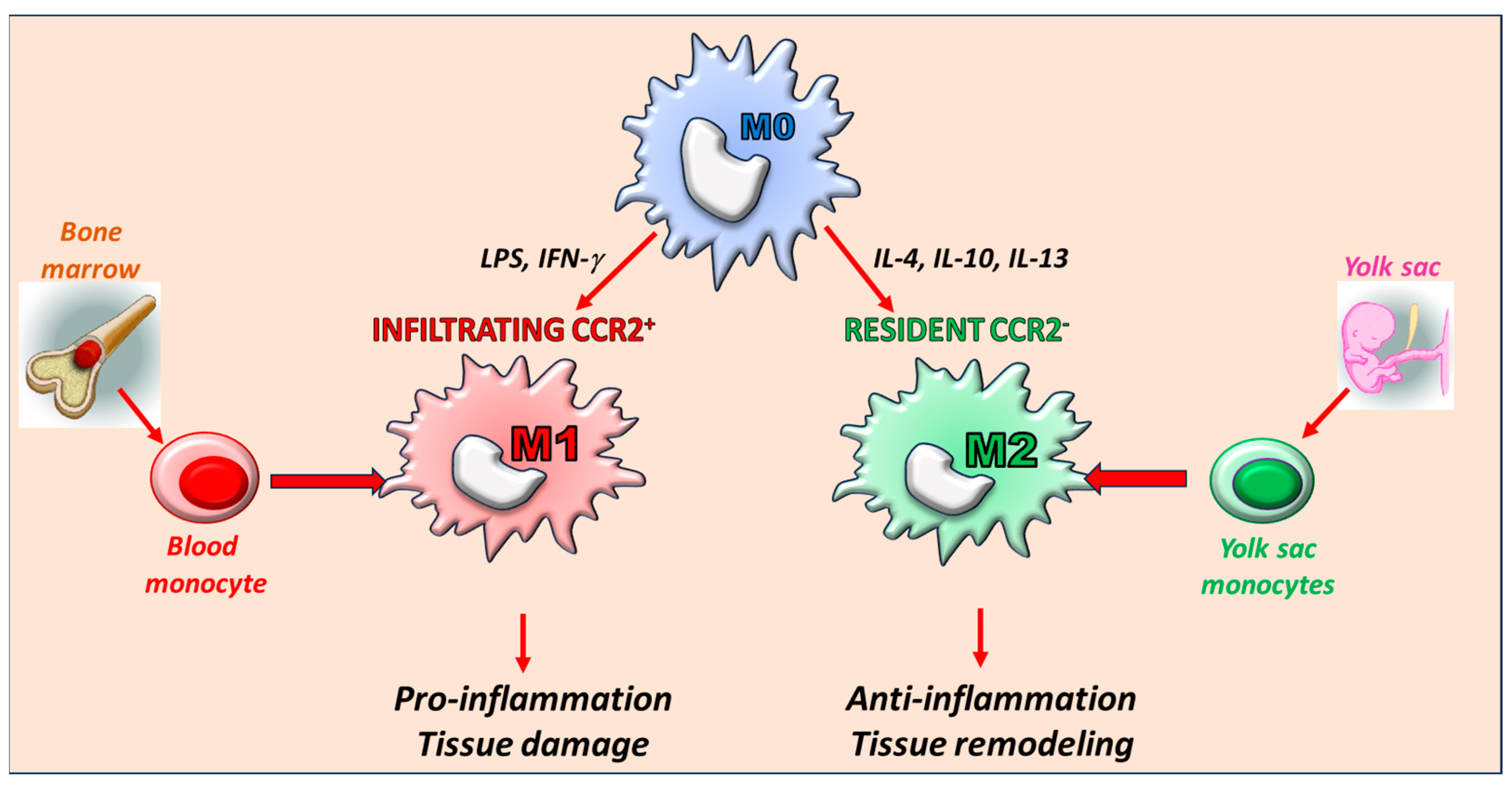
3. The Key Role of Macrophages: Origin and Phenotype

Macrophages and their precursors (monocytes) are ubiquitous key players of the innate immune response that can change their phenotype and function in response to environmental stimulation [36,37].
The M0 phenotype is the state in which macrophages exist in when in a resting condition without any stimulation. In response to specific stimuli, M0 macrophages can polarize in the M1 proinflammatory phenotype (e.g., in response to lipopolysaccharide or interferon-γ) or the anti-inflammatory M2 phenotype (e.g., in response to IL-4, IL-13, IL-10) [38,39] (Figure 2). Beyond this net differentiation of the two macrophage subtypes, the M1/M2 classification represents the two extremes of a sequence of activated states. Furthermore, this classification well reflects their in vitro differentiation. However, in vivo a wide and continuous range of macrophage phenotypes is detectable in dependence of the microenvironmental context. Thus, today the M1/M2 paradigm is considered an oversimplification due to the extremely high level of macrophage plasticity. Alternative nomenclature reflecting the cell surface marker expression and/or cell source is now considered more appropriate. However, taking into account all these limitations, the M1 and M2 classification still is useful to understand the different immunopathological responses mediated by macrophages [40].
These macrophage phenotypic and functional switches have a double opposite role: maintaining cardiac homeostasis by regulating fundamental processes such as cardiac tissue repair, regeneration, and fibrosis; or promoting the development of cardiac damage by uncontrolled production of inflammatory cytokines and growth factors, lack of anti-inflammatory response, and alterations in functional interactions between macrophages and cardiac cells [23,38,41,42].
Cardiac macrophages have different origins that are related to their specific function. Macrophages exist as both circulating and tissue-resident cells [43,44]. Tissue-resident macrophages are of neonatal origin and maintain themselves in adults by self-renewal; during embryonic development, macrophages derive from yolk-sac precursors that migrate to the fetal liver to convert into fetal liver monocytes that subsequently migrate and colonize tissues differentiating into tissue-resident macrophages [44,45]. These cells facilitate coronary development and tissue homeostasis. The tissue can also be infiltrated by macrophages derived from peripheral blood monocytes [46,47]. During myocardial infarction (MI), atherosclerosis, and stroke, monocytes derived from hematopoietic stem cell proliferation in the bone marrow and spleen infiltrate the diseased tissue, differentiate into macrophages, and proliferate locally [39]. These cells have a predominant role in tissue injury and destruction. Recently, CCR2 receptor expression was classified as a marker to distinguish macrophages derived from infiltrating monocytes (CCR2+) or tissue-resident macrophages (CCR2) [38]. Consistently, CCR2 cells are involved in cardiac remodeling and myocardial regeneration by increasing the proliferation and hypertrophy of cardiomyocytes, and CCR2+ macrophages instead trigger the initial processes of the inflammation [23,38,42].

4. The Role of Macrophages in Heart Physiology

Macrophages are essential players in maintaining cardiac homeostasis. They are widely expressed in the heart (among cardiomyocytes and valves, in the atrioventricular node, close to coronary arteries, and in serous fluid) and exert different actions that allow for the correct functioning of the cardiovascular system [24,42,48]. These effects are mainly due to resident macrophages which can remove cell debris [49], regulate extracellular matrix (ECM) [50], promote cell proliferation, migration, and angiogenesis [51,52], and stimulate cardiomyocytes to enter the cell cycle [53].
In addition to the production of pro- and anti-inflammatory cytokines, macrophages are also involved in the electric conduction of cardiomyocytes, efferocytosis, cardiac remodeling, cell proliferation, and migration (Figure 3).
The proof of concept that macrophages participate in the correct functioning of electric conduction in the heart derives from the observation that macrophages are abundant in the atrioventricular (AV) node and are electrically connected to cardiomyocytes via the gap junction protein connexin 43 (Cx43) [48,54]. Macrophages facilitate electrical conduction through the distal AV node since the selective macrophage deletion of Cx43 or macrophage depletion disrupts the cardiac rhythm and leads to atrioventricular block [48,54].
Efferocytosis is a multi-step process that allows for the clearance of apoptotic cells and is finely regulated through specific receptor–ligand interactions [55,56]. This process is due to the phagocytic activity of specialized cells including macrophages [55]. Chemotactic factors induce macrophages to recognize dying cells. Phagocytic receptors of macrophages recognize and bind the apoptotic cells, which are further digested and degraded in macrophages. In the cardiovascular setup, this process allows for the resolution of inflammatory processes, prevents necrosis, and maintains cardiac health. Alterations in any step of this process lead to disease development and progression [57,58,59].
This cleaning process in the cell includes the elimination of damaged mitochondria, of which function is essential to the heart, especially during elevated levels of energy demand. A recent study suggests a non-canonical pathway for the elimination of mitochondria from healthy cardiomyocytes which involves macrophages relying on membranous structures (exophores). This is based on the expulsion of damaged mitochondria in the extracellular space and binding to receptor Mertk on the surface of macrophages for recognition and elimination [60]. This mechanism was further confirmed through macrophage depletion and Mertk knockout. Both conditions lead to a failure in exophores elimination with consequent accumulation of dysfunctional mitochondria and debris, and the activation of the NLRP3 inflammasome, resulting in impaired cardiac function and metabolic derangement [60].
Macrophages are essential to activating repair mechanisms after cardiac injury [61]. In response to ischemic insults (myocardial infarction), the number of resident macrophages decreases in the infarct zone. Monocytes and neutrophils are recruited, through damage-associated molecular patterns (DAMPs)-Toll-like receptor (TLR)-myeloid differentiation factor 88 (MyD88) signaling, for the clearance of cell debris. Monocytes are quickly transformed into mature macrophages which secrete various proinflammatory cytokines (i.e., TNFα, IL-1, IL-6), contributing to the acute inflammatory response [47]. In the peri-infarct zone, the rapid proliferation of resident macrophages contributes to myocardial repair, promoting tissue fibrosis and myocardial remodeling. The recruitment of inflammatory cells is inhibited, anti-inflammatory cytokines are secreted, and infiltrating immune cells regulate inactivation/reduction in inflammation by mediating the fibrotic response, and inflammation and formation of scars are healed [61]. Emerging evidence suggests that metabolic remodeling is central to macrophage polarization and its functions in the heart [62]. Ischemia depletes local concentrations of oxygen and nutrients, and this favors a metabolic switch of macrophages. In the early phase post-ischemia, which is referred to as the inflammatory phase, macrophages metabolically prioritize glycolysis and NFκB and MAPK signaling are activated inducing downstream proinflammatory molecules. In the wound healing phase, the sirtuin and AMPK axis activation promotes the switch from glycolysis to mitochondrial oxidative phosphorylation and polarization towards a reparative phenotype leading to collagen deposition and remodeling of ECM [63].

5. The Role of Macrophages in Heart Disease

In the steady state, macrophages contribute to the homeostasis of cardiac tissue. However, macrophages also take part in adverse cardiac events contributing to the development or the progression of several pathologic conditions [24,42]. Although it is not a complete dissection of the pathological conditions in which macrophages intervene, the following paragraphs will shed light specifically on heart failure, anthracycline-dependent cardiotoxicity, and atherosclerosis.
A crucial part of this story is the characterization of signals able to impact macrophage phenotype and functions, thus favoring inflammation and cardiac damage or efferocytosis and tissue homeostasis. Macrophages acquire pathologic functions when conditioned by proinflammatory cytokines (e.g., IL1, TNFα, IL6) or following exposition to endogenous damage-associated molecular patterns released after cell injury [24,42,64]. Little is known about stimuli able to counterbalance this inflammatory response and to protect the cardiac tissue from damage, although this could be of great interest from a therapeutic point of view.
The activation of the endocannabinoid receptor (CB2) has been described as able to modulate a M1 to M2 macrophage switch that resulted in a reduction in inflammation and cardiac remodeling after myocardial ischemia-reperfusion [65]. Enhanced macrophage-mediated cardiac repair can also be obtained by activating SR-A1 (class A1 scavenger receptor), which is a pattern recognition receptor mainly expressed in macrophages [66]. Formyl peptide receptor 2 (FPR2) is another innate immune receptor signal involved in protective response against cardiac damage. Its activation via specific ligands able to sustain anti-inflammatory and pro-resolving responses exerts cardioprotective effects in several models of cardiac damage by affecting the number and functions of macrophage subpopulations [67]. Another receptor signal described as a potential target to treat cardiac heart failure is the GHSR (growth hormone secretagogue receptor) for ghrelin, which is a gastric hormone with cardiovascular effects [68].

5.1. Macrophages in Heart Failure

Resident macrophages exert an essential role in initiating the cardiac remodeling process in response to non-ischemic insults such as pressure overload. Pressure overload-induced cardiac remodeling profoundly alters the composition of cardiac immune cells: macrophages accumulate in the heart after 1 week and return to baseline levels within 4 weeks. Macrophage depletion by anti-CD115 results in a preferential reduction in resident macrophages, and this treatment does not affect cardiac hypertrophy or cardiac function after TAC, suggesting that resident macrophages do not participate in cardiac damage. These cells may instead have a protective effect since their depletion worsens TAC-induced cardiac fibrosis [69]. These data suggest the key role of resident macrophages early in the remodeling process in preventing deterioration of cardiac function.
In mouse models of heart failure due to pressure overload and in heart failure with preserved ejection fraction, an increase in cardiac macrophages was observed [70,71,72,73]. These are proinflammatory macrophages (CCR2+) that participate in the pathogenesis of fibrosis, hypertrophy, and cardiac dysfunction in response to pressure overload [70,74]. The recruitment of monocytes to the heart in response to insults is sustained by increased levels of IL-1β, which induces bone marrow hematopoietic stem cell (HSC) proliferation after myocardial infarction [75,76,77]. In response to cardiac insults, the selective inhibition of CCR2, which blocked monocyte recruitment to the heart, reduced left ventricular hypertrophy, ameliorated systolic function, and reduced cardiac interstitial fibrosis as well as inflammation [70,74,78], proves that that heart-infiltrating macrophages induce heart damage.
Altogether this evidence suggests that targeting distinct macrophage populations or controlling their phenotype could represent a potential strategy for the treatment of heart failure.

5.2. Macrophages in Anthracycline-Dependent Cardiotoxicity

The functional contribution of inflammatory cells in the setup of heart failure in response to doxorubicin (Dox) has recently become of interest. Direct toxicity of Dox on cardiomyocytes is considered the most relevant mechanism of failure in anthracycline-treated patients due to the impairment of several intracellular pathways [79]. However, it was suggested that macrophages infiltrate the heart in response to chronic treatment with Dox, participating in the development and progression of Dox-induced heart failure (HF) [80,81,82]. Resident macrophages are critical immune modulators in Dox-induced cardiomyopathy (DiCM) even if they are vulnerable to Dox insult. The enhanced proliferation of survived resident macrophages conferred a reparative role since it antagonizes DiCM progression [66]. To evaluate the role of infiltrating macrophages in response to Dox, a very recent paper explored whether crosstalk between macrophages and cardiac cells could affect cardiac function in an acute model of cardiotoxicity both in vitro and in vivo. Dox induces the early recruitment of macrophages in mice hearts which, in turn, releases catecholamines. Macrophage depletion in vivo significantly reduced the levels of circulating catecholamines and reduced Dox-dependent cardiac dysfunction, supporting the crucial role of catecholamines in cardiotoxicity [83]. Catecholamines activate beta-adrenergic receptor (β-AR) signaling in cardiac cells, leading to p53-dependent damage in mitochondria and cell death. Treatment with β-blockers is the commonly used therapy in patients with Dox-induced cardiotoxicity, but these recent findings support the idea that β-blocker intervention should be used to prevent, rather than to treat Dox-induced cardiac damage. Treatment with low doses of β-blockers could therefore be a potential strategy to prevent catecholamines-dependent activation of cardiac cell death in oncologic patients undergoing anthracycline therapy with high cardiovascular risks before clinical signs of cardiotoxicity.

5.3. Macrophages in Atherosclerosis

Atherosclerosis is a chronic inflammatory disease of the vessel wall that contributes to the development of several cardiac pathologies, such as angina pectoris, heart attack, and stroke. It mainly depends on the accumulation and oxidation of lipoproteins in the arterial wall and the formation of atherosclerotic plaque. Endothelial cells (EC), macrophages, and smooth muscle cells (SMC) are the key cellular components of this atherosclerotic lesion [84]. The cytokines IL-6, IL-12, and TNFα contribute to the formation of the fibrous cap, promoting the proliferation and migration of SMCs [85]. Macrophages represent an essential component of the atherosclerotic process. Both M1 and M2 macrophages are involved in the pathogenesis of atherosclerosis with a different distribution within the atherosclerotic plaque. Indeed, M1 and M2 macrophages are both present in human atherosclerotic plaque, with M2 macrophages mainly localized to more stable locations within the lesion. Furthermore, M1 cells are highly expressed in symptomatic plaques, whereas the expression of M2 cells is inversely related to disease progression [86]. Thus, their expression is predictive of adverse outcomes in human atherosclerosis, and the modulation of macrophage activity could represent a promising therapeutic strategy in the treatment of atherosclerosis [87,88].

6. Crosstalk between Inflammation and Cardiovascular Disease: The Functional Role of NFκB

In the above-described association between macrophage-dependent inflammation and cardiovascular diseases, at the molecular level, the transcription factor NFκB seems to be an active player. Indeed, it participates in the development and progression of both inflammation and cardiovascular damage [16].
In resting cells, NFκB localizes in the cytosol bound to its inhibitory protein IκBα in an inactivated state. Extracellular signals induce IKK-dependent phosphorylation of IκBα, leading to its ubiquitination and degradation in the proteasome. NFκB is free to translocate to the nucleus, where it binds to specific DNA sequences and activates gene transcription [89]. Generally, NFκB activation depends on several factors: cytokines, hypoxia, oxidative stress, shear stress, and viral or bacterial infections. The final effect of this activation is the regulation of fundamental processes, such as inflammation, cell survival, cell differentiation, and cell proliferation, that contribute to the pathogenesis of cardiovascular diseases (hypertension, atherosclerosis, ischemia, myocardial infarction, cerebrovascular ischemia and stroke, heart failure, and cardiac hypertrophy).
The pathogenetic role of NFκB in cardiovascular diseases has been extensively proven. Indeed, NFκB signaling is involved in all stages of the development of atherosclerosis and is considered a prime candidate for the diagnosis and control of inflammatory cardiovascular disease [90]. Furthermore, the NFκB target genes are different to the classic proinflammatory cytokines (e.g., IL-1, IL-6) and other signaling-inducing intrinsic apoptotic pathways (e.g., TNFα, and p53). Both the prolonged inflammatory response and the increased apoptotic cell death sustained by chronic activation of NFκB signaling result in heart failure [30].
Thus, in the heart, NFκB exerts a dual effect regulating at least three genetic programs: regulation of immunity by driving the expression of genes involved in inflammation, hypertrophy by inducing the expression of hypertrophic genes, and chronic cytotoxicity promoted by a sustained inflammatory response (cardiotoxicity) [30]. However, the nature of the transcriptional mechanism that determines which program will be executed or repressed is still unknown. The type of activation, the cell type involved, and the activation times can have influence, as well as the specific cofactors of NFκB which are different in the various genetic programs [16,30]. Blood-recruited monocytes/macrophages express a plethora of receptors recognizing pathogen-associated molecular patterns which finally activate NFκB. This allows for not only the polarization of macrophages towards the M1-like phenotype but also regulates macrophages’ functions [91]. The activation of NFκB in macrophages produces an inflammatory environment in the heart with detrimental consequences for the tissue.
NFκB activation in macrophages causes cardiac inflammation and vascular dysfunction. Indeed, NFκB is present in the nuclei of macrophages, as well as other cell types, in atherosclerotic lesions [92]. Accordingly, the inhibition of its activity in macrophages leads to more severe atherosclerosis in mice, probably due to an imbalance between pro- and anti-inflammatory pathways [93]. To tackle metabolic syndromes in chronic inflammatory diseases, the inhibition of sodium-glucose cotransporter 2 (SGLT2) and dipeptidyl peptidase-4 (DPP-4) exerts anti-inflammatory effects which are due to the inhibition of NFκB signaling in macrophages [94]. Metformin, commonly used in treatment for type 2 diabetes, can exert a direct vascular anti-inflammatory effect by inhibiting NFκB through blockade of the PI3K-Akt pathway in different cell types, including macrophages [95].

NFκB as a Potential Therapeutic Target

NFκB is not only a trigger of the release of inflammatory molecules but is itself the target of these molecules in other cell types, as occurs in acute viral myocarditis (VM). VM causes profound cardiac metabolic remodeling, characterized by aberrant mitochondrial metabolism (reduction in oxidative metabolism and ATP production) and cardiac inflammation. These effects are mainly due to TNFα released by inflammatory cells infiltrating the infected myocardium which activates NFκB in cardiomyocytes, leading to the inhibition of PGC-1 signaling. This suggests that NFκB could be a potent therapeutic target in the pathologies characterized by a high activity of this transcription factor and for which inflammation favors organ damage, such as in cardiovascular diseases, encouraging the development of therapeutic strategies based on the inhibition of NFκB at different steps of its intracellular signaling pathway in specific cell types (Table 1).
The complex signaling pathway that leads to NFκB activation can be blocked at several steps: inhibition of IKK and IκBα phosphorylation, inhibition of the proteasome, inhibition of nuclear translocation of NFκB, and inhibition of NFκB binding to DNA sequences (Table 2).
NFκB activation induced in mice by IKK overexpression led to myocarditis and inflammatory dilated cardiomyopathy, which was associated with a severe reduction in myocyte contractility [96]. On the other hand, the deletion of the p50 subunit of NFκB in mice reduced ventricular rupture as well as improved cardiac function and survival after myocardial infarction [97].
Accordingly, the overexpression of IκBα, the NFκB repressor, attenuated cardiac hypertrophy in a gender-specific manner in mice [98] and attenuated postinfarct remodeling in rats [99]. Also, the myocyte-specific overexpression of a phosphorylation-resistant form of IκBα (S32A, S36A) attenuated NFκB activation, improved post-infarction remodeling in vivo [100], and alleviated hydrogen peroxide-induced apoptosis and autophagy in vitro in cardiomyocytes [101].
In addition to the regulation of the expression of the specific molecules involved in NFκB signaling, several biological and small molecule products, as well as peptides/proteins, have been shown to regulate NFκB-dependent signaling pathways, leading to the inhibition of its activity [102].
Table 2. Summary of the main mechanisms of NFκB targeting and involved pathologies.
Mechanisms of NFkB Targeting References
IKK overexpression Refs. [84,96]
Deletion of the p50 subunit of NFkBRef. [97]
Overexpression of IκBαRefs. [98,99]
Overexpression of a phosphorylation-resistant form of IκBαRefs. [100,101]
Nuclear accumulation of IκBα by RH domain of GRK5Refs. [34,103]
Inhibition of NFκB activity by GRK2Refs. [33,95]
Pharmacologic inhibitors of NFκBRefs. [104,105,106,107]
Overexpression of the RH domain of the G protein-coupled receptor kinase 5 (GRK5) inhibited NFκB transcriptional activity by inducing nuclear accumulation of its inhibitor IκBα in endothelial cells [103] and ameliorated cardiac function in two rat models of left ventricular hypertrophy (a spontaneously hypertensive rat and a normotensive rat exposed to chronic administration of phenylephrine) [34]. Also, the inhibition of GRK2 was shown to be a useful therapeutic strategy to treat cardiac dysfunction by inhibiting the activity of NFκB. A specific synthetic inhibitor of this kinase, KRX-C7, was able to reduce cardiac inflammation and oxidative in diabetic cardiomyopathy [95] and improve cardiac function in two different mouse models of hypertrophy [33]. Pharmacologic inhibitors of NFκB are available and were tested in other pathologic conditions [104,105,106,107], but their use is limited due to the lack of specificity and side effects.
Drugs targeting IKKβ, for instance, may produce a beneficial effect in preventing atherosclerosis but also induce several side effects that are difficult to avoid [84].
Further studies are needed to design and synthesize more specific and cell-targeted inhibitors of this transcription factor.

7. Conclusions

Inflammation is an essential mechanism in the development and progression of cardiovascular diseases, thus representing a potential therapeutic target. In this context, macrophages exert an essential role in both maintaining cardiovascular homeostasis as well as in triggering cardiac damage. The great plasticity in macrophages’ function depends on their origin and activation. The resident macrophages promote coronary development and tissue homeostasis, favor electric conduction in cardiomyocytes, and contribute to mitochondrial quality control by efferocytosis. On the other hand, infiltrating cells derived from circulating monocytes have a predominant role in tissue injury and destruction through the release of inflammatory cytokines and catecholamines. At the molecular level, the transcription factor NFκB is involved in both inflammatory and hypertrophic gene transcription, representing a potential therapeutic target in the treatment of cardiovascular pathologies.

Author Contributions

Conceptualization, methodology, writing—original draft preparation, writing—review and editing, visualization, supervision, N.P. and D.S.; project administration, D.S.; funding acquisition, D.S. All authors have read and agreed to the published version of the manuscript.

Funding

This research received no external funding.

Conflicts of Interest

The authors declare no conflict of interest.

References

  1. Scheen, A.J. JUPITER: Reduction by rosuvastatin of cardiovascular events and mortality in healthy subjects without hyperlipidaemia but with elevated C-reactive protein. Rev. Med. Liege 2008, 63, 749–753. [Google Scholar] [PubMed]
  2. Ridker, P.M.; Group, J.S. Rosuvastatin in the primary prevention of cardiovascular disease among patients with low levels of low-density lipoprotein cholesterol and elevated high-sensitivity C-reactive protein: Rationale and design of the JUPITER trial. Circulation 2003, 108, 2292–2297. [Google Scholar] [CrossRef] [PubMed]
  3. Sorriento, D.; De Luca, N.; Trimarco, B.; Iaccarino, G. The Antioxidant Therapy: New Insights in the Treatment of Hypertension. Front. Physiol. 2018, 9, 258. [Google Scholar] [CrossRef]
  4. Grailer, J.J.; Haggadone, M.D.; Sarma, J.V.; Zetoune, F.S.; Ward, P.A. Induction of M2 regulatory macrophages through the beta2-adrenergic receptor with protection during endotoxemia and acute lung injury. J. Innate Immun. 2014, 6, 607–618. [Google Scholar] [CrossRef] [PubMed]
  5. Ohtsuka, T.; Hamada, M.; Hiasa, G.; Sasaki, O.; Suzuki, M.; Hara, Y.; Shigematsu, Y.; Hiwada, K. Effect of beta-blockers on circulating levels of inflammatory and anti-inflammatory cytokines in patients with dilated cardiomyopathy. J. Am. Coll. Cardiol. 2001, 37, 412–417. [Google Scholar] [CrossRef] [PubMed]
  6. Zhou, L.; Li, Y.; Li, X.; Chen, G.; Liang, H.; Wu, Y.; Tong, J.; Ouyang, W. Propranolol Attenuates Surgical Stress-Induced Elevation of the Regulatory T Cell Response in Patients Undergoing Radical Mastectomy. J. Immunol. 2016, 196, 3460–3469. [Google Scholar] [CrossRef] [PubMed]
  7. Balakumar, P.; Sambathkumar, R.; Mahadevan, N.; Muhsinah, A.B.; Alsayari, A.; Venkateswaramurthy, N.; Jagadeesh, G. A potential role of the renin-angiotensin-aldosterone system in epithelial-to-mesenchymal transition-induced renal abnormalities: Mechanisms and therapeutic implications. Pharmacol. Res. 2019, 146, 104314. [Google Scholar] [CrossRef]
  8. Pacurari, M.; Kafoury, R.; Tchounwou, P.B.; Ndebele, K. The Renin-Angiotensin-aldosterone system in vascular inflammation and remodeling. Int. J. Inflam. 2014, 2014, 689360. [Google Scholar] [CrossRef]
  9. Bolla, G.B.; Fedele, A.; Faggiano, A.; Sala, C.; Santangelo, G.; Carugo, S. Effects of Sacubitril/Valsartan on biomarkers of fibrosis and inflammation in patients with heart failure with reduced ejection fraction. BMC Cardiovasc. Disord. 2022, 22, 217. [Google Scholar] [CrossRef]
  10. Jia, T.; Wang, X.; Tang, Y.; Yu, W.; Li, C.; Cui, S.; Zhu, J.; Meng, W.; Wang, C.; Wang, Q. Sacubitril Ameliorates Cardiac Fibrosis Through Inhibiting TRPM7 Channel. Front. Cell Dev. Biol. 2021, 9, 760035. [Google Scholar] [CrossRef]
  11. Ridker, P.M. From C-Reactive Protein to Interleukin-6 to Interleukin-1: Moving Upstream To Identify Novel Targets for Atheroprotection. Circ. Res. 2016, 118, 145–156. [Google Scholar] [CrossRef]
  12. Ridker, P.M.; Everett, B.M.; Thuren, T.; MacFadyen, J.G.; Chang, W.H.; Ballantyne, C.; Fonseca, F.; Nicolau, J.; Koenig, W.; Anker, S.D.; et al. Antiinflammatory Therapy with Canakinumab for Atherosclerotic Disease. N. Engl. J. Med. 2017, 377, 1119–1131. [Google Scholar] [CrossRef]
  13. Ridker, P.M.; Howard, C.P.; Walter, V.; Everett, B.; Libby, P.; Hensen, J.; Thuren, T.; Group, C.P.I. Effects of interleukin-1beta inhibition with canakinumab on hemoglobin A1c, lipids, C-reactive protein, interleukin-6, and fibrinogen: A phase IIb randomized, placebo-controlled trial. Circulation 2012, 126, 2739–2748. [Google Scholar] [CrossRef]
  14. Ridker, P.M.; Libby, P.; MacFadyen, J.G.; Thuren, T.; Ballantyne, C.; Fonseca, F.; Koenig, W.; Shimokawa, H.; Everett, B.M.; Glynn, R.J. Modulation of the interleukin-6 signalling pathway and incidence rates of atherosclerotic events and all-cause mortality: Analyses from the Canakinumab Anti-Inflammatory Thrombosis Outcomes Study (CANTOS). Eur. Heart J. 2018, 39, 3499–3507. [Google Scholar] [CrossRef]
  15. Amin, M.N.; Siddiqui, S.A.; Ibrahim, M.; Hakim, M.L.; Ahammed, M.S.; Kabir, A.; Sultana, F. Inflammatory cytokines in the pathogenesis of cardiovascular disease and cancer. SAGE Open Med. 2020, 8, 2050312120965752. [Google Scholar] [CrossRef]
  16. Fiordelisi, A.; Iaccarino, G.; Morisco, C.; Coscioni, E.; Sorriento, D. NFkappaB is a Key Player in the Crosstalk between Inflammation and Cardiovascular Diseases. Int. J. Mol. Sci. 2019, 20, 1599. [Google Scholar] [CrossRef]
  17. Gambardella, J.; Franco, A.; Giudice, C.D.; Fiordelisi, A.; Cipolletta, E.; Ciccarelli, M.; Trimarco, B.; Iaccarino, G.; Sorriento, D. Dual role of GRK5 in cancer development and progression. Transl. Med. UniSa 2016, 14, 28–37. [Google Scholar]
  18. Tocchetti, C.G.; Ameri, P.; de Boer, R.A.; D’Alessandra, Y.; Russo, M.; Sorriento, D.; Ciccarelli, M.; Kiss, B.; Bertrand, L.; Dawson, D.; et al. Cardiac dysfunction in cancer patients: Beyond direct cardiomyocyte damage of anticancer drugs: Novel cardio-oncology insights from the joint 2019 meeting of the ESC Working Groups of Myocardial Function and Cellular Biology of the Heart. Cardiovasc. Res. 2020, 116, 1820–1834. [Google Scholar] [CrossRef]
  19. Wagner, J.U.G.; Dimmeler, S. Cellular cross-talks in the diseased and aging heart. J. Mol. Cell. Cardiol. 2020, 138, 136–146. [Google Scholar] [CrossRef]
  20. Colliva, A.; Braga, L.; Giacca, M.; Zacchigna, S. Endothelial cell-cardiomyocyte crosstalk in heart development and disease. J. Physiol. 2020, 598, 2923–2939. [Google Scholar] [CrossRef]
  21. Zhang, P.; Su, J.; Mende, U. Cross talk between cardiac myocytes and fibroblasts: From multiscale investigative approaches to mechanisms and functional consequences. Am. J. Physiol. Heart Circ. Physiol. 2012, 303, H1385–H1396. [Google Scholar] [CrossRef]
  22. Jian, Y.; Zhou, X.; Shan, W.; Chen, C.; Ge, W.; Cui, J.; Yi, W.; Sun, Y. Crosstalk between macrophages and cardiac cells after myocardial infarction. Cell Commun. Signal 2023, 21, 109. [Google Scholar] [CrossRef]
  23. Lafuse, W.P.; Wozniak, D.J.; Rajaram, M.V.S. Role of Cardiac Macrophages on Cardiac Inflammation, Fibrosis and Tissue Repair. Cells 2020, 10, 51. [Google Scholar] [CrossRef]
  24. Williams, J.W.; Giannarelli, C.; Rahman, A.; Randolph, G.J.; Kovacic, J.C. Macrophage Biology, Classification, and Phenotype in Cardiovascular Disease: JACC Macrophage in CVD Series (Part 1). J. Am. Coll. Cardiol. 2018, 72, 2166–2180. [Google Scholar] [CrossRef]
  25. Hitscherich, P.; Lee, E.J. Crosstalk Between Cardiac Cells and Macrophages Postmyocardial Infarction: Insights from In Vitro Studies. Tissue Eng. Part. B Rev. 2021, 27, 475–485. [Google Scholar] [CrossRef]
  26. Tattersall, I.W.; Du, J.; Cong, Z.; Cho, B.S.; Klein, A.M.; Dieck, C.L.; Chaudhri, R.A.; Cuervo, H.; Herts, J.H.; Kitajewski, J. In vitro modeling of endothelial interaction with macrophages and pericytes demonstrates Notch signaling function in the vascular microenvironment. Angiogenesis 2016, 19, 201–215. [Google Scholar] [CrossRef]
  27. Butoi, E.; Gan, A.M.; Tucureanu, M.M.; Stan, D.; Macarie, R.D.; Constantinescu, C.; Calin, M.; Simionescu, M.; Manduteanu, I. Cross-talk between macrophages and smooth muscle cells impairs collagen and metalloprotease synthesis and promotes angiogenesis. Biochim. Biophys. Acta 2016, 1863, 1568–1578. [Google Scholar] [CrossRef]
  28. Fernando, M.R.; Giembycz, M.A.; McKay, D.M. Bidirectional crosstalk via IL-6, PGE2 and PGD2 between murine myofibroblasts and alternatively activated macrophages enhances anti-inflammatory phenotype in both cells. Br. J. Pharmacol. 2016, 173, 899–912. [Google Scholar] [CrossRef]
  29. Mitchell, M.D.; Laird, R.E.; Brown, R.D.; Long, C.S. IL-1beta stimulates rat cardiac fibroblast migration via MAP kinase pathways. Am. J. Physiol. Heart Circ. Physiol. 2007, 292, H1139–H1147. [Google Scholar] [CrossRef]
  30. Gordon, J.W.; Shaw, J.A.; Kirshenbaum, L.A. Multiple facets of NF-kappaB in the heart: To be or not to NF-kappaB. Circ. Res. 2011, 108, 1122–1132. [Google Scholar] [CrossRef]
  31. Sorriento, D.; Iaccarino, G. Inflammation and Cardiovascular Diseases: The Most Recent Findings. Int. J. Mol. Sci. 2019, 20, 3879. [Google Scholar] [CrossRef]
  32. Sorriento, D.; Santulli, G.; Ciccarelli, M.; Maione, A.S.; Illario, M.; Trimarco, B.; Iaccarino, G. The Amino-Terminal Domain of GRK5 Inhibits Cardiac Hypertrophy through the Regulation of Calcium-Calmodulin Dependent Transcription Factors. Int. J. Mol. Sci. 2018, 19, 861. [Google Scholar] [CrossRef]
  33. Sorriento, D.; Santulli, G.; Franco, A.; Cipolletta, E.; Napolitano, L.; Gambardella, J.; Gomez-Monterrey, I.; Campiglia, P.; Trimarco, B.; Iaccarino, G.; et al. Integrating GRK2 and NFkappaB in the Pathophysiology of Cardiac Hypertrophy. J. Cardiovasc. Transl. Res. 2015, 8, 493–502. [Google Scholar] [CrossRef]
  34. Sorriento, D.; Santulli, G.; Fusco, A.; Anastasio, A.; Trimarco, B.; Iaccarino, G. Intracardiac injection of AdGRK5-NT reduces left ventricular hypertrophy by inhibiting NF-kappaB-dependent hypertrophic gene expression. Hypertension 2010, 56, 696–704. [Google Scholar] [CrossRef]
  35. Ciccarelli, M.; Sorriento, D.; Cipolletta, E.; Santulli, G.; Fusco, A.; Zhou, R.H.; Eckhart, A.D.; Peppel, K.; Koch, W.J.; Trimarco, B.; et al. Impaired neoangiogenesis in beta(2)-adrenoceptor gene-deficient mice: Restoration by intravascular human beta(2)-adrenoceptor gene transfer and role of NFkappaB and CREB transcription factors. Br. J. Pharmacol. 2011, 162, 712–721. [Google Scholar] [CrossRef]
  36. Bolego, C.; Cignarella, A.; Staels, B.; Chinetti-Gbaguidi, G. Macrophage function and polarization in cardiovascular disease: A role of estrogen signaling? Arter. Thromb. Vasc. Biol. 2013, 33, 1127–1134. [Google Scholar] [CrossRef]
  37. Shapouri-Moghaddam, A.; Mohammadian, S.; Vazini, H.; Taghadosi, M.; Esmaeili, S.A.; Mardani, F.; Seifi, B.; Mohammadi, A.; Afshari, J.T.; Sahebkar, A. Macrophage plasticity, polarization, and function in health and disease. J. Cell. Physiol. 2018, 233, 6425–6440. [Google Scholar] [CrossRef]
  38. Epelman, S.; Lavine, K.J.; Beaudin, A.E.; Sojka, D.K.; Carrero, J.A.; Calderon, B.; Brija, T.; Gautier, E.L.; Ivanov, S.; Satpathy, A.T.; et al. Embryonic and adult-derived resident cardiac macrophages are maintained through distinct mechanisms at steady state and during inflammation. Immunity 2014, 40, 91–104. [Google Scholar] [CrossRef]
  39. Honold, L.; Nahrendorf, M. Resident and Monocyte-Derived Macrophages in Cardiovascular Disease. Circ. Res. 2018, 122, 113–127. [Google Scholar] [CrossRef]
  40. Strizova, Z.; Benesova, I.; Bartolini, R.; Novysedlak, R.; Cecrdlova, E.; Foley, L.K.; Striz, I. M1/M2 macrophages and their overlaps—Myth or reality? Clin. Sci. 2023, 137, 1067–1093. [Google Scholar] [CrossRef]
  41. Hu, S.; Yang, M.; Huang, S.; Zhong, S.; Zhang, Q.; Ding, H.; Xiong, X.; Hu, Z.; Yang, Y. Different Roles of Resident and Non-resident Macrophages in Cardiac Fibrosis. Front. Cardiovasc. Med. 2022, 9, 818188. [Google Scholar] [CrossRef]
  42. Lavine, K.J.; Pinto, A.R.; Epelman, S.; Kopecky, B.J.; Clemente-Casares, X.; Godwin, J.; Rosenthal, N.; Kovacic, J.C. The Macrophage in Cardiac Homeostasis and Disease: JACC Macrophage in CVD Series (Part 4). J. Am. Coll. Cardiol. 2018, 72, 2213–2230. [Google Scholar] [CrossRef]
  43. Heidt, T.; Courties, G.; Dutta, P.; Sager, H.B.; Sebas, M.; Iwamoto, Y.; Sun, Y.; Da Silva, N.; Panizzi, P.; van der Laan, A.M.; et al. Differential contribution of monocytes to heart macrophages in steady-state and after myocardial infarction. Circ. Res. 2014, 115, 284–295. [Google Scholar] [CrossRef]
  44. Moskalik, A.; Niderla-Bielinska, J.; Ratajska, A. Multiple roles of cardiac macrophages in heart homeostasis and failure. Heart Fail. Rev. 2022, 27, 1413–1430. [Google Scholar] [CrossRef]
  45. Alvarez-Argote, S.; O’Meara, C.C. The Evolving Roles of Cardiac Macrophages in Homeostasis, Regeneration, and Repair. Int. J. Mol. Sci. 2021, 22, 7923. [Google Scholar] [CrossRef]
  46. Nielsen, M.C.; Andersen, M.N.; Moller, H.J. Monocyte isolation techniques significantly impact the phenotype of both isolated monocytes and derived macrophages in vitro. Immunology 2020, 159, 63–74. [Google Scholar] [CrossRef]
  47. Zaman, R.; Hamidzada, H.; Epelman, S. Exploring cardiac macrophage heterogeneity in the healthy and diseased myocardium. Curr. Opin. Immunol. 2021, 68, 54–63. [Google Scholar] [CrossRef]
  48. Gao, Y.; Qian, N.; Xu, J.; Wang, Y. The Roles of Macrophages in Heart Regeneration and Repair After Injury. Front. Cardiovasc. Med. 2021, 8, 744615. [Google Scholar] [CrossRef]
  49. Swirski, F.K.; Nahrendorf, M. Cardioimmunology: The immune system in cardiac homeostasis and disease. Nat. Rev. Immunol. 2018, 18, 733–744. [Google Scholar] [CrossRef]
  50. Godwin, J.W.; Debuque, R.; Salimova, E.; Rosenthal, N.A. Heart regeneration in the salamander relies on macrophage-mediated control of fibroblast activation and the extracellular landscape. NPJ Regen. Med. 2017, 2, 22. [Google Scholar] [CrossRef] [PubMed]
  51. Fantin, A.; Vieira, J.M.; Gestri, G.; Denti, L.; Schwarz, Q.; Prykhozhij, S.; Peri, F.; Wilson, S.W.; Ruhrberg, C. Tissue macrophages act as cellular chaperones for vascular anastomosis downstream of VEGF-mediated endothelial tip cell induction. Blood 2010, 116, 829–840. [Google Scholar] [CrossRef] [PubMed]
  52. Leid, J.; Carrelha, J.; Boukarabila, H.; Epelman, S.; Jacobsen, S.E.; Lavine, K.J. Primitive Embryonic Macrophages are Required for Coronary Development and Maturation. Circ. Res. 2016, 118, 1498–1511. [Google Scholar] [CrossRef] [PubMed]
  53. de Couto, G. Macrophages in cardiac repair: Environmental cues and therapeutic strategies. Exp. Mol. Med. 2019, 51, 1–10. [Google Scholar] [CrossRef] [PubMed]
  54. Hulsmans, M.; Clauss, S.; Xiao, L.; Aguirre, A.D.; King, K.R.; Hanley, A.; Hucker, W.J.; Wulfers, E.M.; Seemann, G.; Courties, G.; et al. Macrophages Facilitate Electrical Conduction in the Heart. Cell 2017, 169, 510–522.e520. [Google Scholar] [CrossRef] [PubMed]
  55. Ge, Y.; Huang, M.; Yao, Y.M. Efferocytosis and Its Role in Inflammatory Disorders. Front. Cell Dev. Biol. 2022, 10, 839248. [Google Scholar] [CrossRef]
  56. Kourtzelis, I.; Hajishengallis, G.; Chavakis, T. Phagocytosis of Apoptotic Cells in Resolution of Inflammation. Front. Immunol. 2020, 11, 553. [Google Scholar] [CrossRef]
  57. Arandjelovic, S.; Ravichandran, K.S. Phagocytosis of apoptotic cells in homeostasis. Nat. Immunol. 2015, 16, 907–917. [Google Scholar] [CrossRef]
  58. Doran, A.C.; Yurdagul, A., Jr.; Tabas, I. Efferocytosis in health and disease. Nat. Rev. Immunol. 2020, 20, 254–267. [Google Scholar] [CrossRef]
  59. Wang, Y.; Subramanian, M.; Yurdagul, A., Jr.; Barbosa-Lorenzi, V.C.; Cai, B.; de Juan-Sanz, J.; Ryan, T.A.; Nomura, M.; Maxfield, F.R.; Tabas, I. Mitochondrial Fission Promotes the Continued Clearance of Apoptotic Cells by Macrophages. Cell 2017, 171, 331–345.e322. [Google Scholar] [CrossRef]
  60. Nicolas-Avila, J.A.; Lechuga-Vieco, A.V.; Esteban-Martinez, L.; Sanchez-Diaz, M.; Diaz-Garcia, E.; Santiago, D.J.; Rubio-Ponce, A.; Li, J.L.; Balachander, A.; Quintana, J.A.; et al. A Network of Macrophages Supports Mitochondrial Homeostasis in the Heart. Cell 2020, 183, 94–109.e123. [Google Scholar] [CrossRef]
  61. Bajpai, G.; Bredemeyer, A.; Li, W.; Zaitsev, K.; Koenig, A.L.; Lokshina, I.; Mohan, J.; Ivey, B.; Hsiao, H.M.; Weinheimer, C.; et al. Tissue Resident CCR2− and CCR2+ Cardiac Macrophages Differentially Orchestrate Monocyte Recruitment and Fate Specification Following Myocardial Injury. Circ. Res. 2019, 124, 263–278. [Google Scholar] [CrossRef]
  62. Nassef, M.Z.; Hanke, J.E.; Hiller, K. Mitochondrial metabolism in macrophages. Am. J. Physiol. Cell Physiol. 2021, 321, C1070–C1081. [Google Scholar] [CrossRef]
  63. Zhao, M.; Wang, D.D.; Liu, X.; Tian, R. Metabolic Modulation of Macrophage Function Post Myocardial Infarction. Front. Physiol. 2020, 11, 674. [Google Scholar] [CrossRef]
  64. Chen, G.; Jiang, H.; Yao, Y.; Tao, Z.; Chen, W.; Huang, F.; Chen, X. Macrophage, a potential targeted therapeutic immune cell for cardiomyopathy. Front. Cell Dev. Biol. 2022, 10, 908790. [Google Scholar] [CrossRef]
  65. Duerr, G.D.; Heinemann, J.C.; Suchan, G.; Kolobara, E.; Wenzel, D.; Geisen, C.; Matthey, M.; Passe-Tietjen, K.; Mahmud, W.; Ghanem, A.; et al. The endocannabinoid-CB2 receptor axis protects the ischemic heart at the early stage of cardiomyopathy. Basic. Res. Cardiol. 2014, 109, 425. [Google Scholar] [CrossRef]
  66. Zhang, H.; Xu, A.; Sun, X.; Yang, Y.; Zhang, L.; Bai, H.; Ben, J.; Zhu, X.; Li, X.; Yang, Q.; et al. Self-Maintenance of Cardiac Resident Reparative Macrophages Attenuates Doxorubicin-Induced Cardiomyopathy Through the SR-A1-c-Myc Axis. Circ. Res. 2020, 127, 610–627. [Google Scholar] [CrossRef]
  67. Prevete, N.; Poto, R.; Marone, G.; Varricchi, G. Unleashing the power of formyl peptide receptor 2 in cardiovascular disease. Cytokine 2023, 169, 156298. [Google Scholar] [CrossRef]
  68. Wang, M.; Qian, L.; Li, J.; Ming, H.; Fang, L.; Li, Y.; Zhang, M.; Xu, Y.; Ban, Y.; Zhang, W.; et al. GHSR deficiency exacerbates cardiac fibrosis: Role in macrophage inflammasome activation and myofibroblast differentiation. Cardiovasc. Res. 2020, 116, 2091–2102. [Google Scholar] [CrossRef]
  69. Prabhu, S.D.; Frangogiannis, N.G. The Biological Basis for Cardiac Repair After Myocardial Infarction: From Inflammation to Fibrosis. Circ. Res. 2016, 119, 91–112. [Google Scholar] [CrossRef]
  70. Chen, B.; Frangogiannis, N.G. The Role of Macrophages in Nonischemic Heart Failure. JACC Basic. Transl. Sci. 2018, 3, 245–248. [Google Scholar] [CrossRef]
  71. Hulsmans, M.; Sager, H.B.; Roh, J.D.; Valero-Munoz, M.; Houstis, N.E.; Iwamoto, Y.; Sun, Y.; Wilson, R.M.; Wojtkiewicz, G.; Tricot, B.; et al. Cardiac macrophages promote diastolic dysfunction. J. Exp. Med. 2018, 215, 423–440. [Google Scholar] [CrossRef]
  72. Lim, G.B. Heart failure: Macrophages promote cardiac fibrosis and diastolic dysfunction. Nat. Rev. Cardiol. 2018, 15, 196–197. [Google Scholar] [CrossRef]
  73. Xia, Y.; Lee, K.; Li, N.; Corbett, D.; Mendoza, L.; Frangogiannis, N.G. Characterization of the inflammatory and fibrotic response in a mouse model of cardiac pressure overload. Histochem. Cell Biol. 2009, 131, 471–481. [Google Scholar] [CrossRef]
  74. Patel, B.; Bansal, S.S.; Ismahil, M.A.; Hamid, T.; Rokosh, G.; Mack, M.; Prabhu, S.D. CCR2+ Monocyte-Derived Infiltrating Macrophages Are Required for Adverse Cardiac Remodeling During Pressure Overload. JACC Basic. Transl. Sci. 2018, 3, 230–244. [Google Scholar] [CrossRef]
  75. Bujak, M.; Frangogiannis, N.G. The role of IL-1 in the pathogenesis of heart disease. Arch. Immunol. Ther. Exp. (Warsz) 2009, 57, 165–176. [Google Scholar] [CrossRef]
  76. Mai, W.; Liao, Y. Targeting IL-1beta in the Treatment of Atherosclerosis. Front. Immunol. 2020, 11, 589654. [Google Scholar] [CrossRef]
  77. Sager, H.B.; Heidt, T.; Hulsmans, M.; Dutta, P.; Courties, G.; Sebas, M.; Wojtkiewicz, G.R.; Tricot, B.; Iwamoto, Y.; Sun, Y.; et al. Targeting Interleukin-1beta Reduces Leukocyte Production After Acute Myocardial Infarction. Circulation 2015, 132, 1880–1890. [Google Scholar] [CrossRef]
  78. Lavine, K.J.; Epelman, S.; Uchida, K.; Weber, K.J.; Nichols, C.G.; Schilling, J.D.; Ornitz, D.M.; Randolph, G.J.; Mann, D.L. Distinct macrophage lineages contribute to disparate patterns of cardiac recovery and remodeling in the neonatal and adult heart. Proc. Natl. Acad. Sci. USA 2014, 111, 16029–16034. [Google Scholar] [CrossRef]
  79. Renu, K.; Abilash, V.G.; Tirupathi Pichiah, P.B.; Arunachalam, S. Molecular mechanism of doxorubicin-induced cardiomyopathy—An update. Eur. J. Pharmacol. 2018, 818, 241–253. [Google Scholar] [CrossRef]
  80. Nozaki, N.; Shishido, T.; Takeishi, Y.; Kubota, I. Modulation of doxorubicin-induced cardiac dysfunction in toll-like receptor-2-knockout mice. Circulation 2004, 110, 2869–2874. [Google Scholar] [CrossRef]
  81. Pecoraro, M.; Del Pizzo, M.; Marzocco, S.; Sorrentino, R.; Ciccarelli, M.; Iaccarino, G.; Pinto, A.; Popolo, A. Inflammatory mediators in a short-time mouse model of doxorubicin-induced cardiotoxicity. Toxicol. Appl. Pharmacol. 2016, 293, 44–52. [Google Scholar] [CrossRef]
  82. Shaker, R.A.; Abboud, S.H.; Assad, H.C.; Hadi, N. Enoxaparin attenuates doxorubicin induced cardiotoxicity in rats via interfering with oxidative stress, inflammation and apoptosis. BMC Pharmacol. Toxicol. 2018, 19, 3. [Google Scholar] [CrossRef]
  83. Gambardella, J.; Fiordelisi, A.; Cerasuolo, F.A.; Buonaiuto, A.; Avvisato, R.; Viti, A.; Sommella, E.; Merciai, F.; Salviati, E.; Campiglia, P.; et al. Experimental evidence and clinical implications of Warburg effect in the skeletal muscle of Fabry disease. iScience 2023, 26, 106074. [Google Scholar] [CrossRef]
  84. Gan, J.; Guo, L.; Zhang, X.; Yu, Q.; Yang, Q.; Zhang, Y.; Zeng, W.; Jiang, X.; Guo, M. Anti-inflammatory therapy of atherosclerosis: Focusing on IKKbeta. J. Inflamm. 2023, 20, 8. [Google Scholar] [CrossRef]
  85. Zhu, Y.; Xian, X.; Wang, Z.; Bi, Y.; Chen, Q.; Han, X.; Tang, D.; Chen, R. Research Progress on the Relationship between Atherosclerosis and Inflammation. Biomolecules 2018, 8, 80. [Google Scholar] [CrossRef]
  86. Bouhlel, M.A.; Derudas, B.; Rigamonti, E.; Dievart, R.; Brozek, J.; Haulon, S.; Zawadzki, C.; Jude, B.; Torpier, G.; Marx, N.; et al. PPARgamma activation primes human monocytes into alternative M2 macrophages with anti-inflammatory properties. Cell Metab. 2007, 6, 137–143. [Google Scholar] [CrossRef]
  87. Blagov, A.V.; Markin, A.M.; Bogatyreva, A.I.; Tolstik, T.V.; Sukhorukov, V.N.; Orekhov, A.N. The Role of Macrophages in the Pathogenesis of Atherosclerosis. Cells 2023, 12, 522. [Google Scholar] [CrossRef]
  88. de Gaetano, M.; Crean, D.; Barry, M.; Belton, O. M1- and M2-Type Macrophage Responses Are Predictive of Adverse Outcomes in Human Atherosclerosis. Front. Immunol. 2016, 7, 275. [Google Scholar] [CrossRef]
  89. Hoesel, B.; Schmid, J.A. The complexity of NF-kappaB signaling in inflammation and cancer. Mol. Cancer 2013, 12, 86. [Google Scholar] [CrossRef]
  90. Matsumori, A. Nuclear Factor-kappaB is a Prime Candidate for the Diagnosis and Control of Inflammatory Cardiovascular Disease. Eur. Cardiol. 2023, 18, e40. [Google Scholar] [CrossRef]
  91. Mussbacher, M.; Derler, M.; Basilio, J.; Schmid, J.A. NF-kappaB in monocytes and macrophages—An inflammatory master regulator in multitalented immune cells. Front. Immunol. 2023, 14, 1134661. [Google Scholar] [CrossRef] [PubMed]
  92. Brand, K.; Page, S.; Rogler, G.; Bartsch, A.; Brandl, R.; Knuechel, R.; Page, M.; Kaltschmidt, C.; Baeuerle, P.A.; Neumeier, D. Activated transcription factor nuclear factor-kappa B is present in the atherosclerotic lesion. J. Clin. Investig. 1996, 97, 1715–1722. [Google Scholar] [CrossRef] [PubMed]
  93. Kanters, E.; Pasparakis, M.; Gijbels, M.J.; Vergouwe, M.N.; Partouns-Hendriks, I.; Fijneman, R.J.; Clausen, B.E.; Forster, I.; Kockx, M.M.; Rajewsky, K.; et al. Inhibition of NF-kappaB activation in macrophages increases atherosclerosis in LDL receptor-deficient mice. J. Clin. Investig. 2003, 112, 1176–1185. [Google Scholar] [CrossRef] [PubMed]
  94. Lee, N.; Heo, Y.J.; Choi, S.E.; Jeon, J.Y.; Han, S.J.; Kim, D.J.; Kang, Y.; Lee, K.W.; Kim, H.J. Anti-inflammatory Effects of Empagliflozin and Gemigliptin on LPS-Stimulated Macrophage via the IKK/NF-kappaB, MKK7/JNK, and JAK2/STAT1 Signalling Pathways. J. Immunol. Res. 2021, 2021, 9944880. [Google Scholar] [CrossRef] [PubMed]
  95. Isoda, K.; Young, J.L.; Zirlik, A.; MacFarlane, L.A.; Tsuboi, N.; Gerdes, N.; Schonbeck, U.; Libby, P. Metformin inhibits proinflammatory responses and nuclear factor-kappaB in human vascular wall cells. Arter. Thromb. Vasc. Biol. 2006, 26, 611–617. [Google Scholar] [CrossRef]
  96. Maier, H.J.; Schips, T.G.; Wietelmann, A.; Kruger, M.; Brunner, C.; Sauter, M.; Klingel, K.; Bottger, T.; Braun, T.; Wirth, T. Cardiomyocyte-specific IkappaB kinase (IKK)/NF-kappaB activation induces reversible inflammatory cardiomyopathy and heart failure. Proc. Natl. Acad. Sci. USA 2012, 109, 11794–11799. [Google Scholar] [CrossRef]
  97. Kawano, S.; Kubota, T.; Monden, Y.; Tsutsumi, T.; Inoue, T.; Kawamura, N.; Tsutsui, H.; Sunagawa, K. Blockade of NF-kappaB improves cardiac function and survival after myocardial infarction. Am. J. Physiol. Heart Circ. Physiol. 2006, 291, H1337–H1344. [Google Scholar] [CrossRef] [PubMed]
  98. Zelarayan, L.; Renger, A.; Noack, C.; Zafiriou, M.P.; Gehrke, C.; van der Nagel, R.; Dietz, R.; de Windt, L.; Bergmann, M.W. NF-kappaB activation is required for adaptive cardiac hypertrophy. Cardiovasc. Res. 2009, 84, 416–424. [Google Scholar] [CrossRef]
  99. Trescher, K.; Bernecker, O.; Fellner, B.; Gyongyosi, M.; Krieger, S.; Demartin, R.; Wolner, E.; Podesser, B.K. Adenovirus-mediated overexpression of inhibitor kappa B-alpha attenuates postinfarct remodeling in the rat heart. Eur. J. Cardiothorac. Surg. 2004, 26, 960–967. [Google Scholar] [CrossRef]
  100. Hamid, T.; Guo, S.Z.; Kingery, J.R.; Xiang, X.; Dawn, B.; Prabhu, S.D. Cardiomyocyte NF-kappaB p65 promotes adverse remodelling, apoptosis, and endoplasmic reticulum stress in heart failure. Cardiovasc. Res. 2011, 89, 129–138. [Google Scholar] [CrossRef]
  101. Han, M.; Chen, X.C.; Sun, M.H.; Gai, M.T.; Yang, Y.N.; Gao, X.M.; Ma, X.; Chen, B.D.; Ma, Y.T. Overexpression of IkappaBalpha in cardiomyocytes alleviates hydrogen peroxide-induced apoptosis and autophagy by inhibiting NF-kappaB activation. Lipids Health Dis. 2020, 19, 150. [Google Scholar] [CrossRef] [PubMed]
  102. Gilmore, T.D.; Herscovitch, M. Inhibitors of NF-kappaB signaling: 785 and counting. Oncogene 2006, 25, 6887–6899. [Google Scholar] [CrossRef] [PubMed]
  103. Sorriento, D.; Ciccarelli, M.; Santulli, G.; Campanile, A.; Altobelli, G.G.; Cimini, V.; Galasso, G.; Astone, D.; Piscione, F.; Pastore, L.; et al. The G-protein-coupled receptor kinase 5 inhibits NFkappaB transcriptional activity by inducing nuclear accumulation of IkappaB alpha. Proc. Natl. Acad. Sci. USA 2008, 105, 17818–17823. [Google Scholar] [CrossRef] [PubMed]
  104. Gambardella, J.; Ciccarelli, M.; Del Giudice, C.; Fiordelisi, A.; De Rosa, M.; Sala, M.; Pacelli, R.; Campiglia, P.; Trimarco, B.; Iaccarino, G.; et al. A Novel Small Peptide Inhibitor of NFkappaB, RH10, Blocks Oxidative Stress-Dependent Phenotypes in Cancer. Oxid. Med. Cell. Longev. 2018, 2018, 5801807. [Google Scholar] [CrossRef] [PubMed]
  105. Karunaweera, N.; Raju, R.; Gyengesi, E.; Munch, G. Plant polyphenols as inhibitors of NF-kappaB induced cytokine production-a potential anti-inflammatory treatment for Alzheimer’s disease? Front. Mol. Neurosci. 2015, 8, 24. [Google Scholar] [CrossRef]
  106. Sorriento, D.; Illario, M.; Finelli, R.; Iaccarino, G. To NFkappaB or not to NFkappaB: The Dilemma on How to Inhibit a Cancer Cell Fate Regulator. Transl. Med. UniSa 2012, 4, 73–85. [Google Scholar]
  107. Zanotto-Filho, A.; Delgado-Canedo, A.; Schroder, R.; Becker, M.; Klamt, F.; Moreira, J.C. The pharmacological NFkappaB inhibitors BAY117082 and MG132 induce cell arrest and apoptosis in leukemia cells through ROS-mitochondria pathway activation. Cancer Lett. 2010, 288, 192–203. [Google Scholar] [CrossRef]
Figure 1. Effects of cardiovascular risk factors (obesity, genetic factors, hypertension, dyslipidemia, infections, smoking) on NFκB-dependent cardiac damage (hypertrophy and heart failure).
Jmp 04 00026 g001
Figure 2. Macrophages origin and polarization towards pro- and anti-inflammatory phenotypes.
Jmp 04 00026 g002
Figure 3. Macrophages regulate cardiac homeostasis by favoring electric conduction, cardiac remodeling, angiogenesis, and through cytokines production and activation of efferocytosis.
Jmp 04 00026 g003
Table 1. The main cardiovascular pathologies that are affected by NFκB.
Cardiovascular Pathologies Affected by NFkB
Myocarditis
Myocardial infarction
Left ventricular hypertrophy
Cardiac inflammation
Diabetic cardiomyopathy
Atherosclerosis
Disclaimer/Publisher’s Note: The statements, opinions and data contained in all publications are solely those of the individual author(s) and contributor(s) and not of MDPI and/or the editor(s). MDPI and/or the editor(s) disclaim responsibility for any injury to people or property resulting from any ideas, methods, instructions or products referred to in the content.

