Review

STING-Associated Vasculopathy with Onset in Infancy: A Review Focusing on Pathophysiology and Treatment Options

by
Konstantinos Drougkas
1,†,
Roubini Smerla
1,†,
Charalampos Skarlis
1 and
Clio P. Mavragani
1,2,*
1
Department of Physiology, School of Medicine, National and Kapodistrian University of Athens, 11527 Athens, Greece
2
Joint Academic Rheumatology Program, National and Kapodistrian University of Athens ( NKUA), 11527 Athens, Greece
*
Author to whom correspondence should be addressed.
These authors contributed equally to this work.
J. Mol. Pathol. 2023, 4(4), 294-306; https://doi.org/10.3390/jmp4040024
Submission received: 1 October 2023 / Revised: 8 November 2023 / Accepted: 9 November 2023 / Published: 13 November 2023
(This article belongs to the Collection Feature Papers in Journal of Molecular Pathology)

Abstract

:
STING-associated vasculopathy with onset in infancy (SAVI) is a rare type Ι interferonopathy caused by gain of function mutations in an encoding stimulator of interferon genes (STING) protein 1. SAVI is characterized by neonatal or infantile-onset systemic inflammation, mainly affecting peripheral cutaneous blood vessels, skin, and lungs. The main disease manifestations include recurrent febrile episodes, cough, dyspnea, and failure to thrive, in association with progressive interstitial lung disease, polyarthritis, and cold-induced red violet plaques or papules on fingers, knees, toes, heels, nasal tip, and ears that can lead to distal ulcerations, skin necrosis, tissue loss, and autoamputation. For the management of SAVI, JAK inhibitors can be a valuable therapeutic intervention that hampers disease progression, while conventional immunosuppressive treatments have shown minimal efficacy. This review aims to describe the underlying pathophysiologic mechanisms of SAVI, highlighting the main clinical manifestations and discussing the current treatment approaches.

1. Introduction

Type I interferonopathies represent a sub-group of autoinflammatory disorders marked by excessive activation of the type I interferon (IFN) pathway [1]. Physiologically, type I IFNs (mainly IFNα and IFNβ) are robust antiviral cytokines secreted by almost all types of cells upon detecting microbial products (e.g., lipopolysaccharide) and foreign nucleic acids. It is well recognized that plasmacytoid dendritic cells (pDCs) are the primary producers of IFNα, while various other cell types, including epithelial cells, dendritic cells, phagocytes, fibroblasts, and synoviocytes, produce IFNβ [1,2]. In type I interferonopathies—first introduced as a term in 2011 [2]—the fundamental basis of pathophysiology lies in monogenic abnormalities affecting the distinction between self and non-self nucleic acids [3]. Furthermore, disease improvement in patients with type I interferonopathies who were treated with Janus kinase (JAK) inhibitors provided evidence supporting the concept that type I IFNs play a pivotal role in the development of these diseases [4]. Through expert clinical phenotyping, the utilization of screening assays, and the advancements in next-generation sequencing, the number of identified type I interferonopathies has expanded significantly from seven to nearly forty distinct genotypes [3]. Moreover, this progress has led to the recognition of a wide range of associated phenotypes ranging from neurological and cutaneous manifestations to multi-organ failure [5].
STING-associated vasculopathy with onset in infancy (SAVI) is a novel type I interferonopathy that was initially identified in 2014 in children with cutaneous vasculitis findings and interstitial lung disease (ILD) [6]. SAVI is characterized by systemic inflammation that typically begins in neonatal or early infancy stages, primarily impacting peripheral cutaneous blood vessels, skin, and lungs [7]. The cornerstone of disease pathogenesis is gain-of-function mutations in the stimulator of interferon genes (STING)-1 gene (previously known as TMEM173) that results in the increased stimulation of the type I IFN pathway [8].
In this review, we aim to provide an overview of the main clinical manifestations of SAVI, describe the underlying pathophysiologic mechanisms, and discuss the potential differential diagnoses and treatment options.

2. Clinical Manifestations and Diagnostic Approach

SAVI is a monogenic autoinflammatory disorder of neonatal-onset, characterized by recurrent febrile episodes, prominent peripheral cutaneous vasculopathy with severe tissue loss, necrosis, and ulcerating skin lesions (red-violet patches and plaques) in cold-sensitive areas (digits, face, nasal tip, ears). Life-threatening pulmonary involvement is also present with ILD as the most prevalent finding [1,2] (Table 1, Figure 1).

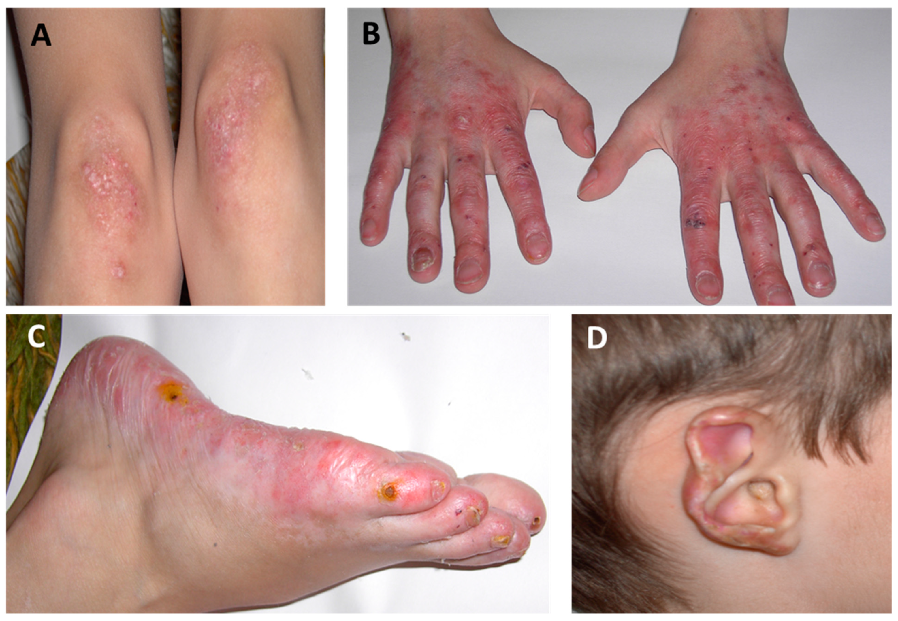
2.1. Cutaneous Manifestations

Inflammatory skin lesions typically present between birth and 6 months of age as cold-induced acral purplish red plaques, papules, and nodules. Erythematous rash of the cheeks, facial and/or acral telangiectasia, pustular lesions, chilblains of the feet, and digital edema are also characteristic. Progressively, cutaneous lesions lead to ulceration of the extremities, eschar formation, nasal and ear tissue loss with nasal septum perforation and disfiguring scarring of the ears, respectively, as well as nail dystrophy or loss and digital gangrene or autoamputation [3]. Cases of livedo reticularis, acral ischemia presenting as Raynaud’s phenomenon, and oral mucosal lesions (gingivostomatitis, aphtosis) have also been described. Paradoxically, a small percentage of patients never demonstrate cutaneous involvement [4].

2.2. Musculoskeletal Manifestations

Articular involvement in the form of polyarthritis is seen in almost one-third of SAVI patients and is positively associated with rheumatoid factor (RF). Interestingly, the course of the arthritis in SAVI can become destructive over time, especially during childhood [5]. Along with articular manifestations, episodes of myositis, and muscle atrophy have been reported [1]. Lastly, bone demineralization with decreased bone mineral density (BMD) and failure to thrive, as well as acro-osteolysis with bone resorption of distal phalanges, are also common findings among SAVI patients [4].

2.3. Pulmonary Manifestations

Pulmonary disease is present in all SAVI patients and is associated with increased disability and mortality. Signs and symptoms such as tachypnea, cough, and shortness of breath on exertion typically occur early in the course of the disease. The majority of patients develop ILD, which requires supplementary oxygen therapy and ultimately progresses to end-stage respiratory failure in adolescence or early adulthood if lung transplantation is not performed [6]. Respiratory findings may also include wheezing, lung fibrosis, paratracheal adenopathy, multifocal lymphoid formations, pulmonary hypertension, emphysema, obliterative bronchiolitis, and recurrent lung infections [7].

2.4. Pulmonary Function Tests (PFTs)

PFTs typically display a restrictive pattern with a reduction in total lung capacity (TLC) and a normal or slightly increased forced expiratory volume in the first second (FEV1) / forced vital capacity (FVC) ratio. However, a small number of patients also show features of pulmonary obstruction and/or hyperinflation [7]. Diffusing capacity for carbon monoxide (DLCO) is significantly decreased in most cases (below 80% of the predicted value). Additionally, the walking distance in the 6 min walking test is frequently reduced and is accompanied by a remarkable decrease in oxygen saturation. In most patients, when PFTs are performed later in the course of the disease, all functional parameters deteriorate [4].

2.5. High-Resolution Computed Tomography (HRCT)

Chest HRCT scanning can exhibit ground-glass and/or reticular opacities, areas of honeycombing, hyperinflation, cystic airspaces, traction bronchiectasis, and lung volume reduction, as well as enlarged hilar and mediastinal lymph nodes [2]. Compared to the lesions observed in ILD that are associated with connective tissue disorders, those in SAVI patients are frequently asymmetrical with findings of lung fibrosis being commonly present from an early age [4].

2.6. Laboratory Findings

Laboratory investigations exhibit elevated acute phase reactants including C-reactive protein (CRP) and/or erythrocyte sedimentation rate (ESR) as well as anemia of chronic disease. The counts of lymphocyte subsets, including CD3+, CD4+, and CD8+ positive, can be normal or mildly decreased. In addition, hypergammaglobulinemia with high IgG and IgA titers are frequently observed while IgM levels often vary [6]. Autoimmune workup in most patients reveals the presence of autoantibodies, mostly antineutrophil cytoplasmic autoantibodies (ANCA), antinuclear antibodies (ANA), antiphospholipid antibodies, and RF, while anti-double-stranded DNA (anti-dsDNA) and anticardiolipin antibodies as well as lupus anticoagulants can also be present [8].

2.7. Diagnostic Approach

In the presence of the aforementioned clinical symptoms and laboratory findings that are suggestive of SAVI, and after the exclusion of other more frequent alternative diagnoses (see “Differential diagnosis”), diagnostic evaluation and confirmation should focus on the measurement of peripheral blood IFN score and genetic testing [9]. The presence of persistently elevated IFN signature is a key finding in patients with type I interferonopathies including SAVI [10]. A peripheral blood IFN signature can be assessed using various methods. These methods include a 28-gene IFN scoring system utilizing NanoString technology or quantitative reverse transcriptase polymerase chain reaction to analyze specific gene products [11]. To establish chronic elevation, these measurements should be taken repeatedly over time [12]. One practical challenge is the limited availability of centers with the capability to assess IFN signatures. Consequently, a chronically elevated peripheral blood IFN signature is not an absolute requirement for diagnosis, but can be highly valuable in raising suspicion of interferonopathy [9]. Regarding genetic confirmation, the recommended approach for screening pathogenic variants is to utilize next-generation sequencing techniques, such as targeted gene panels, whole exome sequencing, or whole genome sequencing, rather than relying on single-gene Sanger sequencing [9]. Sanger sequencing of individual genes can still be a cost-effective option for patients with known familial disease, while in cases where next-generation sequencing is not readily available to the patient, it may be the only viable option [13]. However, it is important to note that this traditional “gene by gene” method is becoming outdated and could potentially lead to diagnostic delays [14]. If a patient with suggestive phenotypic features does not receive a molecular diagnosis following a routine genetic workup, referral to a specialized research center of excellence for additional evaluation and investigation should be considered [9]. Ultimately, having a high level of clinical suspicion in the early stages is essential as it can aid in a prompt diagnostic evaluation and disease confirmation, thereby enabling an early initiation of appropriate treatment.

3. Differential Diagnosis

3.1. Childhood Granulomatosis with Polyangiitis (GPA)

In SAVI cases, the association of cutaneous manifestations (Table 2) with later-developed ILD and subsequent nasal involvement can imply the presence of childhood GPA. However, several clinical, serological, and histopathological findings are incompatible with this diagnosis, including the rarity of neonatal-onset GPA, the absence of some distinctive skin lesions observed in childhood GPA (such as pyoderma gangrenosum-like ulcerations, erythema nodosum, and acneiform granulomatous folliculitis), the absence of granulomatous inflammation in histopathological skin lesions, and the negative titers of cytoplasmic ANCA (c-ANCA) in SAVI patients [15]. Nevertheless, serum ANCA is not always a reliable marker for the discrimination between SAVI and GPA, as positive c-ANCA titers have been described in some SAVI cases [8].

3.2. Cystic Fibrosis (CF)

The prominent lung manifestations in SAVI patients, with ILD progressing to end-stage respiratory failure in adolescence or early adulthood, can be similar in CF. CF is a monogenic disease caused by mutations in the cystic fibrosis transmembrane regulator (CFTR) gene, leading to impaired mucus hydration and clearance. Typically, CF manifests as chronic and recurrent pulmonary inflammation (e.g., Pseudomonas aeruginosa and Staphylococcus aureus infections), pancreatic exocrine insufficiency, male infertility, and is associated with comorbidities such as cystic fibrosis-related diabetes and CF-related liver disease [16]. Immunoreactive trypsinogen measurements, sweat chloride tests, and genetic tests are used for screening. The absence of CF-specific multiorgan manifestations combined with normal sweat chloride tests are useful diagnostic indicators for the discrimination of SAVI and CF [17].

3.3. Primary Immunodeficiency Diseases (PIDDs)

PIDDs are also characterized by early-onset pulmonary manifestations. Nevertheless, the respiratory findings in SAVI (predominantly ILD lesions) are relatively discordant with the pulmonary inflammation observed in PIDDs. PIDDs in children frequently manifest as respiratory infections (e.g., rhinosinusitis, otitis media, bronchitis, and pneumonia) with the associated complications (e.g., bronchiectasis, lung abscesses, empyema, and pneumatoceles), airway structural abnormalities (e.g., bronchial wall thickening) and lymphoproliferative diseases (e.g., lymphoma and lymphadenopathy) [18]. Furthermore, similarly to CF, most patients with PIDDs do not develop cutaneous vasculitic manifestations.

3.4. Aicardi-Goutières Syndrome (AGS)
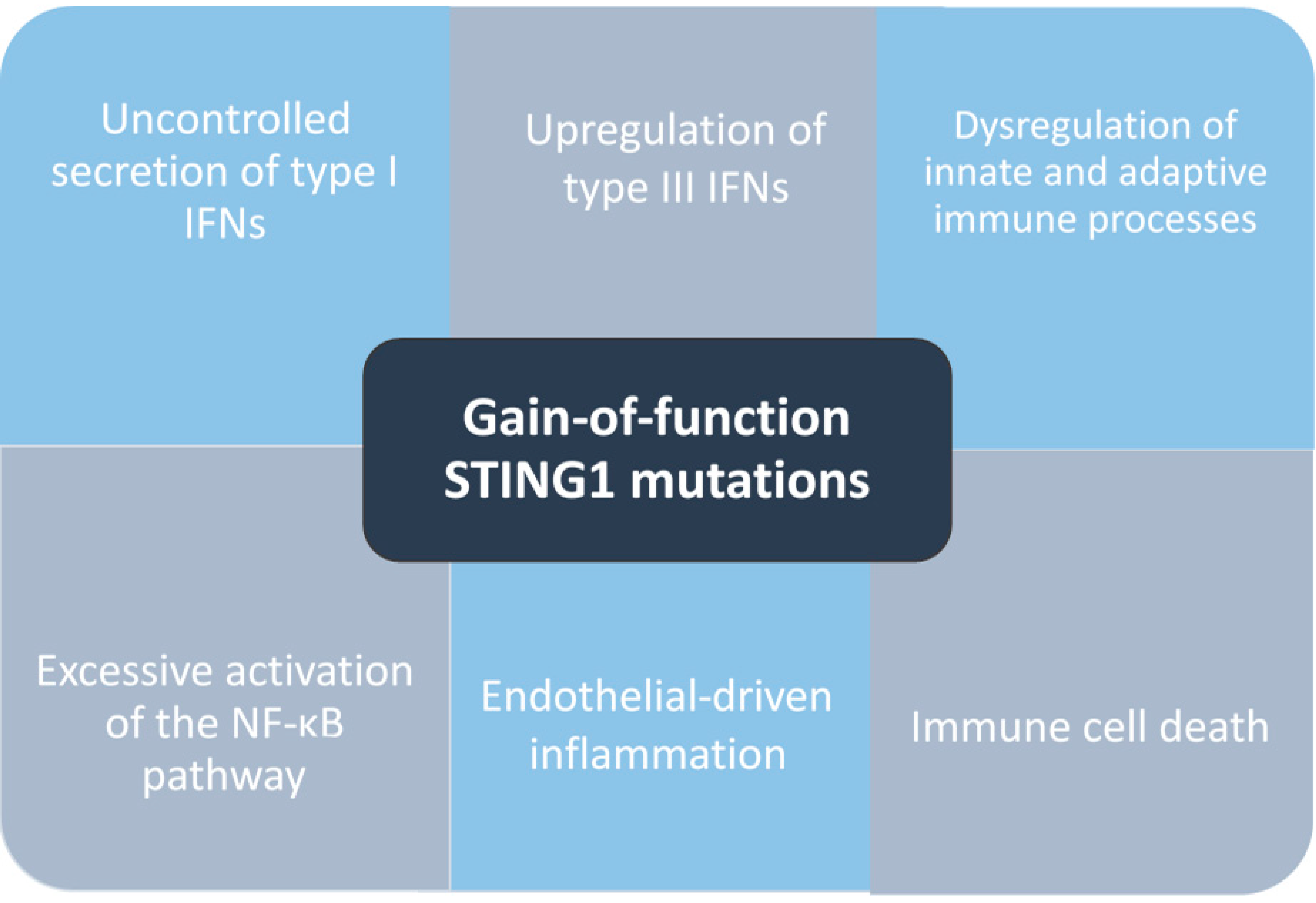
The presence of neurological manifestations include developmental delay, irritability, neurological impairment, dystonia, spasticity, focal motor findings, microcephaly, seizures, and the lack of ILD in AGS essentially differentiates AGS from SAVI. It is worth mentioning that, while SAVI is not typically associated with brain involvement, there have been rare reports of basal ganglia calcifications in SAVI patients [9].

4. Pathogenesis

The term interferonopathies was first introduced to describe and unify a group of rare disorders characterized by IFN pathway dysregulation [19,20,21]. IFNs are members of the class II cytokine family which also includes molecules related to interleukin-10 (IL-10). In humans, type I IFNs [IFN-α (13 subtypes), single IFN-β, IFN-ε, IFN-κ, and IFN-ω] play a critical role in the antiviral defense. These cytokines can be produced and secreted by almost all nucleated cells, mainly after activation of pattern recognition receptors (PRRs) by foreign or self-derived nucleic acids. Following secretion, they bind the IFN-α/β receptor (IFNAR) which is composed of two subunits, IFNAR1 and IFNAR2. Upon IFNAR activation, the intracellular JAK1 and tyrosine kinase 2 (TYK2) receptor autophosphorylate, resulting in transcription modulation of a variety of IFN-stimulated genes (ISGs) via the further activation of the signal transducers and activators of the transcription (STAT) pathway [22,23]. Although the term interferonopathies first entered the medical lexicon in 2011, disorders that are now classified as type I interferonopathies have attracted the interest of the scientific community for almost a century. They are characterized by significant pathogenetic and phenotypic diversity, with almost 20 different genes implicated in type I IFNs signaling dysregulation and the subsequent emergence of autoinflammation [24]. The prototypic disease with this aberrant type I IFN activation is AGS, a systemic inflammatory disorder with onset in early infancy. AGS predominantly arises as a leukoencephalopathy with basal ganglia calcifications white matter abnormalities and progressive brain atrophy, along with chronic lymphocytosis and elevated IFN-α in cerebrospinal fluid (CSF) [25].
SAVI is a monogenic autoinflammatory disorder caused by gain-of-function mutations in the STING1 gene (previously named TMEM173), encoding for the transmembrane protein, called STING [7]. SAVI, similarly to other interferonopathies, is characterized by the dysregulation of type I IFN pathway, resulting in uncontrolled type I IFN secretion [19,26]. STING is a PRR-sensing cytosolic double-strand DNA. Typically, STING activates through the binding of cyclic GMP-AMP (cGAMP), which is produced by cGAMP synthase (cGAS), once cGAS binds to DNA of microbial or host origin. Consequently, activated STING transmigrates from the endoplasmic reticulum (ER) membrane to the Golgi system, where it activates the IκB kinase-related kinases (IKK),serine/threonine-protein kinase named TANK-binding kinase 1 (TBK1), and the interferon regulatory factor 3 (IRF3). The latter leads to the overexpression of ISGs resulting in the production of type I interferons [20,27]. The normal regulation of STING trafficking involves retrograde transport to the ER through coatomer-associated protein subunit alpha (COPA) and transportation to the autophagosome, where subsequent degradation occurs.
The most common STING1 variants (V155M, N154S, and V147M) are located in the same mutation cluster (connector helix loop) and are thought to trigger the constitutive activation of STING via inducing a 180° rotation in the ligand-binding domain that consequently leads to STING1 oligomerization [28]. This activation process occurs independently of any interaction with its ligand cGAMP. Other substitutions within the second mutation cluster (C206, R281, and R284) are located in the polymerization interface and are assumed to inhibit the auto-suppression of STING oligomerization [28]. STING1 mutations (Table 3) result in a constitutively active STING protein, with the uncontrolled secretion and activation of type I IFN through the cGAS-STING- TBK1-IRF3 pathway [1,21]. In SAVI, there is no apparent genotype-phenotype association, although some differences in clinical presentation have been described between patients with mutations occurring in exon 5 (first mutation cluster) and those in exons 6 and 7 (second mutation cluster) [4]. Nearly 60% of documented cases have either the heterozygous p.V155M or p.N154S substitution, with two distinct family lineages, each carrying a different substitution, accounting for 16% of the reported cases, thus limiting the ability to establish substantial genotype-phenotype associations [2]. In a recent systematic review, Dai et al. compared the clinical features of patients with p.N154S and p.V155M mutations with the following results: Patients with the p.N154S mutation exhibited earlier disease onset and more severe cutaneous lesions compared to those with the p.V155M mutation, while there was no significant difference in respiratory manifestations [29].
An analysis of the transcriptome in the whole blood of three SAVI patients, contrasted with healthy controls, revealed the differential expression of 119 genes. These genes primarily included ISGs, along with genes participating in various other innate and adaptive immune functions [36]. Apart from type I interferons, the potential pathogenic role of type III IFNs (IFN-λ1-4) is also considered in SAVI. STING has been identified as a driver of IFN-λ1 production in human cells upon detecting viral DNA, while secretion of IFN-λs upon viral infection is a characteristic response of respiratory epithelial cells [52,53]. Furthermore, gain-of-function mutations in STING1 have been linked to the excessive activation of the nuclear factor kappa B (NF-κB) pathway and the autophagy process. This is attributed to the reported increase in NF-κB-related proteins (e.g., IL-6) and the observed T-cell deficiency in the whole blood of SAVI patients [7]. Within the same context, it is known that severe ER stress in SAVI T lymphocytes can result in programmed cell death and peripheral lymphopenia [54]. Another significant observation in SAVI mice is the lack of lymph nodes, along with the absence of Innate Lymphoid Cell 3 (ILC3) and lymphotoxin inducer cells. This points toward the possibility that lymphatic dysregulation could contribute to the development of lung disease [55]. However, it is important to note that while ILC3 deficiency is also observed in SAVI patients, the absence of lymph nodes is not, indicating that this effect is not responsible for pulmonary disease in humans [56]. Lastly, single-cell RNA sequencing analysis of lung tissue indicates that endothelial cells exhibit elevated expression levels of STING [57,58], while in bulk RNA sequencing of SAVI lung endothelial cells, an increase in the expression of inflammatory genes is observed [59]. These findings suggest that endothelial-driven inflammation may be responsible for lung pathology in SAVI [56] (Figure 2).

5. Therapeutic Strategy

Type I interferonopathies, and therefore SAVI, are associated with high morbidity and mortality, while conventional immunosuppressive treatments have demonstrated minimal efficacy. Considering the well-established association between aberrant activation of the JAK kinases pathway (including JAK1-3 and TYK2) and type I interferon-related disorders development, the inhibition of the JAK/STAT axis represents a promising therapeutic approach.
JAK inhibitors block downstream signaling of several cytokine receptors, including IFN receptors; however, it is not yet proven that the blockade of IFN signaling is the principal responsible mechanism for their efficacy [17]. Regardless of the underlying mechanism, JAK inhibitors are reported to be beneficial in the control of inflammatory responses and in hampering the progression of end-organ damage in type I Interferonopathies [9].
Ruxolitinib is a selective JAK1/JAK2 inhibitor (Table 4) achieving remarkable improvement in patient-reported well-being and clinical pictures. Thus, the rapid clearance of fever, fewer vasculitis flares, the almost complete resolution of skin manifestations, the prevention of spontaneous amputations/the development of gangrene, and amelioration of ILD by preserving pulmonary function suggest a potentially beneficial role for SAVI patients [36,54]. Interestingly, these improvements were not associated with marked ISG downregulation, further enhancing the hypothesis that the beneficial effects of ruxolitinib are mediated mainly by IL-6, IL12/23, and IFNγ suppression [33].
Similar promising results on the associated systemic, cutaneous, and, to some extent, pulmonary manifestations were described with other JAK inhibitors, such as tofacitinib and baricitinib. Tofacitinib preferentially inhibits JAK1 and JAK3, with a minor affinity for JAK2 and TYK2 (Table 4). Through JAK1/JAK3 inhibition tofacitinib downregulates IL-2, IL-4, IL-7 signaling, while through JAK-1/JAK2 interaction blocks IL-6 and IFNγ signaling [60]. The use of tofacitinib for treating SAVI is limited. Several recent reports have indicated that tofacitinib demonstrated robust suppression of IFN levels and led to improvements in acral ischemia and skin lesions [44,49]. Similarly in a recent case report, tofacitinib improved the cutaneous manifestations and ILD within 3 months, while recurrent oral ulcers and growth retardation remained [61]. However, two further studies failed to demonstrate the beneficial effects of tofacitinib in disease progression or pulmonary disease [37,62]. Baricitinib preferentially inhibits JAK1 and JAK2, showing a much lesser affinity for JAK3 and TYK2 [63,64], with promising results demonstrated in case series of pediatric patients with several autoinflammatory diseases, including SAVI [34,65,66,67].
However, severe lung involvement often exhibits limited treatment response and can eventually result in advanced respiratory failure, necessitating the use of oxygen therapy, non-invasive ventilation, and, in some cases, lung transplantation. In the literature, four SAVI patients have undergone lung transplantation. Three of those patients died (immediately, one year, and eight years after transplantation), while the fourth was still alive two years post-transplantation [4,5]. These data regarding severe fibrotic lung disease in SAVI patients highlight the significance of early diagnosis and intervention, along with the pressing need to explore alternative novel therapeutic approaches [23].

6. Conclusions

SAVI is a monogenic autoinflammatory disorder caused by a gain-of-function mutation in the STING-1 gene. The disease provides a unique model to investigate the process of nucleic acid sensing through STING and its association with human pathophysiology. While type I IFNs play a significant role in the disease pathogenesis, given the substantial clinical improvement with JAK inhibitors, the exact pathogenesis of SAVI is not fully understood. SAVI was first identified in patients exhibiting early-onset cutaneous vasculopathy, ILD, and prominent systemic features. Recent cases have highlighted a broader range of clinical manifestations with multiple organ involvement. Interestingly, even in the presence of identical mutations, there is considerable variability observed between affected patients mandating a thorough differential diagnosis necessary to minimize misdiagnosis. Despite this wide range of clinical presentations, certain red flags that should raise clinical suspicion for SAVI include early-onset (neonatal or infantile) recurrent fevers, failure to thrive, respiratory symptoms, polyarthralgia, and vasculopathic skin lesions. These symptoms, in combination with minimal response to conventional immunosuppressive treatments and chronically elevated IFN levels, are indicative of SAVI. JAK inhibitors are the current treatment of choice, but severe lung involvement frequently displays limited response to treatment. Enhancing our understanding of disease pathogenesis and elucidating distinct dysregulated immune system pathways, along with a collaborative approach and extensive patient registries, will be instrumental in developing effective treatment strategies, ultimately improving outcomes in SAVI patients.

Author Contributions

Study design (K.D., C.P.M., R.S. and C.S.); Analysis and interpretation of data (K.D. and R.S.); Drafting/revising (K.D., R.S., C.S. and C.P.M.), Final approval of the last version (C.P.M., R.S., C.S. and K.D.). All authors have read and agreed to the published version of the manuscript.

Funding

This research did not receive any specific grant from any funding agency in the public, commercial, or not-for-profit sectors.

Data Availability Statement

The authors alone are responsible for the content and writing of this article.

Conflicts of Interest

The authors declare no conflict of interest.

References

  1. Liu, Y.; Jesus, A.A.; Marrero, B.; Yang, D.; Ramsey, S.E.; Montealegre Sanchez, G.A.; Tenbrock, K.; Wittkowski, H.; Jones, O.Y.; Kuehn, H.S.; et al. Activated STING in a Vascular and Pulmonary Syndrome. N. Engl. J. Med. 2014, 371, 507–518. [Google Scholar] [CrossRef] [PubMed]
  2. Frémond, M.-L.; Crow, Y.J. STING-Mediated Lung Inflammation and Beyond. J. Clin. Immunol. 2021, 41, 501–514. [Google Scholar] [CrossRef] [PubMed]
  3. Chia, J.; Eroglu, F.K.; Özen, S.; Orhan, D.; Montealegre-Sanchez, G.; De Jesus, A.A.; Goldbach-Mansky, R.; Cowen, E.W. Failure to Thrive, Interstitial Lung Disease, and Progressive Digital Necrosis with Onset in Infancy. J. Am. Acad. Dermatol. 2016, 74, 186–189. [Google Scholar] [CrossRef] [PubMed]
  4. Frémond, M.-L.; Hadchouel, A.; Berteloot, L.; Melki, I.; Bresson, V.; Barnabei, L.; Jeremiah, N.; Belot, A.; Bondet, V.; Brocq, O.; et al. Overview of STING-Associated Vasculopathy with Onset in Infancy (SAVI) Among 21 Patients. J. Allergy Clin. Immunol. Pract. 2021, 9, 803–818.e11. [Google Scholar] [CrossRef] [PubMed]
  5. Clarke, S.L.N.; Robertson, L.; Rice, G.I.; Seabra, L.; Hilliard, T.N.; Crow, Y.J.; Ramanan, A.V. Type 1 Interferonopathy Presenting as Juvenile Idiopathic Arthritis with Interstitial Lung Disease: Report of a New Phenotype. Pediatr. Rheumatol. 2020, 18, 37. [Google Scholar] [CrossRef]
  6. Wang, Y.; Wang, F.; Zhang, X. STING-Associated Vasculopathy with Onset in Infancy: A Familial Case Series Report and Literature Review. Ann. Transl. Med. 2021, 9, 176. [Google Scholar] [CrossRef] [PubMed]
  7. David, C.; Frémond, M.-L. Lung Inflammation in STING-Associated Vasculopathy with Onset in Infancy (SAVI). Cells 2022, 11, 318. [Google Scholar] [CrossRef]
  8. Staels, F.; Betrains, A.; Doubel, P.; Willemsen, M.; Cleemput, V.; Vanderschueren, S.; Corveleyn, A.; Meyts, I.; Sprangers, B.; Crow, Y.J.; et al. Adult-Onset ANCA-Associated Vasculitis in SAVI: Extension of the Phenotypic Spectrum, Case Report and Review of the Literature. Front. Immunol. 2020, 11, 575219. [Google Scholar] [CrossRef]
  9. Cetin Gedik, K.; Lamot, L.; Romano, M.; Demirkaya, E.; Piskin, D.; Torreggiani, S.; Adang, L.A.; Armangue, T.; Barchus, K.; Cordova, D.R.; et al. The 2021 European Alliance of Associations for Rheumatology/American College of Rheumatology Points to Consider for Diagnosis and Management of Autoinflammatory Type I Interferonopathies: CANDLE/PRAAS, SAVI and AGS. Ann. Rheum. Dis. 2022, 81, 601–613. [Google Scholar] [CrossRef]
  10. Volpi, S.; Picco, P.; Caorsi, R.; Candotti, F.; Gattorno, M. Type I Interferonopathies in Pediatric Rheumatology. Pediatr. Rheumatol. 2016, 14, 35. [Google Scholar] [CrossRef]
  11. Rice, G.I.; Melki, I.; Frémond, M.-L.; Briggs, T.A.; Rodero, M.P.; Kitabayashi, N.; Oojageer, A.; Bader-Meunier, B.; Belot, A.; Bodemer, C.; et al. Assessment of Type I Interferon Signaling in Pediatric Inflammatory Disease. J. Clin. Immunol. 2017, 37, 123–132. [Google Scholar] [CrossRef] [PubMed]
  12. Kim, H.; De Jesus, A.A.; Brooks, S.R.; Liu, Y.; Huang, Y.; VanTries, R.; Montealegre Sanchez, G.A.; Rotman, Y.; Gadina, M.; Goldbach-Mansky, R. Development of a Validated Interferon Score Using NanoString Technology. J. Interferon Cytokine Res. 2018, 38, 171–185. [Google Scholar] [CrossRef] [PubMed]
  13. Rama, M.; Mura, T.; Kone-Paut, I.; Boursier, G.; Aouinti, S.; Touitou, I.; Sarrabay, G. Is Gene Panel Sequencing More Efficient than Clinical-Based Gene Sequencing to Diagnose Autoinflammatory Diseases? A Randomized Study. Clin. Exp. Immunol. 2020, 203, 105–114. [Google Scholar] [CrossRef] [PubMed]
  14. Omoyinmi, E.; Standing, A.; Keylock, A.; Price-Kuehne, F.; Melo Gomes, S.; Rowczenio, D.; Nanthapisal, S.; Cullup, T.; Nyanhete, R.; Ashton, E.; et al. Clinical Impact of a Targeted Next-Generation Sequencing Gene Panel for Autoinflammation and Vasculitis. PLOS ONE 2017, 12, e0181874. [Google Scholar] [CrossRef]
  15. Wright, A.C.; Gibson, L.E.; Davis, D.M.R. Cutaneous Manifestations of Pediatric Granulomatosis with Polyangiitis: A Clinicopathologic and Immunopathologic Analysis. J. Am. Acad. Dermatol. 2015, 72, 859–867. [Google Scholar] [CrossRef]
  16. Shteinberg, M.; Haq, I.J.; Polineni, D.; Davies, J.C. Cystic Fibrosis. Lancet 2021, 397, 2195–2211. [Google Scholar] [CrossRef]
  17. Cao, Y.; Jiang, L. The Challenge of Diagnosing SAVI: Case Studies. Pediatr. Allergy Immunol. Pulmonol. 2019, 32, 167–172. [Google Scholar] [CrossRef]
  18. Jesenak, M.; Banovcin, P.; Jesenakova, B.; Babusikova, E. Pulmonary Manifestations of Primary Immunodeficiency Disorders in Children. Front. Pediatr. 2014, 2, 77. [Google Scholar] [CrossRef]
  19. Crow, Y.J. Type I Interferonopathies: A Novel Set of Inborn Errors of Immunity. Ann. N. Y. Acad. Sci. 2011, 1238, 91–98. [Google Scholar] [CrossRef]
  20. Savic, S.; Caseley, E.A.; McDermott, M.F. Moving towards a Systems-Based Classification of Innate Immune-Mediated Diseases. Nat. Rev. Rheumatol. 2020, 16, 222–237. [Google Scholar] [CrossRef]
  21. Skarlis, C.; Mavragani, C.P. Immune Dysfunction and Drug Targets in Autoinflammatory Syndromes. In Comprehensive Pharmacology; Elsevier: Amsterdam, The Netherlands, 2022; pp. 479–491. ISBN 978-0-12-820876-2. [Google Scholar]
  22. Platanias, L.C. Mechanisms of Type-I- and Type-II-Interferon-Mediated Signalling. Nat. Rev. Immunol. 2005, 5, 375–386. [Google Scholar] [CrossRef] [PubMed]
  23. Papadopoulos, V.E.; Skarlis, C.; Evangelopoulos, M.-E.; Mavragani, C.P. Type I Interferon Detection in Autoimmune Diseases: Challenges and Clinical Applications. Expert Rev. Clin. Immunol. 2021, 17, 883–903. [Google Scholar] [CrossRef] [PubMed]
  24. Davidson, S.; Steiner, A.; Harapas, C.R.; Masters, S.L. An Update on Autoinflammatory Diseases: Interferonopathies. Curr. Rheumatol. Rep. 2018, 20, 38. [Google Scholar] [CrossRef] [PubMed]
  25. Crow, Y.J.; Manel, N. Aicardi–Goutières Syndrome and the Type I Interferonopathies. Nat. Rev. Immunol. 2015, 15, 429–440. [Google Scholar] [CrossRef]
  26. Georgin-Lavialle, S.; Fayand, A.; Rodrigues, F.; Bachmeyer, C.; Savey, L.; Grateau, G. Autoinflammatory Diseases: State of the Art. Presse Médicale 2019, 48, e25–e48. [Google Scholar] [CrossRef]
  27. Ablasser, A.; Chen, Z.J. CGAS in Action: Expanding Roles in Immunity and Inflammation. Science 2019, 363, eaat8657. [Google Scholar] [CrossRef]
  28. Shang, G.; Zhang, C.; Chen, Z.J.; Bai, X.; Zhang, X. Cryo-EM Structures of STING Reveal Its Mechanism of Activation by Cyclic GMP–AMP. Nature 2019, 567, 389–393. [Google Scholar] [CrossRef]
  29. Dai, Y.; Liu, X.; Zhao, Z.; He, J.; Yin, Q. Stimulator of Interferon Genes-Associated Vasculopathy With Onset in Infancy: A Systematic Review of Case Reports. Front. Pediatr. 2020, 8, 577918. [Google Scholar] [CrossRef]
  30. Lin, B.; Torreggiani, S.; Kahle, D.; Rumsey, D.G.; Wright, B.L.; Montes-Cano, M.A.; Silveira, L.F.; Alehashemi, S.; Mitchell, J.; Aue, A.G.; et al. Case Report: Novel SAVI-Causing Variants in STING1 Expand the Clinical Disease Spectrum and Suggest a Refined Model of STING Activation. Front. Immunol. 2021, 12, 636225. [Google Scholar] [CrossRef]
  31. Abid, Q.; Best Rocha, A.; Larsen, C.P.; Schulert, G.; Marsh, R.; Yasin, S.; Patty-Resk, C.; Valentini, R.P.; Adams, M.; Baracco, R. APOL1-Associated Collapsing Focal Segmental Glomerulosclerosis in a Patient with Stimulator of Interferon Genes (STING)-Associated Vasculopathy with Onset in Infancy (SAVI). Am. J. Kidney Dis. 2020, 75, 287–290. [Google Scholar] [CrossRef]
  32. Munoz, J.; Rodière, M.; Jeremiah, N.; Rieux-Laucat, F.; Oojageer, A.; Rice, G.I.; Rozenberg, F.; Crow, Y.J.; Bessis, D. Stimulator of Interferon Genes–Associated Vasculopathy with Onset in Infancy: A Mimic of Childhood Granulomatosis with Polyangiitis. JAMA Dermatol. 2015, 151, 872. [Google Scholar] [CrossRef] [PubMed]
  33. Volpi, S.; Insalaco, A.; Caorsi, R.; Santori, E.; Messia, V.; Sacco, O.; Terheggen-Lagro, S.; Cardinale, F.; Scarselli, A.; Pastorino, C.; et al. Efficacy and Adverse Events During Janus Kinase Inhibitor Treatment of SAVI Syndrome. J. Clin. Immunol. 2019, 39, 476–485. [Google Scholar] [CrossRef] [PubMed]
  34. Balci, S.; Ekinci, R.M.K.; De Jesus, A.A.; Goldbach-Mansky, R.; Yilmaz, M. Baricitinib Experience on STING-Associated Vasculopathy with Onset in Infancy: A Representative Case from Turkey. Clin. Immunol. 2020, 212, 108273. [Google Scholar] [CrossRef]
  35. Shoman, W.; El Chazli, Y.; ElSawy, I.; Aróstegui, J. First Egyptian Patient with STING-Associated Vasculopathy with Onset in Infancy. Scand. J. Rheumatol. 2019, 48, 338–339. [Google Scholar] [CrossRef]
  36. Frémond, M.-L.; Rodero, M.P.; Jeremiah, N.; Belot, A.; Jeziorski, E.; Duffy, D.; Bessis, D.; Cros, G.; Rice, G.I.; Charbit, B.; et al. Efficacy of the Janus Kinase 1/2 Inhibitor Ruxolitinib in the Treatment of Vasculopathy Associated with TMEM173 -Activating Mutations in 3 Children. J. Allergy Clin. Immunol. 2016, 138, 1752–1755. [Google Scholar] [CrossRef] [PubMed]
  37. Tang, X.; Xu, H.; Zhou, C.; Peng, Y.; Liu, H.; Liu, J.; Li, H.; Yang, H.; Zhao, S. STING-Associated Vasculopathy with Onset in Infancy in Three Children with New Clinical Aspect and Unsatisfactory Therapeutic Responses to Tofacitinib. J. Clin. Immunol. 2020, 40, 114–122. [Google Scholar] [CrossRef]
  38. Jeremiah, N.; Neven, B.; Gentili, M.; Callebaut, I.; Maschalidi, S.; Stolzenberg, M.-C.; Goudin, N.; Frémond, M.-L.; Nitschke, P.; Molina, T.J.; et al. Inherited STING-Activating Mutation Underlies a Familial Inflammatory Syndrome with Lupus-like Manifestations. J. Clin. Investig. 2014, 124, 5516–5520. [Google Scholar] [CrossRef]
  39. Caorsi, R.; Rice, G.; Cardinale, F.; Volpi, S.; Buoncompagni, A.; Crow, Y.; Martini, A.; Gattorno, M.; Picco, P. AB1014 Enlarging the Clinical Spectrum of Sting-Associated Vasculopathy with Onset in Infancy (SAVI). Ann. Rheum. Dis. 2015, 74, 1237–1238. [Google Scholar] [CrossRef]
  40. Clarke, S.L.N.; Pellowe, E.J.; De Jesus, A.A.; Goldbach-Mansky, R.; Hilliard, T.N.; Ramanan, A.V. Interstitial Lung Disease Caused by STING-Associated Vasculopathy with Onset in Infancy. Am. J. Respir. Crit. Care Med. 2016, 194, 639–642. [Google Scholar] [CrossRef]
  41. Picard, C.; Thouvenin, G.; Kannengiesser, C.; Dubus, J.-C.; Jeremiah, N.; Rieux-Laucat, F.; Crestani, B.; Belot, A.; Thivolet-Béjui, F.; Secq, V.; et al. Severe Pulmonary Fibrosis as the First Manifestation of Interferonopathy (TMEM173 Mutation). Chest 2016, 150, e65–e71. [Google Scholar] [CrossRef]
  42. Dagher, R.; Ghiye, R.; Nicolas, G.; Feghali, H.; Khalife, M.F.; Seabra, L.; Crow, Y. THU0528 Sting-Associated Vasculopathy with Onset in Infancy (SAVI): A Differential Diagnosis of Inflammatory Interstitial Lung Disease. In Proceedings of the Poster Presentations; BMJ Publishing Group Ltd and European League Against Rheumatism: London, UK, 2017; p. 406. [Google Scholar]
  43. Raffaele, C.G.L.; Messia, V.; Moneta, G.; Caiello, I.; Federici, S.; Pardeo, M.; Bracaglia, C.; De Benedetti, F.; Insalaco, A. A Patient with Stimulator of Interferon Genes–Associated Vasculopathy with Onset in Infancy without Skin Vasculopathy. Rheumatology 2020, 59, 905–907. [Google Scholar] [CrossRef] [PubMed]
  44. König, N.; Fiehn, C.; Wolf, C.; Schuster, M.; Cura Costa, E.; Tüngler, V.; Alvarez, H.A.; Chara, O.; Engel, K.; Goldbach-Mansky, R.; et al. Familial Chilblain Lupus Due to a Gain-of-Function Mutation in STING. Ann. Rheum. Dis. 2017, 76, 468–472. [Google Scholar] [CrossRef] [PubMed]
  45. Manoussakis, M.N.; Mavragani, C.P.; Nezos, A.; Zampeli, E.; Germenis, A.; Moutsopoulos, H.M. Type I Interferonopathy in a Young Adult. Rheumatology 2017, 56, 2241–2243. [Google Scholar] [CrossRef]
  46. Melki, I.; Rose, Y.; Uggenti, C.; Van Eyck, L.; Frémond, M.-L.; Kitabayashi, N.; Rice, G.I.; Jenkinson, E.M.; Boulai, A.; Jeremiah, N.; et al. Disease-Associated Mutations Identify a Novel Region in Human STING Necessary for the Control of Type I Interferon Signaling. J. Allergy Clin. Immunol. 2017, 140, 543–552.e5. [Google Scholar] [CrossRef] [PubMed]
  47. Gallagher, K.; Brogan, P.; Burrows, N.; Gass, J.; Bale, P.; Armon, K. P04 STING: Associated Vasculopathy with Onset in Infancy (SAVI). Rheumatology 2018, 57. [Google Scholar] [CrossRef]
  48. Keskitalo, S.; Haapaniemi, E.; Einarsdottir, E.; Rajamäki, K.; Heikkilä, H.; Ilander, M.; Pöyhönen, M.; Morgunova, E.; Hokynar, K.; Lagström, S.; et al. Novel TMEM173 Mutation and the Role of Disease Modifying Alleles. Front. Immunol. 2019, 10, 2770. [Google Scholar] [CrossRef] [PubMed]
  49. Seo, J.; Kang, J.-A.; Suh, D.I.; Park, E.-B.; Lee, C.-R.; Choi, S.A.; Kim, S.Y.; Kim, Y.; Park, S.-H.; Ye, M.; et al. Tofacitinib Relieves Symptoms of Stimulator of Interferon Genes (STING)–Associated Vasculopathy with Onset in Infancy Caused by 2 de Novo Variants in TMEM173. J. Allergy Clin. Immunol. 2017, 139, 1396–1399.e12. [Google Scholar] [CrossRef]
  50. Lin, B.; Berard, R.; Al Rasheed, A.; Aladba, B.; Kranzusch, P.J.; Henderlight, M.; Grom, A.; Kahle, D.; Torreggiani, S.; Aue, A.G.; et al. A Novel STING1 Variant Causes a Recessive Form of STING-Associated Vasculopathy with Onset in Infancy (SAVI). J. Allergy Clin. Immunol. 2020, 146, 1204–1208.e6. [Google Scholar] [CrossRef]
  51. Saldanha, R.G.; Balka, K.R.; Davidson, S.; Wainstein, B.K.; Wong, M.; Macintosh, R.; Loo, C.K.C.; Weber, M.A.; Kamath, V.; CIRCA; et al. A Mutation Outside the Dimerization Domain Causing Atypical STING-Associated Vasculopathy With Onset in Infancy. Front. Immunol. 2018, 9, 1535. [Google Scholar] [CrossRef]
  52. Sui, H.; Zhou, M.; Imamichi, H.; Jiao, X.; Sherman, B.T.; Lane, H.C.; Imamichi, T. STING Is an Essential Mediator of the Ku70-Mediated Production of IFN-Λ1 in Response to Exogenous DNA. Sci. Signal. 2017, 10, eaah5054. [Google Scholar] [CrossRef]
  53. Mahlakõiv, T.; Hernandez, P.; Gronke, K.; Diefenbach, A.; Staeheli, P. Leukocyte-Derived IFN-α/β and Epithelial IFN-λ Constitute a Compartmentalized Mucosal Defense System That Restricts Enteric Virus Infections. PLoS Pathog. 2015, 11, e1004782. [Google Scholar] [CrossRef] [PubMed]
  54. Wu, J.; Chen, Y.-J.; Dobbs, N.; Sakai, T.; Liou, J.; Miner, J.J.; Yan, N. STING-Mediated Disruption of Calcium Homeostasis Chronically Activates ER Stress and Primes T Cell Death. J. Exp. Med. 2019, 216, 867–883. [Google Scholar] [CrossRef] [PubMed]
  55. Bennion, B.G.; Croft, C.A.; Ai, T.L.; Qian, W.; Menos, A.M.; Miner, C.A.; Frémond, M.-L.; Doisne, J.-M.; Andhey, P.S.; Platt, D.J.; et al. STING Gain-of-Function Disrupts Lymph Node Organogenesis and Innate Lymphoid Cell Development in Mice. Cell Rep. 2020, 31, 107771. [Google Scholar] [CrossRef] [PubMed]
  56. Gao, K.M.; Marshak-Rothstein, A.; Fitzgerald, K.A. Type-1 Interferon-Dependent and -Independent Mechanisms in Cyclic GMP–AMP Synthase–Stimulator of Interferon Genes-Driven Auto-Inflammation. Curr. Opin. Immunol. 2023, 80, 102280. [Google Scholar] [CrossRef]
  57. The Tabula Muris Consortium; Overall coordination; Logistical coordination; Organ collection and processing; Library preparation and sequencing; Computational data analysis; Cell type annotation; Writing group; Supplemental text writing group; Principal investigators. Single-Cell Transcriptomics of 20 Mouse Organs Creates a Tabula Muris. Nature 2018, 562, 367–372. [Google Scholar] [CrossRef]
  58. Digre, A.; Lindskog, C. The Human Protein Atlas—Spatial Localization of the Human Proteome in Health and Disease. Protein Sci. 2021, 30, 218–233. [Google Scholar] [CrossRef]
  59. Gao, K.M.; Motwani, M.; Tedder, T.; Marshak-Rothstein, A.; Fitzgerald, K.A. Radioresistant Cells Initiate Lymphocyte-Dependent Lung Inflammation and IFNγ-Dependent Mortality in STING Gain-of-Function Mice. Proc. Natl. Acad. Sci. USA 2022, 119, e2202327119. [Google Scholar] [CrossRef]
  60. Bechman, K.; Yates, M.; Galloway, J.B. The New Entries in the Therapeutic Armamentarium: The Small Molecule JAK Inhibitors. Pharmacol. Res. 2019, 147, 104392. [Google Scholar] [CrossRef]
  61. Shen, D.; Fan, X.; Zhou, Q.; Xu, X.; Lu, M. Use of Tofacitinib for Infant-Onset STING-Associated Vasculopathy: A Case Report from China. Medicine 2022, 101, e31832. [Google Scholar] [CrossRef]
  62. Li, W.; Wang, W.; Wang, W.; Zhong, L.; Gou, L.; Wang, C.; Ma, J.; Quan, M.; Jian, S.; Tang, X.; et al. Janus Kinase Inhibitors in the Treatment of Type I Interferonopathies: A Case Series From a Single Center in China. Front. Immunol. 2022, 13, 825367. [Google Scholar] [CrossRef]
  63. Clark, J.D.; Flanagan, M.E.; Telliez, J.-B. Discovery and Development of Janus Kinase (JAK) Inhibitors for Inflammatory Diseases: Miniperspective. J. Med. Chem. 2014, 57, 5023–5038. [Google Scholar] [CrossRef] [PubMed]
  64. Hadjadj, J.; Frémond, M.-L.; Neven, B. Emerging Place of JAK Inhibitors in the Treatment of Inborn Errors of Immunity. Front. Immunol. 2021, 12, 717388. [Google Scholar] [CrossRef]
  65. Kim, H.; Brooks, K.M.; Tang, C.C.; Wakim, P.; Blake, M.; Brooks, S.R.; Montealegre Sanchez, G.A.; De Jesus, A.A.; Huang, Y.; Tsai, W.L.; et al. Pharmacokinetics, Pharmacodynamics, and Proposed Dosing of the Oral JAK1 and JAK2 Inhibitor Baricitinib in Pediatric and Young Adult CANDLE and SAVI Patients. Clin. Pharmacol. Ther. 2018, 104, 364–373. [Google Scholar] [CrossRef] [PubMed]
  66. Sanchez, G.A.M.; Reinhardt, A.; Ramsey, S.; Wittkowski, H.; Hashkes, P.J.; Berkun, Y.; Schalm, S.; Murias, S.; Dare, J.A.; Brown, D.; et al. JAK1/2 Inhibition with Baricitinib in the Treatment of Autoinflammatory Interferonopathies. J. Clin. Investig. 2018, 128, 3041–3052. [Google Scholar] [CrossRef] [PubMed]
  67. Kanazawa, N.; Ishii, T.; Takita, Y.; Nishikawa, A.; Nishikomori, R. Efficacy and Safety of Baricitinib in Japanese Patients with Autoinflammatory Type I Interferonopathies (NNS/CANDLE, SAVI, And AGS). Pediatr. Rheumatol. 2023, 21, 38. [Google Scholar] [CrossRef]
Figure 1. Clinical manifestations of SAVI. (A) Psoriasiform dermatitis on the knees. (B,C) Acral violaceous patches and plaques with accompanying nail dystrophy/loss. (D) Deforming fibrotic lesion of the ear’s pinnae.
Jmp 04 00024 g001
Figure 2. Main pathophysiologic effects driven by gain-of-function STING1 mutations in SAVI. Stimulator of interferon genes 1: STING1; STING-associated vasculopathy with onset in infancy: SAVI; Interferons: IFNs; Nuclear factor kappa B: NF-Κβ.
Jmp 04 00024 g002
Table 1. The main clinical manifestations in STING-associated vasculopathy with onset in infancy.
Systemic ManifestationsCutaneous ManifestationsMusculoskeletal ManifestationsPulmonary Manifestations
Neonatal-onset systemic inflammation

Growth retardation (failure to thrive)

Recurrent febrile episodes
Acral violaceous patches and plaques (cold-sensitive areas)

Violaceous/erythematous rash

Telangiectasia

Chilblains

Ulcerations

Tissue loss/skin necrosis
Nasal septum perforation
Disfiguring scarring of the ears
Digital gangrene/autoamputation
Polyarthralgia/Polyarthritis

Myositis/Muscle atrophy

Acro-osteolysis of distal phalanges

Bone demineralization
Interstitial lung disease

Pulmonary fibrosis

Pulmonary hypertension

Paratracheal adenopathy

Emphysema

Obliterative bronchiolitis

Recurrent lung infections
Table 2. The main differential diagnosis for STING-associated vasculopathy with onset in infancy.
Childhood GPACFPIDDsAGS
Overlapping CharacteristicsDifferentiating FactorsOverlapping CharacteristicsDifferentiating FactorsOverlapping CharacteristicsDifferentiating FactorsOverlapping CharacteristicsDifferentiating Factors
Interstitial lung disease

Cutaneous manifestations
Reddish purple patches and plaques
Erythematous/Purpuric rash
Ulcerations
Nasal involvement
Childhood GPA-specific skin lesions
Pyoderma gangrenosum-like ulcerations
Erythema nodosum
Acneiform granulomatous folliculitis
Granulomatous inflammation on histopathologic analysis

Positive serum c-ANCA titers
Interstitial lung disease

End-stage respiratory failure in adolescence
CF-specific multiorgan manifestations
Pancreatic exocrine insufficiency
Male infertility
CF liver disease
CF-related diabetes
Chilblain-like cutaneous manifestations (absent in CF)

CF screening tests
Trypsinogen measurements
Sweat chloride tests
Genetic tests
Early-onset pulmonary manifestationsPIDDs respiratory findings
Respiratory infections
Airway structural abnormalities
Lymphoproliferative diseases
Chilblain-like cutaneous manifestations (absent in PIDDs)
Cutaneous manifestations
Vasculopathic skin lesions
Acral ischemia
Chilblain-like lesions
Pulmonary hypertension

Musculoskeletal manifestations

Arthralgia / Arthritis
Myositis
Growth retardation
Basal ganglia calcifications

Elevated peripheral blood IFN signature
AGS neurological manifestations
Developmental delay
Irritability
Neurological impairment
Dystonia
Spasticity
Focal motor findings
Microcephaly
Seizures
Interstitial lung disease (absent in AGS)
Table 3. Identified STING1-activating mutations in SAVI patients.
STING1-Activating MutationsLocationReferences
H72Nexon 3[30]
V147Lexon 5[1,31]
V147Mexon 5[32]
F153Vexon 5[30]
N154Sexon 5[1,3,17,33,34,35]
V155Mexon 5[1,33,36,37,38,39,40,41,42,43]
G158Aexon 5[30]
G166Eexon 5[44]
C206Gexon 6[45]
C206Yexon 6[46,47]
G207Eexon 6[48]
F279Lexon 7[49]
R281Wexon 7[50]
R281Qexon 7[33,46]
R284Gexon 7[46]
R284Sexon 7[51]
STING-associated vasculopathy with onset in infancy: SAVI.
Table 4. Main therapeutic agents for STING-associated vasculopathy with onset in infancy.
Therapeutic AgentMechanism of ActionCommon Adverse ReactionsTherapeutic Use in Autoinflammatory Diseases
TofacitinibJAK1/2/3 inhibitionUpper respiratory tract infections

Diarrhea

Headache
SAVI, AGS, CANDLE, COPA, FCL
BaricitinibJAK1/2 inhibitionUpper respiratory tract infections
RuxolitinibJAK1/2 inhibition
Janus kinase: JAK; STING-associated vasculopathy with onset in infancy: SAVI; Aicardi-Gutieres Syndrome: AGS; Chronic atypical neutrophilic dermatosis with lipodystrophy and elevated temperature: CANDLE; Coatomer-associated protein subunit alpha: COPA; Familial chilblain lupus: FCL.
Disclaimer/Publisher’s Note: The statements, opinions and data contained in all publications are solely those of the individual author(s) and contributor(s) and not of MDPI and/or the editor(s). MDPI and/or the editor(s) disclaim responsibility for any injury to people or property resulting from any ideas, methods, instructions or products referred to in the content.

