Review

Micro-RNA in Cholangiocarcinoma: Implications for Diagnosis, Prognosis, and Therapy

by
Anna Barbato
1,
Fabiola Piscopo
1,
Massimiliano Salati
2,
Luca Reggiani-Bonetti
3,
Brunella Franco
1,4,5 and
Pietro Carotenuto
1,5,*
1
TIGEM, Telethon Institute of Genetics and Medicine, 80078 Naples, Italy
2
Division of Oncology, Department of Oncology and Hematology, University Hospital of Modena, Modena Cancer Centre, 41125 Modena, Italy
3
Section of Pathology, Azienda Ospedaliero—Universitaria Policlinico di Modena, 41125 Modena, Italy
4
Scuola Superiore Meridionale, School for Advanced Studies, 80138 Naples, Italy
5
Medical Genetics, Department of Translational Medical Science, University of Naples “Federico II”, 80138 Naples, Italy
*
Author to whom correspondence should be addressed.
J. Mol. Pathol. 2022, 3(2), 88-103; https://doi.org/10.3390/jmp3020009
Submission received: 25 February 2022 / Revised: 14 April 2022 / Accepted: 27 April 2022 / Published: 9 May 2022
(This article belongs to the Special Issue Molecular Pathology in Solid Tumors)

Abstract

:
Bile-duct cancers (BDC) are a group of solid tumors arising from the biliary tree. Despite their classification as rare cancers, the incidence of BDC is increasing worldwide. Poor prognosis is a common feature of this type of cancer and is mainly determined by the following factors: late diagnosis, lack of effective therapeutic approaches, and resistance to conventional treatments. In the past few years, next-generation sequencing technologies has allowed us to study the genome, exome, and transcriptome of BDC deeper, revealing a previously underestimated class of RNA: the noncoding RNA (ncRNA). MicroRNAs (miRNAs) are small ncRNAs that play an important regulatory role in gene expression. The aberrant expression of miRNAs and their pivotal role as oncogenes or tumor suppressors in biliary carcinogenesis has been widely described in BDC. Due to their ability to regulate multiple gene networks, miRNAs are involved in all cancer hallmarks, including sustaining proliferative signaling, evading growth suppressors, resisting cell death, enabling replicative immortality, inducing/accessing vasculature, activating invasion and metastasis, reprogramming cellular metabolism, and avoiding immune destruction. Their use as diagnostic, prognostic, and predictive biomarkers has been widely explored in several human cancers, including BDC. Furthermore, miRNA-based therapeutic strategies are currently the subject of numerous clinical trials that are providing evidence of their efficacy as potent anticancer agents. In this review, we will provide a detailed update of miRNAs affecting BDC, discussing their regulatory function in processes underlying the molecular pathology of BDC. Finally, an overview of their potential use as biomarkers or therapeutic tools in BDC will be further addressed.

1. Introduction

Bile-duct cancers (BDC) are a group of rare solid tumors originating from the biliary system. A commonly used anatomical classification subdivides BDC into intra-hepatic (iBDC) and extra-hepatic (eBDC), originating respectively from the biliary tree within and outside the liver parenchyma. The eBDC is further subdivided into distal and perihilar BDC (dBDC, pBDC). Each subtype is characterized by specific molecular and epidemiological features [1,2]. Epidemiologic studies have suggested an increasing incidence of iBDC in most parts of the world. Globally, the incidence and mortality rates of BDC show substantial geographical variations, which reflect, at least partially, differences in geographical, environmental, and genetic risk factors [2,3].
BDCs are mainly characterized by a poor prognosis and a survival limited to a few months. The determinants of the terribly poor prognosis are essentially late diagnoses, the absence of efficient therapies, and drug-resistance. International practice guidelines recommend surgical resection followed by adjuvant therapy as the standard curative approach. Although surgery is the preferred treatment option for all BDC subtypes, only a minority of patients (approximately 30%) are suitable for this treatment. However, even in these cases, only a small percentage (20–40%) benefit from the treatment in terms of overall survival. Moreover, a large part of BDC patients are diagnosed at an advanced stage and are not suitable for surgical treatment. Currently, the combination of gemcitabine and cisplatin is the first-line chemotherapy for BDC patients with advanced-stage BDC who cannot be subjected to surgical resection [3]. However, the response rate is frequently characterized by a progressive disease associated with a poor clinical outcome.
In recent years, the scientific community has put a great deal of effort into the discovery of new therapeutic approaches for BDC, and into the identification of novel diagnostic, prognostic, and predictive biomarkers. The successful applications of genomic technology to BDC molecular pathology led to the discovery of targeted therapy as a promising therapeutic approach [4]. In fact, next-generation sequencing platforms allowed us to identify actionable drivers in BDC including genetic alterations in IDH (Isocitrate Dehydrogenase), FGFR2 (Fibroblast Growth Factor Receptor 2), and RAF genes. Pemigatinib was the first therapeutic agent targeting the FGFR2 to be approved in BDC, while other drugs targeting IDH and RAF are under investigation in clinical trials [4,5,6,7,8,9]. Immunotherapy, with immune checkpoint inhibitors, and tumor vaccines are emerging therapeutic strategies currently undergoing clinical trials [10].
In the past few years, genomic analysis—extensively investigating the genome, exome, and transcriptome—unveiled a novel class of RNA: the noncoding RNAs (ncRNA).
MicroRNAs (miRNAs) are small ncRNAs that play a pivotal role in regulating gene expression. We and others have demonstrated that miRNAs are aberrantly expressed in BDC and promote biliary carcinogenesis [11]. Dysregulated miRNA expression in cancer derives from alterations in various molecular mechanisms, including deletion or amplification of miRNA-genes, altered transcriptional control of miRNAs, irregular epigenetic changes, and defects in the biogenesis machinery [12].
Similar to other cancers, miRNAs are implicated in all steps of biliary carcinogenesis by functioning as oncogenes (onco-miRNAs) or onco-suppressor (oncosuppressor-miRNA) genes (Figure 1). Their pro- and anti-tumorigenic molecular function greatly influences all the cancer hallmarks, including supporting proliferative signaling, evading cell death, and activating invasion and metastasis. Several clinical investigations proposed miRNAs as potential biomarkers in cancer and highlighted their diagnostic and prognostic power [12]. Furthermore, miRNA-based therapeutic strategies have been recently developed and currently several clinical trials are assessing their efficacy and safety [12].
In this review, we will provide a detailed update of the miRNAs involved in pathogenesis of BDC; we will discuss their onco-genetic and onco-suppressing functions in the processes underlying the molecular pathology of BDC. We will also review their potential implication in diagnosis, precision medicine, and clinical management of BDC patients.

2. The Impact of the microRNA in Molecular Pathology of BDC
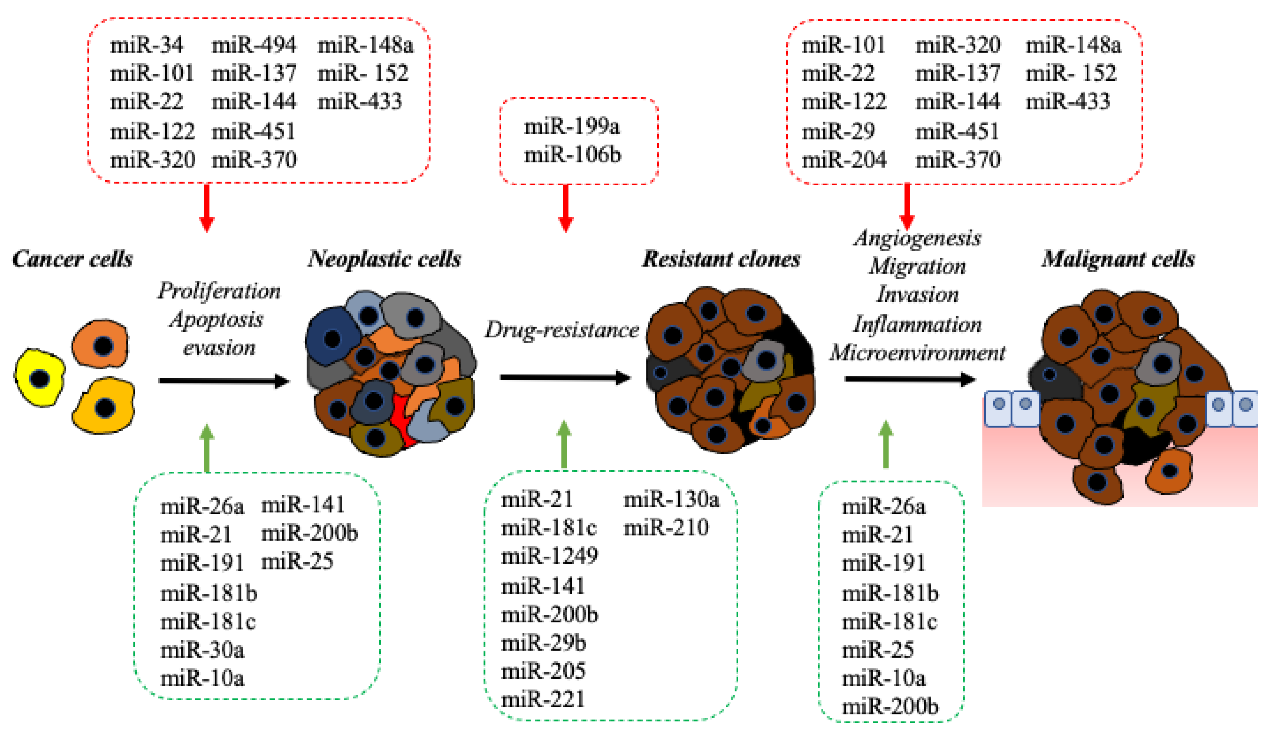
In recent years, the development of powerful technological platforms, capable of performing high-throughput RNA sequencing, allowed us to greatly expand our knowledge of the transcriptome and to reveal its complexity. In particular, the complexity of the eukaryotic transcriptome was made evident by the identification of a new class of RNAs with a structure and function clearly different from those previously known. Based on current knowledge, the eukaryotic transcriptome can be divided into messenger (mRNA) or coding RNAs and a novel class of non-coding RNAs (ncRNAs) [12,13].
The ncRNA represents a class of RNA that is transcribed but not translated into proteins. According to their length, they can be further divided into two subtypes: (a) small ncRNAs ranging from 18 to 200 nucleotides and (b) long ncRNA with length greater than 200 nucleotides [12,13]. NcRNAs influences several physiological and disease processes by playing a pivotal role in the regulation of gene expression.
The most widely studied class of ncRNAs is certainly the microRNA (miRNA). MiRNAs are small non-coding RNA (18 to 24 nucleotides) transcribed by the RNA pol II (RNA polymerase II) and further processed in a small, functionally mature RNA. The mature miRNA regulates target genes by binding to specific miRNA-responsive regions in the 3′UTR (untranslated region). The binding between miRNA and its pair target gene causes target-mRNA degradation and/or translational inhibition [14]. Several protein complexes take part in miRNA-mediated gene expression regulation, including via the RISC (RNA-induced silencing complex) which participates in miRNA-binding and further mRNA degradation [14]. The imperfect base pairing between miRNA and the miRNA-binding site makes miRNA an extremely complex and versatile regulatory agent, capable of fine-regulating hundreds of target genes [13,14,15].
The Croce group identified two miRNA genes at the chromosome 13q14 region in B-cell chronic lymphocytic leukemia cells, providing the earliest evidence of miRNA involvement in human cancer [16]. The identification of two miRNA genes, miR-15a and miR-16-1 initiated a series of further studies that revealed the key role of these miRNAs in tumor suppression [16].
In the following years, advanced technologies to profile miRNAs facilitated the accumulation of evidence regarding their aberrant expression in cancer. The miRNA expression profile has also been proposed as potential diagnostic and prognostic biomarker [15].
Regarding their link to BDC, miRNAs have been shown to be implicated in almost all steps of biliarycarcinogenesis by acting either as oncogenes (onco-miRNAs) or tumor suppressors (oncosuppressor-miRNAs) (Figure 1). An extensive report of the miRNAs involved in BDC, which is comprehensive of their mechanism of action and their targets, is presented in Table 1 and Table 2.

2.1. Onco-miRNAs

MiRNAs presenting abnormally high expression in tumors are commonly classified as oncogenic. MiRNAs with oncogenic function, called “onco-mirs”, usually promote tumor development by negatively inhibiting tumor suppressor genes and/or genes that control cell differentiation or apoptosis [54,55].
The earliest evidence for the role of ncRNAs in biliary-carcinogenesis came from a study by Meng et al., who first reported an aberrant miRNA expression profile in BDC cells compared with normal cholangiocytes [22]. The aberrant overexpression of miR-141, miR-21, and miR-200b, and their locations within genomic gain regions in BDC, caught the attention of authors who described the functional role for these miRNAs in regulating cell survival and drug resistance [22,56,57]. Further experiments showed that miR-141, miR-21, and miR-200b exert their oncogenic role by specifically targeting tumor-suppressor genes including CLOCK (Clock Circadian Regulator), PTPN12 (Protein Tyrosine Phosphatase Non-Receptor Type 12), and PTEN (Phosphatase And Tensin Homolog) [22,56,57].
Interestingly, miR-141 and miR-200b belong to the miR-200 family, which also includes miR-200a, miR-200c and miR-429. The miR-200 family is reported to regulate the formation of cancer stem cells and the regulation of the EMT (Epithelial-Mesenchimal-Transition, but their role in BDC progression has not been clearly understood [56,57].
Further studies investigating the expression of the miRNAome in cell lines and in primary BDC patient samples, identified miRNA-93, -25, -27a, and -21 as the most overexpressed [20]. In particular, quantitative RT-PCR (Real-Time Polymerase Chain Reaction) analysis of miR-21 on 18 primary BDCs and 12 normal liver specimens confirmed miR-21 overexpression. The oncogenic role of miR-21 was showed to be related to the inhibition of PDCD4 (programmed cell death 4), TIMP3 (metallopeptidase inhibitor 3) [20,21,22] and 15-PGDH/HPGD (NAD+-linked 15-hydroxyprostaglandin dehydrogenase) expression [20,21,22]. Further investigations revealed functional and mechanistic links between miR-21 and tumor suppressor genes, PTPN14 (Protein Tyrosine Phosphatase Non-Receptor Type 14) and PTEN, in the pathogenesis of iBDC [20,21,22]. To date, several pieces of evidence have indicated that overexpression of miR-21 significantly promoted cell migration, invasion, and xenograft growth [20,21,22]. More recent studies conducted on a large iBDC dataset indicated that higher miR-21 expression was significantly correlated with a larger tumor size and a poor outcome [20,21,22].
The oncogenic role of miR-26a in BDC is highlighted by observations that miR-26a overexpression promotes BDC cell proliferation, clonogenic formation, migration, and tumor growth in in vivo models [17,18,19]. Evidence has also suggested that miR-26a enhances BDC progression by targeting GSK-3β (Glycogen Synthase Kinase 3 Beta) [17,18,19] and KRT19 (Keratin 19) [17]. In a recent study, high miR-26 expression in BDC serum samples was correlated with a poor outcome, thus confirming its oncogenic role [19].
The oncogenic role of miR-191 in apoptosis, proliferation, invasion, and metastatic behaviors of BDC cells has been recently investigated by Li et al. [23] who reported a marked overexpression of miR-191 in iBDC tissues. They also described a key role of miR-191 in promoting tumor proliferation, migration, and invasion by targeting TET1 (Tet Methylcytosine Dioxygenase 1) both in vitro and in vivo [23].
The roles of several members of the miR-181 family have also been investigated in BDC [24,25,26,27]. Data reported by Jiang et al. [25] indicated that miR-181b-5p acts as an onco-miRNA through sustaining tumor cell proliferation, migration, and invasion of BDC by targeting PARKK (Parkin RBR E3 Ubiquitin Protein Ligase), a gene belonging to the PTEN/PI3K/AKT signaling pathway [25]. Recent evidence supports an oncogenic role for miR-181c in sustaining tumor proliferation, drug-resistance, and metastasis by targeting the tumor suppressor NDRG2 (N-myc downstream-regulated gene 2) [26,27]. Furthermore, elevated levels of miR-181c expression were associated with poor overall survival in patients with BDC [26,27].
MiR-30a-5p, a member of the mir-30 family, plays a crucial role in tumor growth and metastasis in several cancers, including BDC. MiR-30a-5p was found to be overexpressed and significantly associated with tumor size and nodules of BDC patients. In vitro experiments showed that miR-30a-5p contributes to tumor growth by directly targeting the SOCS3 (Suppressor Of Cytokine Signaling 3) gene [28].
Several results highlighted the oncogenic function of miR-25 in BDC [29,30]. Elevated expression of miR-25 was correlated with poor prognosis in BDC patients. Furthermore, the overexpression of miR-25 promotes the proliferation, migration, and invasion of BDC cells. MiR-25 was also reported to negatively regulate apoptosis signaling by directly targeting the death receptor DR4 in BDC cells [30].
An emerging role in the molecular pathology of BDC and in numerous other types of cancer has been described for miR-10a and miR-10b, belonging to miR-10 family. Several reports support the miR-10a family as a key player in cell proliferation and migration. The role of miR-10a-5p in BDC was explored by Gao et al. [31], who showed that miR-10a-5p acts as an onco-miRNA by directly targeting the tumor suppressor gene PTEN in BDC cell lines [31].

2.2. Oncosuppressor-miRNAs

Several experimental and clinic analyses suggested that downregulated miRNAs may function as a novel class of tumor suppressor genes in BDC. Onco-suppressor-miRNAs usually prevent tumor development by negatively inhibiting oncogenes [32,33,34,35,36,37,38,39,40,41,42,43,44,45,46,47,48,49,50,51,52,53,54].
MiR-34a has been studied for a long time and its function as a onco-suppressor-miRNA is widely recognized [32,33]. Several pieces of evidence suggested that its anti-tumor efficiency may be largely attributed to its capacity to suppress the expression of multiple oncogenic pathways. Direct targeting between miR-34a and several key cancer-related genes, including MYC (MYC Proto-Oncogene, BHLH Transcription Factor), MET (MET Proto-Oncogene, Receptor Tyrosine Kinase), CDK4/6 (Cyclin Dependent Kinase 4/6), NOTCH1 (Notch Receptor 1), BCL2 (BCL2 Apoptosis Regulator), and CD44 (CD44 Molecule) has been shown [32,33]. The biological function of miR-34a in BDC was explored by Kwon and colleagues, who used several experimental in vitro and in vivo models to demonstrate that the tumor-suppressive function is mediated by direct targeting of the Notch pathway-related genes (NOTCH1—Notch Receptor 1, NOTCH2—Notch Receptor 2, and JAGGED1—Jagged Canonical Notch Ligand 1) [33].
MiR-101 has been shown to function as an oncosuppressor-microRNA in several cancers by regulating the expression of different genes including EZH2 (Enhancer Of Zeste 2 Polycomb Repressive Complex 2 Subunit) [34,35], COX2 (Cytochrome C Oxidase Subunit 2) [35], APP (amyloid precursor protein) [36,37], and MCL-1 (myeloid cell leukemia sequence-1) [34,35,36,37]. In BDC the miR-101 expression was found to be decreased in 43% of patients and its tumor suppressor function was correlated with the inhibition of angiogenesis mediated by direct targeting of VEGF (Vascular Endothelial Growth Factor A) [35,36,37].
MiR-22 has been shown to play an important role in many cancer types, working as an oncosuppressor-miRNA [39]. In fact, miR-22 activates apoptosis by inducing p53 expression and concomitantly triggering senescence, and inhibiting tumor growth, invasion, and metastasis [38,39]. The activation of the pRb signaling pathway and the direct targeting of SIRT1 (Sirtuin 1), CDK6 (Cyclin Dependent Kinase 6), and Sp1 (Sp1 Transcription Factor) was described [38,39]. Recent work from Gradilone et al. assessed the role of miR-22 in BDC. They demonstrated that overexpression of miR-22 and miR-433 suppresses BDC cell proliferation and migration by targeting HDAC6 (Histone Deacetylase 6) [39].
A liver-specific miRNA, miR-122, has shown to be a key regulator in liver diseases and an oncosuppressor-miRNA in several tumors, including BDC [40,41,42]. Several findings showed that miR-122 can suppress tumor growth and invasion of BDC cells by targeting ALDOA (Aldolase, Fructose-Bisphosphate A) [40,41,42] and CLIC1 (chloride intracellular channel 1) [40,41,42].
A recent report described the role of the miR-29-3p-family in BDC, confirming that members of the miR-29-3p-family act as tumor-suppressors. The miR-29-3p-family plays a pivotal role in regulating multiple oncogenic pathways, including focal adhesion, ECM-receptor, endocytosis, PI3K–AKT signaling, and Hippo signaling. The tumor suppressor function of the MiR-29-3p-family has been linked to the direct regulation of two genes involved in cancer cell migration and invasiveness: ITGA6 (integrin alfa-6) and ITGB1 (integrin beta-1) [43].
A study from Chen L et al. analyzed the miRNA profile in iBDC and identified miR-204 as one of the most down-regulated [44] both in BDC tissues and cell lines. MiR-204 has been shown to work as a potential tumor suppressor in several cancers, including endometrial cancer [44,45], peripheral nerve tumors [44,45], gastric cancer [44,45], and hepatocellular carcinoma [44,45]. In iBDC, miR-204 plays a pivotal tumor suppressor role in inhibiting cell migration and invasion, by directly targeting the gene SLUG (Snail Family Transcriptional Repressor 2), a main component of the epithelial-mesenchymal transition process (EMT) [44,45].
Several studies have also reported a key role for miR-320 in targeting the EMT, tumor migration, and metastasis [46,47]. The significant down-regulation of miR-320 was described in BDC samples [46,47]. Moreover, miR-320 inhibits BDC growth both in vitro and in vivo by regulating the VEGF (Vascular endothelial growth factor) pathway through direct targeting of NRP-1 (Neuropilin-1) [47].
Furthermore, miR-494 emerged as an important regulator of BDC growth. It was shown to be down-regulated in BDC and that its overexpression inhibited cancer cell growth through multiple targets involved in cell cycle regulation, including CDK4/6 (cyclin-dependent kinase 4/6), CCND1 (cyclin D1), CCNE2 (cyclin E2), HDAC1 (histone deacetylase 1), RB1 (RB transcriptional corepressor 1), PLK1 (polo-like kinase 1), PTTG1 (pituitary tumor-transforming 1), CCNB1 (cyclin B1), CDK2 (cyclin-dependent kinase 2), and TOP2A (DNA topoisomerase Iiα) [48].
A recent study investigated the expression of miR-137 in BDC tissues and cell lines demonstrating that miR-137 acts as a suppressor in BDC by suppressing the expression of WNT2B and WNT-pathway-related genes [49].
Recently, miR-144-5p and miR-451a were found to play a common and synergic tumor suppressor role in BDC. In fact, miR-144-5p and miR-451a were downregulated in BDC patient samples and their concomitant overexpression suppresses proliferation, invasion, and migration of BDC cells by directly targeting the ST8SIA4 (ST8 alpha-N-acetyl-neuraminide alpha-2,8-sialyltransferase 4) gene [50].
Several reports have shown that IL-6 (Interleukine 6) may serve as a pro-oncogenic factor by triggering the epigenetic silencing of several oncosuppressor-miRNAs, including miR-370 [51,52], miR-148a, and miR-152 [53]. These miRNAs are, respectively, involved in the regulation of several known oncogenes including MAP3K8 (mitogen-activated protein kinase kinase kinase 8), RASSF1 (Ras association domain family member 1), and CDKN2A (cyclin-dependent kinase inhibitor 2α) [53].

3. The Impact of miRNAs in Precision Medicine of BDC

In the last few years, the recent advances in the genomic knowledge of BDC have sped up the idea of ‘precision medicine’. A precision medicine-based approach aims to select the most appropriate treatment dependent on the specific molecular alterations displayed by a single patient. Due to the heterogeneity of BDC, the absence of diagnostic predictive tools, and the inefficacy and toxicity of current treatment, a precision medicine approach could greatly benefit the clinical management of BDC. Moreover, the specificity and sensitivity of available biomarkers determined in serum and biopsy samples are not sufficient to assist in the clinical management of BDC. The availability of predictive biomarkers, which could aid patient stratification and treatment response identification, still represents an unmet clinical need. Extensive research is being carried out to identify novel biomarkers that could contribute to a better understanding of the molecular pathogenesis of BDC, as well as to provide new diagnostic, prognostic, and predictive tools [5,26,58,59,60].
A huge number of studies proposed the application of miRNAs to the precision medicine of BDC as a promising tool to improve the management of cancer patients. Next, we discuss the impact of miRNAs in precision medicine of BDC, and the challenges associated with translating these findings into the clinic [27,58,59,60,61,62,63].

3.1. miRNAs as Biomarkers for BDC

Current biomarkers used in clinical management of BDC have showed low sensitivity and specificity. Actually, the CA19-9 (Carbohydrate antigen 19-9), a non-specific tumor biomarker, is currently in use in the diagnosis of BDC, but it lacks sensitivity and specificity. Currently, the clinical management needs biomarkers to improve early-stage diagnosis and prognosis prediction. In recent years, many studies have proposed miRNAs as potential biomarkers as diagnostic, prognostic, and predictive tools in BDC.
miRNAs were first identified as biomarkers for cancer in 2008 by Lawrie et al. in the serum of large B-cell lymphoma patients [60], and since then, their potential use as biomarkers has been investigated for numerous diseases [61]. This novel class of molecules possesses an array of advantages that make them very close to ideal candidates for biomarkers: (a) accessibility; miRNAs can be easily extracted and detected through liquid biopsies from blood, urine, and other bodily fluids. (b) Specificity/sensitivity; miRNAs possess a high specificity and sensitivity for the tissue or cell type of origin. (c) Time- and cost-effectiveness; miRNA detection requires less time and lower costs in comparison to other available biomarkers. (d) Multi-marker detection; the use of miRNA signatures provides a non-invasive method for the diagnosis and prediction of disease progression and treatment efficacy. Circulating- or tissue-derived miRNAs are promising diagnostic tools for various kinds of cancers.
Although there are an increasing number of studies reporting their potential use in BDC, the diagnostic efficacy has not been established yet. Two miRNAs—miR-21 and miR-122—are the most extensively studied as potential biomarkers. The diagnostic value of both miRNAs was investigated in plasma of 1001 iBDC samples and compared to the traditional marker CA19-9, by the Zheou group [58]. They established a diagnostic three-marker model combining plasma miR-21, miR-122, and CA19-9, discriminating iBDC from controls with high accuracy [58,59].
The diagnostic and prognostic role of miR-21 has been assessed in several kinds of cancers. Importantly, serum miR-21 has shown to be a promising biomarker for early detection and prognosis in patients with colorectal cancer [62]. Several studies have reported the use of miR-21 as a biomarker in BDC for both diagnosis [20,21,22,62] and prognosis [27]. A study conducted by Selaru et al. reported elevated sensitivity (95%) and specificity (100%) of miR-21 in distinguishing between BDC and normal tissues [63]. Similar results were obtained by analyzing urine- [64] and plasma-derived [65] miR-21 of BDC patients.
Wang et al. proposed miR-21 as an independent prognostic and predictive factor in iBDC [20]. The prognostic relevance of miR-21 has been further assessed by a recent meta-analysis of 31 studies that confirmed the association between high miR-21 expression and poor outcome in patients with BDC [26]. The meta-analysis also identified other miRNAs as potential prognostic biomarkers, including high-expressed miR-26a, miR-29a, miR-181c, miR-191, miR-192, miR-200c, and miR-221, and low-expressed miR-34a, miR-106a, miR-203, and miR-373 [26].
The diagnostic and prognostic value of miR-26a in BDC has been assessed by comparing tissue and serum analysis between the pre-operative serum and post-operative patients [8,26]. Accumulating evidence showed high sensitivity and specificity of serum miR-26a as a BDC biomarker. In fact, the levels of serum miR-26a were significantly higher than that of healthy controls [8,26]. It was also found to be an independent predictor of poor overall and progression-free survival and it was significantly correlated with clinical stage, distant metastasis, differentiation status, and poor survival [8,26], and proposes serum miR-26a as a potential noninvasive biomarker candidate for the early detection of BDC.
Numerous investigations extensively explored the prognostic significance of miR-25 as a biomarker in BDC [29]. Studies comparing the relative expression of miR-25 in BDC tissues and cell lines with paired normal tissues and a normal cell line, reported that miR-25 is upregulated in both malignant BDC cell lines and patient tissue [9,10]. Furthermore, high miR-25 expression was significantly associated with TNM stage and lymph node metastasis, and overall survival of BDC patients [29,30].
Aberrant expression of miR-29a has been found in several types of tumor, providing evidence that it can be proposed as a novel cancer biomarker [66]. Results obtained by Deng et al. [67], by using qRT-PCR, demonstrated that miR-29a overexpression correlated with lymph node metastasis, clinical stage, differentiation, and poor overall survival of BDC, allowing the authors to propose miR-29a expression as an independent prognostic factor.
Li et al. [23] demonstrated that miR-191 is an independent prognostic factor in patients with iBDC. In fact, high expression levels of serum and tissue miR-191 were found to be associated with advanced iBDC, as well as overall and disease-free survival. Interestingly, they also showed that a combination of miR-191 with its direct target TET1 has a more prognostic and predictive value [23].
The overexpression of miR-181c in clinical human BDC samples was associated with poorer BDC patient overall survival. By combining the expression of both miR-181c and its target NDRG2, a high accuracy and efficacy for the diagnosis of BDC were found [27].
miRNA-150 expression has been shown to be a potent diagnostic tool in myeloid leukemia [68]. The diagnostic value of serum miR-150-5p was also evaluated in BDC patients, alone or in combination with CA19-9 expression, showing elevated sensitivity and specificity [69].
Circulating miR-106a was described by Cheng et al. [70] as a novel biomarker in BDC. Down-regulation of miR-106a in serum of BDC patients was correlated with poor prognosis, but the diagnostic value of miR-106a was moderate and requires large-scale prospective validation [70].
The expression of miR-146a in BDC tissues was found to be an independent prognostic biomarker in patients subjected to surgical treatment [71], and both plasma and tissue miR-146a expression correlated with favorable overall survival.
The expression of miR-203 was dramatically decreased in BDC tissues. Low expression of miR-203 was significantly associated with tumor progression and predicted poor prognosis in BDC patients. These findings indicated that miR-203 serves as a novel prognostic marker in BDC [72]. Similar results were obtained for serum miR-195, which showed a relatively high clinical value in diagnosis and prognosis [73].
Recently, using high-throughput functional studies, the Braconi group identified miR-1249 as a novel independent BDC prognostic biomarker [11]. High expression of miR1249 was found in 32% of BDC and was associated with a worse prognosis, independent of adjuvant chemotherapy at multivariate analysis (considering TNM stage, adjuvant chemotherapy, and MIR1249 tumor expression) [11].
Using a statistically robust approach in clinically relevant samples, Meijer et al. discovered a novel two-miRNA panel consisting of miR-16 and miR-877 to detect distal BDC in plasma. The two-miRNA signature can discriminate distal BDC from other cancers with elevated diagnostic power [74].
Similarly, the two-miRNA signature consisting of miR-151-3p and miR-126 showed significant prognostic power [75]. In fact, the molecular profile of miR-151-3p and miR-126 correlated with an improved survival in resected BDC [75].
A recent study described a novel three-miRNA signature (miR-10b, miR-22, and miR-551b) and evaluated its prognostic performance using high-throughput data downloaded from the TCGA database [76], demonstrating that it could be considered an alternative prognostic marker in BDC [76].
Finally, extracellular vesicles (EVs) are attracting great interest as potential disease biomarker containers. Recently, microRNAs contained in a bile-extracted extracellular vesicle were postulated as an option for BDC diagnosis. A novel BDC-based diagnostic panel with potential clinical utility has been investigated by Li et al. [77]. They described a five-miR panel including miR-191, miR-486-3p, miR-1274b, miR-484, and miR-16, with superior diagnostic accuracy, in comparison to the currently available diagnostic methods in BDC [77].

3.2. Implication of MiRNAs in Therapy Sensitivity and Resistance of BDC

A combination of gemcitabine (Gem) and cisplatin (Cis) is the first-line systemic therapy for patients with BDC, who are diagnosed at advanced stages and are not suitable for surgical treatment [4,6,7,78,79,80]. Other therapeutic regimens including FOLFIRINOX (a combination of 5-fluorouracil, leucovorin, oxaliplatin, and irinotecan) or combined treatments with Gem, Cis, and nab-paclitaxel have been shown to have a moderate effect on patient survival [1,4,78]. Unfortunately, drug resistance remains a key issue during such treatments [1]. Promising data are being obtained from ongoing clinical trials and that will potentially provide new therapies to increase the survival of advanced BDC [79,80]. Emerging therapies, including FGFR inhibitors [8,9], IDH1 and/or IDH2 inhibitors [7,80], and immunotherapies [10,81] have shown a marked efficiency. The identification of biomarkers associated with both chemo- and target-therapies can be useful to predict the efficiency, sensitivity, resistance, or toxicity of pharmacological regimes [1,79,80].
Several reports have suggested that miRNAs play a key role in the development of drug-sensitivity or -resistance in several kinds of cancers [11,12,13,82]. Numerous studies evidenced that miRNAs regulate several mechanisms driving drug-resistance [5]. Moreover, therapeutic strategies aiming to overexpress oncosuppressor-miRNAs or inhibit onco-miRs may represent efficient approaches to overcome drug-resistance or increasing the efficacy of therapeutic regimens. The advent of precision medicine has paved the way for the introduction of miRNAs as biomarkers to predict therapeutic responses and cancer patient survival [11,12,13,14,15,82].
The role of miRNAs in regulating the drug-response to chemotherapy in BDC was recently explored by the Braconi group. They took advantage of using a high-throughput screening of miRNA-inhibitors to identify miRNAs implicated in the response to chemotherapy in BDC cells [11]. In particular, this study led to the identification of eleven miRNAs modulating chemotherapy-response in BDC cells, including miR-1249, miR-133b, miR-1247, miR-1224-3p, miR-1228, miR-1234, miR-1280, miR-2196, miR-566, miR-877, and miR-885-5p [11]. MiR-1249 was further demonstrated to be a key molecular player in driving the emergence of chemo-resistance mediated by the proliferation of cancer stem cell [11].
Okamoto and colleagues performed a correlation analysis between miRNA expression profiles and response to Gem in BDC cell lines [83]. They proposed miR-29b, miR-125a-5p, miR-205, and miR-221 as diagnostic biomarkers of sensitivity to Gem treatment [83]. The above listed miRNAs were showed to be molecular modulators of the apoptotic pathway and potent regulators of the resistance of BDC cells to chemotherapy [83].
Meng et al. reported that miR-21 and miR-200b expression is correlated with Gem-resistance in cell lines derived from gallbladder carcinoma [22]. The same authors reported that miR-21 plays a key role in modulating Gem-induced apoptosis by direct targeting of PTEN (phosphatase and tensin homolog) signaling both in in vitro and in vivo models [22].
The high expression of miR-210 has shown to be a potential biomarker of Gem resistance in BDC cells [84]. Elevated expression levels of miR-210 has been reported in most solid tumors, and high levels are correlated with a poor clinical outcome for patients. In BDC cells, miR-210 has been reported to modulate the sensitivity to Gem by sustaining HIF-1α (Hypoxia Inducible Factor 1 Subunit Alpha) activity [84].
Asukai et al. showed that miR-130a-3p modulates Gem-resistance in BDC by regulating the PPARγ (peroxisome proliferator-activated receptor-γ) gene [85].
The implication of miRNAs in regulating the response to Cis was explored by Li Q. et al., who described the role of miR-199a-3p in modulating Cis-sensitivity in BDC cells by inhibiting the expression of key molecular drivers of drug-resistance, such as MDR1 (Multidrug Resistance Protein 1) and the mTOR (Mechanistic Target of Rapamycin Kinase) gene network [86].
MiR-106b was recently proposed as a prognostic biomarker in BDC, and its role in modulating the efficacy of 5-FU (5-Fluorouracyl) treatment was explored [87]. In particular, overexpression of miR-106b may overcome 5-FU resistance in BDC cells, acting as a regulator of ZBTB7A (Zinc Finger and BTB Domain Containing 7A) expression [87].
The role of miRNA as an efficacy regulator of targeted therapy was also explored [88]. It has recently been reported that HSP90 (heat shock protein 90) inhibition could offer a promising therapeutic strategy for BDC [88]. Moreover, as a partner of the FGFR family, HSP90 helps in the folding and protein packaging of FGFR. The onco-miR MiR-21 has been shown to drive resistance to HSP90 inhibition in human BDC cell lines, 3D organoid cultures, and patient-derived xenografts [88].
Recently, immunotherapy is emerging as a promising treatment option in BDC. Immune checkpoint inhibitors (ICI) represented by monoclonal antibodies directed against CTLA-4 (cytotoxic T lymphocyte antigen 4) or PD-1 (programmed cell death 1) or its ligand (programmed cell death ligand-1, PD-L1) are currently under investigation in several clinical trials. KEYNOTE-158 (NCT02628067) and KEYNOTE-028 (NCT02054806) studies evaluated the efficiency and safety data from patients with advanced BDC receiving pembrolizumab, a monoclonal antibody (mAb) that works by binding to the PD-1 receptor and preventing its interaction with ligands. Other studies in BDC have investigated the anti-tumor activity of different immune-therapeutic strategies combining PD-1/L1 and CTL-4 inhibitors such as Nivolumab in combination with the ipilimumab, and Durvalumab plus tremelimumab [89]. Nevertheless, the limited response to ICI in advanced BDC highlighted the need for predictive biomarkers for immunotherapeutic treatment responses. Analysis evaluating genomic instability/Tumor Mutational Burden (TMB), the immune contexture, PD-L1 Expression, mutations in DNA damage repair (DDR) genes, and Microsatellite Instable Tumors (MSI) have been investigated for their predictive value in treatment of BDC patients with ICI and their combinations. Some miRNAs have been found to modulate several aspects of the antitumor immune response, including immune checkpoints (PD-1, PD-L1, and CTL-A4), immune cells (macrophages, MDSCs, and NKs), and tumor antigen-processing machinery [53], thus suggesting that miRNAs represent potential biomarkers to predict the effective response to ICI.
Future studies comparing the miRNA expression profiles of patients who respond to such therapies and those of non-responders will be beneficial to disclose the potential benefit of miRNAs in this regard.
More data is then needed to evaluate the potential benefit of the use of miRNAs as biomarkers for immune therapies in BDC patients with MSI, especially whether a combination (anti PD-1 and anti-CTLA4) could be more effective in these patients.
Although preclinical, these above-described observations suggest miRNAs may be involved with chemo- and target-therapy sensitivity or resistance, and that the translational potential of miRNAs should be investigated in wider clinical trials. Identification of miRNAs as potential predictive biomarker for immunotherapy response would allow better patient selection and improvement of therapeutic efficacy. Furthermore, miRNA can be easily detected in the blood of BDC patients, rationalizing their applicability in BDC precision medicine.

4. The Impact of miRNA-Based Therapeutic in BDC

MiRNA-based therapy is emerging as a promising novel strategy to treat cancers. miRNA-based therapeutic approaches operate by silencing overexpressed onco-miRNAs or replacing downregulated oncosuppressor-miRNAs [15]. Several miRNA-based therapeutics are under investigation in clinical trials according to www.clinicaltrials.gov (accessed on 22 January 2022) [90].
From the methodological point of view, the following strategies were adopted to efficiently perform onco-miRNA silencing both in vitro and in vivo: anti-miRNA oligonucleotides (AMOs), anti-miRNA locked nucleic acid (LNA), anti-miRNA sponges, and genetic knockouts based on the CRISPR/Cas9 genome-editing technologies [15]. On the other hand, miRNA replacement therapy has generally achieved restoration of onco-suppressor-miRNA by introducing synthetically modified oligonucleotides (miRNA mimics) or viral vectors [15].
However, while promising, miRNA-based therapeutics has raised the following issues to be considered: delivery, selectivity to specific target cells, degradation, and toxicity. Chemical modification of nucleotides or of the RNA backbone through methylation or LNAs, together with the development of vehicles to encapsulate the RNAs, are the main strategies put in place to protect miRNA from degradation [15]. Moreover, toxicity and side effects represent perhaps the biggest obstacle so far encountered in miRNA-based therapeutics [15]. Viral and non-viral vectors have been developed to improve delivery efficiencies to target cells, but the risk of adverse immunogenicity has restricted their use. Lipid-based and polymer-based nanoparticles (NPs) resulted as promising technical approaches as they guarantee efficient delivery and a good safety profile [15,91].
MRX34, a formulation based on miR-34 mimics in liposomal particles, was the first miRNA restoration strategy performed, subsequently entering a clinical trial recruiting patients with solid tumors, including hepatocellular carcinoma [92]. Preliminary results emerging from clinical investigations reported a good safety profile and significant anticancer activity for MRX34 treatment [92]. Recent findings showing the inhibition of BDC cell growth by the miR-34 mimic strongly suggested that miRNA-34-based therapy may be a potential efficient and safe therapeutic approach in BDC [32].
Li et al. first found that stroma-derived extracellular vesicles (EVs) containing miR-195 inhibit the proliferation, migration, and metastasis of BDC cells [91]. Furthermore, they observed that the systemic injection of miR-195-loaded EVs inhibited BDC tumor growth and prolonged survival in animal models [91].
Xie et al. explored a new therapeutic approach based on the use of nanoparticles loaded with a cholesterol-modified polymeric antagonist of CXCR4 (C-X-C receptor type 4) and anti-miR-210 [93]. They showed that CXCR4 antagonist- and anti-miR-210-loaded nanoparticles cooperate synergistically in inducing apoptosis and sensitizing BDC cells to Gem/Cis treatment. They presented results showing that the novel nano-therapeutic approach combining the silencing of both CXCR4 and miR-210 suppress tumor growth in BDC cell lines and animal models [93].
Another miRNA-based therapeutic approach is represented by the combination of chemotherapy and miRNA to establish a synergistic antitumor effect. Recently, Zhang et al. assembled NPs loaded with gemcitabine—oleic acid prodrugs (GOA) and miR-122 to form GOA/miRNAs NPs [18]. They demonstrated that GOA/miR-122-loaded NPs were efficiently delivered to the tumor area and inhibited hepatocellular carcinoma tumor growth in vivo, without significantly affecting the biosafety profile [18].
To date, miRNA-based therapeutics have been investigated by a huge number of preclinical studies, but only a small number have moved up to clinical trials. Major obstacles are still represented by the delivering of miRNA-based drugs, their stability, and safety profile. However, promising novel strategies are emerging, combining novel delivery platforms and low-toxicity profiles, providing a basis for innovative miRNA-based therapeutic approaches.

5. Conclusions

The majority of BDC patients encounter poor prognoses, largely due to late diagnosis, inefficient treatments, and therapy-resistance. These unmet clinical needs urge faster discovery of novel prognostic and predictive biomarkers for earlier diagnosis, tumor aggressiveness, and response to treatment. Furthermore, novel therapeutic strategies that can guarantee more clinical benefits to patient survival are much needed. Recent studies revealed the key function of miRNAs in molecular processes driving BDC carcinogenesis. miRNAs are emerging as promising tumor diagnostic and prognostic biomarkers to be incorporated into the clinical management of BDC patients. Their predictive value in assessing the efficacy of chemo- and targeted-therapy has been also largely investigated in BDC. Besides the promising findings currently emerging, more investigations are needed to demonstrate their value in the clinical management of BDC patients. The application of miRNA-based therapeutics to BDC is still in its infancy, but it represents an attractive anti-tumor approach as a new strategic option for cancer therapy.

Author Contributions

Study concept and design: P.C., B.F., M.S. and L.R.-B.; analysis and interpretation of data: P.C., B.F., M.S. and L.R.-B.; drafting of the manuscript: P.C., B.F., M.S., L.R.-B., A.B. and F.P.; critical revision of the manuscript for important intellectual content: P.C., B.F., M.S. and L.R.-B.; obtained funding: P.C. and B.F. All authors have read and agreed to the published version of the manuscript.

Funding

This study was kindly supported by POR CAMPANIA FESR 2014/2020 grant (Project title: “Dalla genomica alla terapia di tumori rari”—and “Combattere la resistenza tumorale: piattaforma integrata multidisciplinare per un approccio tecnologico innovativo alle oncoterapie; CUP B61G18000470007) and by the Italian Association for Cancer Research (AIRC) (grant n. IG 17711/2015 to B.F.). PC is a current recipient of a Marie Skłodowska-Curie Career Re-Integration fellowship funded by AIRC and the European Union’s Horizon 2020 research and innovation programme (Grant agreement No 800924).

Conflicts of Interest

Authors declare no conflict of interest.

References

  1. Rizvi, S.; Khan, S.A.; Hallemeier, C.L.; Kelley, R.K.; Gores, G.J. Cholangiocarcinoma—Evolving concepts and therapeutic strate-gies. Nat. Rev. Clin. Oncol. 2018, 15, 95–111. [Google Scholar] [CrossRef] [PubMed] [Green Version]
  2. Khan, S.A.; Tavolari, S.; Brandi, G. Cholangiocarcinoma: Epidemiology and risk factors. Liver Int. 2019, 39, 19–31. [Google Scholar] [CrossRef] [PubMed] [Green Version]
  3. Bertuccio, P.; Malvezzi, M.; Carioli, G.; Hashim, D.; Boffetta, P.; El-Serag, H.B.; Negri, E. Global trends in mortality from intrahepatic and extrahepatic cholangiocarcinoma. J. Hepatol. 2019, 71, 104–114. [Google Scholar] [CrossRef] [PubMed]
  4. Kelley, R.K.; Bridgewater, J.; Gores, G.J.; Zhu, A.X. Systemic therapies for intrahepatic cholangiocarcinoma. J. Hepatol. 2020, 72, 353–363. [Google Scholar] [CrossRef] [Green Version]
  5. Braconi, C.; Roessler, S.; Kruk, B.; Lammert, F.; Krawczyk, M.; Andersen, J.B. Molecular perturbations in cholangiocarcinoma: Is it time for precision medicine? Liver Int. 2019, 39, 32–42. [Google Scholar] [CrossRef] [Green Version]
  6. Adeva, J.; Sangro, B.; Salati, M.; Edeline, J.; La Casta, A.; Bittoni, A.; Berardi, R.; Bruix, J.; Valle, J.W. Medical treatment for cholangiocarcinoma. Liver Int. 2019, 39, 123–142. [Google Scholar] [CrossRef] [Green Version]
  7. De la Fouchardiere, C. Towards greater clarity in the treatment of cholangiocarcinoma. Lancet Oncol. 2020, 21, 738–739. [Google Scholar] [CrossRef]
  8. Salati, M.; Caputo, F.; Baldessari, C.; Carotenuto, P.; Messina, M.; Caramaschi, S.; Dominici, M.; Bonetti, L.R. The Evolving Role of FGFR2 Inhibitors in Intrahepatic Cholangiocarcinoma: From Molecular Biology to Clinical Targeting. Cancer Manag. Res. 2021, 13, 7747–7757. [Google Scholar] [CrossRef]
  9. Franco, B.; Clarke, P.; Carotenuto, P. Pemigatinib. Fibroblast growth factor receptor inhibitor, Treatment of cholangiocarci-noma. Drugs Future 2019, 44, 923. [Google Scholar] [CrossRef]
  10. Guo, X.; Shen, W. Latest evidence on immunotherapy for cholangiocarcinoma (Review). Oncol. Lett. 2020, 20, 381. [Google Scholar] [CrossRef]
  11. Carotenuto, P.; Hedayat, S.; Fassan, M.; Cardinale, V.; Lampis, A.; Guzzardo, V.; Braconi, C. Modulation of Biliary Cancer Chemo-Resistance Through MicroRNA-Mediated Rewiring of the Expansion of CD133+ Cells. Hepatology 2020, 72, 982–996. [Google Scholar] [CrossRef] [PubMed]
  12. Salati, M.; Braconi, C. Noncoding RNA in Cholangiocarcinoma. Semin. Liver Dis. 2018, 39, 013–025. [Google Scholar] [CrossRef] [PubMed] [Green Version]
  13. Anastasiadou, E.; Jacob, L.S.; Slack, F.J. Non-coding RNA networks in cancer. Nat. Rev. Cancer 2018, 18, 5–18. [Google Scholar] [CrossRef] [PubMed]
  14. Gebert, L.F.R.; Macrae, I.J. Regulation of microRNA function in animals. Nat. Rev. Mol. Cell Biol. 2019, 20, 21–37. [Google Scholar] [CrossRef] [PubMed]
  15. Rupaimoole, R.; Slack, F.J. MicroRNA therapeutics: Towards a new era for the management of cancer and other diseases. Nat. Rev. Drug Discov. 2017, 16, 203–222. [Google Scholar] [CrossRef]
  16. Adrian, C.G.; Dan, D.C.; Shimizu, M.; Bichi, R.; Zupo, S.; Noch, E.; Croce, C.M. Frequent Deletions and Down-Regulation of Micro-RNA Genes miR15 and miR16 at 13q14 in Chronic Lymphocytic Leukemia [Internet]. Available online: https://www.pnas.org/doi/abs/10.1073/pnas.242606799 (accessed on 22 January 2022).
  17. Wang, P.; Lv, L. miR-26a induced the suppression of tumor growth of cholangiocarcinoma via KRT19 approach. Oncotarget 2016, 7, 81367–81376. [Google Scholar] [CrossRef] [Green Version]
  18. Zhang, H.-T.; Sun, J.; Yan, Y.; Cui, S.-H.; Wang, H.; Wang, C.-H.; Zhang, Q. Encapsulated microRNA by gemcitabine prodrug for cancer treatment. J. Control. Release 2019, 316, 317–330. [Google Scholar] [CrossRef]
  19. Zhang, J.; Han, C.; Wu, T. MicroRNA-26a Promotes Cholangiocarcinoma Growth by Activating β-catenin. Gastroenterology 2012, 143, 246–256.e8. [Google Scholar] [CrossRef] [Green Version]
  20. Wang, L.-J.; He, C.-C.; Sui, X.; Cai, M.-J.; Zhou, C.-Y.; Ma, J.-L.; Zhu, Q. MiR-21 Promotes Intrahepatic Cholangiocarcinoma Proliferation and Growth In Vitro and In Vivo by Targeting PTPN14 and PTEN [Internet]. Volume 6. Available online: www.impactjournals.com/oncotarget (accessed on 22 January 2022).
  21. Liu, Z.; Jin, Z.-Y.; Liu, C.-H.; Xie, F.; Lin, X.-S.; Huang, Q. MicroRNA-21 regulates biological behavior by inducing EMT in human cholangiocarcinoma. Int. J. Clin. Exp. Pathol. 2015, 8, 4684–4694. [Google Scholar]
  22. Meng, F.; Henson, R.; Lang, M.; Wehbe, H.; Maheshwari, S.; Mendell, J.T.; Jiang, J.; Schmittgen, T.D.; Patel, T. Involvement of Human Micro-RNA in Growth and Response to Chemotherapy in Human Cholangiocarcinoma Cell Lines. Gastroenterology 2006, 130, 2113–2129. [Google Scholar] [CrossRef] [Green Version]
  23. Li, H.; Zhou, Z.-Q.; Yang, Z.-R.; Tong, D.-N.; Guan, J.; Shi, B.-J.; Nie, J.; Ding, X.-T.; Li, B.; Zhou, G.-W.; et al. MicroRNA-191 acts as a tumor promoter by modulating the TET1-p53 pathway in intrahepatic cholangiocarcinoma. Hepatology 2017, 66, 136–151. [Google Scholar] [CrossRef] [PubMed] [Green Version]
  24. Indrieri, A.; Carrella, S.; Carotenuto, P.; Banfi, S.; Franco, B. The Pervasive Role of the miR-181 Family in Development, Neurodegeneration, and Cancer. Int. J. Mol. Sci. 2020, 21, 2092. [Google Scholar] [CrossRef] [PubMed] [Green Version]
  25. Jiang, Z.L.; Zhang, F.X.; Zhan, H.L.; Yang, H.J.; Zhang, S.Y.; Liu, Z.H.; Ke, R.S. miR-181b-5p Promotes the Progression of Cholangiocarcinoma by Targeting PARK2 via PTEN/PI3K/AKT Signaling Pathway. Biochem. Genet. 2022, 60, 223–240. [Google Scholar] [CrossRef] [PubMed]
  26. Sun, C.; Zhu, J.; Wu, B.; Chen, J.; Zhu, Z.; Cai, P.; Guo, W.; Gu, Z.; Wang, J.; Huang, S. Diagnostic and prognostic value of microRNAs in cholangiocarcinoma: A systematic review and meta-analysis. Cancer Manag. Res. 2018, 10, 2125–2139. [Google Scholar] [CrossRef] [Green Version]
  27. Wang, J.; Xie, C.; Pan, S.; Liang, Y.; Han, J.; Lan, Y.; Liu, L. N-myc downstream-regulated gene 2 inhibits human cholangiocarci-noma progression and is regulated by leukemia inhibitory factor/MicroRNA-181c negative feedback pathway. Hepatology 2016, 64, 1606–1622. [Google Scholar] [CrossRef] [Green Version]
  28. Zhang, J.W.; Wang, X.; Li, G.C.; Wang, D.; Han, S.; Zhang, Y.D.; Luo, C.H.; Wang, H.W.; Jiang, W.J.; Li, C.X.; et al. MiR-30a-5p promotes cholangiocarcinoma cell proliferation through targeting SOCS3. J. Cancer 2020, 11, 3604–3614. [Google Scholar] [CrossRef] [Green Version]
  29. Liu, H.; Ma, L.; Wang, J. Overexpression of miR-25 is associated with progression and poor prognosis of cholangiocarcinoma. Exp. Ther. Med. 2019, 18, 2687–2694. [Google Scholar] [CrossRef] [Green Version]
  30. Razumilava, N.; Bronk, S.F.; Smoot, R.L.; Fingas, C.D.; Werneburg, N.W.; Roberts, L.; Mott, J.L. miR-25 targets TNF-related apoptosis inducing ligand (TRAIL) death receptor-4 and promotes apoptosis resistance in cholangiocarcinoma. Hepatology 2011, 55, 465–475. [Google Scholar] [CrossRef]
  31. Gao, L.; Yang, X.; Zhang, H.; Yu, M.; Long, J.; Yang, T. Inhibition of miR-10a-5p suppresses cholangiocarcinoma cell growth through downregulation of Akt pathway. OncoTargets Ther. 2018, 11, 6981–6994. [Google Scholar] [CrossRef] [Green Version]
  32. Han, Y.; Meng, F.; Venter, J.; Wu, N.; Wan, Y.; Standeford, H.; Alpini, G. miR-34a-dependent overexpression of Per1 decreases chol-angiocarcinoma growth. J. Hepatol. 2016, 64, 1295–2130. [Google Scholar] [CrossRef] [Green Version]
  33. Slabáková, E.; Culig, Z.; Remšík, J.; Souček, K. Alternative mechanisms of miR-34a regulation in cancer. Cell Death Dis. 2017, 8, e3100. [Google Scholar] [CrossRef] [PubMed]
  34. Au, S.L.-K.; Wong, C.C.L.; Lee, J.M.-F.; Fan, D.N.-Y.; Tsang, F.H.; Ng, I.O.-L.; Wong, C.-M. Enhancer of zeste homolog 2 epigenetically silences multiple tumor suppressor microRNAs to promote liver cancer metastasis. Hepatology 2012, 56, 622–631. [Google Scholar] [CrossRef]
  35. Strillacci, A.; Griffoni, C.; Sansone, P.; Paterini, P.; Piazzi, G.; Lazzarini, G.; Tomasi, V. MiR-101 downregulation is involved in cycloox-ygenase-2 overexpression in human colon cancer cells. Exp. Cell Res. 2009, 315, 1439–1447. [Google Scholar] [CrossRef] [PubMed]
  36. Su, H.; Yang, J.-R.; Xu, T.; Huang, J.; Xu, L.; Yuan, Y.; Zhuang, S.M. MicroRNA-101, down-regulated in hepatocellular carcinoma, pro-motes apoptosis and suppresses tumorigenicity. Cancer Res. 2009, 69, 1135–1142. [Google Scholar] [CrossRef] [Green Version]
  37. Zhang, J.; Han, C.; Zhu, H.; Song, K.; Wu, T. miR-101 Inhibits Cholangiocarcinoma Angiogenesis through Targeting Vascular Endothelial Growth Factor (VEGF). Am. J. Pathol. 2013, 182, 1629–1639. [Google Scholar] [CrossRef] [PubMed] [Green Version]
  38. Wang, J.; Li, Y.; Ding, M.; Zhang, H.; Xu, X.-M.; Tang, J. Molecular mechanisms and clinical applications of miR-22 in regulating malignant progression in human cancer (Review). Int. J. Oncol. 2016, 50, 345–355. [Google Scholar] [CrossRef] [PubMed] [Green Version]
  39. Mansini, A.P.; Pisarello, M.J.L.; Thelen, K.M.; Cruz-Reyes, M.; Peixoto, E.; Jin, S.; Howard, B.N.; Trussoni, C.E.; Gajdos, G.B.; LaRusso, N.F.; et al. MicroRNA (miR)-433 and miR-22 dysregulations induce histone-deacetylase-6 overexpression and ciliary loss in cholangiocarcinoma. Hepatology 2018, 68, 561–573. [Google Scholar] [CrossRef] [PubMed]
  40. Fu, X.; Calin, G.A. miR-122 and hepatocellular carcinoma: From molecular biology to therapeutics. EBioMedicine 2018, 37, 17–18. [Google Scholar] [CrossRef] [Green Version]
  41. Xu, Z.; Liu, G.; Zhang, M.; Zhang, Z.; Jia, Y.; Peng, L.; Zhu, Y.; Hu, J.; Huang, R.; Sun, X. miR-122-5p Inhibits the Proliferation, Invasion and Growth of Bile Duct Carcinoma Cells by Targeting ALDOA. Cell. Physiol. Biochem. 2018, 48, 2596–2606. [Google Scholar] [CrossRef]
  42. Kong, L.; Wu, Q.; Zhao, L.; Ye, J.; Li, N.; Yang, H. Upregulated lncRNA-UCA1 contributes to metastasis of bile duct carcinoma through regulation of miR-122/CLIC1 and activation of the ERK/MAPK signaling pathway. Cell Cycle 2019, 18, 1212–1228. [Google Scholar] [CrossRef]
  43. Hozaka, Y.; Seki, N.; Tanaka, T.; Asai, S.; Moriya, S.; Idichi, T.; Wada, M.; Tanoue, K.; Kawasaki, Y.; Mataki, Y.; et al. Molecular Pathogenesis and Regulation of the miR-29-3p-Family: Involvement of ITGA6 and ITGB1 in Intra-Hepatic Cholangiocarcinoma. Cancers 2021, 13, 2804. [Google Scholar] [CrossRef] [PubMed]
  44. Zhang, L.; Wang, X.; Chen, P. MiR-204 down regulates SIRT1 and reverts SIRT1-induced epithelial-mesenchymal transition, anoikis resistance and invasion in gastric cancer cells. BMC Cancer 2013, 13, 290–299. [Google Scholar] [CrossRef] [PubMed] [Green Version]
  45. Chung, T.; Lau, T.; Cheung, T.; Yim, S.; Lo, K.; Siu, N.; Chan, L.; Yu, M.; Kwong, J.; Doran, G.; et al. Dysregulation of microRNA-204 mediates migration and invasion of endometrial cancer by regulating FOXC1. Int. J. Cancer 2011, 130, 1036–1045. [Google Scholar] [CrossRef] [PubMed]
  46. Liang, Y.; Li, S.; Tang, L. MicroRNA 320, an Anti-Oncogene Target miRNA for Cancer Therapy. Biomedicines 2021, 9, 591. [Google Scholar] [CrossRef]
  47. Zhu, H.; Jiang, X.; Zhou, X.; Dong, X.; Xie, K.; Yang, C.; Jiang, H.; Sun, X.; Lu, J. Neuropilin-1 regulated by miR-320 contributes to the growth and metastasis of cholangiocarcinoma cells. Liver Int. 2017, 38, 125–135. [Google Scholar] [CrossRef]
  48. Olaru, A.V.; Ghiaur, G.; Yamanaka, S.; Luvsanjav, D.; An, F.; Popescu, I.; Selaru, F.M. MicroRNA down-regulated in human cholangio-carcinoma control cell cycle through multiple targets involved in the G1/S checkpoint. Hepatology 2011, 54, 2089–2098. [Google Scholar] [CrossRef] [Green Version]
  49. Chen, T.; Lei, S.; Zeng, Z.; Pan, S.; Zhang, J.; Xue, Y.; Sun, Y.; Lan, J.; Xu, S.; Mao, D.; et al. MicroRNA-137 suppresses the proliferation, migration and invasion of cholangiocarcinoma cells by targeting WNT2B. Int. J. Mol. Med. 2020, 45, 886–896. [Google Scholar] [CrossRef]
  50. Fu, W.; Yu, G.; Liang, J.; Fan, P.; Dong, K.; Zhang, B.; Chen, X.; Zhu, H.; Chu, L. miR-144-5p and miR-451a Inhibit the Growth of Cholangiocarcinoma Cells Through Decreasing the Expression of ST8SIA4. Front. Oncol. 2021, 10, 3104. [Google Scholar] [CrossRef]
  51. Meng, F.; Wehbe-Janek, H.; Henson, R.; Smith, H.; Patel, T. Epigenetic regulation of microRNA-370 by interleukin-6 in malig-nant human cholangiocytes. Oncogene 2008, 27, 378–386. [Google Scholar] [CrossRef] [Green Version]
  52. An, F.; Yamanaka, S.; Allen, S.; Roberts, L.R.; Gores, G.J.; Pawlik, T.M.; Xie, Q.; Ishida, M.; Mezey, E.; Ferguson-Smith, A.C.; et al. Silencing of miR-370 in Human Cholangiocarcinoma by Allelic Loss and Interleukin-6 Induced Maternal to Paternal Epigenotype Switch. PLoS ONE 2012, 7, e45606. [Google Scholar] [CrossRef] [Green Version]
  53. Omar, H.A.; El-Serafi, A.T.; Hersi, F.; Arafa, E.A.; Zaher, D.M.; Madkour, M.; Arab, H.H.; Tolba, M.F. Immunomodulatory MicroRNAs in cancer: Targeting immune checkpoints and the tumor microenvironment. FEBS J. 2019, 286, 3540–3557. [Google Scholar] [CrossRef] [PubMed] [Green Version]
  54. Shi, T.; Morishita, A.; Kobara, H.; Masaki, T. The Role of microRNAs in Cholangiocarcinoma. Int. J. Mol. Sci. 2021, 22, 7627. [Google Scholar] [CrossRef] [PubMed]
  55. Chen, L.; Yan, H.-X.; Yang, W.; Hu, L.; Yu, L.-X.; Liu, Q.; Li, L.; Huang, D.-D.; Ding, J.; Shen, F. The role of microRNA expression pattern in human intrahepatic cholangiocarcinoma. J. Hepatol. 2009, 50, 358–369. [Google Scholar] [CrossRef] [PubMed]
  56. Peng, F.; Jiang, J.; Yu, Y.; Tian, R.; Guo, X.; Li, X.; Qin, R. Direct targeting of SUZ12/ROCK2 by miR-200b/c inhibits cholangiocarcinoma tumourigenesis and metastasis. Br. J. Cancer 2013, 109, 3092–3104. [Google Scholar] [CrossRef] [Green Version]
  57. Park, S.-M.; Gaur, A.B.; Lengyel, E.; Peter, M.E. The miR-200 family determines the epithelial phenotype of cancer cells by target-ing the E-cadherin repressors ZEB1 and ZEB2. Genes Dev. 2008, 22, 894–907. [Google Scholar] [CrossRef] [Green Version]
  58. Hu, J.; Wang, Y.-N.; Song, D.-J.; Tan, J.-P.; Cao, Y.; Fan, J.; Zhou, J. A High-Accuracy Model Based on Plasma miRNAs Diagnoses In-trahepatic Cholangiocarcinoma: A Single Center with 1001 Samples. Diagnostics 2021, 11, 610. [Google Scholar] [CrossRef]
  59. Macias, R.I.; Banales, J.; Sangro, B.; Muntané, J.; Avila, M.; Lozano, E.; Perugorria, M.J.; Padillo, F.J.; Bujanda, L.; Marin, J. The search for novel diagnostic and prognostic biomarkers in cholangiocarcinoma. Biochim. Biophys. Acta (BBA)—Mol. Basis Dis. 2018, 1864, 1468–1477. [Google Scholar] [CrossRef]
  60. Lawrie, C.H.; Gal, S.; Dunlop, H.M.; Pushkaran, B.; Liggins, A.P.; Pulford, K.; Banham, A.H.; Pezzella, F.; Boultwood, J.; Wainscoat, J.S.; et al. Detection of elevated levels of tumour-associated microRNAs in serum of patients with diffuse large B-cell lymphoma. Br. J. Haematol. 2008, 141, 672–675. [Google Scholar] [CrossRef]
  61. Mitchell, P.S.; Parkin, R.K.; Kroh, E.M.; Fritz, B.R.; Wyman, S.K.; Pogosova-Agadjanyan, E.L.; Tewari, M. Circulating microRNAs as stable blood-based markers for cancer detection. Proc. Nat. Acad. Sci. USA 2008, 105, 10513–10518. [Google Scholar] [CrossRef] [Green Version]
  62. Toiyama, Y.; Takahashi, M.; Hur, K.; Nagasaka, T.; Tanaka, K.; Inoue, Y.; Goel, A. Serum miR-21 as a diagnostic and prognostic biomarker in colorectal cancer. J. Natl. Cancer Inst. 2013, 105, 849–859. [Google Scholar] [CrossRef] [Green Version]
  63. Selaru, F.M.; Olaru, A.V.; Kan, T.; David, S.; Cheng, Y.; Mori, Y.; Meltzer, S.J. MicroRNA-21 is overexpressed in human cholangiocarci-noma and regulates programmed cell death 4 and tissue inhibitor of metalloproteinase 3. Hepatology 2009, 49, 1595–1601. [Google Scholar] [CrossRef]
  64. Silakit, R.; Loilome, W.; Yongvanit, P.; Thongchot, S.; Sithithaworn, P.; Boonmars, T.; Namwat, N. Urinary microRNA-192 and mi-croRNA-21 as potential indicators for liver fluke-associated cholangiocarcinoma risk group. Parasitol. Int. 2017, 66, 479–485. [Google Scholar] [CrossRef] [PubMed]
  65. Kishimoto, T.; Eguchi, H.; Nagano, H.; Kobayashi, S.; Akita, H.; Hama, N.; Wada, H.; Kawamoto, K.; Tomokuni, A.; Tomimaru, Y.; et al. Plasma miR-21 is a novel diagnostic biomarker for biliary tract cancer. Cancer Sci. 2013, 104, 1626–1631. [Google Scholar] [CrossRef] [PubMed]
  66. Wang, J.-Y.; Zhang, Q.; Wang, D.-D.; Yan, W.; Sha, H.-H.; Zhao, J.-H.; Yang, S.-J.; Zhang, H.-D.; Hou, J.-C.; Xu, H.-Z.; et al. MiR-29a: A potential therapeutic target and promising biomarker in tumors. Biosci. Rep. 2018, 38, BSR20171265. [Google Scholar] [CrossRef] [Green Version]
  67. Deng, Y.; Chen, Y. Increased Expression of miR-29a and Its Prognostic Significance in Patients with Cholangiocarcinoma. Oncol. Res. Treat. 2017, 40, 128–132. [Google Scholar] [CrossRef]
  68. Wang, F.; Ren, X.; Zhang, X. Role of microRNA-150 in solid tumors. Oncol. Lett. 2015, 10, 11–16. [Google Scholar] [CrossRef] [Green Version]
  69. Salem, P.E.S.; Ghazala, R.A.; El Gendi, A.M.; Emara, D.M.; Ahmed, N.M. The association between circulating MicroRNA-150 level and cholangiocarcinoma. J. Clin. Lab. Anal. 2020, 34, e23397. [Google Scholar] [CrossRef]
  70. Cheng, Q.; Feng, F.; Zhu, Z.; Zheng, Y.; Luo, X.; Liu, C.; Jiang, X. Circulating miR-106a is a Novel Prognostic and Lymph Node Me-tastasis Indicator for Cholangiocarcinoma. Sci. Rep. 2015, 5, 16103. Available online: www.nature.com/scientificreports (accessed on 22 January 2022). [CrossRef]
  71. Zhang, R.-X.; Zheng, Z.; Li, K.; Wu, X.-H.; Zhu, L. Both plasma and tumor tissue miR-146a high expression correlates with prolonged overall survival of surgical patients with intrahepatic cholangiocarcinoma. Medicine 2017, 96, e8267. [Google Scholar] [CrossRef]
  72. Li, J.; Gao, B.; Huang, Z.; Duan, T.; Li, D.; Zhang, S.; Cheng, K. Prognostic significance of microRNA-203 in cholangiocarcinoma [Internet]. Int. J. Clin. Exp. Pathol. 2015, 8, 9512. Available online: www.ijcep.com/ (accessed on 22 January 2022).
  73. Chen, Q.; Wang, C.; Zhang, H.; Li, Y.; Cao, Y.; Zhang, Y.; Han, X. Expression levels of serum miRNA-195 in different types of pa-tients with cholangiocarcinoma and its value to determine the prognosis thereof. Oncol. Let. 2018, 15, 5947–5951. [Google Scholar]
  74. Meijer, L.L.; Puik, J.R.; le Large, T.Y.S.; Heger, M.; Dijk, F.; Funel, N. Unravelling the Diagnostic Dilemma: A MicroRNA Panel of Circulating MiR-16 and MiR-877 as A Diagnostic Classifier for Distal Bile Duct Tumors. Cancers. 2019, 11, 1181. Available online: www.mdpi.com/journal/cancers (accessed on 22 January 2022). [CrossRef] [PubMed] [Green Version]
  75. McNally, M.E.; Collins, A.; Wojcik, S.E.; Liu, J.; Henry, J.C.; Jiang, J.; Bloomston, M. Concomitant dysregulation of microRNAs miR-151-3p and miR-126 correlates with improved survival in resected cholangiocarcinoma. HPB 2013, 15, 260–264. [Google Scholar] [CrossRef] [PubMed] [Green Version]
  76. Cao, J.; Sun, L.; Li, J.; Zhou, C.; Cheng, L.; Chen, K.; Duan, W. A novel three miRNA signature predicts survival in cholangiocarcino-ma based on RNA Seq data. Oncol. Rep. 2018, 40, 1422–1434. [Google Scholar]
  77. Li, L.; Masica, D.; Ishida, M.; Tomuleasa, C.; Umegaki, S.; Kalloo, A.N.; Georgiades, C.; Singh, V.K.; Khashab, M.; Amateau, S. Human Bile Contains MicroRNA-Laden Extracellu-lar Vesicles That Can Be Used for Cholangiocarcinoma Diagnosis. Hepatology 2014, 60, 896–907. [Google Scholar] [CrossRef]
  78. Salati, M.; Rizzo, A.; Merz, V.; Messina, C.; Francesco, C.; Gelsomino, F.; Dominici, M. Third-line chemotherapy in advanced biliary cancers (ABC): Pattern of care, treatment outcome and prognostic factors from a multicenter study. Expert Rev. Gastroenterol. Hepatol. 2022, 16, 73–79. [Google Scholar] [CrossRef]
  79. Rizvi, S.; Gores, G.J. Emerging molecular therapeutic targets for cholangiocarcinoma. J. Hepatol. 2017, 67, 632–644. [Google Scholar] [CrossRef]
  80. Personeni, N.; Lleo, A.; Pressiani, T.; Colapietro, F.; Openshaw, M.R.; Stavraka, C.; Pouptsis, A.; Pinato, D.J.; Rimassa, L. Biliary Tract Cancers: Molecular Heterogeneity and New Treatment Options. Cancers 2020, 12, 3370. [Google Scholar] [CrossRef]
  81. Ghidini, M.; Cascione, L.; Carotenuto, P.; Lampis, A.; Trevisani, F.; Previdi, M.C.; Hahne, J.C.; Said-Huntingford, I.; Raj, M.; Zerbi, A.; et al. Characterisation of the immune-related transcriptome in resected biliary tract cancers. Eur. J. Cancer 2017, 86, 158–165. [Google Scholar] [CrossRef] [Green Version]
  82. Ma, J.; Dong, C.; Ji, C. MicroRNA and drug resistance. Cancer Gene Ther. 2010, 17, 523–531. Available online: www.nature.com/cgt (accessed on 22 January 2022). [CrossRef] [Green Version]
  83. Okamoto, K.; Miyoshi, K.; Murawaki, Y. miR-29b, miR-205 and miR-221 Enhance Chemosensitivity to Gemcitabine in HuH28 Human Cholangiocarcinoma Cells. PLoS ONE 2013, 8, e77623. [Google Scholar] [CrossRef] [PubMed]
  84. Silakit, R.; Kitirat, Y.; Thongchot, S.; Loilome, W.; Techasen, A.; Ungarreevittaya, P.; Khuntikeo, N.; Yongvanit, P.; Yang, J.H.; Kim, N.H.; et al. Potential role of HIF-1-responsive microRNA210/HIF3 axis on gemcitabine resistance in cholangiocarcinoma cells. PLoS ONE 2018, 13, e0199827. [Google Scholar] [CrossRef] [PubMed]
  85. Asukai, K.; Kawamoto, K.; Eguchi, H.; Konno, M.; Asai, A.; Iwagami, Y.; Yamada, D.; Asaoka, T.; Noda, T.; Wada, H.; et al. Micro-RNA-130a-3p Regulates Gemcitabine Resistance via PPARG in Cholangiocarcinoma. Ann. Surg. Oncol. 2017, 24, 2344–2352. [Google Scholar] [CrossRef]
  86. Li, Q.; Xia, X.; Ji, J.; Ma, J.; Tao, L.; Mo, L.; Chen, W. Oncotarget 33621 MiR-199a-3p enhances cisplatin sensitivity of cholangiocarcinoma cells by inhibiting mTOR signaling pathway and expression of MDR1. Oncotarget 2017, 8, 33621. [Google Scholar] [CrossRef] [Green Version]
  87. Jiao, D.; Yan, Y.; Shui, S.; Wu, G.; Ren, J.; Wang, Y.; Han, X. miR-106b regulates the 5-fluorouracil resistance by targeting Zbtb7a in cholangiocarcinoma. Oncotarget 2017, 8, 52913. [Google Scholar] [CrossRef] [Green Version]
  88. Lampis, A.; Carotenuto, P.; Vlachogiannis, G.; Cascione, L.; Hedayat, S.; Burke, R.; Clarke, P.A.; Bosma, E.; Simbolo, M.; Scarpa, A.; et al. MIR21 Drives Resistance to Heat Shock Protein 90 Inhibition in Cholangiocarcinoma. Gastroenterology 2018, 154, 1066–1079.e5. [Google Scholar] [CrossRef] [Green Version]
  89. Boilève, A.; Hilmi, M.; Smolenschi, C.; Ducreux, M.; Hollebecque, A.; Malka, D. Immunotherapy in Advanced Biliary Tract Cancers. Cancers 2021, 13, 1569. [Google Scholar] [CrossRef]
  90. Clinical Trial Database. Available online: www.clinicaltrials.gov (accessed on 22 January 2022).
  91. Li, L.; Piontek, K.; Ishida, M.; Fausther, M.; Dranoff, J.A.; Fu, R.; Selaru, F.M. Extracellular vesicles carry micro RNA -195 to intrahepatic cholangiocarcinoma and improve survival in a rat model. Hepatology 2017, 65, 501–514. [Google Scholar] [CrossRef]
  92. Hong, D.S.; Kang, Y.-K.; Brenner, A.J.; Sachdev, J.C.; Ejadi, S.; Borad, M.J.; Kim, T.-Y.; Lim, H.Y.; Park, K.; Becerra, C.; et al. MRX34, a liposomal miR-34 mimic, in patients with advanced solid tumors: Final dose-escalation results from a first-in-human phase I trial of microRNA therapy. J. Clin. Oncol. 2016, 34, 2508. [Google Scholar] [CrossRef]
  93. Xie, Y.; Wang, Y.; Li, J.; Hang, Y.; Jaramillo, L.; Wehrkamp, C.J.; Phillippi, M.A.; Mohr, A.M.; Chen, Y.; Talmon, G.A.; et al. Cholangiocarcinoma therapy with nanoparticles that combine downregulation of MicroRNA-210 with inhibition of cancer cell invasiveness. Theranostics 2018, 8, 4305–4320. [Google Scholar] [CrossRef]
Figure 1. The impact of the miRNA in BDC carcinogenesis.
Jmp 03 00009 g001
Table 1. MiRNAs acting as onco-miRNAs in BDC: their targets and molecular function.
miRNATarget GeneMechanismReferences
miR-26aGSK-3β; KRT19Proliferation, migration, and invasion[17,18,19]
miR-21PTEN; PDCD4; TIMP3; PTPN14; 15-PGDH/HPGDProliferation, apoptosis, EMT, inflammation[20,21,22]
miR-191TETProliferation, invasion, and migration[23]
miR-181b-5pPARK2Proliferation, migration, and invasion[24,25,26]
miR-181cNDRG2Proliferation, drug-resistance, and metastasis[27]
MiR-30a-5pSOCS3Proliferation[28]
miR-25DR4Proliferation, invasion, and apoptosis[29,30]
miR-10a-5pPTENProliferation [31]
Table 2. MiRNAs acting as oncosuppressor-miRNAs in BDC: their targets and molecular function.
miRNATarget GeneMechanismReferences
miR-34MYC, MET, CDK4/6, BCL2, CD44, NOTCH1, NOTCH2, JAGGED1Proliferation, apoptosis[32,33]
miR-101EZH2, COX-2, APP, MCL-1, VEGFProliferation, apoptosis; angiogenesis, inflammation; transcriptional repression[34,35,36,37]
miR-22SIRT1, CDK6, SP1, HDAC6Proliferation, senescence, invasion, metastasis; ciliogenesis, histone modifications[38,39]
miR-122ALDOA, CLIC1Proliferation and invasion[40,41,42]
miR-29-3pITGA6, ITGB1Cell migration and invasion[43]
miR-204SLUGCell migration, invasion, EMT[44,45]
miR-320VEGF, NRP-1Proliferation, invasion, EMT, tumor migration, and metastasis[46,47]
miR-494CCNB1, CDK2, CDK4, CDK6, CCND1, CCNE2, HDAC1, RB1, PLK1, PTTG1, TOP2AProliferation, cell cycle[48]
miR-137WNT2BProliferation, migration, and invasion[49]
miR-144-5p/miR-451ST8SIA4 Proliferation, migration, and invasion[50]
miR-370MAP3K8Proliferation, inflammation, tumor microenvironment[51,52]
miR-148a RASSF1Proliferation, inflammation, tumor microenvironment[53,54]
miR-152 CDKN2AProliferation, inflammation, tumor microenvironment[53,54]
Publisher’s Note: MDPI stays neutral with regard to jurisdictional claims in published maps and institutional affiliations.

