Proceeding Paper

HOXB7 siRNA Delivered by Hybrid Nanoparticles and the Co-Therapy with Tamoxifen: Promising Strategy against Hormone Receptor-Positive Breast Cancer †

by
Ana Beatriz Caribé dos Santos Valle
1,
Ana Cristina Gualberto
1,
Kézia Cristine Barbosa Ferreira
2,
Tânia Beatriz Creczynski-Pasa
3,
Jacy Gameiro
1,
Guilherme Diniz Tavares
2 and
Frederico Pittella
1,2,*
1
Graduate Program in Biological Sciences, Federal University of Juiz de Fora, Juiz de Fora, MG 36036-330, Brazil
2
Graduate Program in Pharmaceutical Sciences, Federal University of Juiz de Fora, Juiz de Fora, MG 36036-330, Brazil
3
Graduate Program in Pharmacy, Federal University of Santa Catarina, Florianópolis, SC 88037-000, Brazil
*
Author to whom correspondence should be addressed.
Presented at the 2nd International Online-Conference on Nanomaterials, 15–30 November 2020; Available online: https://iocn2020.sciforum.net/.
Mater. Proc. 2021, 4(1), 69; https://doi.org/10.3390/IOCN2020-07845
Published: 11 November 2020
(This article belongs to the Proceedings of The 2nd International Online-Conference on Nanomaterials)

Abstract

:
Breast cancer is the most common type of cancer that affects and kills women annually in the world. It impacts more than two million women and is responsible for the death of approximately 25% of them. Almost 70% of breast cancer diagnoses are positive for hormone receptor and have a good prognosis. However, resistance to drugs used in hormone therapy, such as tamoxifen, is usual and about 40% of recurrences do not respond to it. In some cases, the overexpression of the HOXB7 gene is related to this mechanism and its silencing can reverse the response to tamoxifen. Here, we used copolymer-coated calcium phosphate nanoparticles to deliver HOXB7 siRNA and restore the sensitization of MCF7 cells to tamoxifen. Nanoparticle synthesis and characterization were performed, and cell viability and gene expression were evaluated. Hybrid nanoparticle presented a Z-average diameter of 83 nm and polydispersity index (PdI) of 0.07, while showing good entrapment of siRNA molecules. We also observed a decrease in HOXB7 gene expression (~65%) promoted by the siRNA molecule delivered by the nanoparticles. The gene silencing has good correlation to the cell viability assay: a reduction in breast cancer viability was observed in 48 (31%) and 72 (38%) hours. As for the co-treatment with tamoxifen, cell viability started dropping after 15 h, which did not occur in the treatment only with Tamoxifen at the same concentration. This result indicates that the biological effect was possibly related to RNAi effect and suggests that HOXB7 may be promoting cell sensitization to tamoxifen without reducing cell viability. Overall, these results suggest that the nanostructured system was effective in promoting gene silencing and that the co-therapy can be a promising tool for the treatment of hormone receptor-positive breast cancers.

1. Introduction

Cancer is the second worldwide leading cause of fatalities, responsible for one out of six deaths in the world. This corresponds to an almost 10 million deaths per year due to cancer [1]. Breast cancer is the most common type of cancer that affects and kills women around the world [2]. Hormone receptor-positive breast cancer types Luminal A and B are responsible for 70% of breast cancer diagnoses [3].
Several genes are involved in vital stages of breast cancer. Homeobox genes encode transcription factors that play a crucial role in several processes of embryogenesis [4]. Changes in the expression of members of this family are already known to cause neoplastic anomalies [5]. Among homeobox genes, HOXB7 is known to be overexpressed in some cancer cell lines (melanoma, ovarian, breast cancer and others) and acts in several metabolic pathways of cancer such as cell proliferation, angiogenesis, invasion, DNA repair, cell survival and drug resistance [6,7,8,9]. Studies show the relationship between overexpressed-HOXB7 and resistance of estrogen receptor-positive tumors to tamoxifen (TAM) [10,11]. TAM is a selective estrogen receptor modulator that acts as its antagonist in the breast tissue, blocking the signaling cascade of cell proliferation triggered by the binding of estrogen to its receptor [12]. HOXB7 promotes TAM resistance, playing a role in two main pathways: EGFR and ER [10,11]. Thus, the overexpression of HOXB7 promotes HOXB7, correlated with clinical progression, poor outcome of breast cancer patients [10,13] and shorter relapse-free survival [14]
Targeted therapies such as RNA interference (RNAi) therapy provide new perspectives for the treatment of several diseases, including breast cancer [15]. This became more tangible with the approval of the first RNAi-based medicine ONPATTRO® by the USA Food and Drug Administration, 20 years after the elucidation of the RNAi mechanism by Fire et al. [16]. RNAi therapy is based on this mechanism, where small RNA molecules (siRNA) sequence specifically binds to messenger RNAs (mRNAs), resulting in the cleavage and degradation of the targeted mRNA to inhibit protein synthesis [15].
A combined therapy of subtoxic concentrations of TAM with HOXB7 silencing is a promising therapy against ER-positive breast cancer [11]. Therefore, in this study, hybrid nanoparticles were adapted to carry siRNA molecules for HOXB7 gene silencing in MCF7 human breast cancer cells. The RNAi effect alone and combined to tamoxifen was evaluated.

2. Materials and Methods

2.1. Materials

Calcium chloride and tamoxifen were purchased from Sigma Aldrich (San Luis, Missouri, MO, USA). Dibasic sodium phosphate was purchased from Proquimios (Rio de Janeiro, Rio de Janeiro, RJ, Brazil). Tris (hydroxymethyl) aminomethane (TRIS) and (4-(2-hydroxyethyl)-1-piperazine ethanesulfonic acid) (HEPES) were acquired from Synth. Block copolymer poly(ethylene glycol)-block-poly-L-glutamic acid (PEG-p(Glu)) was purchased from Alamanda Polymers, Inc. (Huntsville, AL, USA). Roswell Park Memorial Institute culture medium (RPMI); fetal bovine serum (FBS) and trypsin were acquired from Gibco® (Waltham, MA, USA). Trypan blue and 3-(4,5-dimethylthiazol-2-yl)-2,5-diphenyltetrazolium bromide (MTT) were acquired from Invitrogen (Waltham, MA, USA). RNeasy® Plus Mini Kit was purchased from Qiagen (Venlo, The Netherlands). The high-capacity cDNA reverse transcription kit was purchased from Applied Biosystems (Foster City, CA, USA). The qPCR-SYBR-Green mix was acquired from Ludwig Biotecnologia Ltda. (Alvorada, Rio Grande do Sul, RS, Brazil). The siRNA and primer sequences were synthesized by Sigma-Aldrich. HOXB7 siRNA sequences (5′-3′): ACCUACCACUCGCGUGUUC[dT][dT] (sense sequence) and GAACACGCGAGUGGUAGGU[dT][dT] (antisense sequence). Scrambled siRNA sequences (5′-3′): GGAGUCGACGAGCAUAGGU[dT][dT] (sense sequence) and ACCUAUGCUCGUCGACUCC[dT][dT] (antisense sequence). Primer sequences (5′-3′): CCAACCGCGAGAAGATGA (β-actin forward), CCAGAGGCGTACAGGGATA (β-actin reverse), GCCTACAAATCATCCGGCCA (HOXB7 forward) and GGTTGGAAGCAAACGCACAA (HOXB7 reverse).

2.2. Preparation of Hybrid Nanoparticles

A 2.5 M CaCl2 solution was diluted in 10 mM Tris buffer (pH 10) to a final concentration of 0.2 M. Another solution containing PEG-PGlu in 50 mM/15 mM Hepes-phosphate buffer (pH 7.2) was mixed with siRNA solution to obtain 3 µM of siRNA. The former solution was homogenized with the latter solution for around 30 s. Mock nanoparticles were prepared by replacing the siRNA for Hepes-phosphate buffer. Each sample solution was used immediately after preparation. All solutions used were previously sterilized.

2.3. Nanoparticle Physicochemical Characterization

2.3.1. Dynamic Light Scattering (DLS)

The determination of average hydrodynamic diameter (Z-average), polydispersity index (PdI) and size distribution of the hybrid nanoparticles were performed by the dynamic light scattering (DLS) technique. DLS measurements were carried out at 25 °C using Zetasizer Nano Z (Malvern Instruments, Malvern, UK) with a He-Ne laser (633) as incident beam and detection angle of 173°.

2.3.2. Zeta Potential

Zeta potential (ZP) values were determined using the electrophoretic mobility technique. These measurements were performed using Zetasizer Nano Z (Malvern Instruments, Malvern, Worcestershire, UK) equipment with an established potential of ±150 mV.

2.3.3. Transmission Electron Microscopy (TEM)

The morphology analysis was carried out by transmission electron microscopy observations using JEM-1011 (Jeol Ltd., Tokyo, Japan) operated at 80 kV acceleration voltage. Briefly, 2 µL of nanoparticle suspension were placed on amorphous carbon-coated Parlodion® 200 mesh (CF200-Ni EMS) nickel grids and then dried out for 24 h at room temperature. The microscope was operated in bright field mode at a magnification of 50,000× g. ImageJ software was used for image processing.

2.3.4. Determination of siRNA Encapsulation in Hybrid Nanoparticles

The estimated amount of siRNA encapsulation in hybrid nanoparticles was evaluated indirectly by the ultrafiltration/centrifugation technique. Briefly, the nanoparticle suspension was transferred to an Amicon® 10,000 MW device (Millipore, Burlington, MA, USA) and centrifuged at 15,000× g rpm for 20 min. The filtered sample was collected to determine the non-encapsulated siRNA concentration by measurement of absorbance at 260 nm in NanoDrop Lite Spectrophotometer. The percentage of loaded siRNA was calculated using the following formula:
Encapsulated percentage (%) = total drug content − free drug/total drug content × 100

2.4. Cell Viability Assay

Human breast cell line, MCF7 (ER+, PR+/−, HER2−; ATCC number: HTB-22) were seeded in a 96-well plate (5,000 cells/well) and incubated for 24 h at 37 °C under 5% CO2. Nanoparticle suspensions containing siRNA (10 to 150 nM siRNA) and controls were added with fresh medium and the cell viability was evaluated after 20 h, 48 h and 72 h incubation by the MTT assay [17]. The absorbance was measured at 540 nm.
The co-treatment NP-siHOXB7 and TAM followed similar proceedings. NP-siHOXB7 (final concentration 100 nM) and TAM at different concentrations (0.3 to 30 µM) were added at the same time to the plate and analyzed after 15 h.

2.5. Real-Time PCR (qPCR)

MCF7 cells were cultured on a six-well plate at a density of 1 × 10⁶ cells/well. Fresh medium with hybrid nanoparticles containing siRNA (siHOXB7 or siScr) or free siHOXB7 were applied to each well to a final siRNA concentration of 150 nM. After 20 h, the cells were harvested and RNA was extracted using RNeasy Plus Mini Kit (Qiagen, Venlo, The Netherlands) according to the manufacturer’s instructions. The extracted RNA was quantified and standardized for cDNA synthesis using a high-capacity cDNA reverse transcription kit (Applied Biosystems, Foster City, CA, USA). Real-time PCR was carried out using StepOne Plus Real-Time (Applied Biosystems, Foster City, CA, USA) and qPCR-SYBR-Green mix (Ludwig Biotecnologia Ltda, Alvorada, Rio Grande do Sul, RS, Brazil). Beta actin was used as reference gene and the obtained data were normalized before statistical analysis.

2.6. Statistical Analysis

Analysis of variance (ANOVA) followed by Tukey post-hoc test was performed to test the treatment effects and compare individual treatment groups, respectively, using the software GraphPad Prisma 5.0 (GraphPad Software, Inc., San Diego, CA, USA). Statistical significance is represented as * for p < 0.05, ** for p < 0.01 and *** for p < 0.001. The results reported were expressed as mean values (±SEM).

3. Results and Discussion

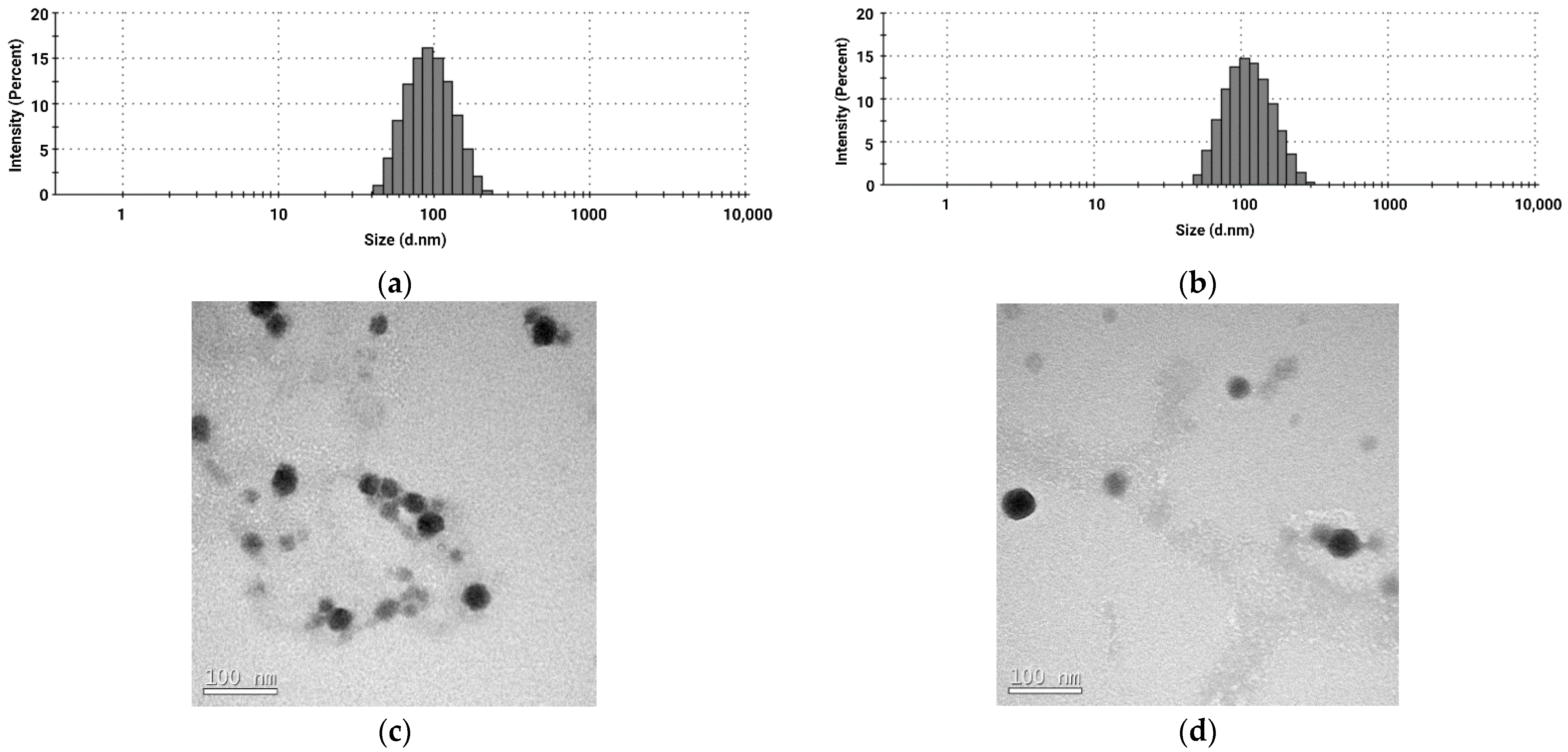
3.1. Preparation and Characterization of Hybrid Nanoparticles

Hybrid nanoparticles carrying siRNA sequences complementary to HOXB7 gene (NP-siHOXB7) and empty hybrid nanoparticles (NP-mock) were prepared by self-assembly of inorganic and organic components. According to the size distribution histogram weighted by intensity (Figure 1a,b), the formulations NP-siHOXB7 and NP-mock presented a Z-average of 87.9 ± 0.54 and 104.96 ± 0.23 nm and PdI of 0.100 ± 0.005 and 0.123 ± 0.004, respectively. Furthermore, TEM images were obtained and showed a homogeneous and spherical morphology (Figure 1c,d).
In addition, the histogram showed a unique narrow pick with height intensity distribution, implying a monodispersed suspension. This assumption was confirmed by the low number of PdI of approximately 0.1 for both formulations [18]. Similar results were obtained by other authors that used PEG-polyanion calcium phosphate nanoparticles [19,20,21].
In addition to the size, PdI and morphology, Zeta potential (ZP) measurements revealed a neutral-like charged suspension of 0.015 ± 0.04 and −1.28 ± 0.1 for NP-siHOXB7 and NP-mock, respectively. This neutral ZP was expected due to the presence of the PEG corona on the surface of the nanoparticle and it is considered an advantageous feature since it prevents non-specific interactions between nanoparticles and culture medium components [22]. Furthermore, the hydrophilic PEG corona promotes steric stabilization, which prevents nanoparticle agglomeration and calcium phosphate crystals growth [19].
Encapsulation efficiency (EE) was analyzed by spectrophotometry by an indirect method using the ultrafiltration/centrifugation technique. The percentage of EE was approximately 65%. The percentage of encapsulated siRNA is inversely proportional to the PEG-polyanion concentration [23]. This relationship is due to the competition between Ca2+ ions and the polyanionic block and siRNA, both negatively charged [19,24].

3.2. Cell Viability and Gene Knockdown

The MCF7 human breast cancer cell line incubated with NP-siHOXB7 were analyzed by MTT assay to evaluate the cytotoxic effect with and without TAM co-treatment. Figure 2 shows cell viability 20, 48 and 72 h after application of NP-siHOXB7 in four different siRNA concentrations: 10, 50, 100 and 150 nM. Treatments employing hybrid nanoparticle with siScramble (NP-siScr), empty nanoparticle (NP-mock) and free siHOXB7 (naked siRNA) were used as controls.
Cell viability was dependent of incubation time and concentration. None of the mentioned treatments and controls showed cytotoxicity after 20 h of incubation. However, after 48 h a reduction of 31% (p < 0.001) in cell viability was observed at 150 nM siRNA. After 72 h, the reduction increased to 38% (p < 0.001) at the same concentration. The same pattern was observed for the treatment with 100 nM siHOXB7. After 48 h, viability was reduced by 12.8%, while in 72 h, the reduction reached 20%. Ma et al. [18] also observed reduced cell viability after siRNA (400 nM) treatment in 48 h.
HOXB7 siRNA sequences were loaded into hybrid nanoparticles and the HOXB7 expression was evaluated after 20 h of incubation by quantitative PCR. The result showed a reduction of approximately 65% (p < 0.05) of the gene expression (Figure 3a). The expressive silencing rate can be related to a high encapsulation rate of siRNA molecules by the nanoparticles and its effectiveness in acting as a transfection agent to MCF7 cells. This result also shows that the designed siRNA sequence was effective in promoting gene silencing.
HOXB7 is a gene that encodes a transcription factor that acts on several metabolic pathways in cancer cell proliferation and survival, which makes the silencing of this gene relevant as an RNAi treatment option. The qPCR result explains what was observed in cell viability experiments. At 20 h, there was HOXB7 gene silencing (Figure 3a), without affecting cell viability as observed in the 20 h MTT assay with NP-siHOXB7 treatment (Figure 2a). Over the time, we could observe the reduction in viability, possibly due to the consummation of already produced HOXB7 protein associated to the gene knockdown.
It is noteworthy that the NP-mock and NP-siScr treatments did not present cytotoxic effects in any incubation period, assuring that the used hybrid nanosystem per se is biocompatible and does not present cell toxicity, as also shown by other authors [20,21,22]. Therefore, we can infer that the cell viability reduction is entirely due to sequence-specific silencing of the HOXB7 gene by siRNA molecule.
Figure 3b shows the results of co-treatment of NP-siHOXB7 at 100 nM combined with different TAM concentrations (0.3 to 30 µM). In a 15 h incubation time, there was a significant difference in cell viability between TAM treatment and co-treatment with NP-siHOXB7 at TAM concentration of 3 µM. The difference between both treatments was a reduction of about 40% (p < 0.001) in cell viability. It is noteworthy that HOXB7 gene silencing sensitizes MCF7 cells to TAM without reducing cell viability as observed in NP-siHOXB7 treatment alone (Figure 2a). The decreased gene expression effect is also observed in co-therapy cell viability assay at 15 h (Figure 3b). Therefore, this result also supports a sensibilization of MCF7 cells to tamoxifen due to HOXB7 silencing.
Some authors have showed HOXB7 gene knockdown in order to better understand its action on cancer pathways. Gene silencing was observed by Ma et al. [18] at an siRNA final concentration of 200 and 400 nM and even higher silencing rates (>80%) were calculated by Wu et al. [7]. However, transfection agents used by these and other studies [7,8,10,18] are toxic and demand a change of culture medium periodically [25]. Other reports used retroviral vectors as transfection agents to carry an shRNA encoder plasmid as gene silencing strategy, which demands a different intracellular pathway since it has to be delivered inside the cell nucleus [23]. Here, we successfully tested a non-toxic effective transfection agent based on biocompatible components for HOXB7 siRNA delivery to breast cancer cells.
Together, these findings demonstrate the relationship between the HOXB7 gene and TAM resistance in ER-positive breast cancer that has yet to be better elucidated. However, they reassure that combined treatments are a promising strategy for anticancer therapy, especially related to tamoxifen resistance.

4. Conclusions

In this work, the nanoparticle formulation used presented suitable physicochemical characteristics and proved to be an effective transfection agent and a key tool to siRNA delivery in breast cancer cells and gene silencing success. In this way, HOXB7 silencing promoted a reduction in cell viability. Furthermore, HOXB7 silencing enhanced the efficacy of tamoxifen treatment by promoting breast cancer cells sensitization in subtoxic concentrations. Here, we demonstrated that the in vitro knockdown of HOXB7 gene by siRNA delivered by PEG-polyanion-coated hybrid nanoparticles combined with tamoxifen is a promising tool for ER-positive breast cancer treatment and should be analyzed in vivo in future research.

Author Contributions

Conceptualization, F.P. and A.B.C.d.S.V.; methodology, A.B.C.d.S.V., A.C.G., K.C.B.F., T.B.C.-P., J.G., G.D.T. and F.P.; validation, A.B.C.d.S.V., A.C.G.; formal analysis, A.B.C.d.S.V., A.C.G., K.C.B.F. and F.P.; resources, F.P.; writing—original draft preparation, A.B.C.d.S.V.; writing—review and editing, G.D.T. and F.P.; supervision, F.P.; funding acquisition, F.P. All authors have read and agreed to the published version of the manuscript.

Funding

This research was funded by Conselho Nacional de Desenvolvimento Científico e Tecnológico (CNPq), grant numbers 459847/2014-7 and 435395/2018-1 and the Fundação de Amparo à Pesquisa do Estado de Minas Gerais, grant number CDS—APQ-00948-14.

Acknowledgments

A.B.C.d.S.V. acknowledges the fellowship from Coordenação de Aperfeiçoamento de Pessoal de Nível Superior (CAPES) and the cooperation of the Integrated Research Laboratory (Laboratório Integrado de Pesquisa—LIP) of Juiz de For a Federal University.

Conflicts of Interest

The authors declare no conflict of interest.

References

  1. World Health Organization. Cancer: Key Facts. Available online: https://www.who.int/en/news-room/fact-sheets/detail/cancer (accessed on 14 October 2020).
  2. Bray, F.; Ferlay, J.; Soerjomataram, I.; Siegel, R.L.; Torre, L.A.; Jemal, A. Global cancer statistics 2018: GLOBOCAN estimates of incidence and mortality worldwide for 36 cancers in 185 countries. CA Cancer J. Clin. 2018, 68, 394–424. [Google Scholar] [CrossRef] [PubMed]
  3. American Cancer Society. Treatment Types. Available online: https://www.cancer.org/treatment/treatments-and-side-effects/treatment-types.html (accessed on 15 October 2020).
  4. Favier, B.; Dolle, P. Developmental functions of mammalian Hox genes. Mol. Hum. Reprod. 1997, 3, 115–131. [Google Scholar] [CrossRef] [PubMed]
  5. Shah, N.; Sukumar, S. The Hox genes and their roles in oncogenesis. Nat. Rev. 2010, 10, 361–371. [Google Scholar] [CrossRef] [PubMed]
  6. Caré, A.; Silvani, A.; Meccia, E.; Mattia, G.; Stoppacciaro, A.; Parmiani, G.; Peschle, C.; Colombo, M.P. HOXB7 constitutively activates basic fibroblast growth factor in melanomas. Mol. Cell. Biol. 1996, 16, 4842–4851. [Google Scholar] [CrossRef]
  7. Wu, X.; Chen, H.; Parker, B.; Rubin, E.; Zhu, T.; Lee, J.S.; Argani, P.; Sukumar, S. HOXB7, a homeodomain protein, is overexpressed in breast cancer and confers epithelial-mesenchymal transition. Cancer Res. 2006, 66, 9527–9534. [Google Scholar] [CrossRef]
  8. Jin, K.; Sukumar, S. A pivotal role for HOXB7 protein in endocrine resistant breast cancer. Oncoscience 2015, 2, 917–919. [Google Scholar] [CrossRef]
  9. Rubin, E.; Wu, X.; Zhu, T.; Cheung, J.C.; Chen, H.; Lorincz, A.; Pandita, R.K.; Sharma, G.G.; Ha, H.C.; Gasson, J.; et al. A role for the HOXB7 homeodomain protein in DNA repair. Cancer Res. 2007, 67, 1527–1535. [Google Scholar] [CrossRef]
  10. Jin, K.; Kong, X.; Shah, T.; Penet, M.F.; Wildes, F.; Sgroi, D.C.; Ma, X.J.; Huang, Y.; Kallioniemi, A.; Landberg, G.; et al. The HOXB7 protein renders breast cancer cells resistant to tamoxifen through activation of the EGFR pathway. Proc. Natl. Acad. Sci. USA 2012, 109, 2736–2741. [Google Scholar] [CrossRef]
  11. Jin, K.; Park, S.; Teo, W.W.; Korangath, P.; Cho, S.S.; Yoshida, T.; Győrffy, B.; Goswami, C.P.; Nakshatri, H.; Cruz, L.A.; et al. HOXB7 is an ERα cofactor in the activation of HER2 and multiple ER target genes leading to endocrine resistance. Cancer Discov. 2015, 5, 944–959. [Google Scholar] [CrossRef]
  12. American Cancer Society. Available online: https://www.cancer.org (accessed on 15 October 2020).
  13. Hyman, E.; Kauraniemi, P.; Hautaniemi, S.; Wolf, M.; Mousses, S.; Rozenblum, E.; Ringnér, M.; Sauter, G.; Monni, O.; Elkahloun, A.; et al. Impact of DNA Amplification on Gene Expression Patterns in Breast Cancer. Cancer Res. 2002, 62, 6240–6245. [Google Scholar]
  14. Mihály, Z.; Kormos, M.; Lánczky, A.; Dank, M.; Budczies, J.; Szász, M.A.; Győrffy, B. A meta-analysis of gene expression-based biomarkers predicting outcome after tamoxifen treatment in breast cancer. Breast Cancer Res. Treat. 2013, 140, 219–232. [Google Scholar] [CrossRef] [PubMed]
  15. Menck, C.F.M. A nova grande promessa da inovação em fármacos: RNA interferência saindo do laboratório para a clínica. Estud. Avançados 2010, 24, 99–108. [Google Scholar] [CrossRef]
  16. Fire, A.; Xu, S.; Montgomery, M.K.; Kostas, S.A.; Driver, S.E.; Mello, C.C. Potent and specific genetic interference by double-stranded RNA in Caenorhabditis elegans. Nature 1998, 391, 806–811. [Google Scholar] [CrossRef]
  17. Mosmann, T. Rapid colorimetric assay for cellular growth and survival: Application to proliferation and cytotoxicity assays. J. Immunol. Methods 1983, 65, 55–63. [Google Scholar] [CrossRef]
  18. Ma, D. Enhancing Endosomal Escape for Nanoparticle Mediated siRNA. Nanoscale 2014, 6, 6415. [Google Scholar] [CrossRef] [PubMed]
  19. Kakizawa, Y.; Miyata, K.; Furukawa, S.; Kataoka, K. Size-controlled formation of a calcium phosphate-based organic-inorganic hybrid vector for gene delivery using poly(ethylene glycol)-block-poly(aspartic acid). Adv. Mater. 2004, 16, 699–702. [Google Scholar] [CrossRef]
  20. Kakizawa, Y.; Kataoka, K. Block copolymer self-assembly into monodisperse nanoparticles with hybrid core of antisense DNA and calcium phosphate. Langmuir 2002, 18, 4539–4543. [Google Scholar] [CrossRef]
  21. de Mello, L.J.; Souza, G.R.; Winter, E.; Silva, A.H.; Pittella, F.; Creczynski-Pasa, T.B. Knockdown of antiapoptotic genes in breast cancer cells by siRNA loaded into hybrid nanoparticles. Nanotechnology 2017, 28, 175101. [Google Scholar] [CrossRef]
  22. Pittella, F.; Miyata, K.; Maeda, Y.; Suma, T.; Watanabe, S.; Chen, Q.; Christie, R.J.; Osada, K.; Nishiyama, N.; Kataoka, K. Pancreatic cancer therapy by systemic administration of VEGF siRNA contain in calcium phosphate/charge-conversional polymer hybrid nanoparticles. J. Control. Release 2012, 161, 868–874. [Google Scholar] [CrossRef]
  23. Pittella, F.; Zhang, M.; Lee, Y.; Kim, H.J.; Tockary, T.; Osada, K.; Ishii, T.; Miyata, K.; Nishiyama, N.; Kataoka, K. Enhanced endosomal escape of siRNA-incorporating hybrid nanoparticles from calcium phosphate and PEG-block charge conversional polymer for efficient gene knockdown with negligible cytotoxicity. Biomaterials 2011, 32, 3106–3114. [Google Scholar] [CrossRef]
  24. Kakizawa, Y.; Furukawa, S.; Kataoka, K. Block copolymer-coated calcium phosphate nanoparticles sensing intracellular environment for oligodeoxynucleotide and siRNA delivery. J. Control. Release 2004, 97, 345–356. [Google Scholar] [CrossRef] [PubMed]
  25. Wang, T.; Larcher, L.M.; Ma, L.; Veedu, R.N. Systematic Screening of Commonly Used Commercial Transfection Reagents towards Efficient Transfection of Single-Stranded Oligonucleotides. Molecules 2018, 23, 2564. [Google Scholar] [CrossRef] [PubMed]
Figure 1. Size distribution histogram of hybrid nanoparticles and TEM images. (a) Histogram of hybrid nanoparticles carrying siRNA; (b) Histogram of empty hybrid nanoparticles; (c,d) TEM images of hybrid nanoparticles.
Materproc 04 00069 g001
Figure 2. Cell viability evaluation after NP-siHOXB7 treatment on MCF7 breast cancer cells at (a) 20 h, (b) 48 h, and (c) 72 h. One-way ANOVA followed by Tukey test, n = 6, * p < 0.05, *** p < 0.001, compared with control. NP-siHOXB7 = hybrid nanoparticle carrying siHOXB7; NP-siScr = hybrid nanoparticle carrying scrambled siRNA; NP-mock = empty hybrid nanoparticle; Naked = free siHOXB7.
Materproc 04 00069 g002
Figure 3. (a) Knockdown of HOXB7 gene expression in human ER-positive breast cancer cells after 20 h incubation with NP-siHOXB7 (one-way ANOVA followed by Tukey test, n = 3, * p < 0.05, compared with control). (b) Cell viability evaluation after NP-siHOXB7 and tamoxifen co-treatment on MCF7 breast cancer cells at 15 h. One-way ANOVA followed by Tukey test, n = 6, * p < 0.05, *** p < 0.001, compared with free TAM. NP-siHOXB7 = hybrid nanoparticle carrying siHOXB7; NP-siScr = hybrid nanoparticle carrying scrambled siRNA; Naked = free siHOXB7; TAM = tamoxifen.
Materproc 04 00069 g003
Publisher’s Note: MDPI stays neutral with regard to jurisdictional claims in published maps and institutional affiliations.

