
Green Synthesis of ZnO-NPs by Juglans regia Green Husk Aqueous Extract †


Abstract
:1. Introduction
2. Materials and Methods
2.1. Materials
2.2. Biosynthesis of ZnO-NPs
2.3. UV-Vis Spectroscopy
2.4. DLS Analysis
2.5. TEM Analysis
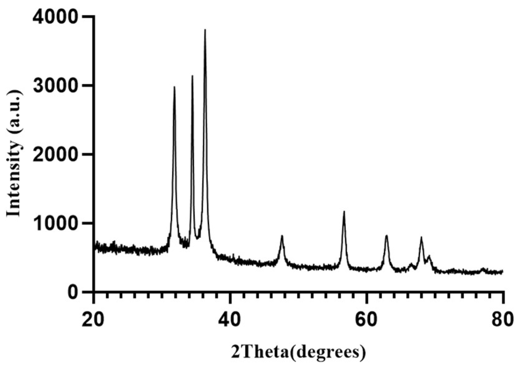
2.6. XRD Analysis
2.7. FT-IR Analysis
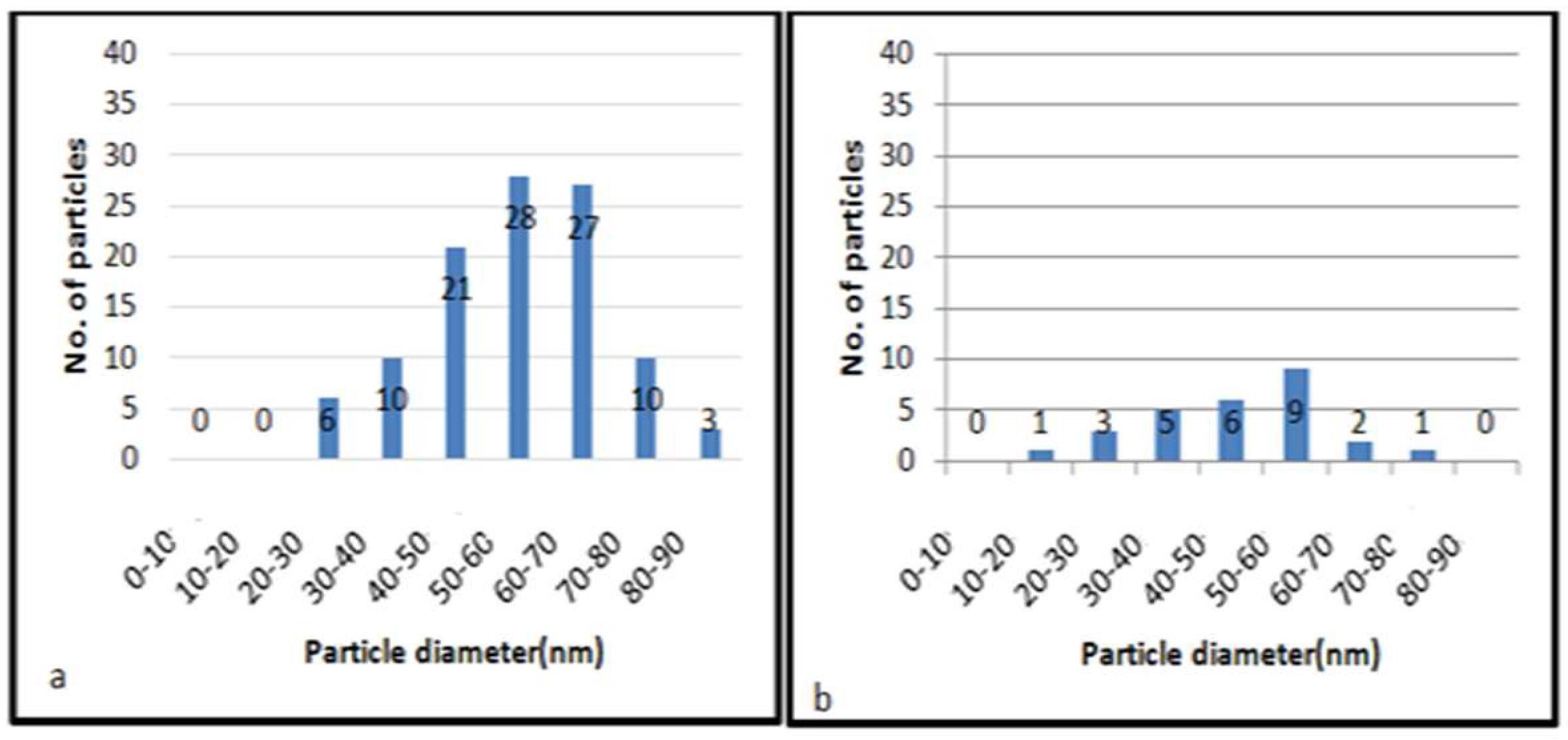
3. Results and Discussion
4. Conclusions
Author Contributions
Funding
Institutional Review Board Statement
Informed Consent Statement
Data Availability Statement
Acknowledgments
Conflicts of Interest
References
- Marchiol, L. Synthesis of metal NPs in living plants. Ital. J. Agron. 2012, 3, 274–282. [Google Scholar]
- Basnet, P.; Chanu, T.; Samanta, D.; Chatterjee, S. A review on bio-synthesized ZnO-NPs using plant extracts as reductants and stabilizing agents. J. Photochem. Photobiol. B Biol. 2018, 183, 201–221. [Google Scholar] [CrossRef]
- Laurent, S.; Forge, D.; Port, M.; Roch, A.; Robic, C.; Vander, E.L.; Muller, R. Magnetic Iron Oxide NPs: Synthesis, Stabilization, Vectorization, Physicochemical Characterizations, and Biological Applications. Chem. Rev. 2010, 108, 2064–2110. [Google Scholar] [CrossRef]
- Ali, S.H.; Khan, I.; Khan, S.; Sohail, M. Electrocatalytic performance of Ni@Pt core–shell NPs supported on carbon nanotubes for methanol oxidation reaction. J. Electroanal. Chem. 2017, 795, 17–25. [Google Scholar] [CrossRef]
- Sun, S.H.; Murray, C.B.; Weller, D.; Folks, L.; Moser, A. Monodisperse FePt NPs and Ferromagnetic FePt Nanocrystal Superlattices. Science 2000, 80, 1989–1992. [Google Scholar] [CrossRef]
- Bacaksiz, E.; Parlak, M.; Tomakin, M.; Özçelik, A.; Karakız, M.; Altunbaş, M. The effect of zinc nitrate, zinc acetate and zinc chloride precursors on investigation of structural and optical properties of ZnO thin films. J. Alloy. Compd. 2008, 466, 447–450. [Google Scholar] [CrossRef]
- Suresh, D.; Nethravathi, P.C.; Udayabhanu, H.; Nagabhushana, H.; Sharma, S.C. Green synthesis of multifunctional zinc oxide (ZnO) NPs using Cassia fistula plant extract and their photodegradative, antioxidant and antibacterial activities. Mater. Sci. Semicond. Process. 2015, 31, 446–454. [Google Scholar] [CrossRef]
- Narendhran, S.; sivaraj, R. Biogenic ZnO NPs synthesized using L. aculeata leaf extract and their antifungal activity against plant fungal pathogens. Indian Acad. Sci. 2015, 39, 1–5. [Google Scholar]
- Nagajyothi, P.C.; Ju Cha, S.; Jun Yang, I.; Sreekanth, T.V.M.; Joong Kim, K.; Mook Shin, H. Antioxidant and anti-inflammatory activities of ZnO-NPs synthesized using Polygala tenuifolia root extract. J. Photochem. Photobiol. B Biol. 2015, 146, 10–17. [Google Scholar] [CrossRef]
- Mirzaei, H.; Darroudi, M. ZnO-NPs: Biological synthesis and biomedical applications. Ceram. Int. 2017, 43, 907–914. [Google Scholar] [CrossRef]
- Tamer, T.M.; Abou-Taleb, V.M.; Roston, G.D.; Mohyeldin, M.S.; Omer, A.M.; Hafez, A.M. Formation of ZnO-NPs using alginate as a template for purification of waste water. Environ. Nanotechnol. Monit. Manag. 2018, 10, 112–121. [Google Scholar]
- Iravani, S. Green synthesis of metal NPs using plants. Green Chem. 2011, 13, 2638–2650. [Google Scholar] [CrossRef]
- Kalaiarasi, R.; Jayallakshmi, N.; Venkatachalam, P. Phytosynthesis of NPs and its applications. Plant Cell Biotechnol. Mol. Biol. 2010, 11, 1–16. [Google Scholar]
- Dobrucka, R.; Dugaszewska, J. Biosynthesis and antibacterial activity of ZnO NPs using Trifolium pratense flower extract. Saudi J. Biol. Sci. 2015, 23, 517–523. [Google Scholar] [CrossRef]
- Ali, K.; Dwivedi, S.; Azam, A.; Saquib, Q.; Al-Said, M.S.; Alkhedhairy, A.A.; Musarrat, J. Aloe vera extract functionalized ZnO-NPs as nanoantibiotics against multi-drug resistant clinical bacterial isolates. J. Colloid Interface Sci. 2016, 472, 145–156. [Google Scholar] [CrossRef]
- Abbasi, Z.; Feizi, S.H.; Taghipour, E.; Ghadam, P. Green synthesis of silver NPs using aqueous extract of dried Juglans regia green husk and examination of its biological properties. Green Process. Synth. 2017, 6, 477–485. [Google Scholar] [CrossRef]
- Darroudi, M.; Sabouri, Z.; Kazemi Oskuee, R.; Kargar, H.; Ali Hosseini, H. Neuronal toxicity of biopolymer-template synthesized ZnO NPs. Nanomed. J. 2013, 2, 88–93. [Google Scholar]
- Shah, M.; Fawcett, D.; Sharma, S.; Kumar, T.; Gérrard Eddy Jai Poinern, G. Green Synthesis of Metallic NPs via Biological Entities. Materials 2015, 11, 7278–7308. [Google Scholar] [CrossRef]
- Zhai, T.; Fang, X.; Liao, M.; Xu, X.; Zeng, H.; Yoshio, B.; Golberg, D. A Comprehensive Review of One-Dimensional Metal-Oxide Nanostructure Photodetectors. Sensors 2009, 9, 6504–6529. [Google Scholar] [CrossRef]
- Glaspell, G.; Dutta, P.; Manivannan, A. A Room-Temperature and Microwave Synthesis of M-Doped ZnO (M=Co, Cr, Fe, Mn & Ni). J. Clust. Sci. 2005, 16, 524–536. [Google Scholar]
- Delaviz, H.; Mohammadi, J.; Ghalamfarsa, G.H.; Mohammadi, B.; Farhadi, N. A Review Study on Phytochemistry and Pharmacology Applications of Juglans Regia Plant. Pharmacogen 2017, 22, 145–152. [Google Scholar]
- Rana, N.; Chand, S.; Gathania, A.K. Green synthesis of zinc oxide nano-sized spherical particles using Terminalia chebula fruits extract for their photocatalytic applications. Int. Nano Lett. 2015, 6, 91–98. [Google Scholar] [CrossRef]
- Sharma, D.; Sabela, M.I.; Kanchi, S.; Mdluli, P.S.; Singh, G.; Stenström, T.A.; Bisetty, K. Biosynthesis of ZnO NPs using Jacaranda mimosifolia flowers extract: Synergistic antibacterial activity and molecular simulated facet specific adsorption studies. J. Photochem. Photobiol. B Biol. 2016, 162, 199–207. [Google Scholar] [CrossRef] [PubMed]
- Elumalaia, K.; Velmurugan, S. Green synthesis, characterization and antimicrobial activities of zincoxide NPs from the leaf extract of Azadirachta indica (L.). Appl. Surf. Sci. 2015, 345, 329–336. [Google Scholar] [CrossRef]
- Kerker, M. The Scattering of Light and Other Electromagnetic Radiation; Academic Press: New York, NY, USA, 1969. [Google Scholar]
- Kreibig, U.; Vollmer, M. Optical Properties of Metal Clusters; Springer: Berlin/Heidelberg, Germany, 1995. [Google Scholar]
- Liz-Marzan, L.M. Tailoring Surface Plasmons through the Morphology and Assembly of Metal NPs. Langmuir 2005, 22, 32–41. [Google Scholar] [CrossRef]
- Sangeetha, G.; Rajeshwari, S.; Venckatesh, R. Green synthesis of ZnO-NPs by aloe barbadensis miller leaf extract: Structure and optical properties. Mater. Res. Bull. 2011, 46, 2560–2566. [Google Scholar] [CrossRef]
- Fowsiya, J.; Madhumitha, G.; Al-Dhabi, N.A.; Valan Arasu, M. Photocatalytic degradation of Congo red using Carissa edulis extract capped ZnO-NPs. J. Photochem. Photobiol. B Biol. 2016, 162, 395–401. [Google Scholar] [CrossRef]
- Mourdikoudis, S.; Pallares, R.M.; Thanh, N.T.K. Characterization techniques for NPs: Comparison and complementarity upon studying NP properties. R. Soc. Chem. Nanoscale 2018, 10, 12871–12934. [Google Scholar] [CrossRef]
- Jafarirad, S.; Mehrabi, M.; Divband, B.; Kosari-Nasab, M. Biofabrication of ZnO-NPs using fruit extract of Rosa canina and their toxic potential against bacteria: A mechanistic approach. Mater. Sci. Eng. C 2015, 59, 296–302. [Google Scholar] [CrossRef]
- Çolak, H.; Karaköse, E. Green synthesis and characterization of nanostructured ZnO thin films using Citrus aurantifolia (lemon) peel extract by spin-coating method. J. Alloy. Compd. 2016, 690, 658–662. [Google Scholar] [CrossRef]
- Ramesh, M.; Anbuvannan, M.; Viruthagiri, G. Green synthesis of ZnO NPs using Solanum nigrum leaf extract and their antibacterial activity. Mol. Biomol. Spectrosc. 2014, 136, 864–870. [Google Scholar] [CrossRef] [PubMed]
- Rajiv, P.; Rajeshwari, S.; Venckatesh, R. Bio-Fabrication of ZnO-NPs using leaf extract of Parthenium hysterophorus L. and its size-dependent antifungal activity against plant fungal pathogens. Spectrochim. Acta Part A Mol. Biomol. Spectrosc. 2013, 112, 384–387. [Google Scholar] [CrossRef] [PubMed]


Disclaimer/Publisher’s Note: The statements, opinions and data contained in all publications are solely those of the individual author(s) and contributor(s) and not of MDPI and/or the editor(s). MDPI and/or the editor(s) disclaim responsibility for any injury to people or property resulting from any ideas, methods, instructions or products referred to in the content. |
© 2023 by the authors. Licensee MDPI, Basel, Switzerland. This article is an open access article distributed under the terms and conditions of the Creative Commons Attribution (CC BY) license (https://creativecommons.org/licenses/by/4.0/).
Share and Cite
Dehghani, M.; Ghadam, P. Green Synthesis of ZnO-NPs by Juglans regia Green Husk Aqueous Extract. Mater. Proc. 2023, 14, 7. https://doi.org/10.3390/IOCN2023-14444
Dehghani M, Ghadam P. Green Synthesis of ZnO-NPs by Juglans regia Green Husk Aqueous Extract. Materials Proceedings. 2023; 14(1):7. https://doi.org/10.3390/IOCN2023-14444
Chicago/Turabian StyleDehghani, Marzieh, and Parinaz Ghadam. 2023. "Green Synthesis of ZnO-NPs by Juglans regia Green Husk Aqueous Extract" Materials Proceedings 14, no. 1: 7. https://doi.org/10.3390/IOCN2023-14444