1. Introduction
The human epidermal growth factor receptor 2 (HER2) is the 185 kDa transmembrane glycoprotein member of the HER family essential for normal cell growth, differentiation, and migration [
1]. HER2 overexpression has been identified in several solid tumors such as breast, prostate, and gastric cancer, and it is associated with aggressive behavior, resistance, and a poor prognosis [
2]. Therefore, the monoclonal antibody trastuzumab (Herceptin) is used as standard targeted therapy for HER2-positive breast cancer. Nonetheless, it has been associated with resistance, low response, and cardiotoxicity [
3].
Recent advances in nanotechnology have highlighted the potential of its application in biomedicine for diagnosis, drug administration, and imaging studies for various human diseases, materials called “theranostics”. Within metal-based nanosystems, gold nanoparticles (AuNPs) are widely used due to their unique optical properties such as localized surface plasmon resonance (LSPR) and the ability to modify their surface for a particular interest [
4]. Surface coating with polymers such as polyethylene glycol (PEG) is widely used as it reduces toxicity and improves stability. AuNPs have been conjugated with different molecules for diagnoses [
5] and targeted cancer treatment [
6].
Aptamers are short single-stranded deoxyribonucleic acid (DNA) or ribonucleic acid (RNA) oligonucleotides, capable of forming tertiary structures that bind to specific targets, from small molecules to proteins and cells. Compared to antibodies, aptamers have more stability, low immune reaction, lower production cost, and the ability to cross biological barriers [
7]. The use of aptamers as recognition molecules in the creation of sensors for detecting cancer cells that express different biomarkers has been reported [
8]. This study investigated the potential of aptamer-functionalized AuNPs as a theranostic tool, comparing their effect with the monoclonal antibody trastuzumab and the ability to detect HER2-overexpressing cells.
2. Materials and Methods
2.1. Nanosystem Assembly
AuNP-PEG-AptHer2 nanosystem conjugation was carried out through a maleimide-thiol link between a 20 nm gold nanoparticle coated with 5 kDa PEG from the Cytodiagnostics conjugation kit OLIGOREADY (Burlington, ON, Canada) and AptHer2 aptamer previously reported with the following sequence: 5′-/5ThioMC6-D/TCT AAA AGG ATT CTT CCC AAG GGG ATC CAA TTC AAA CAG/3ATTO647NN/-3′ (Integrated DNA Technologies, Coralville, IA, USA) [
9].
2.2. Characterization
AuNP-PEG-AptHer2 absorption spectra were obtained with UV-Vis spectroscopy using a Nanodrop ND-(Thermo Fisher Scientific, Inc., Waltham, MA, USA). The hydrodynamic diameter by dynamic light scattering (DLS) and ζ-potential measurements were carried out on a Zetasizer Nano ZS instrument (Malvern). Morphology and size were analyzed in the electron transmission microscope (TEM) JEM-2200FS (Jeol Ltd., Tokyo, Japan).
2.3. Viability Assay
The nanosystem effect on cellular viability was tested on the Vero CCL-81 cell line as a non-cancerous control, and three cancerous cell lines were used: LNCaP with low HER2 expression, ZR-75-30 with HER2 overexpression and HCC1954 with HER2 overexpression and resistance to trastuzumab. In total, 10,000 cells per well seeded in 96-well plates were incubated for 24 h at standard conditions of 37 °C with 5% CO2 in a humidified incubator. The cells were treated with a nanosystem (0.05, 0.5, and 5 µg/mL) or trastuzumab (Roche, Tuas Bay Link, Singapore) (0.01, 0.1, and 1 µg/mL) and incubated for 24 h at 37 °C. After 24 h of treatment, 100 µL of a 10% MTT (3-[4, 5-dimethylthiazol-2-yl]-2,5-diphenyltetrazolium bromide) solution was added to each well and incubated for 3 h at 37 °C. Formazan crystals were resuspended adding 100 µL of acidic isopropanol. The absorbance was measured at 570 nm and 651 nm using the Cytation 3 multi-mode reader (Bio-Tek, Winooski, VT, USA) .
2.4. Hemolysis Assay
From a globular package, an erythrocyte suspension was made by taking 100 µL of the erythrocyte concentrate and 9800 µL of PBS 1X. In total, 16 µL of the nanosystem (final concentration of 0.05, 0.5, and 5 µg/mL), trastuzumab (final concentration of 0.01, 0.1, and 1 µg/mL), distilled H2O (positive control), or PBS (negative control) were incubated with 4 µL of the erythrocyte suspension for 30 min at 37 °C and 300 rpm in a Thermomixer (Eppendorf, Hamburg, Germany). After centrifugation at 14,000 rpm for 3 min, the release of hemoglobin was read in a NanoDrop ND-100 (Thermo Fisher Scientific, Inc., Waltham, MA, USA) at 415 nm.
2.5. Fluorescence Detection
We evaluated the nanosystem’s in vitro binding capacity and specificity, depending on the HER2 expression level; 100 µL of a Vero CCL-81 or ZR-75-30 cell suspension (1 × 10⁶ cells) were incubated with 200 µL of the nanosystem (5 µg/mL) and incubated for 30 min at 37 °C with 5% CO2. The tubes were centrifuged at 1000 rpm for 5 min, washed with PBS 1X, and resuspended in a fresh culture medium. The fluorescence signal of the fluorophore ATTO 647N present in the AptHer2 aptamer was analyzed at 664 nm in the Countess II FL Automated Cell Counter instrument (Thermo Fisher Scientific, Inc., Waltham, MA, USA).
2.6. Statistical Analysis
Each experiment contained three individual replications. Data are shown as mean ± standard deviation (SD). Statistical analyses were assessed using GraphPad Prism 5.1 software (GraphPad software, Inc., California, USA) and consisted of the two-way analysis of variance (ANOVA) and Bonferroni post hoc, considering significance when p-values were below 0.05.
3. Results and Discussion
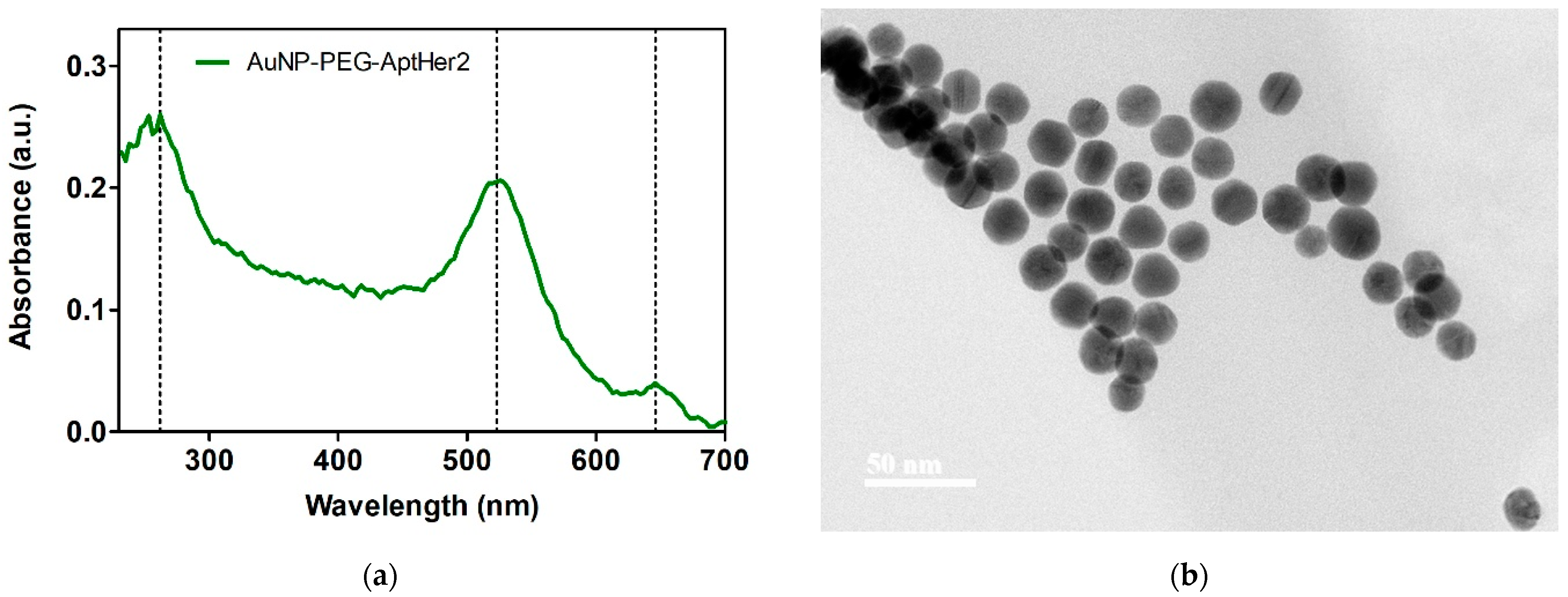
The conjugation of AuNP-PEG with AptHer2 shows an absorption spectrum (
Figure 1a) with three main peaks; the peak at 260 nm corresponding to nucleic acid absorption, the peak at 523 nm corresponding to the localized surface plasmon resonance (LSPR) of gold nanoparticles, and the peak at 646 nm corresponding to the fluorophore ATTO647N absorption.
TEM micrography (
Figure 1b) demonstrates a uniform spherical morphology with an average core diameter of 19.21 ± 1.86 nm. The separation between nanoparticles could be due to electrostatic repulsion between them, giving colloidal stability [
10].
DLS and ζ potential measurements are shown in
Table 1. The nanosystem possessed a hydrodynamic diameter of 65.30 ± 1.12 nm. This increment in the diameter of the 20 nm core gold nanoparticle could be due to the coating of PEG 5 kDa and conjugation with the AptHer2 aptamer to the surface [
11]. Furthermore, ζ potential measurements confirmed a superficial negative charge of −17.4 ± 0.68 mV, suggesting the correct conjugation of the HER2-specific aptamer to AuNP-PEG [
12].
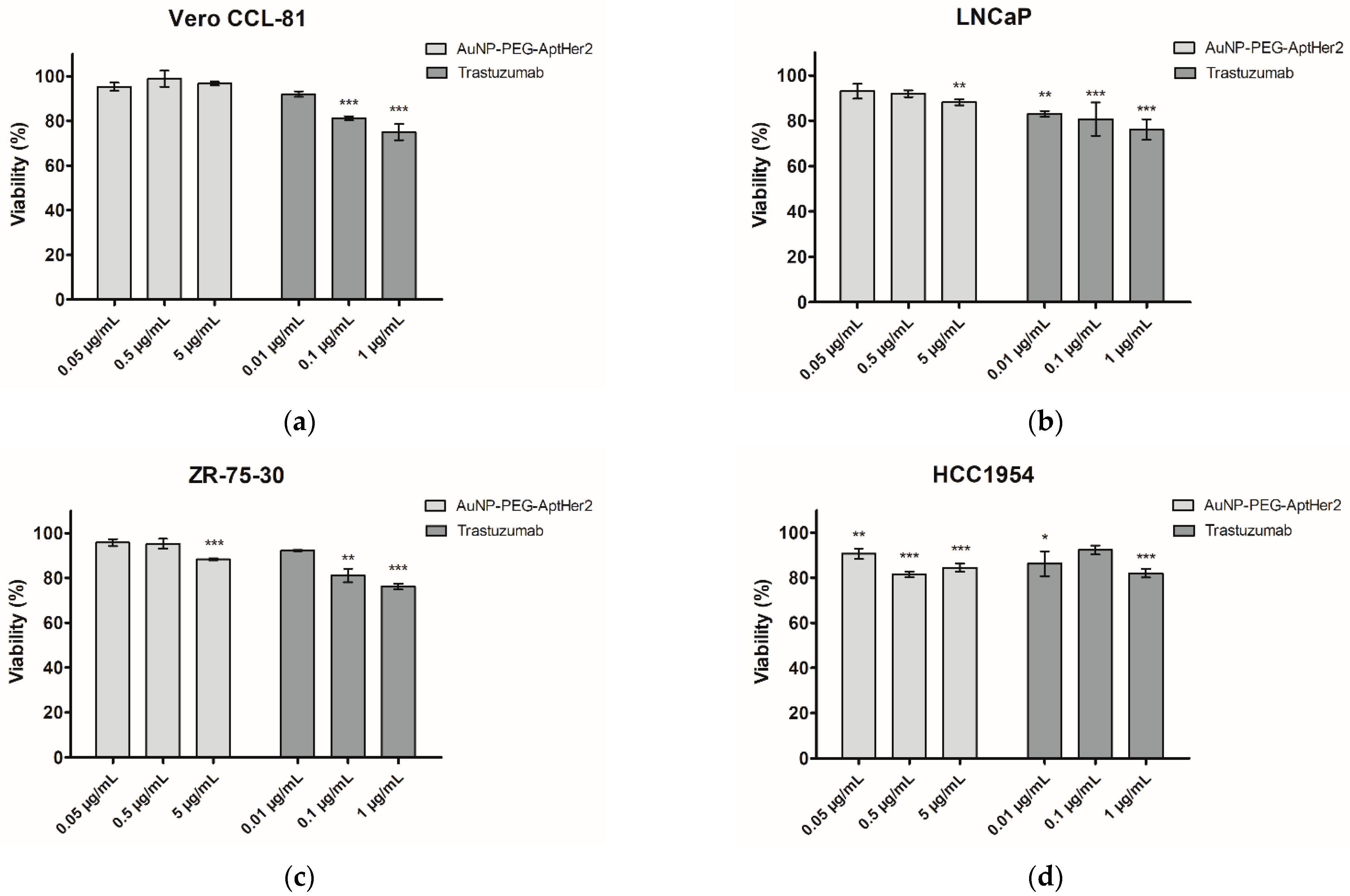
Viability assays in the non-cancerous Vero CCL-81 cell line demonstrated a nonspecific cytotoxic effect with trastuzumab treatment, exerting a reduction of 25% viability at the greatest evaluated concentration (
Figure 2a). The LNCaP prostate cancer cell line (
Figure 2b) presented a reduction in cell viability when treated with 5 µg/mL of the nanosystem, with a more significant effect when treated with trastuzumab in all concentrations evaluated. The ZR-75-30 breast cancer cell line with HER2 overexpression (
Figure 2c) demonstrated a reduction in viability when treated with the AuNP-PEG-AptHer2 nanosystem and trastuzumab. Interestingly, when treated with the nanosystem at all evaluated concentrations, the HCC1954 breast cancer cell line with HER2 overexpression and trastuzumab resistance (
Figure 2d) presented a significant cell viability reduction.
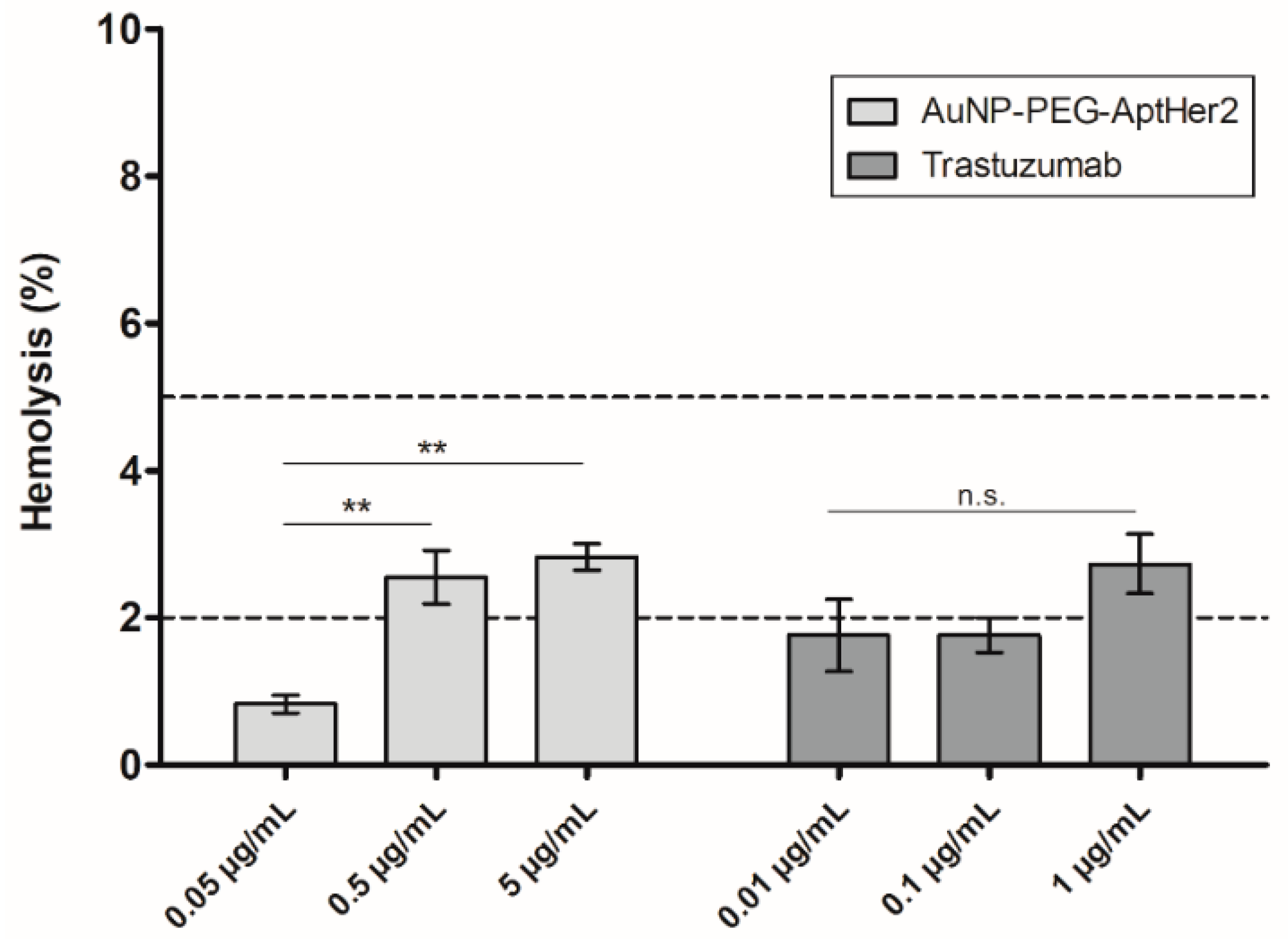
The interpretation of hemolysis results was made based on the ASTM-F756-17 standard practice for the evaluation of the hemolytic properties of materials, where materials are classified as non-hemolytic when they cause hemolysis of less than 2%, slightly hemolytic when hemolysis is less than 5%, and highly hemolytic when the material presents hemolysis greater than 5%.
The hemolysis assay (
Figure 3) of the AuNP-PEG-AptHer2 nanosystem demonstrated no hemolytic activity at 0.05 µg/mL, while it was slightly hemolytic at 0.5 and 5 µg/mL with hemolysis of 2.5% and 2.8%, respectively. Incubation with trastuzumab showed no hemolytic activity at 0.01 and 0.1 µg/mL, while there was slight hemolysis of 2.7% at 1 µg/mL.
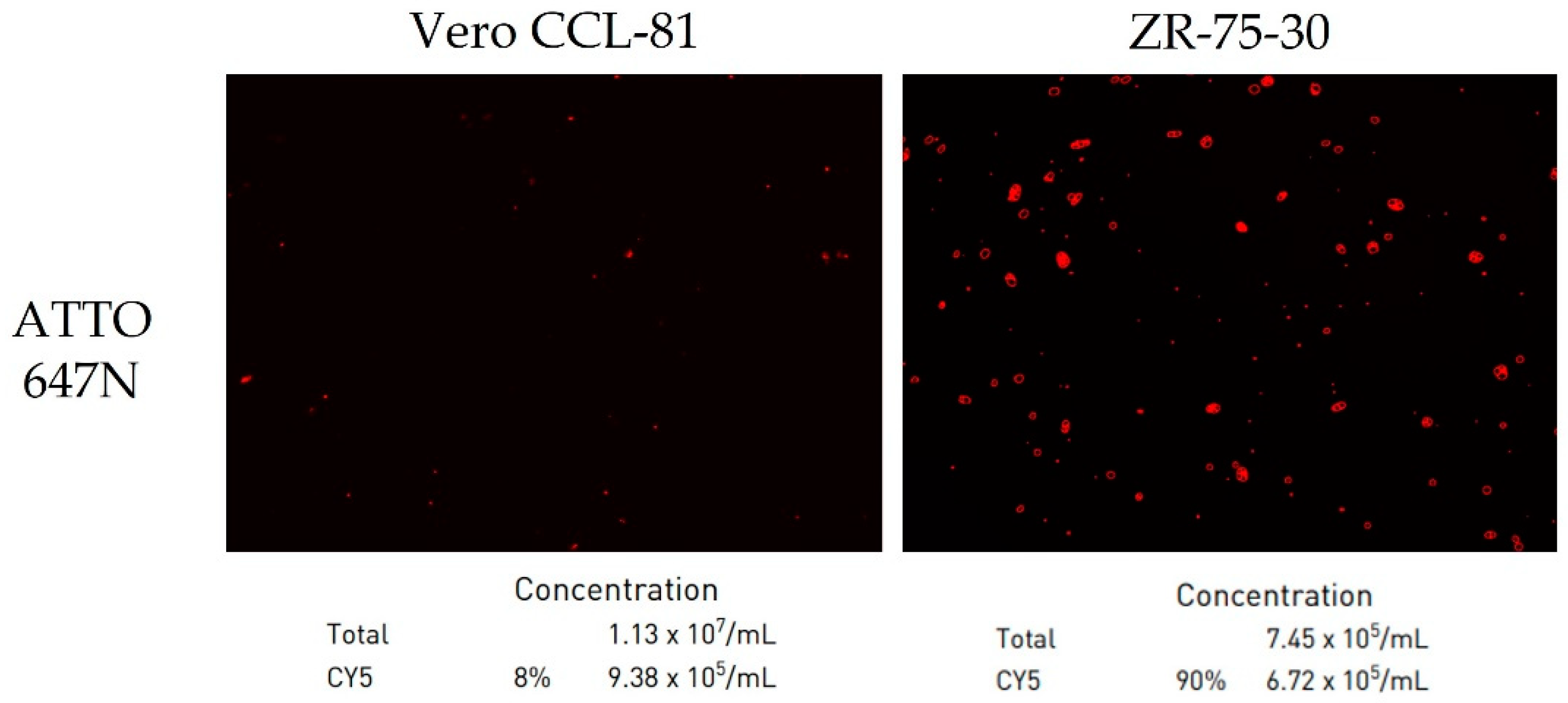
Images of fluorescence detection assays are presented in
Figure 4. Incubation of the non-cancerous control Vero CCL-81 cell line with the AuNP-PEG-AptHer2 nanosystem demonstrated a low fluorescence intensity of 8%, while incubation of the ZR-75-30 HER2-overexpressing cell line with the AuNP-PEG-AptHer2 nanosystem demonstrated a high fluorescence intensity of 90%, suggesting the nanosystem can specifically target and bind to HER2-overexpressing cells.
4. Conclusions
In this study, we assembled a nanosystem based on 5 kDa PEG-coated gold nanoparticles and the HER2-targeting aptamer, obtaining a hydrodynamic diameter of 65.30 ± 1.12 nm and superficial charge of −17.4 ± 0.68 mV, with a spherical morphology and dispersed state.
The AuNP-PEG-AptHer2 nanosystem selectively reduced cell viability in HER2-overexpressing cell lines, while trastuzumab showed a nonspecific effect on low-HER2-expressing cells and non-cancerous control cells. Slight to no hemolytic activity was found at the evaluated concentrations. Additionally, fluorescence detection suggests that nanosystems can specifically bind to targeted cells. These results provide insight into the potential of targeted gold nanoparticles in cancer theranostics.
Author Contributions
Conceptualization, C.N.S.-D. and H.L.G.-B.; methodology, P.Y.C.-G.; validation, P.Y.C.-G. and J.A.R.-P.; formal analysis, P.Y.C.-G., C.N.S.-D. and H.L.G.-B.; investigation, P.Y.C.-G.; resources, C.N.S.-D., H.L.G.-B., E.N.G.-T., J.R.D.-B. and M.S.-D.; data curation, P.Y.C.-G.; writing—original draft preparation, P.Y.C.-G.; writing—review and editing, P.Y.C.-G., C.N.S.-D. and H.L.G.-B.; visualization, P.Y.C.-G., C.N.S.-D. and H.L.G.-B.; supervision, C.N.S.-D. and H.L.G.-B.; project administration, C.N.S.-D. and H.L.G.-B.; funding acquisition, C.N.S.-D. and H.L.G.-B. All authors have read and agreed to the published version of the manuscript.
Funding
This research was funded by Consejo Nacional de Ciencia y Tecnología (CONACYT), grant number A1-S-9859, and PAICYT UANL, grant number 147-CS-2022.
Institutional Review Board Statement
The present study was approved by the Institutional Ethics Committee from the School of Medicine and University Hospital “Dr. José Eleuterio González”, Universidad Autónoma de Nuevo León, México (BI23-00004).
Informed Consent Statement
Not applicable.
Data Availability Statement
The data presented in this study are available on request.
Conflicts of Interest
The authors declare no conflict of interest.
References
- Lv, Q.; Meng, Z.; Yu, Y.; Jiang, F.; Guan, D.; Liang, C.; Zhou, J.; Lu, A.; Zhang, G. Molecular Mechanisms and Translational Therapies for Human Epidermal Receptor 2 Positive Breast Cancer. Int. J. Mol. Sci. 2016, 17, 2095. [Google Scholar] [CrossRef] [PubMed]
- Oh, D.Y.; Bang, Y.J. HER2-Targeted Therapies—A Role beyond Breast Cancer. Nat. Rev. Clin. Oncol. 2020, 17, 33–48. [Google Scholar] [CrossRef] [PubMed]
- Bregni, G.; Galli, G.; Gevorgyan, A.; De Braud, F.; Di Cosimo, S. Trastuzumab Cardiac Toxicity: A Problem We Put Our Heart Into. Tumori 2016, 102, 1–5. [Google Scholar] [CrossRef] [PubMed]
- Singh, P.; Pandit, S.; Mokkapati, V.R.S.S.; Garg, A.; Ravikumar, V.; Mijakovic, I. Gold Nanoparticles in Diagnostics and Therapeutics for Human Cancer. Int. J. Mol. Sci. 2018, 19, 1979. [Google Scholar] [CrossRef] [PubMed]
- Wu, Y.; Feng, Y.; Li, X. Classification of Breast Cancer by a Gold Nanoparticle Based Multicolor Fluorescent Aptasensor. J. Colloid Interface Sci. 2022, 611, 287–293. [Google Scholar] [CrossRef] [PubMed]
- Priya, M.R.K.; Iyer, P.R. Antiproliferative Effects on Tumor Cells of the Synthesized Gold Nanoparticles against Hep2 Liver Cancer Cell Line. Egypt. Liver J. 2020, 10, 15. [Google Scholar] [CrossRef]
- Fu, Z.; Xiang, J. Aptamers, the Nucleic Acid Antibodies, in Cancer Therapy. Int. J. Mol. Sci. 2020, 21, 2793. [Google Scholar] [CrossRef] [PubMed]
- Poturnayová, A.; Dzubinová, L.; Buríková, M.; Bízik, J.; Hianik, T. Detection of Breast Cancer Cells Using Acoustics Aptasensor Specific to HER2 Receptors. Biosensors 2019, 9, 72. [Google Scholar] [CrossRef] [PubMed]
- Gijs, M.; Penner, G.; Blackler, G.B.; Impens, N.R.E.N.; Baatout, S.; Luxen, A.; Aerts, A.M. Improved Aptamers for the Diagnosis and Potential Treatment of HER2-Positive Cancer. Pharmaceuticals 2016, 9, 15–19. [Google Scholar] [CrossRef] [PubMed]
- Stiufiuc, R.; Iacovita, C.; Nicoara, R.; Stiufiuc, G.; Florea, A.; Achim, M.; Lucaciu, C.M. One-Step Synthesis of PEGylated Gold Nanoparticles with Tunable Surface Charge. J. Nanomater. 2013, 2013, 88. [Google Scholar] [CrossRef]
- Wang, W.; Kim, H.J.; Bu, W.; Mallapragada, S.; Vaknin, D. Unusual Effect of Iodine Ions on the Self-Assembly of Poly(Ethylene Glycol)-Capped Gold Nanoparticles. Langmuir 2020, 36, 311–317. [Google Scholar] [CrossRef] [PubMed]
- Liao, G.; Liu, X.; Yang, X.; Wang, Q.; Geng, X.; Zou, L.; Liu, Y.; Li, S.; Zheng, Y.; Wang, K. Surface Plasmon Resonance Assay for Exosomes Based on Aptamer Recognition and Polydopamine-Functionalized Gold Nanoparticles for Signal Amplification. Microchim. Acta 2020, 187, 251. [Google Scholar] [CrossRef] [PubMed]
| Disclaimer/Publisher’s Note: The statements, opinions and data contained in all publications are solely those of the individual author(s) and contributor(s) and not of MDPI and/or the editor(s). MDPI and/or the editor(s) disclaim responsibility for any injury to people or property resulting from any ideas, methods, instructions or products referred to in the content. |
© 2023 by the authors. Licensee MDPI, Basel, Switzerland. This article is an open access article distributed under the terms and conditions of the Creative Commons Attribution (CC BY) license (https://creativecommons.org/licenses/by/4.0/).


 ,
, 


