Proceeding Paper

Cytotoxic Effects of Zinc Oxide Nanoparticles on Human Glial Cells †

by
Vanessa Valdiglesias
1,2,
Assia Touzani
1,2,
Lucía Ramos-Pan
1,2,
Anabel Alba-González
3,
Mónica Folgueira
3,
Jorge Moreda-Piñeiro
4,
Josefina Méndez
1,
Eduardo Pásaro
2,5,
Natalia Fernández-Bertólez
1,2 and
Blanca Laffon
2,5,*
1
Universidade da Coruña, Grupo DICOMOSA, Centro Interdisciplinar de Química e Bioloxía—CICA, Departamento de Psicología, Facultad de Ciencias de la Educación, Campus Elviña s/n, 15071 A Coruña, Spain
2
Instituto de Investigación Biomédica de A Coruña (INIBIC), Complexo Hospitalario Universitario de A Coruña (CHUAC), Sergas. Xubias de Arriba 84, 15006 A Coruña, Spain
3
Universidade da Coruña, Grupo NEUROVER, Centro Interdisciplinar de Química e Bioloxía—CICA, Rúa As Carballeiras, 15071 A Coruña, Spain
4
Universidade da Coruña, Grupo Química Analítica Aplicada (QANAP), Instituto Universitario Medio Ambiente (IUMA), Departamento de Química, Facultad de Ciencias, Campus A Zapateira s/n, 15071 A Coruña, Spain
5
Universidade da Coruña, Grupo NanoToxGen, Centro Interdisciplinar de Química e Bioloxía—CICA, Departamento de Biología, Facultad de Ciencias, Campus A Zapateira s/n, 15071 A Coruña, Spain
*
Author to whom correspondence should be addressed.
Presented at the 4th International Online Conference on Nanomaterials, 5–19 May 2023; Available online: https://iocn2023.sciforum.net.
Mater. Proc. 2023, 14(1), 23; https://doi.org/10.3390/IOCN2023-14509
Published: 5 May 2023
(This article belongs to the Proceedings of The 4th International Online Conference on Nanomaterials)

Abstract

:
Despite being one of the most studied nanomaterials, much remains unknown about the mechanism of action of zinc oxide (ZnO) nanoparticles (NP). The aim of this work was to evaluate the effects on cell viability caused by the exposure of glial cells to ZnO NP, and the role of Zn2+ in the observed effects. The impact of ZnO NP or Zn2+ ions on cell viability was assessed by MTT assay. The exposure to ZnO NP induced a significant decrease in cell viability. The presence of Zn2+ ions released from ZnO NP was not entirely responsible for the observed cytotoxic effects.

1. Introduction

One of the most widely used nanomaterials at present is zinc oxide (ZnO) nanoparticles (NP), with multiple applications in cosmetics, the technological industry and, recently, also in biomedicine [1,2,3,4]. This widespread use means that humans are increasingly being exposed to these NP, raising concerns about how this could affect health. Due to the well-demonstrated ability of these NP to easily reach the brain [3,5], their effects on the nervous system deserve particular attention. Despite being one of the most studied NP from a toxicological point of view, much remains unknown about how they may affect specific cell types, such as glial cells, and about their mechanism of action. On this basis, the aim of this work was to evaluate the effects on cell viability caused by the exposure of human glioblastoma A172 cells to ZnO NP, and the role of Zn2+ in the observed effects.

2. Materials and Methods

A stock suspension of ZnO NP (CAS No. 1314-13-2, Sigma–Aldrich Co. (Madrid, Spain), final concentration 100 µg/mL) was prepared, prior to treatments in A172 cell culture medium, and ultrasonicated (Sonoplus mini 20, Bandelin, Berlin, Germany) as previously described [6]. Average hydrodynamic size and zeta potential of particles were determined by dynamic light scattering (DLS) and mixed mode measurement phase analysis light scattering (M3-PALS), respectively, using a Zetasizer Nano-ZS (model ZEN 3600, Malvern Instruments Ltd., Malvern, UK). The Zn2+ content in the cell culture medium after NP treatments was analysed by flame atomic absorption spectroscopy (FAAS) (PerkinElmer Model 2380 atomic absorption spectrometer, PerkinElmer Instruments, Waltham, MA, USA).
A flow cytometry approach (FACSCalibur, Becton Dickinson, Franklin Lakes, NJ, USA) was employed to assess the potential of the ZnO NP to enter the cells [7], whereas NP effects on viability were evaluated by MTT assay using a SPECTROstar Nano (BMG Labtech, Ortenberg, Germany) microplate reader [8]. For these experiments, cells were incubated at different ZnO NP or ZnSO4 concentrations, or the control solutions (cell culture medium), for different exposures times.
Differences among groups were analysed by Kruskal–Wallis test, with Mann–Whitney U-test for two-by-two comparisons and by employing SPSS for Windows statistical package (version 20.0, IBM, New York, NY, USA). The associations between two variables were analysed by Pearson’s correlation. All experiments were run at least in triplicate. Experimental data were expressed as mean ± standard error and a p-value of <0.05 was considered significant.

3. Results and Discussion

3.1. Nanoparticle Characterization and Cellular Uptake

The ZnO NP employed in the present study were less than 100 nm spherical NP (data provided by the manufacturer). The results obtained from the analysis of hydrodynamic size and zeta potential are collected in Table 1. Despite the variable range obtained for zeta potential, the dispersion of ZnO NP resulted as being quite stable, with slight variations in the hydrodynamic size, ranging between 273.97 nm and 315.01 nm.
Table 2 shows the results obtained from the flow cytometry analysis of cellular uptake of ZnO NP at different concentrations and exposure times.
According to flow cytometry results, NP were effectively internalized by the cells at all exposure times in a dose-dependent manner, although uptake rate was notably much lower at 3 h. These results agree with some previous studies that observed a similar dose- and time-dependent cellular internalization of ZnO NP, evaluated by diverse methods, in other different cell types [9,10,11,12,13]. Nevertheless, current uptake results should be confirmed by means of a suitable microscopy technique.

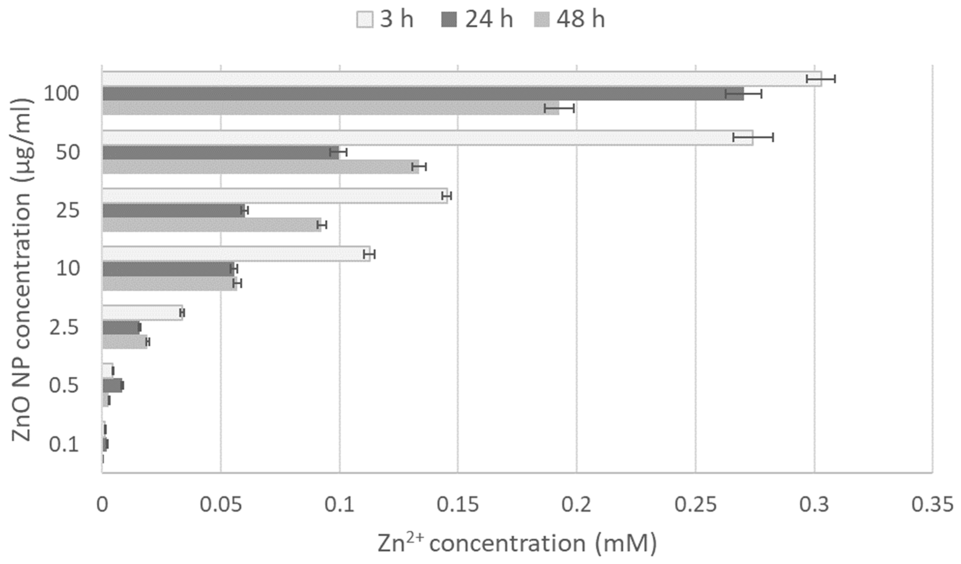
3.2. Zn2+ Ion Release

Results obtained from the FAAS experiments to quantify the release of zinc ions from the ZnO NP are shown in Figure 1.
Increased levels of dissolved Zn2+ ions from the ZnO NP surface were observed (from 2.5 µg/mL on) at all treatment periods (3, 24 and 48 h), but notably higher at the shortest time. The release of Zn2+ ions from ZnO NP is well documented in the literature, and this demonstrates that the solubility of these NP can be highly variable and dependent on both the NP physicochemical properties and the matrix in which they are suspended [14,15,16,17].

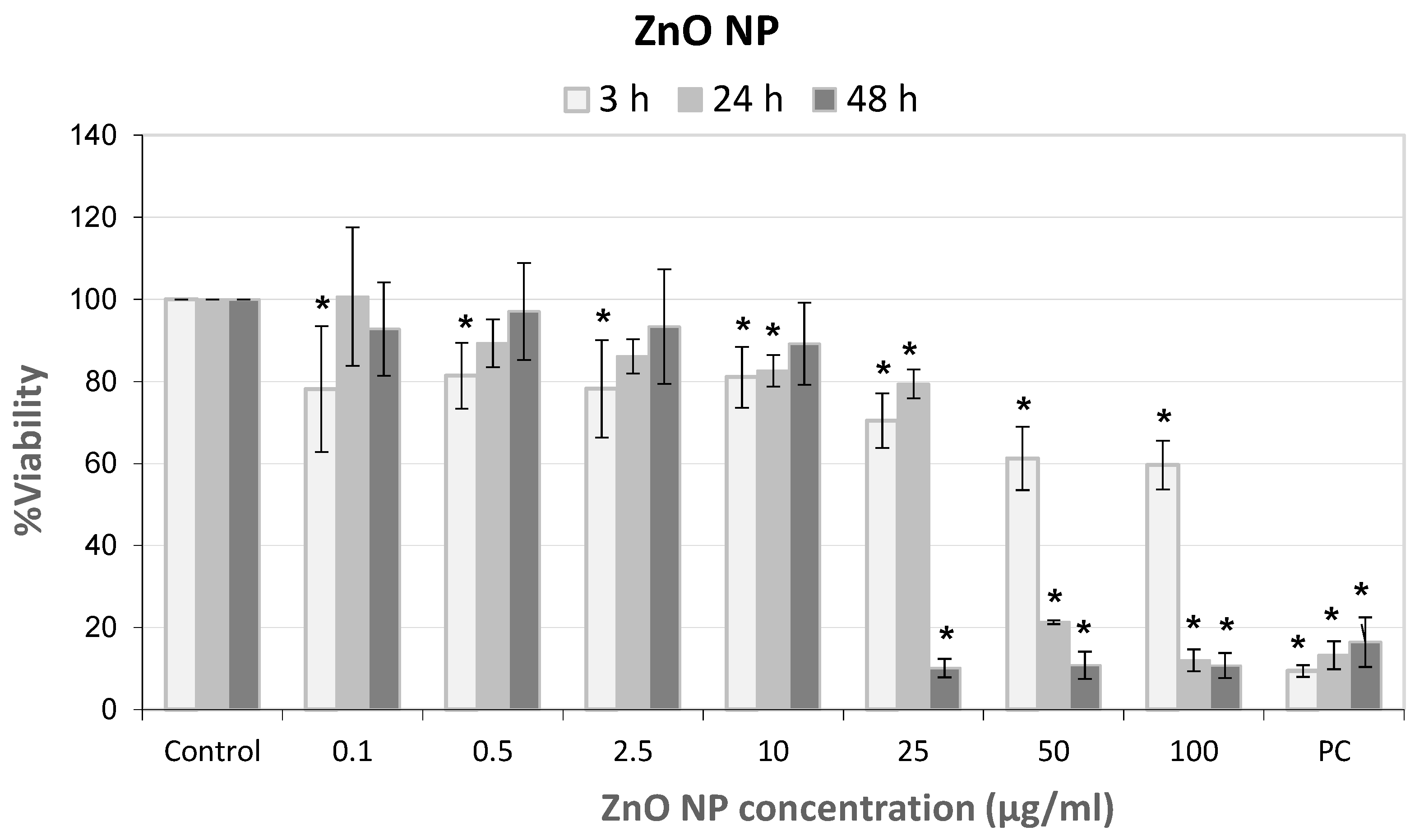
3.3. ZnO NP Effects on Glial Cells

After discarding any potential interference of the NP with MTT assay, the effects of ZnO NP on viability of glial A172 cells were evaluated. The results from these experiments are shown in Figure 2. The effects on cellular viability were moderate after 3 h of exposure, reaching 60% viability only at the highest concentrations. However, after 24 and 48 h of treatments, the decreases in viability were much marked, reaching values close to 90% mortality from 50 or 25 µg/mL on, respectively. Similarly, several authors previously described a dose-dependent decrease in cell viability of various cell types exposed to ZnO NP (reviewed in [18]).

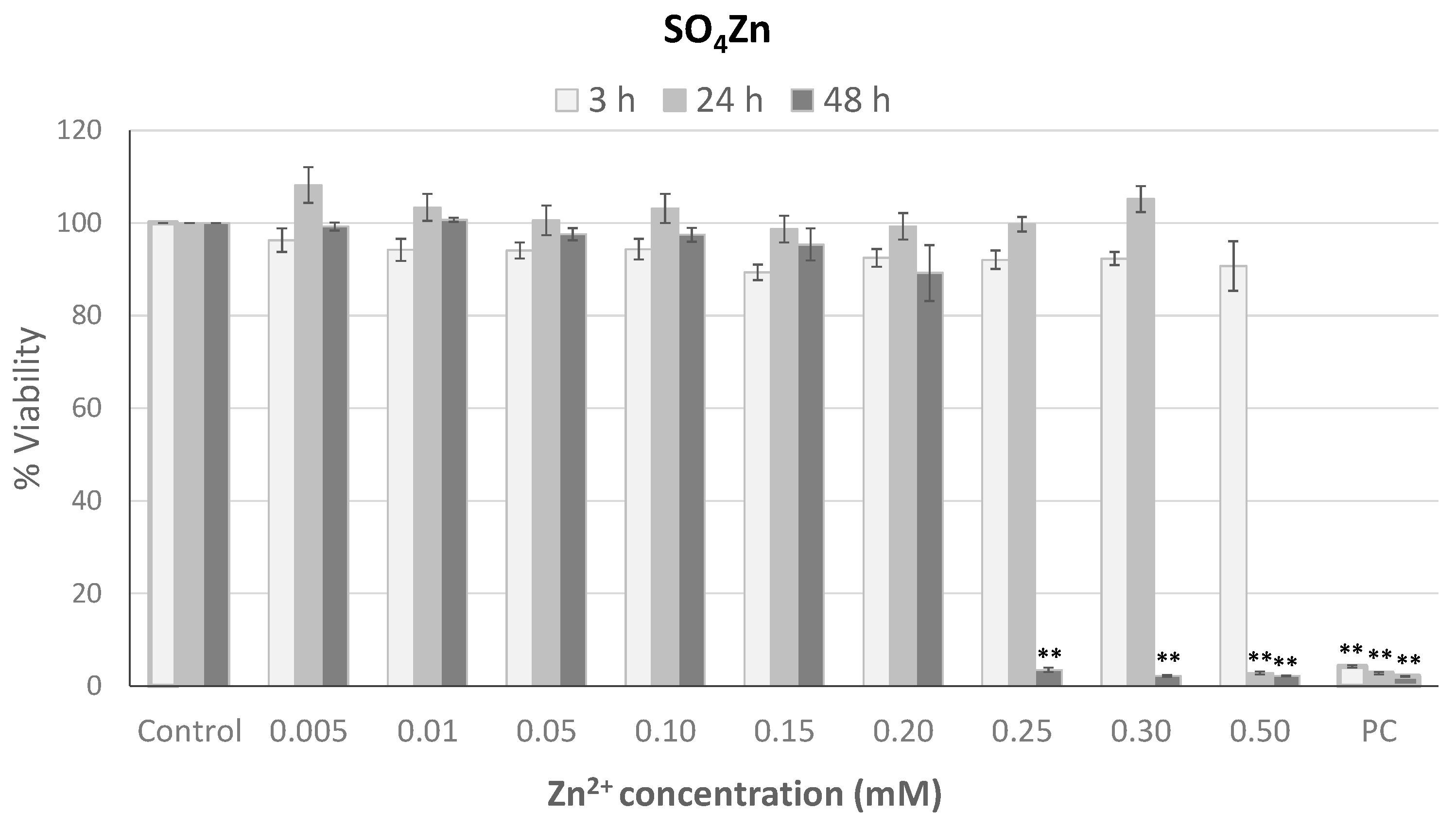
3.4. Role of Zn2+ Ions Released from ZnO NP

In order to test the role of the Zn2+ released from ZnO NP in the cytotoxic effects observed, as previously pointed out [19,20], in the present work, A172 cells were treated with zinc sulfate (ZnSO4) at several concentrations chosen according to FAAS analysis. The results obtained from these analyses showed that a significant decrease in cell viability was just observed at Zn2+ ion concentrations greater than 0.2 and 0.3 mM for 24 and 48 h, respectively (Figure 3). According to this, the release of Zn2+ ions from ZnO NP does not explain, at least completely, their effects on glial cell viability, and hence other different action mechanisms, e.g., the production of ROS or induction of oxidative stress, as previously reported for these NP [21,22,23], should be considered in attempting to explain the observed effects.

4. Conclusions

ZnO NP were effectively internalized by A172 cells and the exposure induced a significant decrease in cell viability, depending on the dose and treatment duration. The ability of ZnO NP to release Zn2+ ions into the medium in a concentration-dependent manner was confirmed; however, their presence was not entirely responsible for the observed cytotoxic effects. These results increase the knowledge about the toxic potential of ZnO NP in human glial cells and the involvement of released Zn2+ ions in the observed effects.

Author Contributions

Conceptualization, V.V., N.F.-B. and B.L.; methodology, A.T., L.R.-P., A.A.-G., J.M.-P., N.F.-B.; formal analysis, M.F., J.M., B.L.; writing—original draft preparation, V.V., E.P.; writing—review and editing, J.M., B.L., M.F.; supervision, V.V., N.F.-B. and B.L.; funding acquisition, E.P., V.V. All authors have read and agreed to the published version of the manuscript.

Funding

This research was funded by Ministry of Science and Innovation: MCIN/AEI/10.13039/501100011033 (Grant PID2020-114908GA-I00), Xunta de Galicia (ED431B 2022/16 and ED481A 2019/003), CICA-Disrupting Project 2021SEM-B2, and Ministry of Education, Culture and Sport [BEAGAL18/00142 to V.V.].

Institutional Review Board Statement

Not applicable.

Informed Consent Statement

Not applicable.

Data Availability Statement

The data presented in this study are available on request from the corresponding author.

Conflicts of Interest

The authors declare no conflict of interest.

References

  1. Du, J.; Tang, J.; Xu, S.; Ge, J.; Dong, Y.; Li, H.; Jin, M. ZnO nanoparticles: Recent advances in ecotoxicity and risk assessment. Drug Chem. Toxicol. 2020, 43, 322–333. [Google Scholar] [CrossRef] [PubMed]
  2. Shi, L.E.; Li, Z.H.; Zheng, W.; Zhao, Y.F.; Jin, Y.F.; Tang, Z.X. Food Additives & Contaminants: Part A Synthesis, antibacterial activity, antibacterial mechanism and food applications of ZnO nanoparticles: A review. Food Addit. Contam. Part A 2014, 31, 173–186. [Google Scholar]
  3. Singh, S. Zinc oxide nanoparticles impacts: Cytotoxicity, genotoxicity, developmental toxicity, and neurotoxicity. Toxicol. Mech. Methods 2019, 29, 300–311. [Google Scholar] [CrossRef] [PubMed]
  4. Smaoui, S.; Chérif, I.; Hlima, H.B.; Khan, M.U.; Rebezov, M.; Thiruvengadam, M.; Sarkar, T.; Shariati, M.A.; Lorenzo, J.M. Zinc oxide nanoparticles in meat packaging: A systematic review of recent literature. Food Packag. Shelf Life 2023, 36, 101045. [Google Scholar] [CrossRef]
  5. Guo, Z.; Zhang, P.; Chakraborty, S.; Chetwynd, A.J.; Monikh, F.A.; Stark, C.; Ali-Boucetta, H.; Wilson, S.; Lynch, I.; Valsami-Jones, E. Biotransformation modulates the penetration of metallic nanomaterials across an artificial blood–brain barrier model. Proc. Natl. Acad. Sci. USA 2021, 118, e2105245118. [Google Scholar] [CrossRef]
  6. Valdiglesias, V.; Costa, C.; Kiliç, G.; Costa, S.; Pásaro, E.; Laffon, B.; Teixeira, J.P.; Paulo, J. Neuronal cytotoxicity and genotoxicity induced by zinc oxide nanoparticles. Environ. Int. 2013, 55, 92–100. [Google Scholar] [CrossRef]
  7. Fernández-Bertólez, N.; Costa, C.; Brandão, F.; Duarte, J.A.; Teixeira, J.P.; Pásaro, E.; Valdiglesias, V.; Laffon, B. Evaluation of cytotoxicity and genotoxicity induced by oleic acid-coated iron oxide nanoparticles in human astrocytes. Environ. Mol. Mutagen 2019, 60, 816–829. [Google Scholar] [CrossRef]
  8. Valdiglesias, V.; Costa, C.; Sharma, V.; Kiliç, G.; Pásaro, E.; Teixeira, J.P.; Dhawan, A.; Laffon, B. Comparative study on effects of two different types of titanium dioxide nanoparticles on human neuronal cells. Food Chem. Toxicol. 2013, 57, 352–361. [Google Scholar] [CrossRef]
  9. Sharma, A.K.; Singh, V.; Gera, R.; Purohit, M.P.; Ghosh, D. Zinc oxide nanoparticle induces microglial death by NADPH-oxidase-independent reactive oxygen species as well as energy depletion. Mol. Neurobiol. 2017, 54, 6273–6286. [Google Scholar] [CrossRef]
  10. Sruthi, S.; Mohanan, P.V. Investigation on cellular interactions of astrocytes with zinc oxide nanoparticles using rat C6 cell lines. Colloids Surf. B Biointerfaces 2015, 133, 1–11. [Google Scholar] [CrossRef]
  11. Heim, J.; Felder, E.; Tahir, M.N.; Kaltbeitzel, A.; Heinrich, U.R.; Brochhausen, C.; Mailänder, V.; Tremel, W.; Brieger, J. Genotoxic effects of zinc oxide nanoparticles. Nanoscale 2015, 7, 8931–8938. [Google Scholar] [CrossRef]
  12. Zhang, J.; Qin, X.; Wang, B.; Xu, G.; Qin, Z.; Wang, J.; Wu, L.; Ju, X.; Bose, D.D.; Qiu, F.; et al. Zinc oxide nanoparticles harness autophagy to induce cell death in lung epithelial cells. Cell Death Dis. 2017, 8, e2954. [Google Scholar] [CrossRef] [PubMed]
  13. Mittag, A.; Hoera, C.; Kämpfe, A.; Westermann, M.; Kuckelkorn, J.; Schneider, T.; Glei, M. Cellular uptake and toxicological effects of differently sized zinc oxide nanoparticles in intestinal cells. Toxics 2021, 9, 96. [Google Scholar] [CrossRef] [PubMed]
  14. Eixenberger, J.E.; Anders, C.B.; Hermann, R.J.; Brown, R.J.; Reddy, K.M.; Punnoose, A.; Wingett, D.G. Rapid dissolution of ZnO nanoparticles induced by biological buffers significantly impacts cytotoxicity. Chem. Res. Toxicol. 2017, 30, 1641–1651. [Google Scholar] [CrossRef] [PubMed]
  15. Liang, Z.; Zhang, F.; Zhang, H. In Vitro response of immune cells on metal oxide nanoparticles with different solubility. J. Nanosci. Nanotechnol. 2016, 16, 5546–5552. [Google Scholar] [CrossRef] [PubMed]
  16. Sharma, V.; Anderson, D.; Dhawan, A. oxide nanoparticles induce oxidative DNA damage and ROS-triggered mitochondria mediated apoptosis in human liver cells (HepG2). Apoptosis Zinc 2012, 17, 852–870. [Google Scholar] [CrossRef] [PubMed]
  17. Yan, Y.; Wang, G.; Huang, J.; Zhang, Y.; Cheng, X.; Chuai, M.; Brand-Saberi, B.; Chen, G.; Jiang, X.; Yang, X. Zinc oxide nanoparticles exposure-induced oxidative stress restricts cranial neural crest development during chicken embryogenesis. Ecotoxicol. Environ. Saf. 2020, 194, 110415. [Google Scholar] [CrossRef]
  18. Pandurangan, M.; Kim, D.H. In vitro toxicity of zinc oxide nanoparticles: A review. J. Nanoparticle Res. 2015, 17, 158. [Google Scholar] [CrossRef]
  19. Bozym, R.A.; Chimienti, F.; Giblin, L.J.; Gross, G.W.; Korichneva, I.; Li, Y.; Libert, S.; Maret, W.; Parviz, M.; Frederickson, C.J.; et al. Free zinc ions outside a narrow concentration range are toxic to a variety of cells in vitro. Exp. Biol. Med. 2010, 235, 741–750. [Google Scholar] [CrossRef]
  20. Holmes, A.M.; Mackenzie, L.; Roberts, M.S. Disposition and measured toxicity of zinc oxide nanoparticles and zinc ions against keratinocytes in cell culture and viable human epidermis. Nanotoxicology 2020, 14, 263–274. [Google Scholar] [CrossRef]
  21. Nair, S.; Sasidharan, A.; Divya Rani, V.V.; Menon, D.; Nair, S.; Manzoor, K.; Raina, S. Role of size scale of ZnO nanoparticles and microparticles on toxicity toward bacteria and osteoblast cancer cells. J. Mater. Sci. Mater. Med. 2009, 20, 235–241. [Google Scholar] [CrossRef] [PubMed]
  22. Patel, P.; Kansara, K.; Senapati, V.A.; Shanker, R.; Dhawan, A.; Kumar, A. Cell cycle dependent cellular uptake of zinc oxide nanoparticles in human epidermal cells. Mutagenesis 2016, 31, 481–490. [Google Scholar] [CrossRef] [PubMed]
  23. Wong, S.W.Y.; Leung, P.T.Y.; Djurišić, A.B.; Leung, K.M.Y. Toxicities of nano zinc oxide to five marine organisms: Influences of aggregate size and ion solubility, in: Analytical and Bioanalytical Chemistry. Anal. Bioanal. Chem. 2010, 396, 609–618. [Google Scholar] [CrossRef] [PubMed]
Figure 1. Analysis of Zn2+ ions released from ZnO NP to the cell culture medium.
Materproc 14 00023 g001
Figure 2. Cytotoxicity of ZnO NP in A172 glial cells at different exposure times. Bars represent mean standard error. * p < 0.05, significant difference with regard to the corresponding control. PC: Positive control: triton X-100 (1%).
Materproc 14 00023 g002
Figure 3. Cytotoxicity of ZnSO4 in A172 glial cells at different exposure times. Bars represent mean standard error. ** p < 0.01, significant difference with regard to the corresponding control. PC: Positive control: triton X-100 (1%).
Materproc 14 00023 g003
Table 1. Physical-chemical characterization of ZnO NP.
Hydrodynamic Diameter (nm) aZeta Potential (mV) a
Time (h)0302.09 ± 0.84−1.73 ± 4.05
3315.01 ± 3.162.79 ± 3.01
24269.16 ± 1.36−20.25 ± 2.03
48273.97 ± 5.91−13.46 ± 2.36
a Mean ± standard deviation.
Table 2. ZnO NP uptake by glial A172 cells (% of cells with NP, mean ± standard deviation).
ZnO NP
Concentration (µg/mL)
Exposure Time
3 h24 h48 h
Control1.06 ± 0.181.13 ± 0.160.76 ± 0.06
0.11.12 ± 1.162.04 ± 0.24 **1.98 ± 0.29 **
0.51.21 ± 0.184.41 ± 1.20 **3.18 ± 0.47 **
2.51.21 ± 0.124.99 ± 1.28 **2.15 ± 0.39 **
101.03 ± 0.137.06 ± 1.80 **4.11 ± 0.61 **
251.05 ± 0.216.79 ± 1.53 **6.42 ± 0.68 **
501.71 ± 0.319.34 ± 1.06 **11.75 ± 0.88 **
1005.28 ± 0.55 **7.92 ± 0.69 **28.06 ± 2.41 **
Positive control42.42 ± 4.06 **64.11 ± 2.25 **71.88 ± 1.21 **
** p < 0.01, significant difference with regard to the corresponding control. Positive control: TiO2 NP (200 µg/mL).
Disclaimer/Publisher’s Note: The statements, opinions and data contained in all publications are solely those of the individual author(s) and contributor(s) and not of MDPI and/or the editor(s). MDPI and/or the editor(s) disclaim responsibility for any injury to people or property resulting from any ideas, methods, instructions or products referred to in the content.

