Proceeding Paper

Microstructure Characterization of Titania-Based Micro-Arc Oxidation Coatings with Nanoparticles †

1
Institute of Metallurgy and Materials Science, Polish Academy of Sciences, 30-059 Krakow, Poland
2
Department of Metallurgical and Materials Engineering, Istanbul Technical University, Istanbul 34467, Turkey
3
Institute of High Pressure Physics, Polish Academy of Sciences, 01-142 Warsaw, Poland
*
Author to whom correspondence should be addressed.
Presented at the 4th International Online Conference on Nanomaterials, 5–19 May 2023; Available online: https://iocn2023.sciforum.net.
Mater. Proc. 2023, 14(1), 14; https://doi.org/10.3390/IOCN2023-14535
Published: 5 May 2023
(This article belongs to the Proceedings of The 4th International Online Conference on Nanomaterials)

Abstract

:
Micro-arc oxidation (MAO) has proven to be a very promising method for the fabrication of functional coatings on titanium-based materials intended for dental implant application. The possibility of mixing the electrolyte components and/or adding the nanopowder opened a way to produce antibacterial surfaces. In the present work, the microstructure study of MAO coatings was carried out with the use of cutting-edge electron microscopes to reveal the way of the incorporation of the metallic and ceramic nanoparticles. These studies showed the possibility of the incorporation of metallic and ceramic nanoparticles into the MAO coating both from the as-supplied nanopowder and from the mix of inorganic salts. The produced nanoparticles locate preferentially on the very top of the coating and areas close to porosity. Furthermore, the use of silver acetate allows the production of ultrafine nanoparticles (with a size <10 nm), unachievable for as-supplied nanopowder.

1. Introduction

Addressing bacterial colonization of biomaterials placed in the human body and the resulting inflammation of the surrounding tissues requires the development of methods of surface modification of the implant materials aimed at obtaining an antibacterial effect [1,2]. Micro-arc oxidation (MAO) turned out to be very popular due to its versatility, controllability and environmental friendliness [3,4]. Simplicity in controlling the electrolyte components makes the incorporation of antibacterial agents possible [5]. Nanoparticles (metallic and ceramic) gained in importance as a large surface-area-to-volume ratio allows substantial amounts of ions of antibacterial additive to be released with the aim to interact with the outer membrane of the bacteria and subsequently with its DNA, disrupting its functioning [6]. Even if many literature reports confirmed the considerable antibacterial effect of the MAO coatings with selected nanoparticles, their microstructure formation remains sparsely investigated [7].
In this work, titanium of commercial purity, subjected to hydrostatic extrusion, has been micro-arc oxidized in the electrolyte bath containing sodium phosphate and as-supplied nanoparticles (ZnO) or acetates (Ag-acetate) to produce titania coatings with embedded nanoparticles. In this way, metallic and ceramic nanoparticles have been incorporated. Advanced methods of scanning and transmission electron microscopy were used to unveil the mechanisms of incorporation of antibacterial additions into the MAO coating.

2. Materials and Methods

In the present work, titanium grade 4 subjected to plastic deformation through hydrostatic extrusion (followed by rotary swaging) was utilized as a substrate material for further MAO processing. Details of this process are mentioned elsewhere [8]. An extruded rod of 5 mm in diameter was cut out with the use of a diamond wire saw into small cylinders (5 mm in height). Its surface was ground and polished with abrasive papers of up to 7000 grit and colloidal silica suspension, respectively.
The MAO processing was carried out in stainless steel container filled with the electrolytes with the composition presented in Table 1. The temperature of the electrolyte was kept at a constant temperature of 22 °C throughout the overall MAO process (3 min). Titanium substrates were immersed in the electrolyte and the electric signal was applied from the bipolar pulsed power supply, i.e., 300 V and 60 V of positive and negative pulse, respectively, with a duration of each pulse of 0.6 ms, separated by breaks of 0.4 ms.
The surface topography observations were performed with the use of a ThermoFisher Scios 2 Dual Beam microscope (Eindhoven, The Netherlands) equipped with a FEG electron gun, Ga+ source and EDS spectrometer. This microscope was also used for the preparation of the lamellae for the TEM studies (focused ion beam (FIB) method). The TEM cross-sectional studies were carried out with the help of a ThermoFisher Titan Themis G2 200 Probe Cs-Corrected transmission electron microscope (Eindhoven, The Netherlands) equipped with a CCD camera for the acquisition of microstructure images and ChemiSTEM EDS system for chemical analysis.

3. Results and Discussion

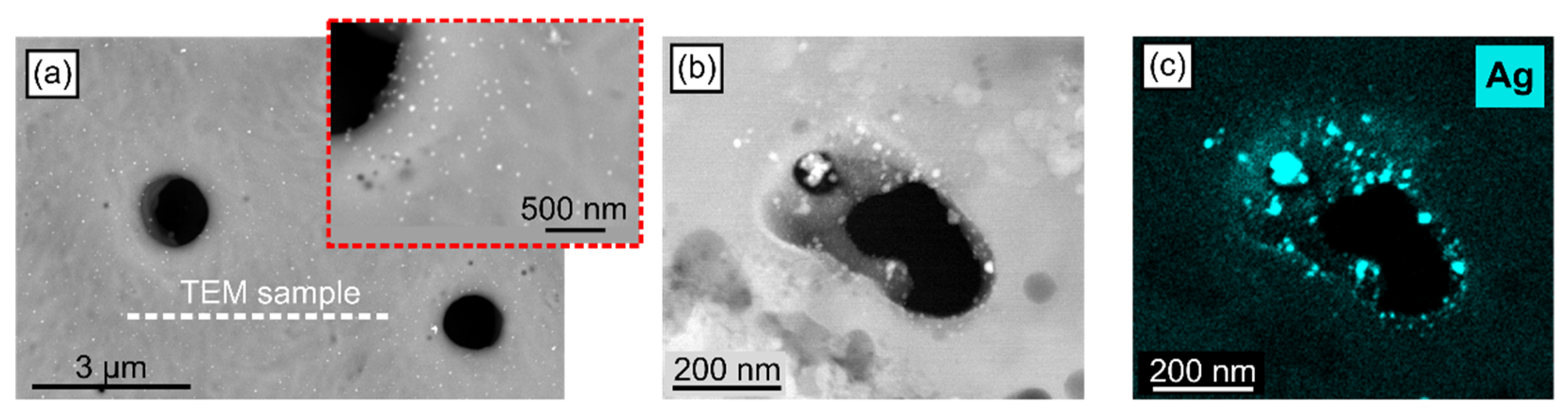
3.1. MAO Coating with Ag Nanoparticles

The SEM/BSE top-view observations of MAO + Ag samples showed a smooth surface of the titanium oxide coating covering the substrate material with embedded ultra-fine particles of high-Z material with a size of tens of nanometers at most, presenting higher density in the areas closer to the porosity/craters. However, due to the very small size of the observed particles and the relatively large volume of interaction between the electron beam and the solid material, it was not possible to determine the type of these particles by SEM/EDS and assign them unambiguously to silver. Therefore, further analyzes were carried out using the STEM method on the lamella cut by the FIB technique along the white dashed line seen in Figure 1a. The HAADF/STEM cross-sectional observations of the cut-out lamella, focused on the areas close to the porosity, revealed a string of bright nanoparticles surrounding the pore (Figure 1b). By combining the HAADF/STEM imaging and EDS mapping, one may assign the particles to the pure silver (Figure 1c).

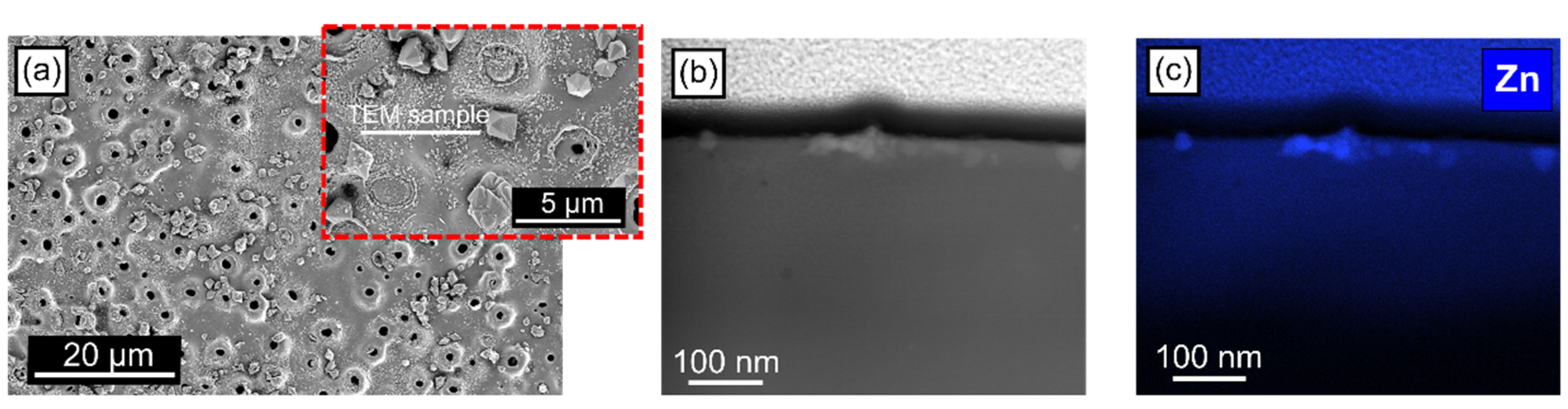
3.2. MAO Coating with ZnO Nanoparticles

The SEM observations of the surface topography of MAO + ZnO coatings revealed that between dispersed craters, several regular crystals are visible as well as a high concentration of irregularly distributed fine particles with an average size <100 nm (Figure 2a). The TEM sample was cut out from this area with the FIB method in order to confirm the nature of the fine particles. The TEM cross-sectional observations of the cut-out lamella proved the embedding of ZnO nanoparticles with a size corresponding to the original nanopowder added to the electrolyte during MAO processing. The nanoparticles are protruding from the surface of the sample. In addition, the presence of some amounts of zinc is confirmed in the surrounding amorphous area of the coating.

4. Conclusions

A microstructure study of MAO coatings, produced on titanium grade 4 with electrolytes containing either as-supplied ZnO nanopowder (average particle size in the range between 20 and 30 nm) or with the addition of the silver acetate, was carried out utilizing cutting-edge electron microscopes, allowing us to conclude that:
  • The incorporation of metallic and ceramic nanoparticles into the MAO coating is possible both from the as-supplied material and from silver acetate. Preferred locations are the very top of the coating and areas close to porosity.
  • The use of silver acetate allows the production of nanoparticles with a diameter of less than 10 nm, which is not possible with the use of as-supplied nanopowder.
  • The TEM method combined with EDS and performed on FIB-produced lamellae allows for unambiguous confirmation of the places of incorporation of nanoparticles into MAO coatings.

Author Contributions

Conceptualization, L.M.; methodology, L.M. and F.M.; investigation, A.T. and M.K.; data curation, A.J.; writing—original draft preparation, L.M. and A.J.; writing—review and editing, H.C. and M.B.; visualization, M.B.; supervision, H.C.; project administration, L.M.; funding acquisition, L.M. All authors have read and agreed to the published version of the manuscript.

Funding

This research was funded by National Science Centre of Poland, grant number UMO- 2020/39/D/ST8/01783.

Institutional Review Board Statement

Not applicable.

Informed Consent Statement

Not applicable.

Data Availability Statement

The data used in this study are available from the corresponding author on request.

Conflicts of Interest

The authors declare no conflict of interest.

References

  1. Aydogan, D.; Muhaffel, F.; Kilic, M.K.; Acar, O.K.; Cempura, G.; Baydogan, M.; Karaguler, N.G.; Kose, G.T.; Czyrska-Filemonowicz, A.; Cimenoglu, H. Optimisation of micro-arc oxidation electrolyte for fabrication of antibacterial coating on titanium. Mater. Technol. 2017, 33, 119–126. [Google Scholar] [CrossRef]
  2. Aydogan, D.; Muhaffel, F.; Acar, O.K.; Topcuoglu, E.N.; Kulekci, H.G.; Kose, G.T.; Baydogan, M.; Cimenoglu, H. Surface modification of Ti6Al4V by micro-arc oxidation in AgC2H3O2-containing electrolyte. Surf. Innov. 2018, 6, 277–285. [Google Scholar] [CrossRef]
  3. Wang, Y.; Yu, H.; Chen, C.; Zhao, Z. A Review of the biocompatibility of micro-arc oxidation coated titanium alloys. Mater. Des. 2015, 85, 640–652. [Google Scholar] [CrossRef]
  4. Simchen, F.; Sieber, M.; Kopp, A.; Lampke, T. Introduction to Plasma Electrolytic Oxidation—An Overview of the Process and Applications. Coatings 2020, 10, 628. [Google Scholar] [CrossRef]
  5. van Hengel, I.; Tierolf, M.W.A.M.; Fratila-Apachitei, L.E.; Apachitei, J.; Zadpoor, A.A. Antibacterial Titanium Implants Biofunctionalized by Plasma Electrolytic Oxidation with Silver, Zinc, and Copper: A Systematic Review. Int. J. Mol. Sci. 2021, 22, 380. [Google Scholar] [CrossRef] [PubMed]
  6. Fattah-alhosseini, A.; Molaei, M.; Attarzadeh, N.; Babaei, K.; Attarzadeh, F. On the enhanced antibacterial activity of plasma electrolytic oxidation (PEO) coatings that incorporate particles: A review. Ceram. Int. 2021, 46, 20587–20607. [Google Scholar] [CrossRef]
  7. Maj, Ł.; Fogarassy, Z.; Wojtas, D.; Jarzębska, A.; Muhaffel, F.; Sulyok, A.; Góral, A.; Kulczyk, M.; Çimenoğlu, H.; Bieda, M. In-situ formation of Ag nanoparticles in the MAO coating during the processing of cp-Ti. Sci. Rep. 2023, 13, 3230. [Google Scholar] [CrossRef] [PubMed]
  8. Maj, Ł.; Wojtas, D.; Jarzębska, A.; Bieda, M.; Trembecka-Wójciga, K.; Chulist, R.; Kozioł, W.; Góral, A.; Trelka, A.; Janus, K.; et al. Titania coating formation on hydrostatically extruded pure titanium by micro-arc oxidation method. J. Mater. Sci. Technol. 2022, 111, 224–235. [Google Scholar] [CrossRef]
Figure 1. SEM/BSE surface image (a), STEM/HAADF cross-sectional image (b) and corresponding EDS map of Ag distribution (c) acquired from MAO + Ag sample.
Materproc 14 00014 g001
Figure 2. SEM/BSE surface image (a), STEM/HAADF cross-sectional image (b) and corresponding EDS map of Zn distribution (c) acquired from MAO + ZnO sample.
Materproc 14 00014 g002
Table 1. Notation of the samples used in the present work.
Notation of SampleBase Aqueous ElectrolyteAdditive
MAO + ZnO14.4 g/L Na2HPO42 g/L of ZnO nanoparticles (avg size: 20–30 nm)
MAO + Ag14.4 g/L Na2HPO40.002 M of AgC2H3O2 (silver acetate)
Disclaimer/Publisher’s Note: The statements, opinions and data contained in all publications are solely those of the individual author(s) and contributor(s) and not of MDPI and/or the editor(s). MDPI and/or the editor(s) disclaim responsibility for any injury to people or property resulting from any ideas, methods, instructions or products referred to in the content.

