Proceeding Paper

Developing a Sensitive Method for the Electrochemical Determination of Tetracycline Using MB-Tagged Aptamers on Gold Electrode Substrates †

by
Stefanos Karapetis
* and
Leda G. Bousiakou
IMD Laboratories Co., R&D Section, Lefkippos Technology Park, NCSR Demokritos, P.O. Box 60037, 15341 Athens, Greece
*
Author to whom correspondence should be addressed.
Presented at the 3rd International Electronic Conference on Biosensors, 8–21 May 2023; Available online: https://iecb2023.sciforum.net/.
Eng. Proc. 2023, 35(1), 38; https://doi.org/10.3390/IECB2023-14597
Published: 12 May 2023
(This article belongs to the Proceedings of The 3rd International Electronic Conference on Biosensors)

Abstract

:
An electrochemical aptasensor for the detection of tetracycline (TET) is prepared based on a methylene blue (MB)-tagged DNA aptamer, with the sequence 5′-MB-CCC CCG GCA GGC CAC GGC TTG GGTTGG TCC CAC TGC GCG-thiol-3′. The DNA aptamer is chemisorbed on a gold electrode and differential pulse voltammetry (DPV) is utilized for the detection. In particular, upon binding of the TET with the purpose-designed aptamer, there is an increase in the current intensity, as a result of the increased proximity of the MB molecule to the gold surface. The sensor is tested using aqueous samples spiked with TET concentrations between 1 and 1000 nM and a limit of detection (LOD) of 1.2 nM is determined. Furthermore, the dissociation constant is estimated to be 1.4 nM using a Lineweaver–Burk plot.

1. Introduction

With the advent of antibiotics, human and veterinary medicine significantly advanced in fighting bacterial disease [1]. Nevertheless, the influx of drugs to the environment can pose a significant threat to aquatic and terrestrial biodiversity as well as to the human food chain [2]. In this regard, tetracycline (TET), a common drug used against intercellular bacteria, such as chlamydia, mycoplasmas, rickettsia, and protozoan parasites, has often been traced in water, food, or milk samples [3,4]. Thus, its qualitative and quantitative monitoring in various systems is important for public health.
Currently, TET is often determined using high-performance or thin-layer liquid chromatography [5,6]. Moreover, enzyme-linked immunosorbent assay (ELISA) kits have also been used for this purpose [7]. Nevertheless, despite their ability to detect TET with both selectivity and sensitivity, they can be either expensive or also time-consuming, requiring in many cases qualified staff. In this regard, biosensors [8] promise a fast, lower-cost technology for the detection of different pharmaceutical compounds. In particular, electrochemical aptasensors [9] provide a robust alternative based on synthetic DNA (or RNA) nucleotides that are easily produced, are target specific, and can be immobilized with ease on a number of surfaces. Moreover, they offer chemical stability and allow for the regeneration of the aptasensors, due to the electrostatic interactions between the aptamers and target.
A number of electrochemical aptasensors have been recently reported as a platform for a robust, low-cost, and often multianalyte analysis, while being compatible with microfabrication technologies [8]. In particular, this work focuses on the detection of TET in an aqueous environment using an electrochemical aptasensor based on a gold working electrode where a methylene blue (MB)-modified DNA synthetically produced oligonucleotide is immobilized via the Au-S bond. In this regard, the limit of detection (LOD) is investigated for our aptasensors, and the dissociation constant Kd is deduced.

2. Materials and Methods

2.1. Reagents and Aptamer Sequence

All reagents were purchased from Sigma-Aldrich. The tetracycline antibiotic (TET, Mw = 444.43 Da) was made from bacteria of the genus Streptomyces. Tris(2-carboxyethyl) phosphine (TCEP) powder was used to break down any disulfide bonding while 6-Mercapto-1-hexanol (MCH) was utilized to avoid any non-specific binding. A MB-tagged thiol-modified DNA aptamer with the composition 5′-MB-CCC CCG GCA GGC CAC GGC TTG GGTTGG TCC CAC TGC GCG-thiol-3′ was used, as described by Niazi et al. [10]. The aptamer was purchased from GeneCust (Boynes, France). The stock solution for the aptamer was based on a TE buffer containing 10 mM Tris-HCl and 1 mM Ethylenediaminetetraacetic acid (EDTA) at pH = 8. Finally, the working buffer was based on 10 mM PBS containing Tris−HCl (20 mM), NaCl (100 mM), MgCl2 (2 mM), KCl (5 mM), and CaCl2 (1 mM) at pH 7.6.

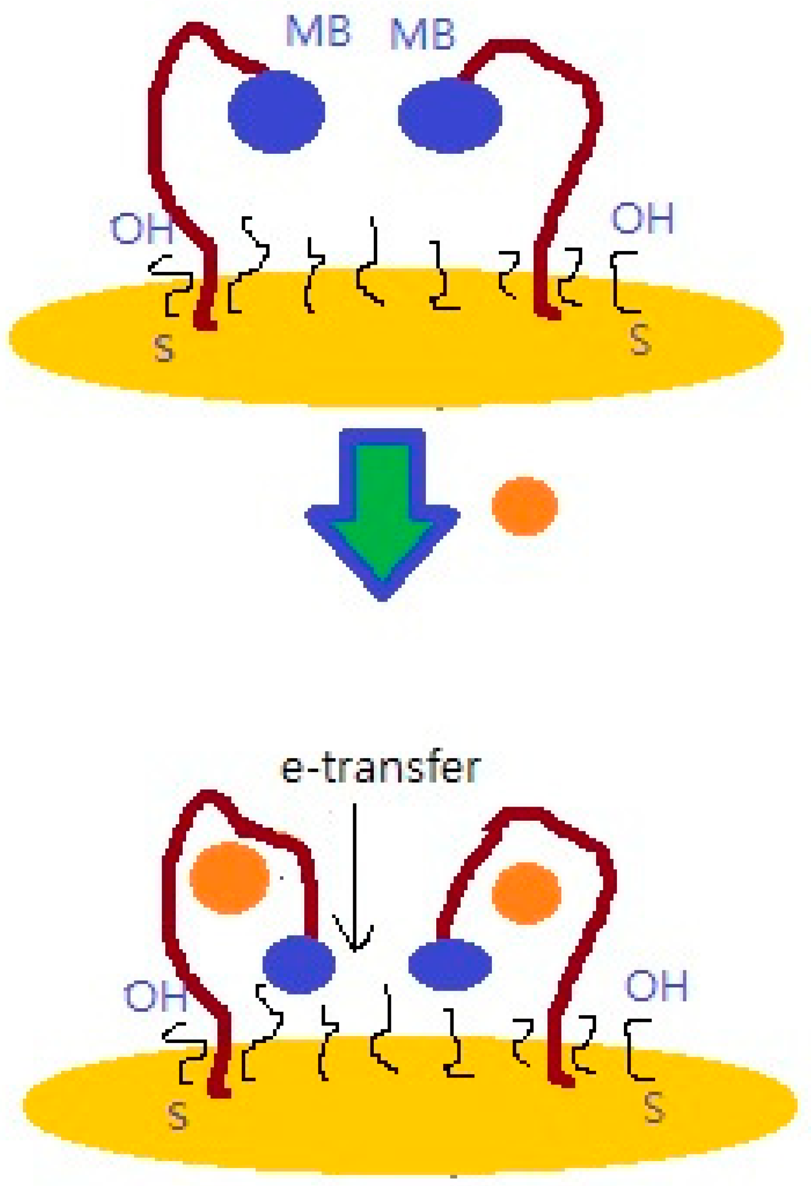
2.2. Preparation of the Aptasensor

The aptasensors used gold electrode disks with a 2 mm diameter that were initially polished using alumina powder and then sonicated 15 min in ethanol. After, the electrodes were rinsed in deionized water and dried under a nitrogen gas flow. In order to prepare the aptamers for immobilization, they were first heated to 90 °C for 10 min and then cooled to room temperature to provide proper aptamer folding. After that, 1 μΜ of aptamer was diluted in the TE buffer and incubated with 10 mM TCEP (1 h) for dissociation of disulfide bonds. For the immobilization, the aptamer was added on the clean Au electrode, until the 2 mm surface was completely covered and incubated for 1 h at room temperature. After that, MCH at a concentration of 2 mM in deionized water was added on the surface overnight to prevent any non-specific binding. After rinsing the aptamer, the sensitized electrode was incubated for 30 min with various concentrations of TET between 1 nM and 1000 nM, which were diluted in the above-mentioned working buffer. The scheme of the sensing surface is presented in Figure 1. The TET detection was performed using differential pulse voltammetry (DPV).

2.3. Electrochemical Measurements

The electrical parameters were determined using a Zahner potentiostat, with a gold working electrode (2 mm), a Ag/AgCl reference, and a Pt wire as a counter. All electrodes were purchased from CH Instruments, USA. The sensor response was studied using differential pulse voltammetry (DPV) with a potential range from 0 V to –0.6 V and a resting time of 60 s.

3. Results and Discussion

3.1. Determination of Different Concentrations of TET Using Aptasensing

The determination of TET was investigated for different concentrations ranging between 1 and 1000 nM in three independent experiments, with good reproducibility. The aptasensor showed selectivity to TET, using spiked samples in an aqueous solution. In particular, the following concentrations were looked at: 1 nM, 5 nM, 10 nM, 30 nM, 100 nM, 300 nM, and 1000 nM. The binding of TET on the aptasensor induced an increase in the oxidation peak, as can be seen in Figure 2 below.
Furthermore, the calibration plot of (Ι-Ιο)/Ιο versus the logarithmic scale of the TET concentrations was examined (Figure 3) and regression was applied to identify the limit of detection (LOD) using the relationship LOD = 3σ/α, where σ denotes the standard deviation of the response and S denotes the slope of the linear calibration plot [11]. The limit of detection was then determined to be 1.2 nM
Our results are comparable with the literature, considering that TET has been detected in aqueous solutions with a LOD varying from 7.8 × 10⁻¹¹ M up to 5 × 10⁻⁹ M, depending on the method used, electrode architecture, aptamer sequence, and immobilization [12].

3.2. Deduction of the Dissociation Constant Kd for Our Aptamer Sequence

In this investigation, we also deduced the dissociation constant (Kd) for our aptamer, under the specific buffer conditions, using a Lineweaver–Burk plot [13], as seen in Figure 4 below:
In particular, Kd shows the binding strength between the aptamer and target, with values ranging from micro to picomolar levels [14]. In general, a low value for the dissociation constant relates to a high affinity between the aptamer, indicating that a low aptamer concentration of the aptamer and target is needed for binding. Linear regression yielded a linear fit, such that y = 1.3897x + 0.9765, thus leading to a value of Kd = 1.3897/0.9579 nM or Kd = 1.4 nM, showing that aptasensors provide a robust structure for TET detection.

4. Conclusions

An electrochemical aptasensor was developed for the detection of TET using a single-stranded DNA aptamer that selectively binds to TET in an aqueous solution. In particular, it was immobilized on a gold electrode and the analysis was performed using DPV. Our results show that the electrochemical aptasensor is sensitive to TET where the limit of detection (LOD) was determined to be 1.2 nM, and the Kd was shown to be 1.4 nM with the help of a Lineweaver–Burk plot. The aptasensor developed in this study can potentially be used for detection of tetracycline in pharmaceutical preparations, drinking water, and contaminated food samples such as milk.

Author Contributions

Conceptualization, S.K. and L.G.B.; methodology, S.K.; validation, S.K.; formal analysis, S.K and L.G.B.; writing—original draft preparation, S.K and L.G.B.; writing—review and editing, S.K. and L.G.B. All authors have read and agreed to the published version of the manuscript.

Funding

All the investigations were funded by the European Union in the framework of H2020-MSCA-RISE-2020 SAFEMILK, Project No. 101007299 under the Marie Sklodowska-Curie grant agreement.

Institutional Review Board Statement

Not applicable.

Informed Consent Statement

Not applicable.

Data Availability Statement

Not applicable.

Acknowledgments

Stefanos Karapetis and Leda G. Bousiakou are grateful to H2020-MSCA-RISE-2020 SAFEMILK, Project No. 101007299 for supporting his secondment in the Functional Interfaces Research Group, Institute of Materials and Environmental Chemistry, RCNS (Research Centre for Natural Sciences) in Hungary to conduct this study.

Conflicts of Interest

The authors declare no conflict of interest.

References

  1. Aminov, R.I. A brief history of the antibiotic era: Lessons learned and challenges for the future. Front. Microbiol. 2010, 1, 134. [Google Scholar] [CrossRef] [PubMed]
  2. Polianciuc, S.I.; Gurzău, A.E.; Kiss, B.; Ştefan, M.G.; Loghin, F. Antibiotics in the environment: Causes and consequences. Med. Pharm. Rep. 2020, 93, 231–240. [Google Scholar] [CrossRef] [PubMed]
  3. Ahmad, F.; Zhu, D.; Sun, J. Environmental fate of tetracycline antibiotics: Degradation pathway mechanisms, challenges and perspectives. Environm. Sci. Eur. 2021, 33, 64. [Google Scholar] [CrossRef]
  4. Roberts, M.C. Tetracycline therapy: Update. Clin. Infect. Dis. 2003, 3, 462–467. [Google Scholar] [CrossRef] [PubMed]
  5. Charoenraks, T.; Chuanuwatanakul, S.; Honda, K.; Yamaguchi, Y.; Chailapakul, O. Analysis of tetracycline antibiotics using HPLC with pulsed amperometric detection. Anal. Sci. 2005, 21, 241–245. [Google Scholar] [CrossRef] [PubMed]
  6. Bhushan, R.; Imran, A. TLC separation of certain tetracycline and amino glycopeptide antibiotics. Biomed. Chromatogr. 1992, 6, 196–197. [Google Scholar] [CrossRef] [PubMed]
  7. Diana, S.A.; Randall, G.; Kulshrestha, P. Application of ELISA in determining the fate of tetracyclines in land-applied livestock wastes. Analyst 2003, 128, 658–662. [Google Scholar]
  8. Naresh, V.; Lee, N. A Review on Biosensors and Recent Development of Nanostructured Materials-Enabled Biosensors. Sensors 2021, 21, 1109. [Google Scholar] [CrossRef] [PubMed]
  9. Mishra, G.K.; Sharma, V.; Mishra, R.K. Electrochemical Aptasensors for Food and Environmental Safeguarding: A Review. Biosensors 2018, 8, 28. [Google Scholar] [CrossRef] [PubMed]
  10. Niazi, J.H.; Lee, S.J.; Gu, B.M. Single-stranded DNA aptamers specific for antibiotics tetracyclines. Bioorganic Med. Chem. 2008, 16, 7245–7253. [Google Scholar] [CrossRef] [PubMed]
  11. Shrivastava, A.; Gupta, V. Methods for the determination of limit of detection and limit of quantitation of the analytical methods. Chron. Young Sci. 2011, 2, 21–25. [Google Scholar] [CrossRef]
  12. Alawad, A.; Istamboulié, G.; Calas-Blanchard, C.; Noguer, T. A reagentless aptasensor based on intrinsic aptamer redox activity for the detection of tetracycline in water. Sens. Actuators B Chemical. 2019, 288, 141–146. [Google Scholar] [CrossRef]
  13. Lineweaver, H.; Burk, D. The Determination of Enzyme Dissociation Constants. J. Am. Chem. Soc. 1934, 56, 658–666. [Google Scholar] [CrossRef]
  14. Jing, M.; Bowser, M.T. Methods for measuring aptamer-protein equilibria: A review. Anal. Chim. Acta 2011, 686, 9–18. [Google Scholar] [CrossRef] [PubMed]
Disclaimer/Publisher’s Note: The statements, opinions, and data contained in all publications are solely those of the individual author(s) and contributor(s) and not of MDPI and/or the editor(s). MDPI and/or the editor(s) disclaim responsibility for any injury to people or property resulting from any ideas, methods, instructions, or products referred to in the content.
Figure 1. A schematical representation of the aptamer conformational changes upon binding to TET. This results in the MB redox tag, coming in close proximity with the gold electrode surface.
Engproc 35 00038 g001
Figure 2. DPV of the aptasensor following with incubation of TET at various concentrations (see inset).
Engproc 35 00038 g002
Figure 3. Calibration plot based on 3 independent measurements at each TET concentration.
Engproc 35 00038 g003
Figure 4. Lineweaver–Burk plot leading to a constant of dissociation, Kd = 1.4 nM.
Engproc 35 00038 g004
Disclaimer/Publisher’s Note: The statements, opinions and data contained in all publications are solely those of the individual author(s) and contributor(s) and not of MDPI and/or the editor(s). MDPI and/or the editor(s) disclaim responsibility for any injury to people or property resulting from any ideas, methods, instructions or products referred to in the content.

